
A Preliminary Study of the Influence of High Intensity Laser Therapy (HILT) on Skin Surface Temperature and Longissimus Dorsi Muscle Tone Changes in Thoroughbred Racehorses with Back Pain
 , , and
Abstract
Simple Summary
Abstract
1. Introduction
2. Materials and Methods
2.1. Horses and Inclusion Criteria
2.2. Study Design
2.3. Radiographic Examination and Palpation
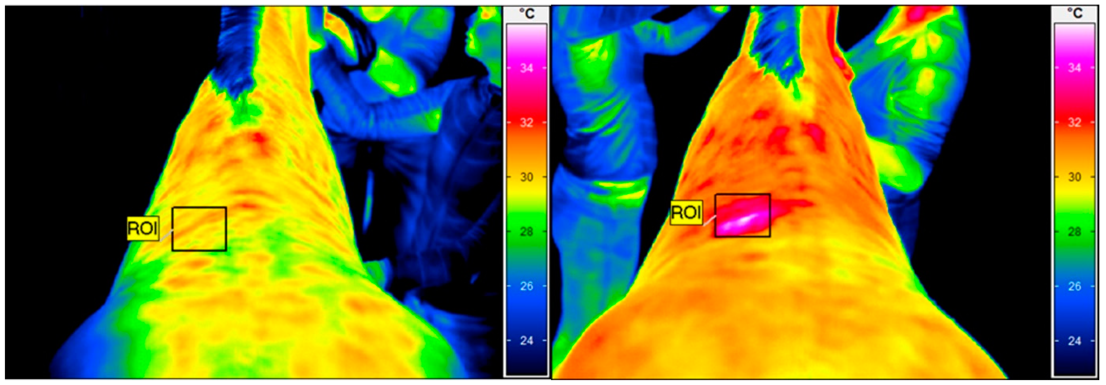
2.4. Thermographic Examination
2.5. High Intensity Laser Therapy
2.6. Statistical Analysis
3. Results
4. Discussion
5. Conclusions
Author Contributions
Funding
Institutional Review Board Statement
Informed Consent Statement
Data Availability Statement
Conflicts of Interest
References
- Jeffcott, L.B. Disorders of the thoracolumbar spine of the horse—A survey of 443 cases. Equine Vet. J. 1980, 12, 197–210. [Google Scholar] [CrossRef] [PubMed]
- Walmsley, J.P.; Pettersson, H.; Winberg, F.; McEvoy, F. Impingement of the dorsal spinous processes in two hundred and fifteen horses: Case selection, surgical technique and results. Equine Vet. J. 2002, 34, 23–28. [Google Scholar] [CrossRef]
- Mayaki, A.M.; Intan-Shameha, A.R.; Noraniza, M.A.; Mazlina, M.; Adamu, L.; Abdullah, R. Clinical investigation of back disorders in horses: A retrospective study (2002–2017). Vet. World 2019, 12, 377–381. [Google Scholar] [CrossRef] [PubMed]
- Greve, L.; Dyson, S. Saddle fit and management: An investigation of the association with equine thoracolumbar asymmetries, horse and rider health. Equine Vet. J. 2015, 47, 415–421. [Google Scholar] [CrossRef] [PubMed]
- Ridgway, K.; Harman, J. Equine back rehabilitation. Vet. Clin. N. Am. Equine Pract. 1999, 15, 263–280. [Google Scholar] [CrossRef] [PubMed]
- Greve, L.; Dyson, S. The horse-saddle-rider interaction. Vet. J. 2013, 195, 275–281. [Google Scholar] [CrossRef]
- Peham, C.; Kotschwar, A.; Borkenhagen, B.; Kuhnke, S.; Molsner, J.; Baltacis, A. A comparison of forces acting on the horse’s back and the stability of the rider’s seat in different positions at the trot. Vet. J. 2010, 184, 56–59. [Google Scholar] [CrossRef]
- Henson, F.M.D.; Kidd, J.A. Overriding dorsal spinous processes. In Equine Back Pathology: Diagnosis and Treatment; Henson, F.M.D., Ed.; Blackwell Publishing: Oxford, UK, 2009; pp. 147–156. [Google Scholar]
- Turner, T.A. Overriding spinous processes (“kissing spines”) in horses: Diagnosis, treatment, and outcome in 212 Cases. In Proceedings of the 57th America Association of Equine Practitioners, San Antonio, TX, USA, 18–22 November 2011; pp. 424–430. [Google Scholar]
- Denoix, J.M.; Dyson, S.J. Thoracolumbar spine. In Diagnosis and Management of Lameness in the Horse, 2nd ed.; Michael, W.R., Dyson, S.J., Eds.; Elsevier: Maryland Heights, MO, USA, 2010; pp. 592–605. [Google Scholar]
- Quiroz-Rothe, E.; Novales, M.; Aguilera-Tejero, E.; Rivero, J.L.L. Polysaccharide storage myopathy in the M. longissimus lumborum of showjumpers and dressage horses with back pain. Equine Vet. J. 2010, 34, 171–176. [Google Scholar] [CrossRef]
- Ranner, W.; Gerhards, H.; Klee, W. Diagnostic validity of palpation in horses with back problems. Berl. Und Munch. Tierarztl. Wochenschr 2002, 115, 420–424. [Google Scholar]
- Varcoe-Cocks, K.; Sagar, K.N.; Jeffcott, L.B.; McGowan, C.M. Pressure algometry to quantify muscle pain in racehorses with suspected sacroiliac dysfunction. Equine Vet. J. 2006, 38, 558–562. [Google Scholar] [CrossRef]
- Haussler, K.K.; Erb, H.N. Pressure algometry for the detection of induced back pain in horses: A preliminary study. Equine Vet. J. 2006, 38, 76–81. [Google Scholar] [CrossRef] [PubMed]
- Sullivan, K.A.; Hill, A.E.; Haussler, K.K. The effects of chiropractic, massage and phenylbutazone on spinal mechanical nociceptive thresholds in horses without clinical signs. Equine Vet. J. 2008, 40, 14–20. [Google Scholar] [CrossRef] [PubMed]
- Bendtsen, L.; Jensen, R.; Jensen, N.K.; Olesen, J. Pressure-controlled palpation: A new technique which increases the reliability of manual palpation. Cephalalgia 1995, 15, 205–210. [Google Scholar] [CrossRef] [PubMed]
- Sakai, F.; Ebihara, S.; Akiyama, M.; Horikawa, M. Pericranial muscle hardness in tension-type headache. A non-invasive measurement method and its clinical application. Brain 1995, 118, 523–531. [Google Scholar] [CrossRef]
- Haussler, K.K. Back problems. Chiropractic evaluation and management. Vet. Clin. N. Am. Equine Pract. 1999, 15, 195–209. [Google Scholar] [CrossRef]
- Martin, B.B.; Klide, A. Diagnosis and treatment of chronic back pain in horses. In Proceedings of the Annual Convention of the AAEP 1997, Phoenix, AZ, USA, 10 December 1997; Volume 43, pp. 310–311. [Google Scholar]
- de Oliveira, K.; Soutello, R.V.G.; da Fonseca, R.; Costa, C.; Paulo, P.R.; Fachiolli, D.F.; Clayton, H.M. Gymnastic training and dynamic mobilization exercises improve stride quality and increase epaxial muscle size in therapy horses. J. Equine Vet. Sci. 2015, 35, 888–893. [Google Scholar] [CrossRef]
- Zielińska, P.; Soroko, M.; Zwyrzykowska, A.; Kiełbowicz, Z. The use of laser biostimulation in human and animal physio-therapy–A review. Acta Vet. Brno 2017, 86, 91–96. [Google Scholar] [CrossRef]
- Santamato, A.; Solfrizzi, V.; Panza, F.; Tondi, G.; Frisardi, V.; Leggin, B.G.; Ranieri, M.; Fiore, P. Short-term efects of high-intensity laser therapy versus ultrasound therapy in the treatment of people with subacromial impingement syndrome: A randomized clinical trial. Phys. Ther. 2009, 89, 643–652. [Google Scholar] [CrossRef]
- Zati, A.; Valent, A. Physical therapy: New technologies in rehabilitation medicine (translated to English). Edizioni Minerva Med. 2006, 2006, 162–185. [Google Scholar]
- Jaafar, S.E.; Al-Bayti, A.A.; Abdullah, S.I. Using short term of high power laser therapy in horse’s tendon injuries. Arch Razi Ins. 2021, 76, 1437–1444. [Google Scholar]
- Zielińska, P.; Nicpoń, J.; Kiełbowicz, Z.; Soroko, M.; Dudek, K.; Zaborski, D. Effects of high intensity laser therapy in the treatment of tendon and ligament injuries in performance horses. Animals 2020, 10, 1327. [Google Scholar] [CrossRef] [PubMed]
- Quiney, L.; Murray, R.; Dyson, S. Management of primary injuries of the medial collateral ligament of the carpus in two horses. J. Equine Vet. Sci. 2019, 86, 102878. [Google Scholar] [CrossRef] [PubMed]
- Pluim, M.; Martens, A.; Vanderperren, K.; Sarrazin, S.; Koene, M.; Luciani, A.; van Weeren, P.; Delesalle, C. Short- and long term follow-up of 150 sports horses diagnosed with tendinopathy or desmopathy by ultrasonographic examination and treated with high-power laser therapy. Res. Vet. Sci. 2018, 119, 232–238. [Google Scholar] [CrossRef]
- Zielińska, P.; Kiełbowicz, Z.; Paczuska, J. High Intensity Laser Therapy (HILT) in treatment of orthopedic diseases in horses. Med. Weter. 2015, 7, 373–376. [Google Scholar]
- Zielińska, P.; Śniegucka, K.; Kiełbowicz, Z. A case series of 11 horses diagnosed with bone spavin treated with high in-tensity laser therapy (HILT). J. Equine Vet. Sci. 2023, 120, 104188. [Google Scholar] [CrossRef]
- Lopes-Martins, R.Á.B.; Marcos, R.L.; Leonardo, P.S.; Prianti, A.C., Jr.; Muscará, M.N.; Aimbire, F.; Frigo, L.; Iversen, V.V.; Bjordal, J.M. Effect of low-level laser (Ga-Al-As 655 nm) on skeletal muscle fatigue induced by electrical stimulation in rats. J. Appl. Physiol. 2006, 101, 283–288. [Google Scholar] [CrossRef]
- Ramos, L.; Junior, E.C.P.L.; Pallotta, R.C.; Frigo, L.; Marcos, R.L.; De Carvalho, M.H.C.; Bjordal, J.M.; Lopes-Martins, R.A. Infrared (810 nm) low-level laser therapy in experimental model of strain-induced skeletal muscle injury in rats: Effects on functional outcomes. Photochem. Photobiol. 2012, 88, 154–160. [Google Scholar] [CrossRef]
- Alayat, M.S.; Elsodany, A.M.; Miyajan, A.F.; Alzhrani, A.A.; Alzhrani, H.M.S.; Maqliyah, A.M. Changes in local skin temperature after the application of a pulsed Nd:YAG laser to healthy subjects: A prospective crossover controlled trial. Lasers Med. Sci. 2019, 34, 1681–1688. [Google Scholar] [CrossRef]
- Lee, S.-K.; Lee, H.-O.; Youn, J.-I. Evaluation of muscle tension in hemiplegia patients with a real-time monitoring system during high intensity laser therapy. J. Opt. Soc. Korea 2015, 19, 277–283. [Google Scholar] [CrossRef]
- Haussler, K.K.; Manchon, P.T.; Donnell, J.R.; Frisbie, D.D. Effects of low-level laser therapy and chiropractic care on back pain in quarter horses. J. Equine Vet. Sci. 2019, 86, 102891. [Google Scholar] [CrossRef] [PubMed]
- Berner, D.; Winter, K.; Brehm, W.; Gerlach, K. Influence of head and neck position on radiographic measurement of inter-vertebral distances between thoracic dorsal spinous processes in clinically sound horses. Equine Vet. J. 2012, 44, 21–26. [Google Scholar] [CrossRef] [PubMed]
- Soroko, M.; Zaborski, D.; Dudek, K.; Yarnell, K.; Górniak, W.; Vardasca, R. Evaluation of thermal pattern distributions in racehorse saddles using infrared thermography. PLoS ONE 2019, 14, e0221622. [Google Scholar] [CrossRef]
- Howell, K.; Dudek, K.; Soroko, M. Thermal camera performance and image analysis repeatability in equine thermography. Infrared Phys. Technol. 2020, 110, 103447. [Google Scholar] [CrossRef]
- Wyche, S. Understanding the Horse´s Back, 1st ed.; Wiltshire: Crowood, UK, 2011; pp. 8–43. [Google Scholar]
- Zimmerman, M.; Dyson, S.; Murray, R. Close, impinging and overriding spinous processes in the thoracolumbar spine: The relationship between radiological and scintigraphic findings and clinical signs. Equine Vet. J. 2011, 44, 178–184. [Google Scholar] [CrossRef]
- Infante, D.; Croxatto, A.; Corrêa, F. Radiologic findings consistent with kissing spines syndrome in Chilean thoroughbreds horses. Sustain. Agric. Food Environ. Res. 2017, 4, 14–17. [Google Scholar] [CrossRef]
- Godlewska, M.; Soroko, M.; Zielińska, P. Assessment of vein diameter and body surface temperature after high-intensity laser therapy (HILT) on the tarsal joint in healthy horses. J. Equine Vet. Sci. 2020, 93, 103198. [Google Scholar] [CrossRef]
- Laakso, L.; Richardson, C.; Cramond, T. Factors affecting low level laser therapy. Aust. J. Physiother. 1993, 39, 95–99. [Google Scholar] [CrossRef]
- Duesterdieck-Zellmer, K.F.; Larson, M.K.; Plant, T.K.; Sundholm-Tepper, A.; Payton, M.E. Ex vivo penetration of low-level laser light through equine skin and flexor tendons. Am. J. Vet. Res. 2016, 77, 991–999. [Google Scholar] [CrossRef]
- Ryan, T.; Smith, R. An investigation into the depth of penetration of low level laser therapy through the equine tendon in vivo. Ir. Vet. J. 2007, 60, 295–299. [Google Scholar] [CrossRef]
- Watson, T. (Ed.) Electrotherapy E-Book: Evidence-Based Practice; Elsevier Health Sciences: St. Louis, MO, USA, 2008. [Google Scholar]
- Kim, G.-J.; Choi, J.; Lee, S.; Jeon, C.; Lee, K. The effects of high intensity laser therapy on pain and function in patients with knee osteoarthritis. J. Phys. Ther. Sci. 2016, 28, 3197–3199. [Google Scholar] [CrossRef] [PubMed]
- Zielińska, P.; Soroko, M.; Howell, K.; Godlewska, M.; Hildebrand, W.; Dudek, K. Comparison of the effect of high-intensity laser therapy (HILT) on skin surface temperature and vein diameter in pigmented and non-pigmented skin in healthy racehorses. Animals 2021, 11, 1965. [Google Scholar] [CrossRef] [PubMed]
- Zielińska, P.; Soroko-Dubrovina, M.; Śniegucka, K.; Dudek, K.; Čebulj-Kadunc, N. Effects of high-intensity laser therapy (HILT) on skin surface temperature and vein diameter in healthy racehorses with clipped and non-clipped coat. Animals 2023, 13, 216. [Google Scholar] [CrossRef] [PubMed]
- Cameron, M.H. Physical agents in Rehabilitation: From Research to Practice; Elsevier Health Sciences: St. Louis, MO, USA, 2012; pp. 221–224. [Google Scholar]
- Kujawa, J.; Zavodnik, L.; Zavodnik, I.; Buko, V.; Lapshyna, A.; Bryszewska, M. Effect of low-intensity (3.75–25 J/cm2) near-infrared (810 nm) laser radiation on red blood cell ATPase activities and membrane structure. J. Clin. Laser Med. Surg. 2004, 22, 111–117. [Google Scholar] [CrossRef] [PubMed]
- Thabet, A.A.E.-M.; Alshehri, M.A. Effect of pulsed high-intensity laser therapy on pain, adhesions, and quality of life in women having endometriosis: A randomized controlled trial. Photomed. Laser Surg. 2018, 36, 363–369. [Google Scholar] [CrossRef] [PubMed]
- Formisano, R.; Pantano, P.; Buzzi, M.G.; Vinicola, V.; Penta, F.; Barbanti, P.; Lenzi, G.L. Late motor recovery is influenced by muscle tone changes after stroke. Arch. Phys. Med. Rehabil. 2005, 86, 308–311. [Google Scholar] [CrossRef] [PubMed]
- Palastanga, N.P. Heat and cold. In Pain Management and Control in Physiotherapy; Wells, P., Frampton, V., Bowsher, D., Eds.; Heineman: London, UK, 1988; pp. 169–180. [Google Scholar]


| Parameters | Without KSS (n = 10) | With KSS (n = 10) | Without KSS vs. With KSS (p-Value) | Power 1 - β |
|---|---|---|---|---|
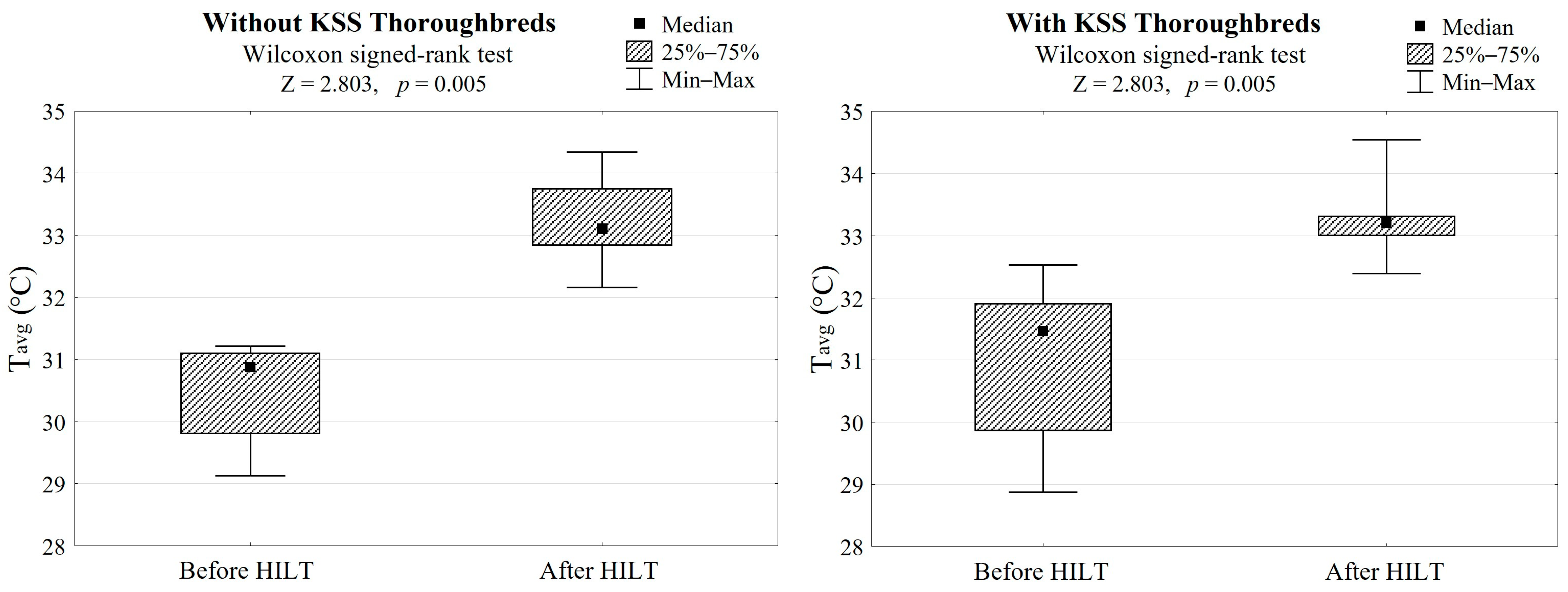
| Tavg before HILT (°C) | 30.9 (29.8 to 31.1) | 31.5 (29.9 to 31.9) | 0.140 | 0.974 |
| Min–Max | 29.1–31.2 | 28.9–32.5 | ||
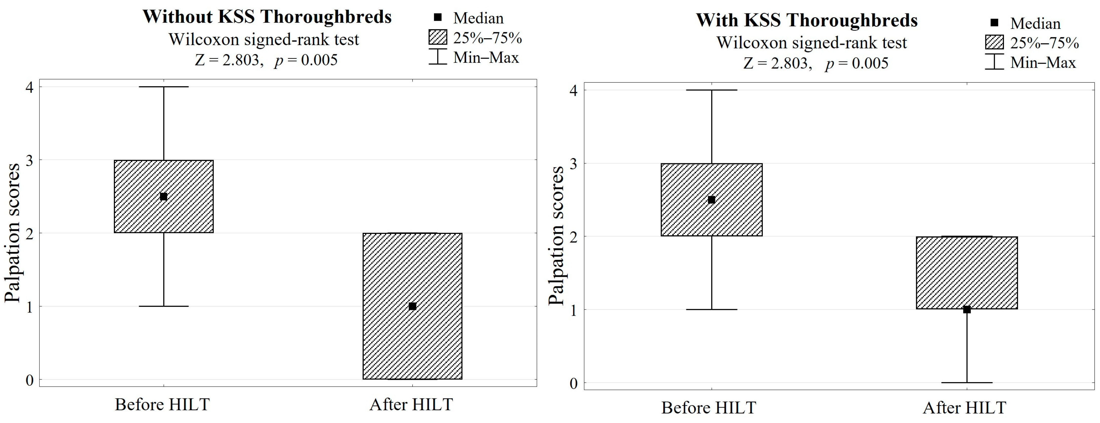
| PS before HILT (score) | 2.5 (2 to 3) | 2.5 (2 to 3) | 0.969 | 0.051 |
| Min–Max | 1–4 | 1–4 | ||
| Tavg after HILT (°C) | 33.1 (32.8 to 33.8) | 33.2 (33.0 to 33.3) | 0.734 | 0.562 |
| Min–Max | 32.2–34.3 | 32.4–34.5 | ||
| PS after HILT (score) | 1 (0 to 2) | 1 (1 to 2) | 0.601 | 0.796 |
| Min–Max | 0–2 | 0–2 | ||
| ΔTavg (°C) | 2.6 (2.3 to 3.1) | 2.4 (1.3 to 3.3) | 0.307 | 0.863 |
| Min–Max | 1.4–3.8 | 0.7–3.5 | ||
| ΔPS (score) | −2 (−2 to −1) | −1 (−2 to −1) | 0.322 | 0.999 |
| Min–Max | −2–−1 | −3–−1 |
| Statistics | Without KSS (n = 10) | With KSS (n = 10) | Without KSS and With KSS (n = 20) |
|---|---|---|---|
| Rho (95% CI) | 0.071 (−0.585 to 0.671) | −0.180 (−0.727 to 0.507) | −0.064 (−0.493 to 0.389) |
| p-value | 0.831 | 0.590 | 0.780 |
Disclaimer/Publisher’s Note: The statements, opinions and data contained in all publications are solely those of the individual author(s) and contributor(s) and not of MDPI and/or the editor(s). MDPI and/or the editor(s) disclaim responsibility for any injury to people or property resulting from any ideas, methods, instructions or products referred to in the content. |
© 2023 by the authors. Licensee MDPI, Basel, Switzerland. This article is an open access article distributed under the terms and conditions of the Creative Commons Attribution (CC BY) license (https://creativecommons.org/licenses/by/4.0/).
Share and Cite
Zielińska, P.; Soroko-Dubrovina, M.; Dudek, K.; Ruzhanova-Gospodinova, I.S. A Preliminary Study of the Influence of High Intensity Laser Therapy (HILT) on Skin Surface Temperature and Longissimus Dorsi Muscle Tone Changes in Thoroughbred Racehorses with Back Pain. Animals 2023, 13, 794. https://doi.org/10.3390/ani13050794
Zielińska P, Soroko-Dubrovina M, Dudek K, Ruzhanova-Gospodinova IS. A Preliminary Study of the Influence of High Intensity Laser Therapy (HILT) on Skin Surface Temperature and Longissimus Dorsi Muscle Tone Changes in Thoroughbred Racehorses with Back Pain. Animals. 2023; 13(5):794. https://doi.org/10.3390/ani13050794
Chicago/Turabian StyleZielińska, Paulina, Maria Soroko-Dubrovina, Krzysztof Dudek, and Iliana Stefanova Ruzhanova-Gospodinova. 2023. "A Preliminary Study of the Influence of High Intensity Laser Therapy (HILT) on Skin Surface Temperature and Longissimus Dorsi Muscle Tone Changes in Thoroughbred Racehorses with Back Pain" Animals 13, no. 5: 794. https://doi.org/10.3390/ani13050794
APA StyleZielińska, P., Soroko-Dubrovina, M., Dudek, K., & Ruzhanova-Gospodinova, I. S. (2023). A Preliminary Study of the Influence of High Intensity Laser Therapy (HILT) on Skin Surface Temperature and Longissimus Dorsi Muscle Tone Changes in Thoroughbred Racehorses with Back Pain. Animals, 13(5), 794. https://doi.org/10.3390/ani13050794