Article

Molecular Identification of Sarcocystis rileyi and Sarcocystis sp. (Closely Related to Sarcocystis wenzeli) in Intestines of Mustelids from Lithuania

by
Petras Prakas
*,
Darija Moskaliova
,
Donatas Šneideris
,
Evelina Juozaitytė-Ngugu
,
Evelina Maziliauskaitė
,
Saulius Švažas
and
Dalius Butkauskas
Nature Research Centre, Akademijos Str. 2, LT-08412 Vilnius, Lithuania
*
Author to whom correspondence should be addressed.
Animals 2023, 13(3), 467; https://doi.org/10.3390/ani13030467
Submission received: 16 December 2022 / Revised: 26 January 2023 / Accepted: 26 January 2023 / Published: 29 January 2023

Abstract

:

Simple Summary

Protozoan parasites of the genus Sarcocystis are characterised by a two-host prey–predator life cycle. To date, more than 25 Sarcocystis species have been confirmed to form sarcocysts in muscles and CNS of birds. Avian Sarcocystis species are transmitted via predatory birds, placental mammals, and opossums. The objective of the study was to examine the role of predatory mammals of the family Mustelidae in the transmission of avian Sarcocystis spp. by means of molecular methods. In total, 115 small intestine samples of mustelids collected in Lithuania were tested for the presence of Sarcocystis spp. using anseriforms and domestic fowl (Gallus domesticus) as their intermediate hosts. Based on the DNA sequence analysis, S. rileyi known as forming macrocysts in muscles of ducks was detected in 11.3% of examined small intestine samples and Sarcocystis sp. was identified in two samples. The latter species was most closely related to Sarcocystis spp. isolates infecting chickens and causing encephalitis. This is the first report of avian Sarcocystis identified by molecular methods in the small intestines of mustelids, indicating the significance of these small predators for the spreading of Sarcocystis spp. using birds as intermediate hosts. Based on current knowledge, canids and mustelids are most likely the definitive hosts of S. rileyi in Europe.

Abstract

The genus Sarcocystis is a group of numerous protozoan parasites having a two-host life cycle. Based on laboratory experiments and/or phylogenetic analysis results it was shown that seven Sarcocystis spp. producing sarcocsyts in bird tissues are transmitted via predatory placental mammals. To date the role of small mammals of the family Mustelidae in the distribution of avian Sarcocystis spp. have not been studied. During the current investigation, intestinal mucosa scrapings of 115 mustelids belonging to five species were tested for S. albifronsi, S. anasi, S. rileyi, and S. wenzeli infecting anseriforms and chickens. Microscopically, free sporocysts, sporulating oocysts, and loose oocysts were found in 61 samples (53.0%). Using nested PCR targeting the ITS1 region and sequencing, S. rileyi was confirmed in eight American minks, two European polecats and single European badger. Sarcocystis sp. was identified in one American mink and one European pine marten. Based on the partial ITS1 region this parasite showed that 100% identity to pathogenic Sarcocystis sp. caused a fatal infection in backyard chickens from Brazil. Phylogenetically, the Sarcocystis sp. identified in our study was most closely related to S. wenzeli parasitising domestic fowl (Gallus domesticus).

1. Introduction

Members of the genus Sarcocystis (Apicomplexa: Sarcocystidae) are protozoan parasites distributed worldwide. The genus Sarcocystis has a broad host spectrum encompassing mammals, birds and reptiles. These parasites are distinguished by an obligatory prey–predator two-host life cycle [1]. Sarcocysts are found mainly in muscles or CNS of intermediate hosts, while endogenous sporulation of oocysts take place in the intestine of the definitive host [2]. To date more than 200 Sarcocystis species are known, some of them being pathogenic for their intermediate host [1,3]. Sarcocystis species are morphologically characterised and described in intermediate hosts, while oocyst and sporocyst of parasite species found in definitive hosts can be differentiated only by molecular methods [4].
Birds serve as intermediate hosts for more than 25 known species of Sarcocystis [5]. Based on laboratory experiments, predatory birds, placental mammals, and opossums of the genus Didelphis are definitive hosts of Sarcocystis species forming sarcocysts in tissues of birds [1,6]. Phylogenetic results indicate that seven species, S. albifronsi, S. anasi, S. atraii, S. chloropusae, S. cristata, S. rileyi, and S. wenzeli are transmitted via predatory mammals of the order Carnivora [1,5,7,8,9,10,11]. Three species, S. albifronsi, S. anasi, and S. rileyi infect muscles of ducks and geese [7,8,12,13], S. wenzeli are found in chickens [11], S. atraii and S. chloropusae were described in birds of order Gruiformes [9,10], and S. cristata was detected in the representative of order Musophagiformes [5]. Of these seven species, three, S. albifronsi, S. anasi, and S. rileyi, were confirmed in Lithuania [7,12].
Sarcocystis species are mostly genetically characterised at nuclear 18S rDNA, 28S rDNA, ITS1, and mitochondrial cox1 [1,5]. The choice of genetic loci for the identification of Sarcocystis species depends on their hosts [14]. For instance, cox1 is most appropriate for the differentiation of Sarcocystis species employing ruminants as intermediate hosts [15,16]. Avian Sarcocystis spp. could be differentiated on the basis of the 28S rDNA and ITS1, and the ITS1 is more variable of these two genetic markers [1,17]. By contrast, 18S rDNA and cox1 appeared to be insufficiently variable for the discrimination of some Sarcocystis spp. employing birds as intermediate hosts [17].
Representatives of the family Mustelidae are widespread in Lithuania [18,19]. They occur in all habitats and with nine species compose most diverse family of the order Carnivora [19,20,21,22,23]. It has been shown that mustelids play a significant role in transmitting Sarcocystis species forming sarcocysts in rodents and ungulates [16,24].
So far, no research has been carried out to find out whether mustelids can contribute to the transmission of Sarcocystis species those intermediate hosts are birds. The invasive American mink (Neovison vison) is an important predator of ducks in wetland habitats of Finland and Denmark [25,26]. In Lithuania and Latvia, the mass killing of incubating females of mallard (Anas platyrhynchos), common pochard (Aythya farina), and tufted duck (Aythya fuligula) by American mink was recorded, particularly on small islands of lakes where ducks breed almost colonially [27]. Birds are an important food component also for other mustelid species in Lithuania [28]. The diet of mustelids includes eggs, young and adult individuals of various waterbird species, and also birds found dead, particularly in winter [18,19]. Taking into account the diet of mustelids and their abundance in Lithuania, the aim of this study was to investigate the potential role of mustelids in spreading Sarcocystis species using birds as intermediate hosts. To achieve this objective, intestinal mucosa scrapings of mustelids collected in Lithuania were tested for the presence of S. albifronsi, S. anasi, S. rileyi, and S. wenzeli by means of molecular methods.

2. Materials and Methods

2.1. Sample Collection and Isolation of Oocysts/Sporocysts

A total of 115 animals (61 American mink, 26 European pine marten Martes martes, 18 European polecat Mustela putorius, 6 European badger Meles meles, and 4 Beech marten Martes foina) from the Mustelidae family were collected in accordance with national and institutional guidelines from licensed third parties. The animals were legally hunted mainly in southern, eastern, and central Lithuania between 2017 and 2021, in September–April and were kept frozen at –20 °C. The small intestine was removed from the animals and cut lengthwise. The intestinal epithelium was lightly scraped using a scalpel and suspended in 50 mL of water. The isolation of oocysts/sporocysts of Sarcocystis spp. was performed using previously described methodology [24].

2.2. Molecular Identification and Phylogenetic Analysis

Genomic DNA extraction was performed using a GeneJET Genomic DNA Purification Kit (Thermo Fisher Scientific Baltics, Vilnius, Lithuania) according to the manufacturer’s instructions. The DNA samples were kept frozen at −20 °C until further molecular analysis.
Nested PCR amplification of internal transcribed spacer 1 (ITS1) partial sequences was performed. In the first step, forward SU1F (5’- GATTGAGTGTTCCGGTGAATTATT -3’) and reverse 5.8SR2 (5’- AAGGTGCCATTTGCGTTCAGAA -3’) primer pair was used [29]. Whereas in the second step, two primer pairs, GsSrilF2 (5’- ACGTTGTTCTATATTATGTGACCATT -3’)/GsSrilR2 (5’- TACTATAGAGGTGAAAGGGAGGTGA -3’) and AZVF1 (5’- TCAAAACGTCCAAATAATGGTAT -3’)/AZVR1 (5’- ACACATTCCTACTGCCTTCCAC -3’) were used. The following primers were designed using the Primers 3 Plus program [30]. In silico, the first primer pair was chosen to amplify ITS1 fragments of S. rileyi, while the second primer pair was selected to amplify fragments of S. albifronsi, S. anasi, and S. wenzeli. Positive controls (DNA of S. albifronsi, S. anasi and S. rileyi extracted from single sarcocysts) were used in each set of PCRs. Three negative controls (nuclease free water instead of target DNA) were used: one for the first amplification step and two for the second step of nested PCR. The third negative control was obtained by transferring two µL from the negative control of the first amplification step to the negative control of the second amplification step.
PCR reactions were carried out using DreamTaq PCR Master Mix (Thermo Fisher Scientific Baltics, Vilnius, Lithuania) according to the manufacturer’s instructions. The PCR cycling conditions were as followed: initial denaturation for 5 min at 95 °C, 35 cycles of 45 s at 94 °C, 45 s at 55, 57, or 63 °C depending on the primer pair, 60 s at 72 °C, and final extension for 10 min at 72 °C. PCR products were observed in agarose gel and purified using Exonuclease I and FastAP Thermosensitive Alkaline Phosphatase (Thermo Fisher Scientific Baltics, Vilnius, Lithuania). Amplified products of the second nested PCR step were sequenced directly with the 3500 Genetic Analyzer (Applied Biosystems, Foster City, CA, USA) using the same forward and reverse primers as for PCR. The obtained ITS1 sequences were deposited in GenBank with accession numbers OP970969–OP970981.
The examined sequences were combined into single fragments, truncated excluding primer-binding nucleotide positions, checked manually for ambiguously placed nucleotides, and compared by BLAST (http://blast.ncbi.nlm.nih.gov/, accessed on 30 November 2022). For phylogenetic analysis sequences were aligned using MUSCLE algorithm loaded in MEGA7 software [31]. Selection of the evolutionary model best fit to the obtained multiple-sequence alignment and construction of phylogenetic tree by Bayesian methods were conducted with the help of TOPALi v2.5 software [32].

2.3. Data Analysis

Sterne’s exact method [33] was used to compute 95% confidence interval (CI) for the prevalence of Sarcocystis spp. in host species and in animals hunted during different months. Differences in the detection of Sarcocystis species in the examined mustelid species were evaluated using a Chi-squared test. The unconditional exact test was used to compare S. rileyi prevalence in animals collected in different months [34]. Statistical tests were carried out using the Quantitative Parasitology 3.0 software [35].

3. Results

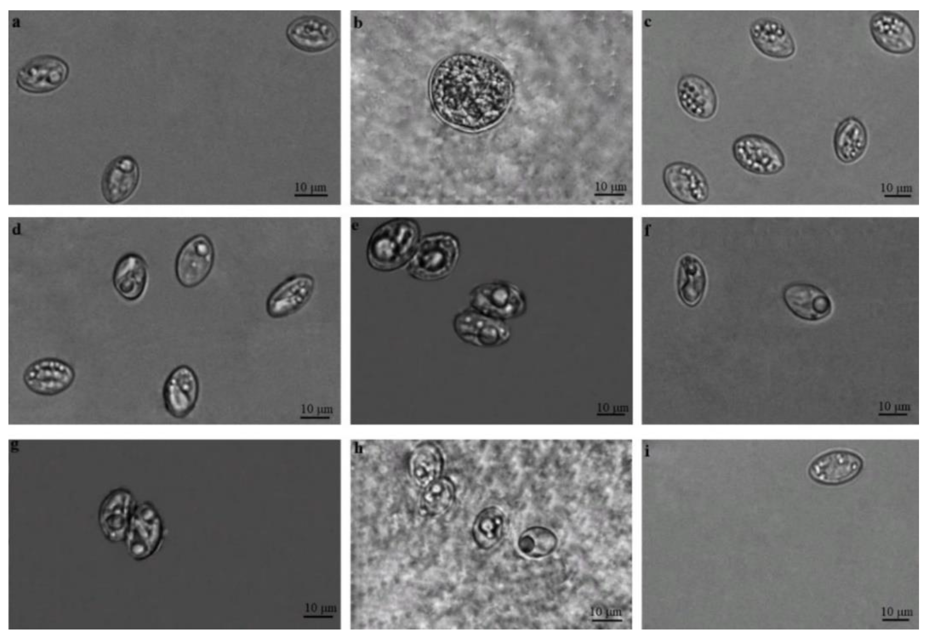
3.1. Microscopical Examination of Sarcocystis spp. Oocysts/Sporocysts

Sarcocystis spp. sporocysts and/or oocysts were noticed in the intestinal epithelium of the small intestines of all five species of mustelids analysed in the current study (Figure 1). In some samples, only a few (one-five) sporocysts and/or oocysts were detected in the area of the 24 × 24 mm coverslip, while in other samples, numerous parasites of different stages were found and it was even difficult to count the exact number of oocysts/sporocysts.
Under a light microscope, oocysts and/or sporocysts of Sarcocystis spp. were detected in 61 of 115 (53.0%, 95% CI = 43.9%–62.2%) analysed samples (Table 1). The differences in Sarcocystis spp. detection rates among five predator species were insignificant (χ2 = 4.47, df = 4, p = 0.349). It should be noted that free sporocysts were seen more often than sporulating oocysts or loose oocysts. Free sporocysts of Sarcocystis spp. measured 11.8 × 8.3 μm (7.1–14.5 × 6.5–10.9 μm; n = 450), whereas ellipsoidal sporulated oocysts were thin-walled, contained two sporocysts, and measured 18.1 × 15.6 μm (17.5–19.0 × 15.1–15.9; n = 130). Oocysts measured 19.9 × 17.1 μm (13.9–23.5 × 12.0–22.4; n = 46) and were seen in the intestinal mucosa of American mink, European pine marten, and European polecat. The morphometric sizes of sporocysts and oocysts found in different predator species overlapped (Table 1). Further molecular analysis was used for the identification of selected parasite species in the examined specimens of intestine mucosal scrapings.

3.2. Sarcocystis Species Identification and Their Distribution in Intestine Samples of Mustelids

Based on the nested PCR targeting the partial ITS1 region, the sequencing of amplified products, and the comparison of obtained sequences, two Sarcocystis species were identified (Table 2). The more common S. rileyi was confirmed in 11 samples (9.6%, 95% CI = 5.1%–16.4%). This Sarcocystis species forming macrocysts in ducks [12,13] was identified in eight American minks (13.1%, 95% CI = 6.2%–24.4%), two European polecats (11.1%, 95% CI = 20.0%–33.0%), and a single European badger (16.7%, 95% CI = 8.6%–58.9%). Meanwhile, undescribed Sarcocystis sp. was established in one American mink (1.6%, 95% CI = 0.9%–8.7%) and one European pine marten (3.8%, 95% CI = 0.2%–18.8%). The ITS1 fragments of S. rileyi and Sarcocystis sp. were amplified using GsSrilF2/GsSrilR2 and AZVF1/AZVR1 primer pairs, respectively. Notably, S. rileyi and Sarcocystis sp. were identified in different animals and the overall prevalence of Sarcocystis spp. in the intestinal mucosa of mustelids accounted for 11.3% (95% CI = 6.3%–18.6%).
Eleven 611–612 bp-long ITS1 sequences of S. rileyi determined in the present investigation displayed 99.18%–100% identity between each other. Of the 11 sequences, 9 identical ones (OP970971-79) showed an 100% match with other sequences of S. rileyi available in GenBank (GU188427, HM185744, KJ396584, MZ151434, MZ468639-40, and LT992314-16). The remaining two sequences of S. rileyi obtained in the current work (OP970980-81) differed from the most common haplotype by two nucleotide substitutions and one deletion and by two nucleotide substitutions, respectively. The sequences of S. rileyi obtained from mucosal scrapings of mustelids showed 91.22%–92.34% similarity to S. atraii from the common coot (Fulica atra) from Egypt and less than 74% similarity compared with the sequences of other Sarcocystis species.
Two 817 bp-long ITS1 sequences of Sarcocystis sp. LT-2022 obtained in the present work (OP970969-70) did not differ from each other. These two ITS1 sequences showed 100% identity with the sequence of Sarcocsytis sp. Chicken-2016-DF-BR (MN846302) isolated from brain tissues of two chickens in Brazil [36], 98.17%–98.66% similarity with the sequences of S. wenzeli (MT756994-98) parasitising chickens [11], and 95.49%–96.99% similarity with the sequences (OP490606-9, OP490613-4) obtained from pooled samples of the brain, pectoral muscle, lung, and heart of native village chickens in Malaysia.
We observed seasonal changes in the abundance of S. rileyi in our sample (Figure 2). S. rileyi was determined by molecular methods in one animal hunted in October, in two animals each hunted in September, November, and December, and in four animals hunted in January. The unconditional exact test showed that the detection of S. rileyi in September–January (15.5%, 95% CI = 8.5%–25.9%) was significantly higher (p = 0.0034) than in the February–April period (0%, 95% CI = 0%–8.5%). Meanwhile, Sarcocystis sp. LT-2022 was established in two animals collected in March and April.

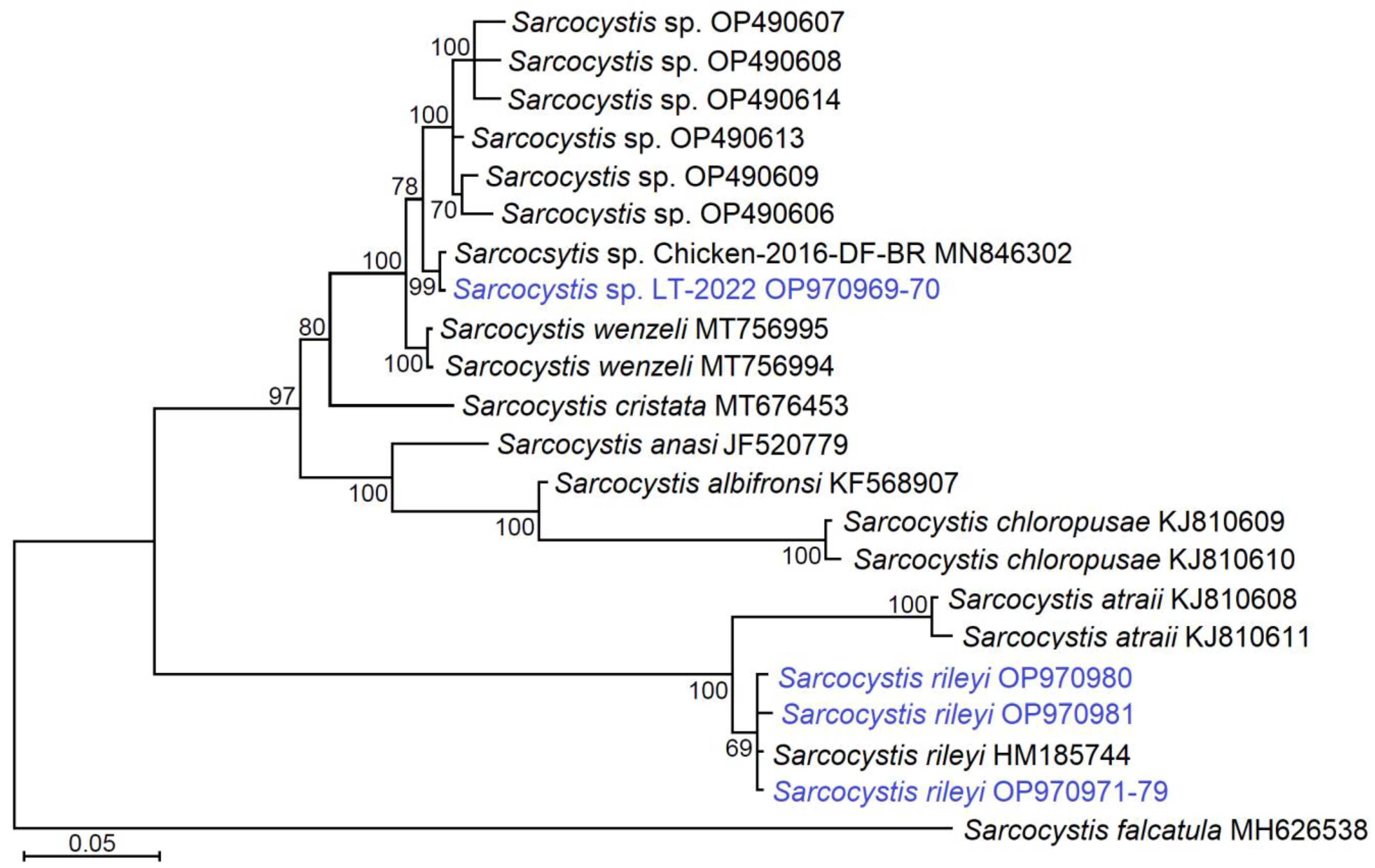
3.3. Phylogenetic Relationships of Identified Sarcocystis Species

Comparing the ITS1 fragments established in the current work, sequences of Sarcocystis sp. were longer at the 5’ end and the sequences of S. rileyi were longer at the 3’ end. Thus, after multiple alignment and sequence truncation, 547 bp-long sequences of S. rileyi and 554 bp-long sequences of Sarcocystis sp. were used for phylogenetic analysis. In the phylogenetic tree, S. rileyi obtained from mustelids grouped with other S. rileyi isolates obtained from various intermediate hosts (Figure 3). Based on ITS1, S. rileyi was the sister taxon to S. atraii and these two species formed separate clusters in the phylogram.
Based on the analysed ITS fragment, Sarcocystis sp. LT-2022 isolated from two representatives of two mustelid species were identical to Sarcocystis sp. Chicken-2016-DF-BR isolated from chickens in Brazil and they were placed in one well-supported cluster together with S. wenzeli and Sarcocystis sp. from chickens from Malaysia. Whereas, S. cristata described in the muscles of the great blue turaco (Corythaeola cristata) was the sister taxon to the Sarcocystis isolates established in Brazil, Malaysia, and Lithuania, and S. wenzeli. The remaining Sarcocystis spp., S. albifornsi, S. anasi from anseriforms, and S. chloropusae from the common moorhen (Gallinula chloropus) made a separate cluster in the phylogenetic tree.

4. Discussion

4.1. The Role of Mustelids in Distribution of Sarcocystis Species

In the present study, free sporocysts, sporulating oocysts, and loose oocysts were found in the intestinal mucosa of the five examined species, American mink, European pine marten, European polecat, European badger, and beech marten (Figure 1 and Table 1). The morphometric sizes of parasite stages detected in five hosts overlapped. Thus, it was impossible to determine whether the studied host species were infected with the same or different Sarcocystis species. Furthermore, it is known that predators can be simultaneously infected with sporocysts of several Sarcocystis species [16,24,38]. Therefore, the identification of Sarcocystis species was performed using molecular methods.
Based on the nested PCR and subsequent BLAST analyses of the obtained DNA sequences, two Sarcocystis species using birds as intermediate hosts were confirmed. The prevalence of Sarcocystis spp. defined by means of molecular examination was relatively low, reaching 11.3% (13/115). By microscopical analysis, sporocysts and/or oocysts of Sarcocystis spp. were noticed in more than half (53.0%, 61/115) of the investigated samples. Thus, the obtained results of the present work indicate that the examined mustelids spread considerably more than Sarcocystis species, employing mammals rather than birds as their definitive hosts. Our previous research on the small intestine samples of mustelids by species-specific PCR revealed a high prevalence (89.3%, 75/84) of Sarcocystis species using cattle as their intermediate hosts [24]. Furthermore, 32 of the 40 (80.0%) examined small intestine samples of American mink tested positive for S. elongata, S. entzerothi, S. japonica, S. silva, and S. truncata by molecular methods, producing sarcocysts in muscles of ungulates of the family Cervidae [16]. It was also confirmed by experimental infection that possible definitive hosts of S. campestris, S. citellivulpes, S. muris, S. putorii, and S. undulati are members of family Mustelidae [39]. Research carried out until now implies that mustelids play a significant role for the transmission of various Sarcocystis species using hosts that belong to different taxonomic groups.

4.2. Mustelids as Possible Definitive Hosts of S. rileyi

Eleven ITS1 sequences obtained in the current study demonstrated 99.18%–100% similarity to the sequences of S. rileyi available in GenBank and showed less than 93% similarity with any other known species of Sarcocystis. Hence, S. rileyi was confirmed in the intestinal mucosa of eight American minks, two European polecats and single European badger (Table 2). The overall prevalence of S. rileyi in the analysed samples of mustelids was 9.6% (95% CI = 5.1%–16.4%). In the present study, the identified S. rileyi is a well-known Sarcocystis species forming macroscopic sarcocysts resembling grains of rice in the muscles of ducks. This species was described in the late nineteenth century [40] and redescribed in 2003, providing detailed morphological characterisation [41]. For a long time, macrocysts in numerous duck species were recorded only in North America [42,43,44,45,46,47,48]. Based on light microscopy, transmission electron microscopy, and molecular characterisation at three genetic loci (18S rDNA, 28S rDNA, and ITS1), S. rileyi was identified in Lithuania in 2011 [12]. According to the current data, the distribution of S. rileyi covers the eastern, northern, and central parts of Europe [12,13,29,49,50,51,52,53]. In this continent, S. rileyi was mostly recorded in mallard [12,13,49,52] and with much less frequency in several other duck species [29,53]. The mallard is the most abundant species of ducks and also one of most important bird game species in Europe [54,55,56]. Sarcocystis rileyi cause economic losses, since hunted duck meat contaminated with macrocysts is not suitable for human consumption [13]. Additionally, severe infection of S. rileyi may result in weakness of hosts, reduced flying capacity, and infected birds may be more easily caught by predators [57]. The stripped skunk (Mephitis mephitis) of the family Mephistidae is an experimentally proved definitive hosts of S. rileyi in North America [58,59]. This small predatory animal lives only in captivity in Europe [60]. Therefore, for a long time it was unclear which predators are responsible for the spread of S. rileyi in Europe. Based on molecular analysis, red foxes (Vulpes vulpes) and raccoon dogs (Nyctereutes procyonoides) of the family Canidae were identified as definitive hosts of S. rileyi in Lithuania and Germany [38,50]. Hence, based on the findings of previous and current investigations, S. rileyi is transmitted in Europe by predators of family Canidae and Mustelidae. It should be noted that Mephistidae and Mustelidae families are closely related and together with Ailuridae and Procyonidae compose a superfamily, Musteloidea [61]. Thus, the current findings indicate the co-evolution of S. rileyi with their definitive hosts. A co-evolution of Sarcocystis spp. with their definitive host rather than the intermediate host has been shown in the phylogenetic investigations of various groups of Sarcocystis species [15,62,63]. The raccoon (Procyon lotor) of the family Procyonidae which is native to North America is now spreading in Lithuania through the western part of the country [64]. Taking into account the close relationship of raccoon with mustelids and mephistids, this invasive predator should be screened for the distribution of S. rileyi.
In general, Sarcocystis species are more host-specific for their intermediate hosts than for their definitive hosts. For instance, S. cruzi, the most common species of Sarcocystis of cattle worldwide, is transmissible via dogs, coyotes, foxes, and wolves [1]. With the exception of S. wenzeli, Sarcocystis species transmitted by canids cannot be transmitted by felids and vice versa [65]. However, laboratory experiments evidenced that some Sarcocystis spp. transmitted via canids or felids can be spread via mustelids [39,66]. Furthermore, on the basis of molecular investigations of small intestine samples it was shown that S. cruzi can be spread not only by canids, but also by mustelids [24]. The current study on the basis of ITS1 sequence analysis also indicates that S. rileyi can be transmitted in Europe by the members of two families, Canidae and Mustelidae.
It should be emphasised that in the present study S. rileyi was identified in mustelids, which were hunted from September to January (15.5%, 11/71). This Sarcocystis species was mostly confirmed in American mink, while S. rileyi was not detected in 44 animals which were collected during February–April (Figure 2). In Lithuania, during September, large flocks of ducks and waders concentrate in partly drained fishponds and these birds form a large part of the diet of invasive American mink [67]. In summer–early autumn, birds are also an important food component for other mustelid species in various regions of Lithuania [18]. Whereas, in late autumn and winter mustelids only occasionally hunt ducks [18,19,20,28,68,69,70]. Oocysts/sporocysts of Sarcocystis spp. are found in the faeces of definitive hosts 7–14 days post infection and excretion of infective parasite stages mostly lasts several months [1]. Thus, the observed variations in the identification of S. rileyi during different months are congruent with the diet of mustelids and the life cycle peculiarities of Sarcocystis parasites. The results of the abundance of S. rileyi depending on the season mustelids were hunted are in congruent with the investigation of S. rileyi in canids. During a previous study conducted in Lithuania, S. rileyi was identified in the small intestines of red foxes and raccoon dogs hunted in November and December, but the parasite was not detected in animals hunted in February and March [50]. Thus, future research on the prevalence of Sarcocystis in predatory mammals through different seasons is needed.

4.3. Detection of Sarcocystis sp. Closely Related to S. wenzeli in Small Intestine of Mustelids

Based on the obtained ITS1 sequence analysis, Sarcocystis sp. was identified in a single American mink and in a single American pine marten (Table 2). Two 817 bp-long ITS1 sequences were 100% identical to the sequence of Sarcocystis sp. Chicken-2016-DF-BR obtained from the brains of two chickens in the midwest of Brazil [36]. This parasite caused fatal outcomes in backyard chickens. The infected chickens suffered from anorexia, weight loss, incoordination, ataxia, and opisthotonos. The histopathological analysis showed necrotizing granulomatous and meningoencephalitis with intralesional Sarcocystis-like schizonts and merozoites. Infected chickens remained free during the day and were kept in the coop at night [36]. It should be noted that severe myositis and encephalitis associated with Sarcocystis parasites has been reported several times in domestic fowl in different geographical regions [71,72].
The ITS1 region is highly variable for Sarcocystis spp. [8]. Due to the large number of indels (insertions/deletions) it is hard to align ITS1 sequences of Sarcocystis spp. sharing relatively low similarity. Therefore, the ITS1 region is not a good choice for the discriminating phylogenetic relationships of genetically remote group of Sarcocystis. However, this genetic locus is suitable for phylogenetic analysis of closely related Sarcocystis species [63,73]. Several studies have demonstrated that ITS1 is an appropriate genetic marker inferring phylogenetic relationships of Sarcocystis spp. using birds as intermediate hosts [5,9,10,11,73,74,75,76,77,78]. Furthermore, for this group of Sarcocystis species, ITS1 and 28S rDNA give congruous topology [73,79]. Based on the ITS1 sequence analysis conducted in the current study, the topology of the examined Sarcocystis species using birds–predatory mammals in their intermediate–definitive host life cycle (Figure 3) in general corresponded to that determined in the latest phylogenetic studies [5,36]. Sarcocystis sp. LT-2022 obtained in the present work and Sarcocystis sp. Chicken-2016-DF-BR were placed in one phylogenetic cluster together with S. wenzeli infecting chickens and Sarcocystis sp. isolated from pooled various tissue samples of native village chickens in Malaysia (Figure 3). Thus, the obtained sequences were grouped with those of Sarcocystis spp. parasitising chickens. Further molecular studies are needed to clarify the number of species that represent S. wenzeli, Sarcocystis sp. Chicken-2016-DF-BR, Sarcocystis sp. from Malaysian chickens, and Sarcocystis sp. LT-2022 identified in the present work.
There is ongoing debate on the classification of Sarcocystis species in chickens [39]. In the latest taxonomic review of the genus Sarcocystis, two species infecting chickens, S. horvathi and S. wenzeli, were distinguished [1]. Sarcocystis wenzeli is characterised morphologically in detail and based on transmission experiments dogs and cats are confirmed as definitive hosts of this species [65], whereas the definitive hosts of S. horvathi are unknown [1].
The data of the current work indicate that mustelids might be involved in the transmission of Sarcocystis species infecting domestic gallinaceous fowl. However, laboratory infection experiments are definitely necessary to test the results obtained. Additionally, here we present the first identification of Sarcocystis sp. in Lithuania, closely related to Sarcocystis parasitizing chickens. Despite extensive studies of Sarcocystis conducted in Lithuania in various groups of wild birds [7,8,12,73,75,76,77,78,79], these parasites have not been studied in poultry so far. In Lithuania, chickens are mainly raised in poultry farms [80]. However, in rural regions small numbers of domestic fowl are kept free. The main predators of backyard chickens in Lithuania are mustelids and red fox [18]. Mustelids can potentially cause the transmission of highly pathogenic Sarcocystis species in poultry farming.

5. Conclusions

Based on the nested PCR and sequencing of the ITS1 region, S. rileyi producing macroscopic sarcocysts in muscles of ducks was for the first time confirmed in small intestine scrapings of three mustelid species collected in Lithuania. The prevalence of S. rileyi in the examined mustelids was 9.6%. According to the current data obtained by molecular investigations, canids and mustelids are responsible for the spread of S. rileyi in Europe.
Undescribed Sarcocystis sp. LT-2022 showed 100% similarity within the 817 bp-long ITS1 fragment with Sarcocystis sp. Chicken-2016-DF-BR, which caused a fatal infection in two backyard chickens in Brazil. The detected Sarcocystis parasite was most closely related to S. wenzeli and Sarcocystis sp. using chickens as their intermediate hosts. Thus, this is the first report of Sarcocystis sp. associated with possible infection in gallinaceous birds in Lithuania.

Author Contributions

Conceptualization, P.P. and D.B.; methodology, P.P.; software, P.P.; validation, P.P., E.J.-N. and S.Š.; formal analysis, P.P., D.M. and D.Š.; investigation, D.M., D.Š., E.J.-N. and E.M.; resources, P.P, S.Š. and D.B.; writing—original draft preparation, P.P., D.Š., E.J.-N. and S.Š.; writing—review and editing, P.P., D.M., D.Š., E.J.-N., E.M., S.Š., D.B.; visualization, P.P. and E.J.-N.; supervision, P.P.; funding acquisition, S.Š. and D.B. All authors have read and agreed to the published version of the manuscript.

Funding

This research received no external funding.

Institutional Review Board Statement

Samples were collected with the permission of the Ministry of Environment of the Republic of Lithuania (2017-03-23 no.26-A4-3119; 2019-03-01 no. 26-A4-1535; 2021-03-31 nr. (26)-SR-89).

Informed Consent Statement

Not applicable.

Data Availability Statement

The ITS1 sequences of S. rileyi and Sarcocystis sp. were submitted in GenBank database with accession numbers OP970969–OP970981.

Acknowledgments

The authors are grateful to Valentinas Pabrinkis (Nature Research Centre, Vilnius, Lithuania) and to Kaunas T. Ivanauskas Zoology Museum for providing samples for the study.

Conflicts of Interest

The authors declare no conflict of interest.

References

  1. Dubey, J.P.; Calero-Bernal, R.; Rosenthal, B.M.; Speer, C.A.; Fayer, R. Sarcocystosis of Animals and Humans, 2nd ed.; CRC Press: Boca Raton, FL, USA, 2015. [Google Scholar]
  2. Mehlhorn, H.; Heydorn, A.O. The Sarcosporidia (Protozoa, Sporozoa): Life Cycle and Fine Structure. Adv. Parasitol. 1978, 16, 43–91. [Google Scholar] [CrossRef] [PubMed]
  3. Tenter, A.M. Current research on Sarcocystis species of domestic animals. Int. J. Parasitol. 1995, 25, 1311–1330. [Google Scholar] [CrossRef]
  4. Juozaitytė-Ngugu, E.; Švažas, S.; Šneideris, D.; Rudaitytė-Lukošienė, E.; Butkauskas, D.; Prakas, P. The Role of Birds of the Family Corvidae in Transmitting Sarcocystis Protozoan Parasites. Animals 2021, 11, 3258. [Google Scholar] [CrossRef] [PubMed]
  5. Máca, O.; González-Solís, D. Sarcocystis cristata sp. nov. (Apicomplexa, Sarcocystidae) in the imported great blue turaco Corythaeola cristata (Aves, Musophagidae). Parasites Vectors 2021, 14, 56. [Google Scholar] [CrossRef]
  6. Gallo, S.S.M.; Lindsay, D.S.; Ederli, N.B.; Matteoli, F.P.; Venancio, T.M.; de Oliveira, F.C.R. Identification of opossums Didelphis aurita (Wied-Neuweid, 1826) as a definitive host of Sarcocystis falcatula-like sporocysts. Parasitol. Res. 2018, 117, 213–223. [Google Scholar] [CrossRef]
  7. Kutkienė, L.; Prakas, P.; Sruoga, A.; Butkauskas, D. Description of Sarcocystis anasi sp. nov. and Sarcocystis albifronsi sp. nov. in birds of the order Anseriformes. Parasitol. Res. 2012, 110, 1043–1046. [Google Scholar] [CrossRef] [PubMed]
  8. Prakas, P.; Oksanen, A.; Butkauskas, D.; Sruoga, A.; Kutkienė, L.; Švažas, S.; Isomursu, M.; Liaugaudaitė, S. Identification and Intraspecific Genetic Diversity of Sarcocystis rileyi from Ducks, Anas spp., in Lithuania and Finland. J. Parasitol. 2014, 100, 657–661. [Google Scholar] [CrossRef]
  9. El-Morsey, A.; El-Seify, M.; Desouky, A.-R.Y.; Abdel-Aziz, M.M.; El-Dakhly, K.M.; Kasem, S.; Abdo, W.; Haridy, M.; Sakai, H.; Yanai, T. Morphologic and molecular characteristics of Sarcocystis atraii n. sp. (Apicomplexa: Sarcocystidae) infecting the common coot (Fulica atra) from Egypt. Acta Parasitol. 2015, 60, 691–699. [Google Scholar] [CrossRef]
  10. El-Morsey, A.; El-Seify, M.; Desouky, A.Y.; Abdel-Aziz, M.M.; Sakai, H.; Yanai, T. Sarcocystis chloropusae (protozoa: Sarcocystidae) n. sp. from the common moorhen (Gallinula chloropus) from Egypt. Parasitology 2015, 142, 1063–1065. [Google Scholar] [CrossRef]
  11. Pan, J.; Ma, C.; Huang, Z.; Ye, Y.; Zeng, H.; Deng, S.; Hu, J.; Tao, J. Morphological and molecular characterization of Sarcocystis wenzeli in chickens (Gallus gallus) in China. Parasites Vectors 2020, 13, 512. [Google Scholar] [CrossRef]
  12. Kutkienė, L.; Prakas, P.; Sruoga, A.; Butkauskas, D. Identification of Sarcocystis rileyi from the mallard duck (Anas platyrhynchos) in Europe: Cyst morphology and results of DNA analysis. Parasitol. Res. 2011, 108, 709–714. [Google Scholar] [CrossRef] [PubMed]
  13. Zuo, S.; Sørensen, S.R.; Kania, P.W.; Buchmann, K. Sarcocystis rileyi (Apicomplexa) in Anas platyrhynchos in Europe with a potential for spread. Int. J. Parasitol. Parasites Wildl. 2021, 15, 270–275. [Google Scholar] [CrossRef] [PubMed]
  14. Prakas, P.; Kirillova, V.; Gavarāne, I.; Grāvele, E.; Butkauskas, D.; Rudaitytė-Lukošienė, E.; Kirjušina, M. Morphological and molecular description of Sarcocystis ratti n. sp. from the black rat (Rattus rattus) in Latvia. Parasitol. Res. 2019, 118, 2689–2694. [Google Scholar] [CrossRef] [PubMed]
  15. Gjerde, B. Phylogenetic relationships among Sarcocystis species in cervids, cattle and sheep inferred from the mitochondrial cytochrome c oxidase subunit I gene. Int. J. Parasitol. 2013, 43, 579–591. [Google Scholar] [CrossRef]
  16. Prakas, P.; Rudaitytė-Lukošienė, E.; Šneideris, D.; Butkauskas, D. Invasive American mink (Neovison vison) as potential definitive host of Sarcocystis elongata, S. entzerothi, S. japonica, S. truncata and S. silva using different cervid species as intermediate hosts. Parasitol. Res. 2021, 120, 2243–2250. [Google Scholar] [CrossRef]
  17. Prakas, P.; Butkauskas, D.; Švažas, S.; Stanevičius, V. Morphological and genetic characterisation of Sarcocystis halieti from the great cormorant (Phalacrocorax carbo). Parasitol. Res. 2018, 117, 3663–3667. [Google Scholar] [CrossRef]
  18. Kontrimavičius, V. Lietuvos Fauna: Žinduoliai; Mokslas: Vilnius, Lithuania, 1988. [Google Scholar]
  19. Balčiauskas, L.; Trakimas, G.; Juškaitis, R.; Ulevičius, A.; Balčiauskienė, L. Atlas of Lithuanian Mammals, Amphibians and Reptiles, 2nd ed.; Akstis: Vilnius, Lithuania, 1999. [Google Scholar]
  20. Baghli, A.; Engel, E.; Verhagen, R. Feeding habits and trophic niche overlap of two sympatric mustelidae, the polecat Mustela putorius and the beech marten Martes foina. Z. Für Jagdwiss. 2002, 48, 217–225. [Google Scholar] [CrossRef]
  21. Koepfli, K.-P.; Deere, K.A.; Slater, G.J.; Begg, C.; Begg, K.; Grassman, L.; Lucherini, M.; Veron, G.; Wayne, R.K. Multigene phylogeny of the Mustelidae: Resolving relationships, tempo and biogeographic history of a mammalian adaptive radiation. BMC Biol. 2008, 6, 10. [Google Scholar] [CrossRef] [Green Version]
  22. Newman, C.; Zhou, Y.-B.; Buesching, C.D.; Kaneko, Y.; Macdonald, D.W. Contrasting Sociality in Two Widespread, Generalist, Mustelid Genera, Meles and Martes. Mammal Study 2011, 36, 169–188. [Google Scholar] [CrossRef]
  23. Law, C.J.; Slater, G.J.; Mehta, R.S. Lineage Diversity and Size Disparity in Musteloidea: Testing Patterns of Adaptive Radiation Using Molecular and Fossil-Based Methods. Syst. Biol. 2018, 67, 127–144. [Google Scholar] [CrossRef]
  24. Prakas, P.; Balčiauskas, L.; Juozaitytė-Ngugu, E.; Butkauskas, D. The Role of Mustelids in the Transmission of Sarcocystis spp. Using Cattle as Intermediate Hosts. Animals 2021, 11, 822. [Google Scholar] [CrossRef]
  25. Bonesi, L.; Palazon, S. The American mink in Europe: Status, impacts, and control. Biol. Conserv. 2007, 134, 470–483. [Google Scholar] [CrossRef]
  26. Holopainen, S.; Väänänen, V.-M.; Vehkaoja, M.; Fox, A.D. Do alien predators pose a particular risk to duck nests in Northern Europe? Results from an artificial nest experiment. Biol. Invasions 2021, 23, 3795–3807. [Google Scholar] [CrossRef]
  27. Viksne, J.; Švažas, S.; Czajkowski, A. Atlas of Duck Populations in Eastern Europe; Akstis Press: Vilnius, Lithuania, 2010. [Google Scholar]
  28. Baltrūnaitė, L. Diet Composition of the Red Fox (Vulpes Vulpes L.), Pine Marten (Martes Martes L.) and Raccoon Dog (Nyctereutes Procyonoides Gray) in Clay Plain Landscape, Lithuania. Acta Zool. Litu. 2002, 12, 362–368. [Google Scholar] [CrossRef]
  29. Gjerde, B. Molecular characterisation of Sarcocystis rileyi from a common eider (Somateria mollissima) in Norway. Parasitol. Res. 2014, 113, 3501–3509. [Google Scholar] [CrossRef] [PubMed]
  30. Rozen, S.; Skaletsky, H. Primer3 on the WWW for general users and for biologist programmers. Bioinform. Methods Protoc. 2000, 132, 365–386. [Google Scholar]
  31. Kumar, S.; Stecher, G.; Tamura, K. MEGA7: Molecular Evolutionary Genetics Analysis Version 7.0 for Bigger Datasets. Mol. Biol. Evol. 2016, 33, 1870–1874. [Google Scholar] [CrossRef] [PubMed] [Green Version]
  32. Milne, I.; Wright, F.; Rowe, G.; Marshall, D.; Husmeier, D.; McGuire, G. TOPALi: Software for automatic identification of recombinant sequences within DNA multiple alignments. Bioinformatics 2004, 20, 1806–1807. [Google Scholar] [CrossRef] [Green Version]
  33. Reiczigel, J. Confidence intervals for the binomial parameter: Some new considerations. Stat. Med. 2003, 22, 611–621. [Google Scholar] [CrossRef]
  34. Reiczigel, J.; Abonyi-Tóth, Z.; Singer, J. An exact confidence set for two binomial proportions and exact unconditional confidence intervals for the difference and ratio of proportions. Comput. Stat. Data Anal. 2008, 52, 5046–5053. [Google Scholar] [CrossRef]
  35. Rózsa, L.; Reiczigel, J.; Majoros, G. Quantifying Parasites in Samples of Hosts. J. Parasitol. 2000, 86, 228–232. [Google Scholar] [CrossRef] [PubMed]
  36. Wilson, T.M.; Sousa, S.K.; Paludo, G.R.; de Melo, C.B.; Llano, H.A.; Soares, R.M.; Castro, M.B. An undescribed species of Sarcocystis associated with necrotizing meningoencephalitis in naturally infected backyard chickens in the Midwest of Brazil. Parasitol. Int. 2020, 76, 102098. [Google Scholar] [CrossRef]
  37. Kimura, M. A simple method for estimating evolutionary rates of base substitutions through comparative studies of nucleotide sequences. J. Mol. Evol. 1980, 16, 111–120. [Google Scholar] [CrossRef]
  38. Moré, G.; Maksimov, A.; Conraths, F.; Schares, G. Molecular identification of Sarcocystis spp. in foxes (Vulpes vulpes) and raccoon dogs (Nyctereutes procyonoides) from Germany. Veter Parasitol. 2016, 220, 9–14. [Google Scholar] [CrossRef] [PubMed]
  39. Odening, K. The present state of species-systematics in Sarcocystis Lankester, 1882 (Protista, Sporozoa, Coccidia). Syst. Parasitol. 1998, 41, 209–233. [Google Scholar] [CrossRef]
  40. Stiles, C.W. On the presence of sarcosporidia in birds. USDA Bur. Anim. Ind. Bull. 1893, 3, 79–89. [Google Scholar]
  41. Dubey, J.P.; Cawthorn, R.J.; Speer, C.A.; Wobeser, G.A. Redescription of the sarcocysts of Sarcocystis rileyi (Apicomplexa: Sarcocystidae). J. Eukaryot. Microbiol. 2003, 50, 476–482. [Google Scholar] [CrossRef] [PubMed]
  42. Erickson, A.B. Sarcocystis in Birds. Auk 1940, 57, 514–519. [Google Scholar] [CrossRef]
  43. Cornwell, G. New Waterfowl Host Records for Sarcocystis rileyi and a Review of Sarcosporidiosis in Birds. Avian Dis. 1963, 7, 212. [Google Scholar] [CrossRef]
  44. Chabreck, R.H. Sarcosporidiosis in Ducks in Louisiana. Trans. N. Am. Wildl. Conf. 1965, 30, 174–184. [Google Scholar]
  45. Drouin, T.E.; Mahrt, J.L. The Prevalence of Sarcocystis Lankester, 1882, in some Bird Species in Western Canada, with Notes on its Life Cycle. Can. J. Zool. 1979, 57, 1915–1921. [Google Scholar] [CrossRef] [PubMed]
  46. Fedynich, A.M.; Pence, D.B. Sarcocystis in Mallards on the Southern High Plains of Texas. Avian Dis. 1992, 36, 1067. [Google Scholar] [CrossRef]
  47. Dubey, J.P.; Rosenthal, B.M.; Felix, T.A. Morphologic and Molecular Characterization of the Sarcocysts of Sarcocystis rileyi (Apicomplexa: Sarcocystidae) from the Mallard Duck (Anas platyrhynchos). J. Parasitol. 2010, 96, 765–770. [Google Scholar] [CrossRef] [PubMed]
  48. Padilla-Aguilar, P.; Romero-Callejas, E.; Osorio-Sarabia, D.; Ramírez-Lezama, J.; Cigarroa-Toledo, N.; Machain-Williams, C.; Manterola, C.; Zarza, H. Detection and Molecular Identification of Sarcocystis rileyi (Apicomplexa: Sarcocystidae) from a Northern Shoveler (Anas clypeata) in Mexico. J. Wildl. Dis. 2016, 52, 931–935. [Google Scholar] [CrossRef] [PubMed]
  49. Kalisinska, E.; Betlejewska, K.M.; Schmidt, M.; Gozdzicka-Jozefiak, A.; Tomczyk, G. Protozoal Macrocysts in the Skeletal Muscle of a Mallard duck in Poland: The First Recorded Case. Acta Parasitol. 2003, 48, 1–5. [Google Scholar]
  50. Prakas, P.; Liaugaudaitė, S.; Kutkienė, L.; Sruoga, A.; Švažas, S. Molecular identification of Sarcocystis rileyi sporocysts in red foxes (Vulpes vulpes) and raccoon dogs (Nyctereutes procyonoides) in Lithuania. Parasitol. Res. 2015, 114, 1671–1676. [Google Scholar] [CrossRef]
  51. Cromie, R.; Ellis, M. Sarcocystis Survey. Sarcocystis Survey Feedback Report–The UK Wildfowl Sarcocystis Survey. 2019. Available online: www.sarcocystissurvey.org.uk/2015-2018-feedback-report/ (accessed on 15 December 2022).
  52. Szekeres, S.; Juhász, A.; Kondor, M.; Takács, N.; Sugár, L.; Hornok, S. Sarcocystis rileyi emerging in Hungary: Is rice breast disease underreported in the region? Acta Vet. Hung. 2019, 67, 401–406. [Google Scholar] [CrossRef]
  53. Muir, A.; Ellis, M.; Blake, D.P.; Chantrey, J.; Strong, E.A.; Reeves, J.P.; Cromie, R.L. Sarcocystis rileyi in UK free-living wildfowl (Anatidae): Surveillance, histopathology and first molecular characterisation. Vet. Rec. 2020, 186, 186. [Google Scholar] [CrossRef] [Green Version]
  54. Mooij, J.H. Protection and use of Waterbirds in the European Union. Beitr. Jagd Wildforschung 2005, 30, 49–76. [Google Scholar]
  55. Hirschfeld, A.; Attard, G.; Scott, L. Bird Hunting in Europe: An Analysis of Bag Figures and the Potential Impact on the Conservation of Threatened Species. Br. Birds 2019, 112, 153–166. [Google Scholar]
  56. Sibille, S.; Griffin, C.; Scallan, D. Europe’s Huntable Birds: A Review of Status and Conservation Priorities. European Federation for Hunting and Conservation (FACE). 2020. Available online: https://www.face.eu/ (accessed on 14 December 2022).
  57. Friend, M.; Franson, J.C. Field Manual of Wildlife Diseases—General Field Procedures and Diseases of Birds; US Geological Survey: Reston, Virginia, 1999. [Google Scholar]
  58. Cawthorn, R.J.; Rainnie, D.; Wobeser, G. Experimental transmission of Sarcocystis sp. (Protozoa: Sarcocystidae) between the shoveler (Anas clypeata) duck and the striped skunk (Mephitis mephitis). J. Wildl. Dis. 1981, 17, 389–394. [Google Scholar] [CrossRef] [PubMed] [Green Version]
  59. Wicht, R.J. Transmission of Sarcocystis rileyi to the striped skunk (Mephitis mephitis). J. Wildl. Dis. 1981, 17, 387–388. [Google Scholar] [CrossRef] [PubMed] [Green Version]
  60. Wilson, D.E.; Reeder, D.M. Mammal Species of the World: A Taxonomic and Geographic Reference, 3rd ed.; The Johns Hopkins University Press: Baltimore, MD, USA, 2005; p. 512. [Google Scholar]
  61. Flynn, J.J.; Finarelli, J.A.; Zehr, S.; Hsu, J.; Nedbal, M.A. Molecular Phylogeny of the Carnivora (Mammalia): Assessing the Impact of Increased Sampling on Resolving Enigmatic Relationships. Syst. Biol. 2005, 54, 317–337. [Google Scholar] [CrossRef] [PubMed] [Green Version]
  62. Šlapeta, J.R.; Modrý, D.; Votýpka, J.; Jirků, M.; Lukeš, J.; Koudela, B. Evolutionary relationships among cyst-forming coccidia Sarcocystis spp. (Alveolata: Apicomplexa: Coccidea) in endemic African tree vipers and perspective for evolution of heteroxenous life cycle. Mol. Phylogenet. Evol. 2003, 27, 464–475. [Google Scholar] [CrossRef]
  63. Morrison, D.A.; Bornstein, S.; Thebo, P.; Wernery, U.; Kinne, J.; Mattsson, J.G. The current status of the small subunit rRNA phylogeny of the coccidia (Sporozoa). Int. J. Parasitol. 2004, 34, 501–514. [Google Scholar] [CrossRef]
  64. Jasiulionis, M.; Stirkė, V.; Balčiauskas, L. Invasive Raccoon Dog (Nyctereutes procyonoides) and Raccoon (Procyon lotor) Monitoring in Lithuania Based on Camera Traps Data. Biol. Life Sci. Forum 2022, 15, 3. [Google Scholar] [CrossRef]
  65. Mao, J.B.; Zuo, Y.X. Studies on the Prevalence and Experimental Transmission of Sarcocystis sp. in Chickens. Acta Vet. Zootech Sin. 1994, 25, 555–559. [Google Scholar]
  66. Pak, S.M.; Perminova, V.V.; Yeshtokina, N.V. Sarcocystis citellivulpes sp. n. from the Yellow Suslik Citellus fulvus Lichtenstain, 1923. In Toksoplazmidy, Protozoologiya; Beyer, T.V., Bezukladnikova, N.A., Galuzo, I.G., Konovalova, S.I., Pak, S.M., Eds.; Akademii Nauk Sovetskoi Sotsialisticheskoi Respubliki: Moscow, Russia, 1979. [Google Scholar]
  67. Švažas, S.; Kozulin, A. Waterbirds of Large Fishponds of Belarus and Lithuania; Institute of Ecology Press: Vilnius, Lithuania, 2002. [Google Scholar]
  68. Lanszki, J.; Heltai, M. Feeding Habits of Sympatric Mustelids in an Agricultural Area of Hungary. Acta Zool. Acad. Sci. Hung. 2011, 57, 291–304. [Google Scholar]
  69. Zschille, J.; Stier, N.; Roth, M.; Mayer, R. Feeding habits of invasive American mink (Neovison vison) in northern Germany—Potential implications for fishery and waterfowl. Acta Theriol. 2013, 59, 25–34. [Google Scholar] [CrossRef]
  70. Tsunoda, H.; Peeva, S.; Raichev, E.; Kronawetter, T.; Kirilov, K.B.; Georgiev, D.; Kaneko, Y. Patterns of spatial distribution and diel activity in carnivore guilds (Carnivora). J. Vertebr. Biol. 2022, 71, 22018-1–22018-11. [Google Scholar] [CrossRef]
  71. Munday, B.L.; Humphrey, J.D.; Kila, V. Pathology Produced by, Prevalence, of, and Probable Life-cycle of a Species of Sarcocystis in the Domestic Fowl. Avian Dis. 1977, 21, 697–703. [Google Scholar] [CrossRef] [PubMed]
  72. Mutalib, A.; Keirs, R.; Maslin, W.; Topper, M.; Dubey, J.P. Sarcocystis-Associated Encephalitis in Chickens. Avian Dis. 1995, 39, 436–440. [Google Scholar] [CrossRef] [PubMed]
  73. Prakas, P.; Kutkienė, L.; Butkauskas, D.; Sruoga, A.; Žalakevičius, M. Molecular and morphological investigations of Sarcocystis corvusi sp. nov. from the jackdaw (Corvus monedula). Parasitol. Res. 2013, 112, 1163–1167. [Google Scholar] [CrossRef] [PubMed]
  74. Olias, P.; Olias, L.; Lierz, M.; Mehlhorn, H.; Gruber, A.D. Sarcocystis calchasi is distinct to Sarcocystis columbae sp. nov. from the wood pigeon (Columba palumbus) and Sarcocystis sp. from the sparrowhawk (Accipiter nisus). Vet. Parasitol. 2010, 171, 7–14. [Google Scholar] [CrossRef] [PubMed]
  75. Kutkienė, L.; Prakas, P.; Butkauskas, D.; Sruoga, A. Description of Sarcocystis turdusi sp. nov. from the common blackbird (Turdus merula). Parasitology 2012, 139, 1438–1443. [Google Scholar] [CrossRef]
  76. Prakas, P.; Butkauskas, D.; Švažas, S.; Juozaitytė-Ngugu, E.; Stanevičius, V. Morphologic and genetic identification of Sarcocystis fulicae n. sp.(Apicomplexa: Sarcocystidae) from the Eurasian coot (Fulica atra). J. Wildl. Dis. 2018, 54, 765–771. [Google Scholar] [CrossRef]
  77. Prakas, P.; Butkauskas, D.; Juozaitytė-Ngugu, E. Molecular identification of four Sarcocystis species in the herring gull, Larus argentatus, from Lithuania. Parasites Vectors 2020, 13, 2. [Google Scholar] [CrossRef]
  78. Juozaitytė-Ngugu, E.; Butkauskas, D.; Švažas, S.; Prakas, P. Investigations on Sarcocystis species in the leg muscles of the bird family Corvidae in Lithuania. Parasitol. Res. 2022, 121, 703–711. [Google Scholar] [CrossRef]
  79. Prakas, P.; Butkauskas, D.; Juozaitytė-Ngugu, E. Molecular and morphological description of Sarcocystis kutkienae sp. nov. from the common raven (Corvus corax). Parasitol. Res. 2020, 119, 4205–4210. [Google Scholar] [CrossRef]
  80. Ministry of Agriculture of the Republic of Lithuania. Lithuanian Agrifood Sector. 2020. Available online: https://zum.lrv.lt/uploads/zum/documents/files/LT_versija/Naujiena/Leidiniai/Lithuanian_agrifood_sector_2020.pdf (accessed on 16 December 2022).
Figure 1. Oocysts/sporocysts found in small intestine mucosal scrapings of Mustelidae species. (a,c,d,f,h,i) Sporocysts. (b) Oocysts. (e,g,h) Sporulated oocysts. Sarcocystis spp. from American mink (a,b), European pine marten (c), European polecat (d,e), European badger (f,g), and Beech marten (h,i).
Animals 13 00467 g001
Figure 2. The molecular identification of S. rileyi in the mucosal scrapings of the mustelids collected during different months in Lithuania.
Animals 13 00467 g002
Figure 3. The phylogenetic tree of the Sarcocystis species based on ITS1 sequences and Bayesian inference. The multiple-sequence alignment contained 609 aligned nucleotide positions; the Kimura’s two-parameter substitution model (K2P) [37] was used for analysis. The tree was rooted on S. falcatula and scaled according to branch length. The posterior probability values supporting branching are shown next to the branches. The sequences determined in the present study are presented in blue.
Animals 13 00467 g003
Table 1. Detection rates and morphology of oocysts/sporocysts found in small intestine mucosal scrapings of Mustelidae species from Lithuania.
Host SpeciesMicroscopical
Detection of Sarcocystis spp.
The Size of SporocystsThe Size of
Sporulating Oocysts
The Size of Free Oocysts
Infected/Investigate (%)95% CI
American mink31/61 (50.8%)38.5–63.210.2–14.1 × 7.1–9.4 (12.3 × 8.3; n = 170)14.5–21.1 × 10.9–17.5 (18.0 × 14.0; n = 30)14.8–23.5 × 13.5–22.4 (21.1 × 18.3; n = 14)
European pine marten15/26 (57.7%)38.3–75.410.1–14.1 × 6.9–10.1 (11.6 × 8.2; n = 115)12.4–19.2 × 10.1–18.3 (14.9 × 12.8; n = 20)17.9–23.1 × 15.5–21.5 (21.8 × 17.6; n = 17)
European polecat11/18 (61.1%)37.4–81.510.0–14.6 × 6.7–9.9 (12.4 × 8.3; n = 140)13.3–19.5 × 11.1–18.0 (17.5 × 13.3; n = 40)13.9–23.0 × 12.0–22.0 (19.2 × 18.5; n = 15)
European badger1/6 (16.7%)8.6–58.910.0–14.1 × 6.4–9.6 (12.7 × 8.1; n = 15)13.5–18.6 × 9.7–16.0 (16.2 × 13.1; n = 17)-
Beech marten3/4 (75.0%)24.9–98.77.0–12.6 × 7.0–8.6 (10.2 × 7.8; n = 10)13.5–23.9 × 10.1–17.5 (16.2 × 13.1; n = 23)-
Overall61/115 (53.0%)43.9–62.27.0–14.6 × 6.4–10.2 (12.1 × 8.2; n = 450)12.4–23.9 × 9.7–18.3 (16.5 × 13.2; n = 130)13.9–23.5 × 12.0–22.2 (20.6 × 17.4; n = 46)
Table 2. Molecular identification of two avian Sarcocystis species in the mucosal scrapings of the examined mustelids.
Host SpeciesNSarcocystis rileyi (%, 95% CI)Sarcocystis sp. (%, 95% CI)
American mink618 (13.1, 6.2–24.4)1 (1.6, 0.9–8.7)
European pine marten2601 (3.8, 0.2–18.8)
European polecat182 (11.1, 20.0–33.0)0
European badger61 (16.7, 8.6–58.9)0
Beech marten400
Overall11511 (9.6, 5.1–16.4)2 (3.3, 0.3–6.3)
Disclaimer/Publisher’s Note: The statements, opinions and data contained in all publications are solely those of the individual author(s) and contributor(s) and not of MDPI and/or the editor(s). MDPI and/or the editor(s) disclaim responsibility for any injury to people or property resulting from any ideas, methods, instructions or products referred to in the content.

