
Usefulness of Tissue Doppler Imaging for the Evaluation of Pulmonary Hypertension in Canine Heartworm Disease
 ,
,  , ,
, ,  and
and 
Abstract
Simple Summary
Abstract
1. Introduction
2. Materials and Methods
2.1. Studied Animals
2.2. Echocardiography
2.3. Statistical Analysis
2.4. Ethical Statement
3. Results
4. Discussion
5. Conclusions
Author Contributions
Funding
Institutional Review Board Statement
Informed Consent Statement
Data Availability Statement
Acknowledgments
Conflicts of Interest
References
- Simón, F.; Siles-Lucas, M.; Morchón, R.; González-Miguel, J.; Mellado, I.; Carretón, E.; Montoya-Alonso, J.A. Human and Animal Dirofilariasis: The Emergence of a Zoonotic Mosaic. J. Clin. Microbiol. 2012, 25, 507–544. [Google Scholar] [CrossRef]
- Reinero, C.; Visser, L.C.; Kellihan, H.B.; Masseau, I.; Rozanski, E.; Clercx, C.; Williams, K.; Abbott, J.; Borgarelli, M.; Scansen, B.A. ACVIM consensus statement guidelines for the diagnosis, classification, treatment, and monitoring of pulmonary hypertension in dogs. J. Vet. Intern. Med. 2020, 34, 549–573. [Google Scholar] [CrossRef]
- Romano, A.E.; Saunders, A.B.; Gordon, S.G.; Wesselowski, S. Intracardiac heartworms in dogs: Clinical and echocardiographic characteristics in 72 cases (2010–2019). J. Vet. Intern. Med. 2021, 35, 88–97. [Google Scholar] [CrossRef] [PubMed]
- Falcón-Cordón, Y.; Montoya-Alonso, J.A.; Caro-Vadillo, A.; Matos, J.I.; Carretón, E. Persistence of pulmonary endarteritis in canine heartworm infection 10 months after the eradication of adult parasites of Dirofilaria immitis. Vet. Parasitol. 2019, 273, 1–4. [Google Scholar] [CrossRef] [PubMed]
- Serrano-Parreño, B.; Carretón, E.; Caro-Vadillo, A.; Falcón-Cordón, Y.; Falcón-Cordón, S.; Montoya-Alonso, J.A. Evaluation of pulmonary hypertension and clinical status in dogs with heartworm by Right Pulmonary Artery Distensibility Index and other echocardiographic parameters. Parasites Vectors 2017, 10, 106–112. [Google Scholar] [CrossRef] [PubMed]
- Venco, L.; Mihaylova, L.; Boon, J.A. Right Pulmonary Artery Distensibility Index (RPAD Index). A field study of an echocardiographic method to detect early development of pulmonary hypertension and its severity even in the absence of regurgitant jets for Doppler evaluation in heartworm-infected dogs. Vet. Parasitol. 2014, 206, 60–66. [Google Scholar] [PubMed]
- Chetboul, V.; Sampedrano, C.C.; Gouni, V.; Concordet, D.; Lamour, T.; Ginesta, J.; Nicolle, A.P.; Pouchelon, J.L.; Lefebvre, H.P. Quantitative Assessment of Regional Right Ventricular Myocardial Velocities in Awake Dogs by Doppler Tissue Imaging: Repeatability, Reproducibility, Effect of Body Weight and Breed, and Comparison with Left Ventricular Myocardial Velocities. J. Vet. Intern. Med. 2005, 19, 837–844. [Google Scholar] [CrossRef] [PubMed]
- Chetboul, V.; Gouni, V.; Sampedrano, C.C.; Tissier, R.; Serres, F.; Pouchelon, J.L. Assessment of regional systolic and diastolic myocardial function using tissue Doppler and strain imaging in dogs with dilated cardiomyopathy. J. Vet. Intern. Med. 2007, 21, 719–730. [Google Scholar] [CrossRef] [PubMed]
- Koffas, H.; Dukes-McEwan, J.; Corcoran, B.M.; Moran, C.M.; French, A.; Sboros, V.; Simpson, K.; McDicken, W.N. Pulsed tissue Doppler imaging in normal cats and cats with hypertrophic cardiomyopathy. J. Vet. Intern. Med. 2006, 20, 65–77. [Google Scholar] [CrossRef]
- Serres, F.; Chetboul, V.; Gouni, V.; Tissier, R.; Sampedrano, C.C.; Pouchelon, J.L. Diagnostic value of echo-Doppler and tissue Doppler imaging in dogs with pulmonary arterial hypertension. J. Vet. Intern. Med. 2007, 21, 1280–1289. [Google Scholar]
- Toaldo, M.; Poser, H.; Menciotti, G.; Battaia, S.; Contiero, B.; Cipone, M.; Diana, A.; Mazzotta, E.; Guglielmini, C. Utility of Tissue Doppler Imaging in the Echocardiographic Evaluation of Left and Right Ventricular Function in Dogs with Myxomatous Mitral Valve Disease with or without Pulmonary Hypertension. J. Vet. Intern. Med. 2016, 30, 697–705. [Google Scholar] [CrossRef] [PubMed]
- Morita, T.; Nakamura, K.; Osuga, T.; Morishita, K.; Sasaki, N.; Ohta, H.; Takiguchi, M. Right ventricular function and dyssynchrony measured by echocardiography in dogs with precapillary pulmonary hypertension. J. Vet. Cardiol. 2019, 23, 1–14. [Google Scholar] [CrossRef] [PubMed]
- Yuchi, Y.; Suzuki, R.; Yasumura, Y.; Saito, T.; Teshima, T.; Matsumoto, H.; Koyama, H. Prognostic value of pulmonary vascular resistance estimated by echocardiography in dogs with myxomatous mitral valve disease and pulmonary hypertension. J. Vet. Intern. Med. 2023, 37, 856–865. [Google Scholar] [CrossRef] [PubMed]
- Morita, T.; Nakamura, K.; Osuga, T.; Takiguchi, M. Incremental predictive value of echocardiographic indices of right ventricular function in the assessment of long-term prognosis in dogs with myxomatous mitral valve disease. J. Vet. Cardiol. 2022, 39, 51–62. [Google Scholar] [CrossRef] [PubMed]
- Tidholm, A.; Ljungvall, I.; Höglund, K.; Westling, A.B.; Häggström, J. Tissue Doppler and strain imaging in dogs with myxomatous mitral valve disease in different stages of congestive heart failure. J. Vet. Intern. Med. 2009, 23, 1197–1207. [Google Scholar] [CrossRef] [PubMed]
- Tuleski, G.; Wolf, M.; Pscheidt, M.; Dos Santos, J.P.; Sousa, M.G. Tissue motion annular displacement to assess the left ventricular systolic function in healthy cats. Vet. Res. Commun. 2022, 8, 10–17. [Google Scholar] [CrossRef] [PubMed]
- Sampedrano, C.C.; Chetboul, V.; Gouni, V.; Nicolle, A.P.; Pouchelon, J.L.; Tissier, R. Systolic and diastolic myocardial dysfunction in cats with hypertrophic cardiomyopathy or systemic hypertension. J. Vet. Intern. Med. 2006, 20, 1106–1115. [Google Scholar] [CrossRef]
- Montoya-Alonso, J.A.; Morchón, R.; García, S.; Falcón-Cordón, Y.; Costa-Rodríguez, N.; Matos, J.; Escolar, I.R.; Carretón, E. Expansion of Canine Heartworm in Spain. Animals 2022, 12, 1268. [Google Scholar] [CrossRef]
- Laflamme, D.P. Development and Validation of a Body Condition Score System for Dogs. Canine Pract. 1997, 22, 10–15. [Google Scholar]
- Visser, L.C.; Im, M.K.; Johnson, L.R.; Stern, J.A. Diagnostic Value of Right Pulmonary Artery Distensibility Index in Dogs with Pulmonary Hypertension: Comparison with Doppler Echocardiographic Estimates of Pulmonary Arterial Pressure. J. Vet. Intern. Med. 2016, 30, 543–552. [Google Scholar] [CrossRef]
- Matos, J.I.; Caro-Vadillo, A.; Falcón-Cordón, Y.; García-Rodríguez, S.N.; Costa-Rodríguez, N.; Carretón, E.; Montoya-Alonso, J.A. Echocardiographic Assessment of the Pulmonary Vein to Pulmonary Artery Ratio in Canine Heartworm Disease. Animals 2023, 13, 703. [Google Scholar] [CrossRef]
- Kellihan, H.B.; Stepien, R.L. Pulmonary hypertension in canine degenerative mitral valve disease. J. Vet. Cardiol. 2012, 14, 149–164. [Google Scholar] [CrossRef] [PubMed]
- Teshima, K.; Asano, K.; Iwanaga, K.; Koie, H.; Uechi, M.; Kato, Y. Evaluation of right ventricular Tei index (index of myocardial performance) in healthy dogs and dogs with tricuspid regurgitation. J. Vet. Med. 2006, 68, 1307–1313. [Google Scholar] [CrossRef] [PubMed][Green Version]
- Morita, T.; Nakamura, K.; Osuga, T.; Lim, S.Y.; Yokoyama, N.; Morishita, K. Repeatability and reproducibility of right ventricular Tei index valves derived from three echocardiographic methods for evaluation of cardiac function in dogs. Am. J. Vet. Res. 2016, 7, 15–20. [Google Scholar] [CrossRef] [PubMed]
- Silva, V.B.C.; Wolf, M.; Lucina, S.B.; Sarraff-Lopes, A.P.; Sousa, M.G. Assessment of right ventricular systolic function by tissue motion annular displacement in healthy dogs. J. Vet. Cardiol. 2020, 32, 40–48. [Google Scholar] [CrossRef]
- Venco, L.; Genchi, C.; Vigevani, D.; Colson, P.; Kramer, L. Relative utility of echocardiography, radiography, serologic testing and microfilariae counts to predict adult worm burden in dogs naturally infected with heartworms. In Recent Advances in Heartworm Disease, Symposium’01; American Heartworm Society: Batavia, IL, USA, 2014; pp. 24–111. [Google Scholar]
- Koenig, T.R.; Mitchell, K.J.; Schwarzwald, C.C. Echocardiographic Assessment of Left Ventricular Function in Healthy Horses and in Horses with Heart Disease Using Pulsed-Wave Tissue Doppler Imaging. J. Vet. Intern. Med. 2017, 31, 556–567. [Google Scholar] [CrossRef] [PubMed]
- Fontes-Sousa, A.P.; Moura, C.; Carneiro, C.S.; Teixeira-Pinto, A.; Areias, J.C.; Leite-Moreira, A.F. Echocardiographic evaluation including tissue Doppler imaging in New Zealand white rabbits sedated with ketamine and midazolam. Vet. J. 2009, 181, 326–331. [Google Scholar] [CrossRef][Green Version]
- Mandour, A.S.; Samir, H.; Yoshida, T.; Matsuura, K.; Abdelmageed, H.A.; Elbadawy, M.; Al-Rejaie, S.; El-Husseiny, H.M.; Elfadadny, A.; Ma, D.; et al. Assessment of the Cardiac Functions Using Full Conventional Echocardiography with Tissue Doppler Imaging before and after Xylazine Sedation in Male Shiba Goats. Animals 2020, 10, 2320. [Google Scholar] [CrossRef]
- Dillon, R.A.; Brawner, W.R.; Hanrahan, L. Influence of number of parasites and exercise on the severity of heartworm disease in dogs. In Proceedings of the Heartworm Symposium’95; Sol, M.D., Knight., D.H., Eds.; American Heartworm Society: Vatavia, IL, USA, 1995; p. 113. [Google Scholar]
- Méndez, J.C.; Carretón, E.; Martínez-Subiela, S.; Tvarijonaviciute, A.; Cerón, J.J.; Montoya-Alonso, J.A. Acute phase protein response in heartworm-infected dogs after adulticide treatment. Vet. Parasitol. 2015, 209, 197–201. [Google Scholar] [CrossRef]


| DOGS (N = 149) | GROUP A (N = 33) | GROUP B (N = 61) | GROUP C (N = 55) | p-Value | |
|---|---|---|---|---|---|
| Age (years) | 7.67 ± 3.50 | 8.51 ± 3.75 | 6.43 ± 2.78 | 8.54 ± 3.64 | 0.01 (0.60) 1 |
| Female: number (%) | 75 (50.33%) | 15 (10.07%) | 36 (24.16%) | 24 (16.11%) | 0.21 3 |
| Body weight (kg) | 18.45 ± 10.34 | 16.44 ± 10.15 | 18.70 ± 10.42 | 19.31 ± 10.40 | 0.47 2 |
| BCS (1–9) | 5.28 ± 0.99 | 5.36 ± 0.99 | 5.31 ± 0.77 | 5.20 ± 1.14 | 0.36 2 |
| Breed: mongrel (%) | 75 (50.34%) | 10 (6.71%) | 38 (25.50%) | 27 (18.12%) | 0.01 (0.61) 3 |
| Respiratory symptom (%) | 69 (46.31%) | 0 (0%) | 21 (14.10%) | 48 (32.21%) | 0.01 (0.68) 3 |
| Right-sided CHF (%) | 17 (11.41%) | 0 (0%) | 1 (0.67%) | 16 (10.74%) | 0.01 (0.43) 3 |
| Parasite burden (1–4) | 1.88 ± 0.91 | 0 ± 0.00 | 2.13 ± 0.87 | 2.78 ± 0.76 | 0.01 (0.79) 3 |
| HR (beats per minute) | 130.71 ± 24.45 | 129.63 ± 21.27 | 132.10 ± 30.51 | 129.82 ± 18.22 | 0.848 1 |
| RPADi (%) | 33.10 ± 12.07 | 41.48 ± 4.85 | 40.32 ± 7.34 | 20.07 ± 7.14 | 0.01 (1.77) 2 |
| TRPG (mmHg) | 24.79 ± 36.92 | 5.15 ± 3.49 | 4.08 ± 3.40 | 59.53 ± 42.10 | 0.01 (−1.47) 2 |
| PT:Ao | 1.05 ± 0.20 | 0.95 ± 0.07 | 0.92 ± 0.10 | 1.25 ± 0.16 | 0.01 (−1.51) 2 |
| PV:PA Ratio | 0.84 ± 0.34 | 1.08 ± 0.12 | 1.05 ± 0.20 | 0.48 ± 0.21 | 0.01 (1.76) 2 |
| TASPSE | 1.45 ± 0.41 | 1.71 ± 0.32 | 1.66 ± 0.27 | 1.06 ± 0.26 | 0.01 (1.58) 2 |
| AT:ET | 0.33 ± 0.10 | 0.39 ± 0.06 | 0.39 ± 0.07 | 0.24 ± 0.06 | 0.01 (1.55) 2 |
| E′ (cm/s) | 8.94 ± 3.16 | 10.67 ± 2.26 | 10.25 ± 2.76 | 6.45 ± 2.42 | 0.01 (1.33) 1 |
| A′ (cm/s) | 10.20 ± 2.98 | 9.97 ± 1.55 | 9.79 ± 3.14 | 10.80 ± 3.38 | 0.17 1 |
| S (cm/s) | 13.44 ± 4.58 | 14.45 ± 2.36 | 16.11 ± 3.84 | 9.85 ± 3.99 | 0.01 (1.00) 1 |
| E′:A′ | 0.92 ± 0.37 | 1.08 ± 0.23 | 1.11 ± 0.37 | 0.61 ± 0.19 | 0.01 (1.26) 1 |
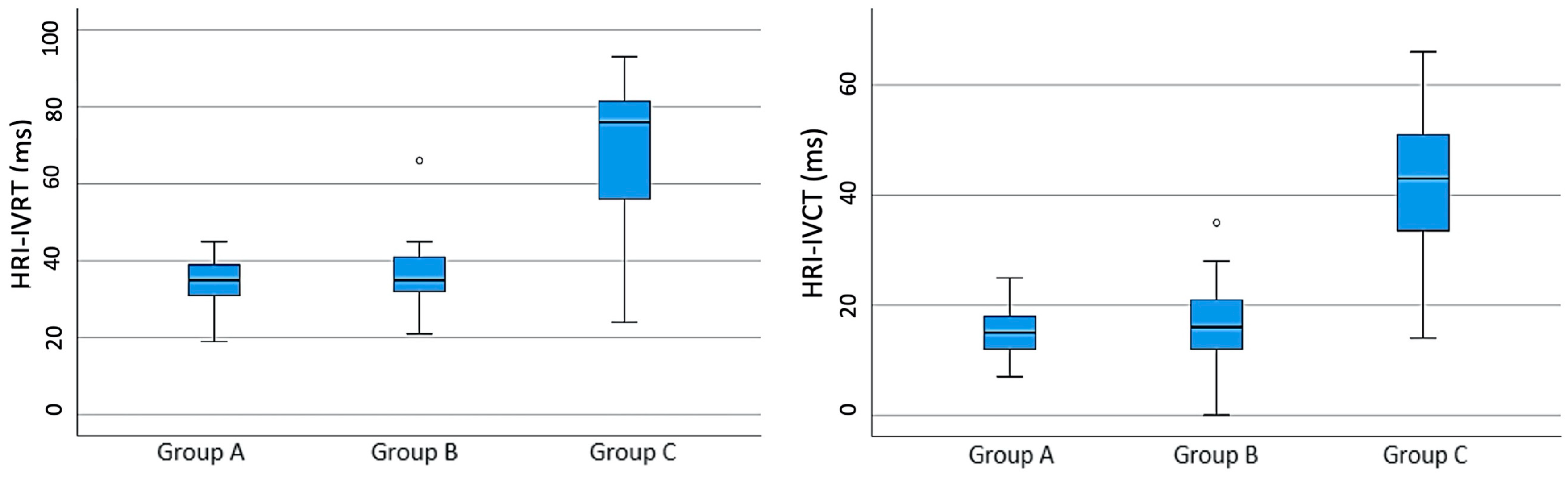
| HRI-IVCT (ms) | 25.01 ± 14.61 | 15.18 ± 4.81 | 16.59 ± 6.04 | 40.45 ± 12.21 | 0.01 (1.21) 1 |
| HRI-IVRT (ms) | 47.93 ± 20.26 | 34.27 ± 6.9 | 35.95 ± 7.13 | 69.42 ± 17.13 | 0.01 (1.73) 1 |
| Global TDI | 12.99 ± 7.83 | 15.55 ± 3.75 | 17.88 ± 7.95 | 6.05 ± 3.03 | 0.01 (1.73) 1 |
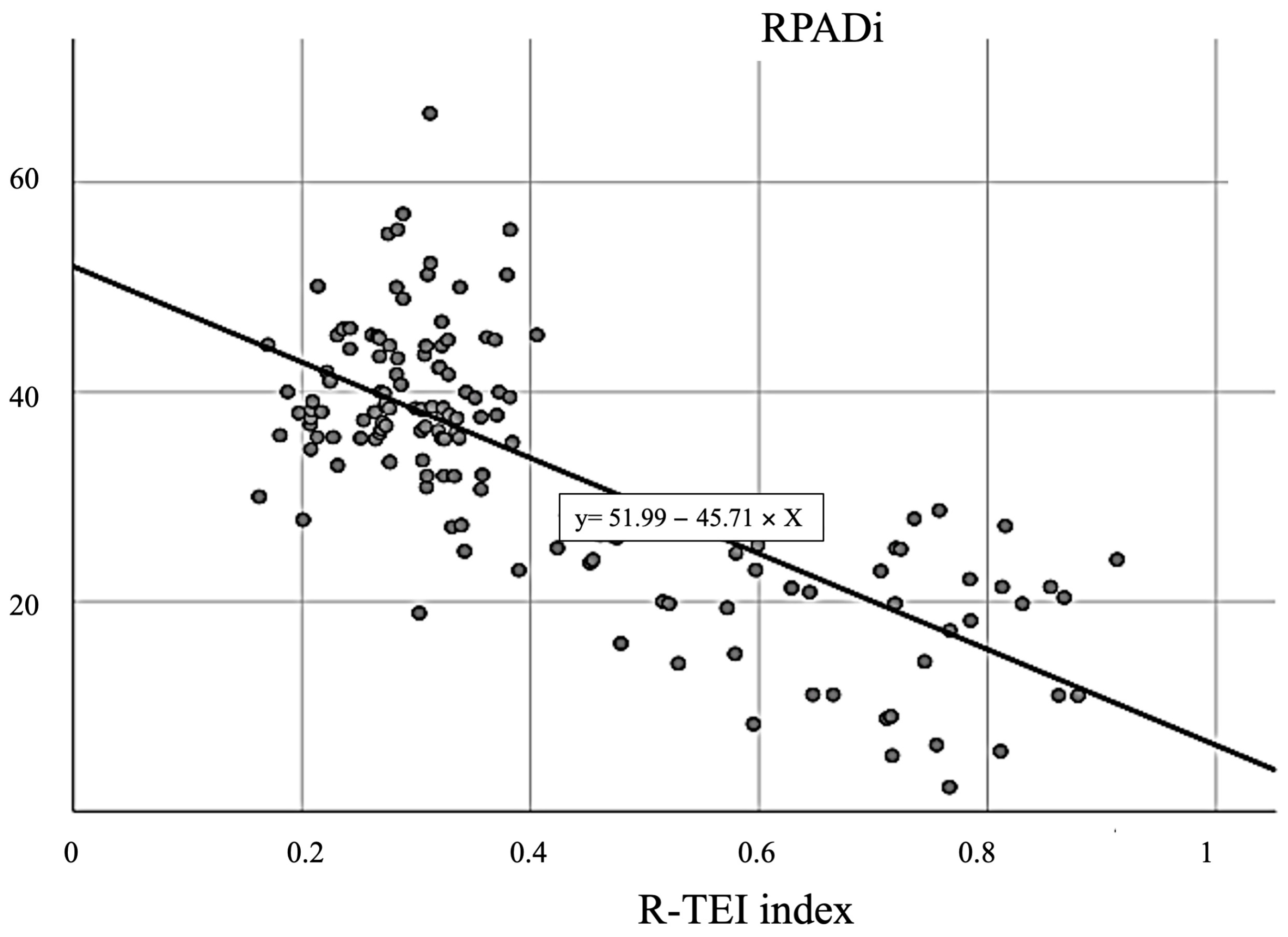
| R-TEI index | 0.41 ± 0.19 | 0.27 ± 0.04 | 0.30 ± 0.06 | 0.626 ± 0.17 | 0.01 (1.80) 1 |
| R2 | AUC | CI 95% | Cut-Off Point | Se | Es | p-Value | Youden Index (Se + Es − 1) | |
|---|---|---|---|---|---|---|---|---|
| E′ (cm/s) | 0.284 | 0.863 | (0.802, 0.924) | ≤8.50 | 0.818 | 0.766 | 0.000 | 0.584 |
| A′ (cm/s) | 0.012 | 0.572 | (0.474, 0.670) | ≥11.50 | 0.455 | 0.755 | 0.093 | 0.210 |
| S (cm/s) | 0.281 | 0.881 | (0.816, 0.947) | ≤12.50 | 0.818 | 0.862 | 0.000 | 0.680 |
| E′:A′ | 0.312 | 0.938 | (0.869, 0.980) | ≤0.839 | 0.873 | 0.926 | 0.000 | 0.798 |
| Global TDI | 0.336 | 0.984 | (0.961, 1.000) | ≤10.20 | 0.945 | 0.989 | 0.000 | 0.935 |
| HRI-IVCT (ms) | 0.513 | 0.958 | (0.926, 0.990) | ≥26.50 | 0.836 | 0.957 | 0.000 | 0.794 |
| HRI-IVRT (ms) | 0.533 | 0.954 | (0.915, 0.994) | ≥46.50 | 0.873 | 0.989 | 0.000 | 0.862 |
| R-TEI index | 0.555 | 0.965 | (0.927, 1.000) | ≥0.387 | 0.909 | 0.989 | 0.000 | 0.898 |
Disclaimer/Publisher’s Note: The statements, opinions and data contained in all publications are solely those of the individual author(s) and contributor(s) and not of MDPI and/or the editor(s). MDPI and/or the editor(s) disclaim responsibility for any injury to people or property resulting from any ideas, methods, instructions or products referred to in the content. |
© 2023 by the authors. Licensee MDPI, Basel, Switzerland. This article is an open access article distributed under the terms and conditions of the Creative Commons Attribution (CC BY) license (https://creativecommons.org/licenses/by/4.0/).
Share and Cite
Matos, J.I.; García-Rodríguez, S.N.; Costa-Rodríguez, N.; Caro-Vadillo, A.; Carretón, E.; Montoya-Alonso, J.A. Usefulness of Tissue Doppler Imaging for the Evaluation of Pulmonary Hypertension in Canine Heartworm Disease. Animals 2023, 13, 3647. https://doi.org/10.3390/ani13233647
Matos JI, García-Rodríguez SN, Costa-Rodríguez N, Caro-Vadillo A, Carretón E, Montoya-Alonso JA. Usefulness of Tissue Doppler Imaging for the Evaluation of Pulmonary Hypertension in Canine Heartworm Disease. Animals. 2023; 13(23):3647. https://doi.org/10.3390/ani13233647
Chicago/Turabian StyleMatos, Jorge Isidoro, Sara Nieves García-Rodríguez, Noelia Costa-Rodríguez, Alicia Caro-Vadillo, Elena Carretón, and José Alberto Montoya-Alonso. 2023. "Usefulness of Tissue Doppler Imaging for the Evaluation of Pulmonary Hypertension in Canine Heartworm Disease" Animals 13, no. 23: 3647. https://doi.org/10.3390/ani13233647
APA StyleMatos, J. I., García-Rodríguez, S. N., Costa-Rodríguez, N., Caro-Vadillo, A., Carretón, E., & Montoya-Alonso, J. A. (2023). Usefulness of Tissue Doppler Imaging for the Evaluation of Pulmonary Hypertension in Canine Heartworm Disease. Animals, 13(23), 3647. https://doi.org/10.3390/ani13233647