
The Thoracic Inlet Length as a Reference Point to Radiographically Assess Cardiac Enlargement in Dogs with Myxomatous Mitral Valve Disease

 ,
, 
Abstract
:Simple Summary
Abstract
1. Introduction
2. Materials and Methods
2.1. Animals
2.2. Echocardiography
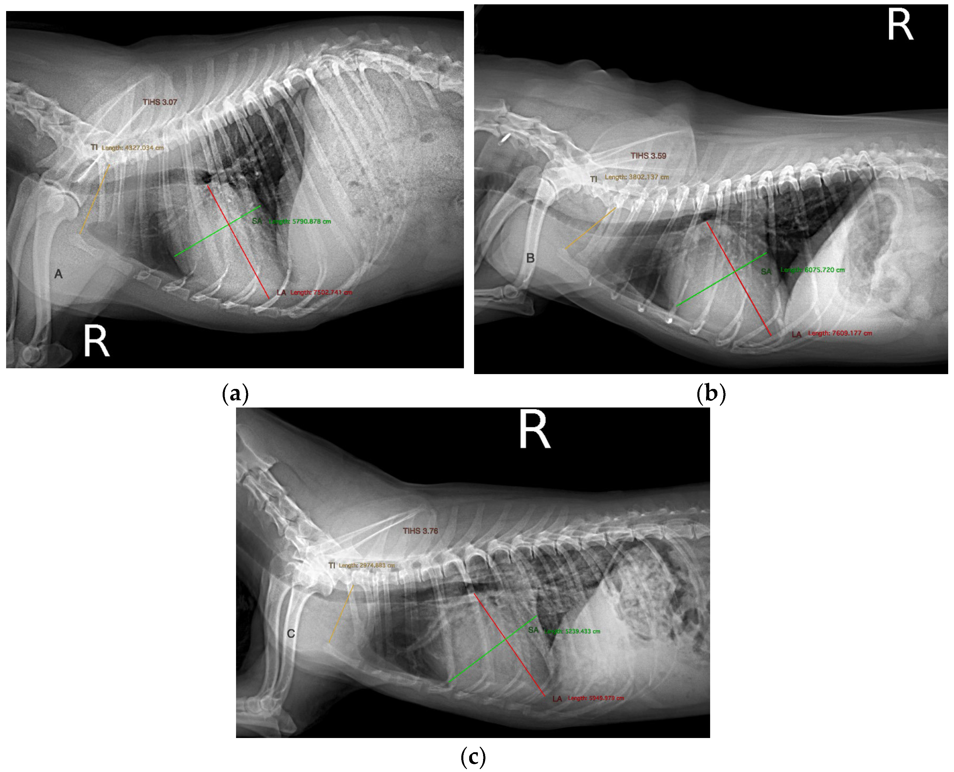
2.3. Radiography
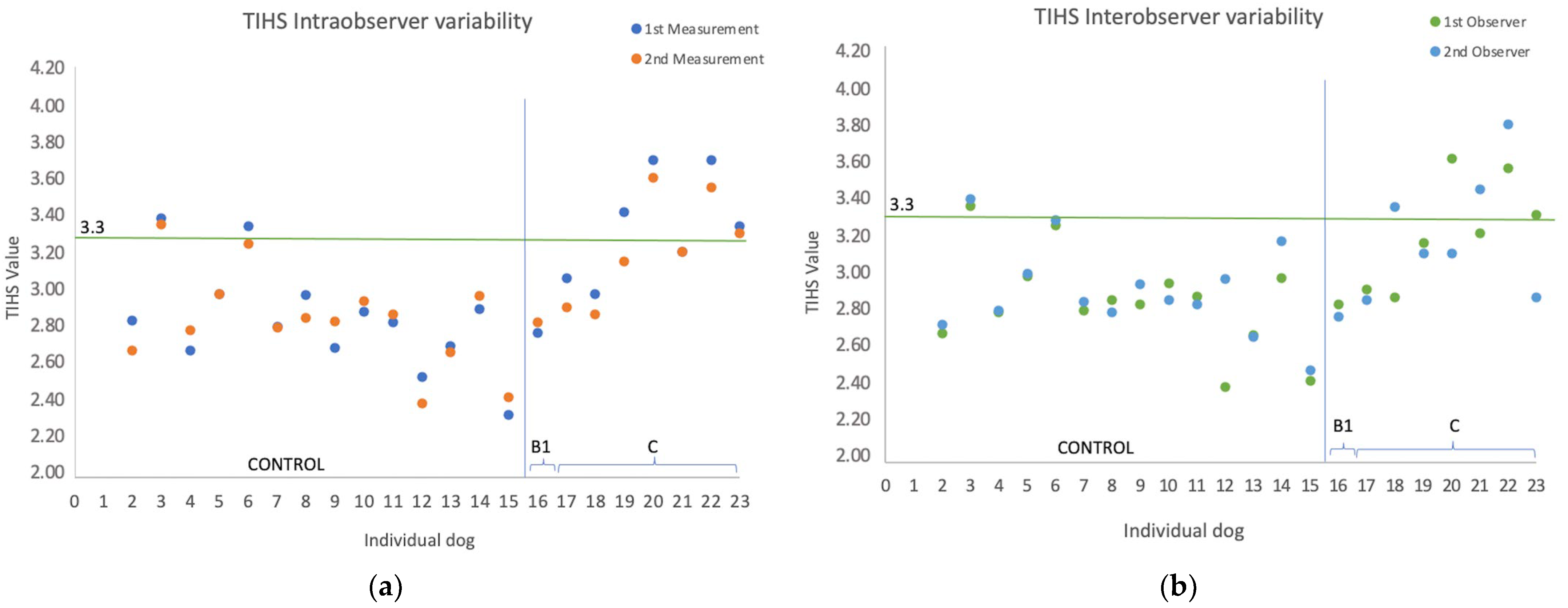
2.4. Statistical Analysis
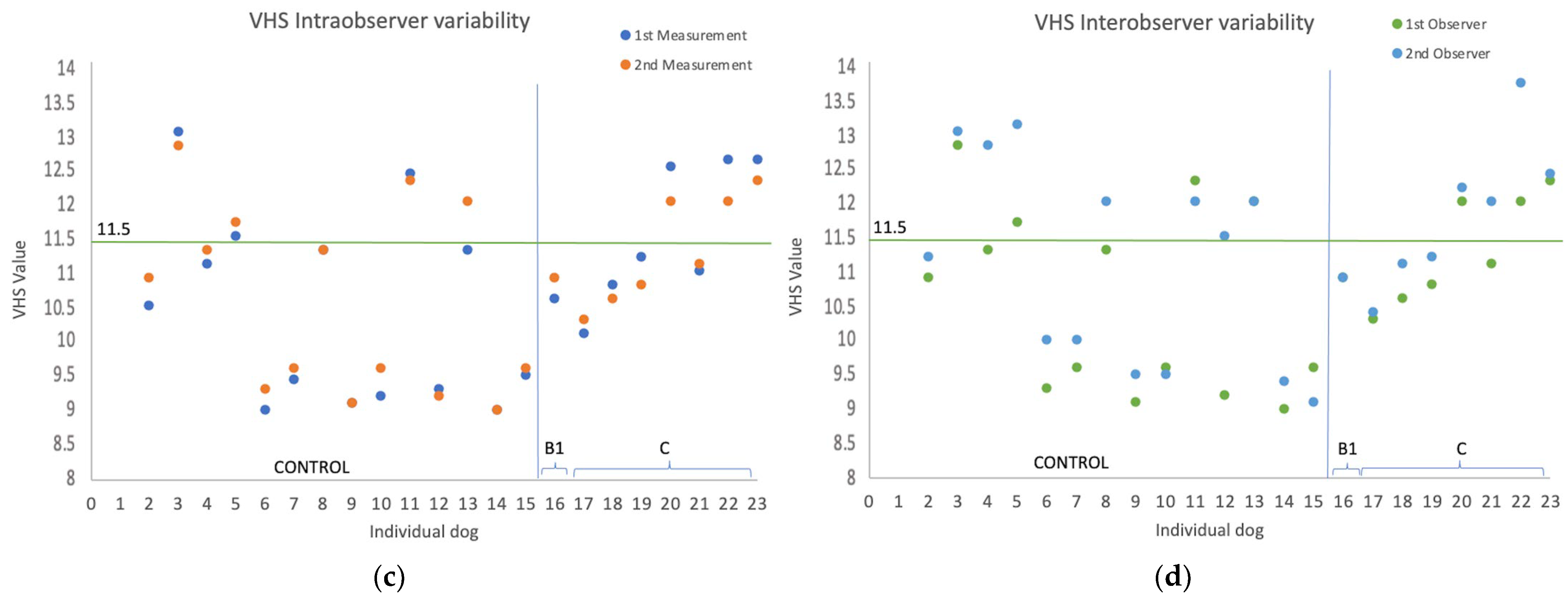
3. Results
4. Discussion
5. Conclusions
Author Contributions
Funding
Institutional Review Board Statement
Informed Consent Statement
Data Availability Statement
Acknowledgments
Conflicts of Interest
References
- Keene, B.W.; Atkins, C.E.; Bonagura, J.D.; Fox, P.R.; Häggström, J.; Fuentes, V.L.; Oyama, M.A.; Rush, J.E.; Stepien, R.; Uechi, M. ACVIM consensus guidelines for the diagnosis and treatment of myxomatous mitral valve disease in dogs. J. Vet. Intern. Med. 2019, 33, 1127–1140. [Google Scholar] [CrossRef]
- Franchini, A.; Borgarelli, M.; Abbott, J.A.; Menciotti, G.; Crosara, S.; Häggström, J.; Lahmers, S.; Rosenthal, S.; Tyrrell, W. The Longitudinal Outcome of Canine (K9) myxomatous mitral valve disease (LOOK Mitral registry): Baseline characteristics. J. Vet. Cardiol. 2023, 6, 32–47. [Google Scholar]
- Buchanan, J.W. Radiology of the heart. In Proceedings of the 35th Annual Meeting of the American Animal Hospital Association, Las Vegas, NV, USA, 21–23 October 1968; pp. 34–35. [Google Scholar]
- Buchanan, J.W.; Bücheler, J. Vertebral scale system to measure canine heart size in radiographs. J. Am. Vet. Med. Assoc. 1995, 206, 194–199. [Google Scholar]
- Torad, F.A.; Hassan, E.A. Two-dimensional cardiothoracic ratio for evaluation of cardiac size in German shepherd dogs. J. Vet. Cardiol. 2014, 16, 237–244. [Google Scholar] [CrossRef]
- Mostafa, A.A.; Berry, C.R. Radiographic assessment of the cardiac silhouette in clinically normal large- and small-breed dogs. Am. J. Vet. Res. 2017, 78, 168–177. [Google Scholar] [CrossRef] [PubMed]
- Costanza, D.; Greco, A.; Piantedosi, D.; Bruzzese, D.; Pasolini, M.P.; Coluccia, P.; Castiello, E.; Baptista, C.S.; Meomartino, L. The heart to single vertebra ratio: A new objective method for radiographic assessment of cardiac silhouette size in dogs. Vet. Radiol. Ultrasound 2023, 64, 378–384. [Google Scholar] [CrossRef] [PubMed]
- Marbella Fernández, D.; García, V.; Santana, A.J.; Montoya-Alonso, J.A. The Thoracic Inlet Heart Size, a New Approach to Radiographic Cardiac Measurement. Animals 2023, 13, 389. [Google Scholar] [CrossRef] [PubMed]
- Le Roux, A.; Rademacher, N.; Saelinger, C.; Rodriguez, D.; Pariaut, R.; Gaschen, L. Value of tracheal bifurcation angle measurement as a radiographic sign of left atrial enlargemen in dogs. Vet. Radiol. Ultrasound 2012, 53, 28–33. [Google Scholar] [CrossRef] [PubMed]
- Malcolm, E.L.; Visser, L.C.; Phillips, K.L.; Johnson, L.R. Diagnostic value of vertebral left atrial size as determined from thoracic radiographs for assessment of left atrial size in dogs with myxomatous mitral valve disease. J. Am. Vet. Med. Assoc. 2018, 253, 1038–1045. [Google Scholar] [CrossRef]
- Sánchez Salguero, X.; Prandi, D.; Llabrés-Díaz, F.; Manzanilla, E.G.; Bussadori, C. A radiographic measurement of left atrial size in dogs. Ir. Vet. J. 2018, 71, 25. [Google Scholar] [CrossRef]
- Sánchez Salguero, X.; Prandi, D.; Llabrés-Díaz, F.; Manzanilla, E.G.; Badiella, L.; Bussadori, C. Heart to spine measurements to detect left atrial enlargement in dogs with mitral valve insufficiency. Ir. Vet. J. 2019, 72, 14. [Google Scholar] [CrossRef] [PubMed]
- Lam, C.; Gavaghan, B.J.; Meyers, F.E. Radiographic quantification of left atrial size in dogs with myxomatous mitral valve disease. J. Vet. Intern. Med. 2021, 35, 747–754. [Google Scholar] [CrossRef] [PubMed]
- An, S.; Hwang, G.; Noh, S.A.; Yoon, Y.; Lee, H.C.; Hwang, T.S. A retrospective study of radiographic measurements of small breed dogs with myxomatous mitral valve degeneration: A new modified vertebral left atrial size. J. Vet. Clin. 2023, 40, 31–37. [Google Scholar] [CrossRef]
- Guglielmini, C.; Diana, A.; Pietra, M.; Di Tommaso, M.; Cipone, M. Use of the vertebral heart score in coughing dogs with chronic degenerative mitral valve disease. J. Vet. Med. Sci. 2009, 71, 9–13. [Google Scholar] [CrossRef]
- Poad, M.H.; Manzi, T.J.; Oyama, M.A.; Gelzer, A.R. Utility of radiographic measurements to predict echocardiographic left heart enlargement in dogs with preclinical myxomatous mitral valve disease. J. Vet. Intern. Med. 2020, 34, 1728–1733. [Google Scholar] [CrossRef]
- Stepien, R.L.; Rak, M.B.; Blume, L.M. Use of radiographic measurements to diagnose stage B2 preclinical myxomatous mitral valve disease in dogs. J. Am. Vet. Med. Assoc. 2020, 256, 1129–1136. [Google Scholar] [CrossRef]
- Duler, L.; Visser, L.C.; Jackson, K.N.; Phillips, K.L.; Pollard, R.E.; Wanamaker, M.W. Evaluation of radiographic predictors of left heart enlargement in dogs with known or suspected cardiovascular disease. Vet. Radiol. Ultrasound 2021, 62, 271–281. [Google Scholar] [CrossRef]
- Wesselowski, S.; Gordon, S.G.; Meddaugh, N.; Saunders, A.B.; Häggström, J.; Cusack, K.; Janacek, B.W.; Matthews, D.J. Prediction of clinically important acquired cardiac disease without an echocardiogram in large breed dogs using a combination of clinical, radiographic and electrocardiographic variables. J. Vet. Cardiol. 2022, 40, 126–141. [Google Scholar] [CrossRef]
- Levicar, C.; Grandos-Soler, J.L.; Freise, F.; Raue, J.F.; Nolte, I.; Bach, J.-P. Comparison of different radiographic scores with associated echocardiographic measurements and prediction of heart enlargement in dogs with and without myxomatous mitral valve disease. J. Vet. Cardiol. 2022, 44, 1–12. [Google Scholar] [CrossRef]
- Mostafa, A.A.; Peper, K.E.; Berry, C.R. Use of cardiac sphericity index and manubrium heart scores to assess radiographic cardiac silhouettes in large-and small-breed dogs with and without cardiac disease. J. Am. Vet. Med. Assoc. 2020, 256, 288–896. [Google Scholar] [CrossRef]
- Mikawa, S.; Nagakawa, M.; Ogi, H.; Akabane, R.; Koyama, Y.; Sakatani, A.; Ogawa, M.; Miyakawa, H.; Shigemoto, J.; Tokuriki, T.; et al. Use of vertebral left atrial size for staging of dogs with myxomatous valve disease. J. Vet. Cardiol. 2020, 30, 92–99. [Google Scholar] [CrossRef]
- Vezzosi, T.; Puccinelli, C.; Citi, S.; Tognetti, R. Two radiographic methods for assessing left atrial enlargement and cardiac remodeling in dogs with myxomatous mitral valve disease. J. Vet. Cardiol. 2021, 34, 55–63. [Google Scholar] [CrossRef] [PubMed]
- Lamb, C.R.; Wikeley, H.; Boswood, A.; Pfeiffer, D.U. Use of breed-specific ranges for the vertebral heart scale as an aid to the radiographic diagnosis of cardiac disease in dogs. Veter Rec. 2001, 148, 707–711. [Google Scholar] [CrossRef] [PubMed]
- Pinto, A.C.B.C.F.; Iwasaki, M. Avaliacão radiográfica da silhueta cardíaca pelo método de mensuracão VHS (vertebral heart size) em cães da raca Poodle clinicamente normais. Braz. J. Vet. Res. Anim. Sci. 2004, 41, 261–267. [Google Scholar] [CrossRef]
- Bavegems, V.; Van Caelenberg, A.; Duchateau, L.; Sys, S.U.; Van Bree, H.; De Rick, A. Vertebral heart size ranges specific for whippets. Vet. Radiol. Ultrasound 2005, 46, 400–403. [Google Scholar] [CrossRef]
- Gülanber, E.G.; Gönenci, R.; Kaya, Ü.; Aksoy, Ö.; Biricik, H.S. Vertebral Scale System to Measure Heart Size in Thoracic Radiographs of Turkish Shepherd (Kangal) Dogs. Turk. J. Vet. Anim. Sci. 2005, 29, 21. [Google Scholar]
- Marin, L.M.; Brown, J.; McBrien, C.; Baumwart, R.; Samii, V.F.; Couto, C.G. Vertebral heart size in retired racing Greyhounds. Vet. Radiol. Ultrasound 2007, 48, 332–334. [Google Scholar] [CrossRef] [PubMed]
- Kraetschmer, S.; Ludwig, K.; Meneses, F.; Nolte, I.; Simon, D. Vertebral heart scale in the beagle dog. J. Small Anim. Pract. 2008, 49, 240–243. [Google Scholar] [CrossRef]
- Gugjoo, M.B.; Hoque, M.; Zama, M.M.S.; Saxena, A.C.; Pawde, A.M.; Ansari, M.M.; Bhat, S.A. Vertebral scale system to measure heart size on thoracic radiographs of Labrador retriever dogs. Indian Vet. J. 2013, 90, 71–73. [Google Scholar]
- Almeida, G.L.; Almeida, M.B.; Santos, A.C.; Mattos, Â.V.; Oliveira, A.C.; Campos, V.D.; Souza, W.N.; Barros, R.S. Vertebral Heart Size in Healthy Belgian Malinois Dogs. J. Vet. Adv. 2015, 5, 1176–1180. [Google Scholar]
- Bodh, D.; Hoque, M.; Saxena, A.C.; Gugjoo, M.B.; Bist, D.; Chaudhary, J.K. Vertebral scale system to measure heart size in thoracic radiographs of Indian Spitz, Labrador retriever and Mongrel dogs. Vet. World 2016, 9, 371–376. [Google Scholar] [CrossRef] [PubMed]
- Birks, R.; Fine, D.M.; Leach, S.B.; Clay, S.E.; Eason, B.D.; Britt, L.G.; Lamb, K.E. Breed-Specific Vertebral Heart Scale for the Dachshund. J. Am. Anim. Hosp. Assoc. 2017, 53, 73–79. [Google Scholar] [CrossRef] [PubMed]
- Luciani, M.G.; Withoeft, J.A.; Mondardo Cardoso Pissetti, H.; Pasini de Souza, L.; Silvestre Sombrio, M.; Bach, E.C.; Mai, W.; Müller, T.R. Vertebral heart size in healthy Australian cattle dog. Anat. Histol. Embryol. 2019, 48, 264–267. [Google Scholar] [CrossRef]
- Taylor, C.J.; Simon, B.T.; Stanley, B.J.; Lai, G.P.; Thieman Mankin, K.M. Norwich terriers possess a greater vertebral heart scale than the canine reference value. Vet. Radiol. Ultrasound 2020, 61, 10–15. [Google Scholar] [CrossRef]
- Puccinelli, C.; Citi, S.; Vezzosi, T.; Garibaldi, S.; Tognetti, R. A radiographic study of breed-specific vertebral heart score and vertebral left atrial size in Chihuahuas. Vet. Radiol. Ultrasound 2021, 62, 20–26. [Google Scholar] [CrossRef]
- Baisan, R.A.; Vulpe, V. Vertebral heart size and vertebral left atrial size reference ranges in healthy Maltese dogs. Vet. Radiol. Ultrasound 2022, 63, 18–22. [Google Scholar] [CrossRef] [PubMed]
- Kallassy, A.; Calendrier, E.; Bouhsina, N.; Fusellier, M. Vertebral Heart Scale for the Brittany Spaniel: Breed-Specific Range and Its Correlation with Heart Disease Assessed by Clinical and Echocardiographic Findings. Vet. Sci. 2021, 8, 300. [Google Scholar] [CrossRef] [PubMed]
- Bagardi, M.; Locatelli, C.; Manfredi, M.; Bassi, J.; Spediacci, C.; Ghilardi, S.; Zani, D.D.; Brambilla, P.G. Breed-specific vertebral heart score, vertebral left atrial size, and radiographic left atrial dimension in Cavalier King Charles Spaniels: Reference interval study. Vet. Radiol. Ultrasound 2021, 8, 300. [Google Scholar] [CrossRef]
- Wiegel, P.S.; Mach, R.; Nolte, I.; Freise, F.; Levicar, C.; Merhof, K.; Bach, J.P. Breed-specific values for vertebral heart score (VHS), vertebral left atrial size (VLAS), and radiographic left atrial dimension (RLAD) in pugs without cardiac disease, and their relationship to Brachycephalic Obstructive Airway Syndrome (BOAS). PLoS ONE 2022, 17, e0274085. [Google Scholar] [CrossRef]
- Hansson, K.; Haggstrom, J.; Kvart, C.; Lord, P. Interobserver variability of vertebral heart size measurements in dogs with normal and enlarged hearts. Vet. Radiol. Ultrasound 2005, 46, 122–130. [Google Scholar] [CrossRef]
- Harvey, C.E.; Fink, E.A. Tracheal diameter: Analysis of radiographic measurements in brachycephalic and nonbrachycephalic dogs. J. Am. Anim. Hosp. Assoc. 1982, 18, 570–576. [Google Scholar]
- Ingman, J.; Näslund, V.; Hansson, K. Comparison between tracheal ratio methods used by three observers at three occasions in English Bulldogs. Acta. Vet. Scand. 2014, 56, 79. [Google Scholar] [CrossRef] [PubMed]
- Mostafa, A.A.; Berry, C.R. Radiographic vertical tracheal diameter assessment at different levels along the trachea as an alternative method for the evaluation of the tracheal diameter in non-brachycephalic small breed dogs. BMC Vet. Res. 2022, 18, 61. [Google Scholar] [CrossRef] [PubMed]
- Cornell, C.C.; Kittleson, M.D.; Della Torre, P.; Häggström, J.; Lombard, C.W.; Pedersen, H.D.; Vollmar, A.; Wey, A. Allometric scaling of M-mode cardiac measurements in normal adult dogs. J. Vet. Intern. Med. 2004, 18, 311–321. [Google Scholar] [CrossRef] [PubMed]
- Hansson, K.; Haggstrom, J.; Kvart, C.; Lord, P. Left atrial to aortic root indices using two-dimensional and M-mode echocardiography in Cavalier King Charles Spaniels with and without left atrial enlargement. Vet. Radiol. Ultrasound 2002, 43, 568–575. [Google Scholar] [CrossRef]
- Jepsen-Grant, K.; Pollard, R.E.; Johnson, L.R. Vertebral heart scores in eight dog breeds. Vet. Radiol. Ultrasound 2013, 54, 3–8. [Google Scholar] [CrossRef]
- Lord, P.F.; Hansson, K.; Carnabuci, C.; Kvart, C.; Häggström, J. Radiographic heart size and its rate of increase as tests for onset of congestive heart failure in Cavalier King Charles Spaniels with mitral valve regurgitation. J. Vet. Intern. Med. 2011, 25, 1312–1319. [Google Scholar] [CrossRef]
- Grosso, G.; Vezzosi, T.; Domenech, O.; Tognetti, R. Prognostic relevance of left cardiac enlargement in dogs with preclinical myxomatous mitral valve disease. J. Vet. Cardiol. 2023, 45, 50–58. [Google Scholar] [CrossRef]
- Timperman, L.; Habing, G.; Green, E. The vertebral heart scale on CT is correlated to radiographs in dogs. Vet. Radiol. Ultrasound 2021, 62, 519–524. [Google Scholar] [CrossRef]
- Levicar, C.; Nolte, L.; Granados-Soler, J.L.; Freise, F.; Raue, J.F.; Bach, J.-P. Methods of heart and left atrial size in dogs with and without myxomatous mitral valve disease: Intra- and Interobserver agreement and practicability of different methods. Animals 2022, 12, 2531. [Google Scholar] [CrossRef]
- Bagardi, M.; Manfredi, M.; Zani, D.D.; Brambilla, P.G.; Locatelli, C. Interobserver variability of radiographic methods for the evaluation of left atrial size in dogs. Vet. Radiol. Ultrasound 2021, 62, 161–174. [Google Scholar] [CrossRef]
- Woolley, R.; Smith, P.; Munro, E.; Smith, S.; Swift, S.; Devine, C.; Corcoran, B.; French, A. Effects of treatment type of vertebral heart size in dogs with myxomatous mitral valve disease. Intern. J. Appl. Res. Vet. Med. 2007, 5, 43–48. [Google Scholar]
- Rishniw, M.; Caivano, D.; Dickson, D.; Vatne, L.; Harris, J.; Matos, J.N. Two-dimensional echocardiographic left- atrial-to-aortic 498 ratio in healthy adult dogs: A reexamination of reference intervals. J. Vet. Cardiol. 2019, 26, 29–38. [Google Scholar] [CrossRef] [PubMed]
- Rishniw, M.; Brown, D. The ACVIM consensus statement definition of left ventricular enlargement in myxomatous mitral valve disease does not always represent left ventricular enlargement. J. Vet. Cardiol. 2022, 42, 92–102. [Google Scholar] [CrossRef] [PubMed]

| n | Control 50 | Stage B1 36 | Stage B2 30 | Stage C 40 |
|---|---|---|---|---|
| Age (years) | 4.8 (1–15.3) a | 11.6 (5.5–15.9) | 11.3 (7.4–18.9) | 11.4 (7.9–17.6) |
| Weight (kg) | 7.40 b (2.10–38.70) | 5.38 (2.60–26.0) | 5.18 (2.0–19.60) | 4.73 (2.80–14.80) |
| Male/Female | 28/22 | 18/18 | 17/13 | 23/17 |
| TI | 29.15 ± 15.11 c | 25.91 ± 9.50 | 26.25 ± 9.55 | 24.13 ± 7.39 |
| SAx | 39.24 ± 20.60 | 36.51 ± 12.53 | 39.84 ± 13.73 | 40.44 ± 11.03 |
| LAx | 45.38 ± 26.81 | 42.09 ± 16.96 | 46.23 ± 17.64 | 45.64 ± 12.13 |
| SAx + LAx | 87.68 ± 50.97 | 78.75 ± 29.30 | 86.24 ± 31.16 | 86.25 ± 22.84 |
| LA/Ao | 1.20 ± 0.18 | 1.36 ± 0.17 | 1.80 ± 0.22 d | 2.08 ± 0.36 d |
| LVIDDN | 1.57 ± 0.20 | 1.57 ± 0.21 | 1.94 ± 0.24 d | 2.08 ± 0.29 d |
| n | Control 50 | Stage B1 36 | Stage B2 30 | Stage C 40 | p |
|---|---|---|---|---|---|
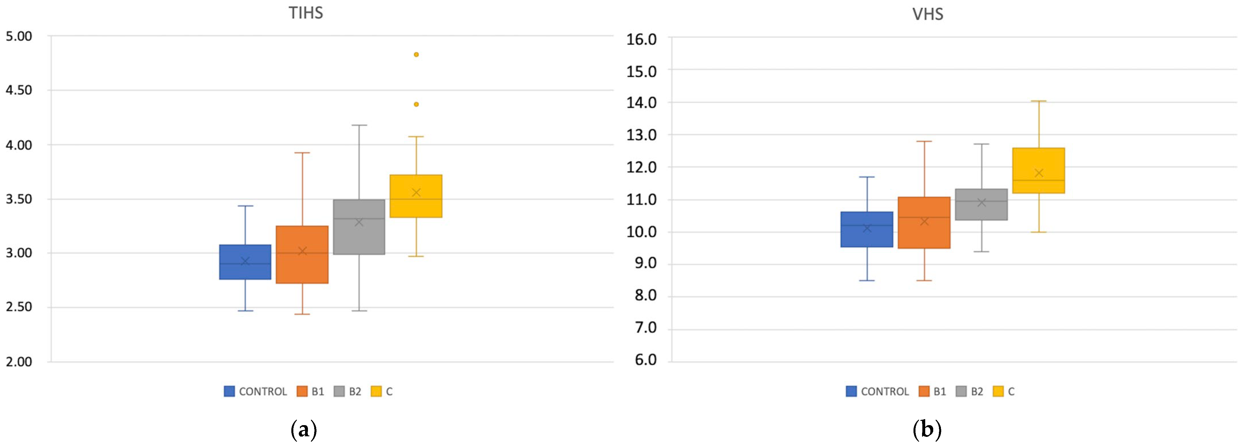
| TIHS (CI) | 2.91 ± 0.23 (2.84–2.98) | 2.98 ± 0.36 a (2.87–3.11) | 3.25 ± 0.34 a (3.09–3.39) | 3.53 ± 0.36 a (3.42–3.64) | <0.01 |
| VHS (CI) | 10.07 ± 0.73 (9.86–10.28) | 10.24 ± 0.95 b (9.93–10.55) | 10.83 ± 0.86 b (10.49–11.25) | 11.74 ± 0.96 b (11.43–12.05) | 0.016 |
| THIS (n) | Male | Female | p | <10 kg | ≥10 kg | p |
|---|---|---|---|---|---|---|
| CONTROL | 2.88 ± 0.23 (28) | 2.94 ± 0.23 (22) | 0.33 | 2.88 ± 0.24 (27) | 2.94 ± 0.22 (23) | 0.41 |
| Stage B1 | 3.01 ± 0.35 (18) | 2.95 ± 0.38 (18) | 0.62 | 3.04 ± 0.35 (26) | 2.84 ± 0.0.35 (10) | 0.13 |
| Stage B2 | 3.25 ± 0.38 (17) | 3.26 ± 0.29 (13) | 0.91 | 3.26 ± 0.37 (25) | 3.22 ± 0.18 (5) | 0.51 |
| Stage C | 3.53 ± 0.37 (23) | 3.53 ± 0.35 (17) | 0.99 | 3.53 ± 0.37 (36) | 3.50 ± 0.21 (4) | 0.78 |
| Chihuahua n | Control 12 | Stage B1 5 | Stage B2 6 | Stage C 9 | p |
|---|---|---|---|---|---|
| LA/Ao | 1.31 ± 0.01 a | 1.33 ± 0.15 a | 1.86 ± 0.18 | 2.07 ± 0.52 | ≤0.001 |
| LVIDDN | 1.37 ± 0.06 b | 1.50 ± 0.24 b | 1.87 ± 0.31 | 2.12 ± 0.28 | ≤0.001 |
| TIHS (CI) | 2.96 ± 0.33 c (2.77–3.15) | 2.92 ± 0.11 c (2.87–3.11) | 3.40 ± 0.28 (3.10–3.70) | 3.61 ± 0.44 (3.27–3.95) | ≤0.01 |
| VHS (CI) | 9.82 ± 0.60 d (9.44–10.2) | 10.33 ± 0.65 d (9.76–10.90) | 11.26 ± 0.71 (10.52–12.0) | 12.08 ± 1.06 (11.27–12.89) | ≤0.001 |
| Radiographic Method | AUC (95% CI) | Se | Sp | Youden Index | PPV | NPV | Cutoff |
|---|---|---|---|---|---|---|---|
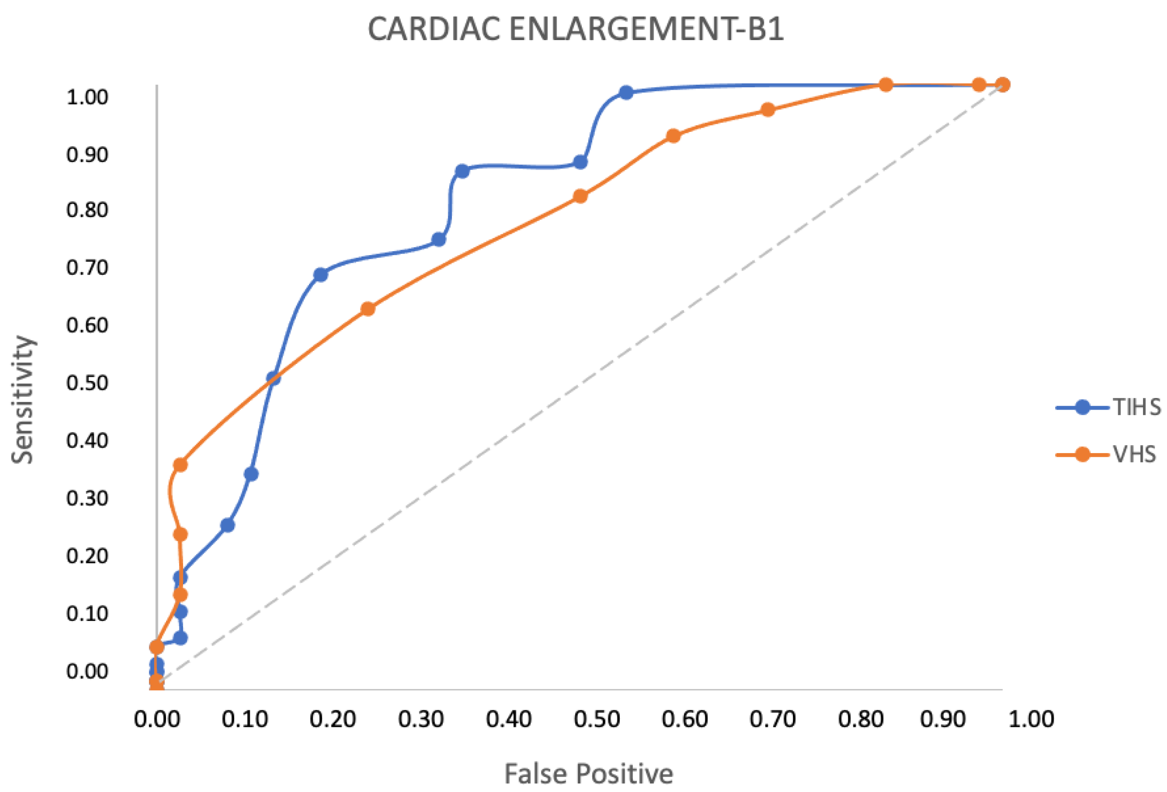
| TIHS | 0.75 | 0.53 0.53 0.43 | 0.75 0.81 0.81 | 0.28 0.33 0.24 | 0.64 0.70 0.65 | 0.66 0.67 0.63 | ≥3.25 ≥3.30 ≥3.35 |
| VHS (v) | 0.74 | 0.50 0.20 0.13 | 0.66 0.94 0.97 | 0.16 0.14 0.10 | 0.65 0.75 0.80 | 0.63 0.59 0.57 | ≥11.0 ≥11.5 ≥12.0 |
| Radiographic Method | AUC (95% CI) | Se | Sp | Youden Index | PPV | NPV | Cutoff |
|---|---|---|---|---|---|---|---|
| TIHS | 0.82 | 0.70 0.69 0.60 | 0.75 0.81 0.81 | 0.45 0.50 0.41 | 0.84 0.87 0.86 | 0.56 0.57 0.51 | ≥3.25 ≥3.30 ≥3.35 |
| VHS (v) | 0.83 | 0.73 0.37 0.21 | 0.66 0.94 0.97 | 0.40 0.31 0.26 | 0.81 0.93 0.95 | 0.56 0.44 0.41 | ≥11.0 ≥11.5 ≥12.0 |
Disclaimer/Publisher’s Note: The statements, opinions and data contained in all publications are solely those of the individual author(s) and contributor(s) and not of MDPI and/or the editor(s). MDPI and/or the editor(s) disclaim responsibility for any injury to people or property resulting from any ideas, methods, instructions or products referred to in the content. |
© 2023 by the authors. Licensee MDPI, Basel, Switzerland. This article is an open access article distributed under the terms and conditions of the Creative Commons Attribution (CC BY) license (https://creativecommons.org/licenses/by/4.0/).
Share and Cite
Marbella Fernández, D.; García, V.; Santana, A.J.; Montoya-Alonso, J.A. The Thoracic Inlet Length as a Reference Point to Radiographically Assess Cardiac Enlargement in Dogs with Myxomatous Mitral Valve Disease. Animals 2023, 13, 2666. https://doi.org/10.3390/ani13162666
Marbella Fernández D, García V, Santana AJ, Montoya-Alonso JA. The Thoracic Inlet Length as a Reference Point to Radiographically Assess Cardiac Enlargement in Dogs with Myxomatous Mitral Valve Disease. Animals. 2023; 13(16):2666. https://doi.org/10.3390/ani13162666
Chicago/Turabian StyleMarbella Fernández, David, Verónica García, Alexis José Santana, and José Alberto Montoya-Alonso. 2023. "The Thoracic Inlet Length as a Reference Point to Radiographically Assess Cardiac Enlargement in Dogs with Myxomatous Mitral Valve Disease" Animals 13, no. 16: 2666. https://doi.org/10.3390/ani13162666
APA StyleMarbella Fernández, D., García, V., Santana, A. J., & Montoya-Alonso, J. A. (2023). The Thoracic Inlet Length as a Reference Point to Radiographically Assess Cardiac Enlargement in Dogs with Myxomatous Mitral Valve Disease. Animals, 13(16), 2666. https://doi.org/10.3390/ani13162666