Article

First Study on Gyrodactylus (Monogenea: Gyrodactylidae) in Morocco, with Description of a New Species from Luciobarbus pallaryi and Luciobarbus ksibi (Actinopterygii: Cyprinidae)

by
Miriam Isoyi Shigoley
1,2,3,*,
Imane Rahmouni
1,
Halima Louizi
1,
Antoine Pariselle
1,4 and
Maarten P. M. Vanhove
2
1
Laboratory of Biodiversity, Ecology and Genome, Research Center Plant and Microbial Biotechnology, Biodiversity and Environment, Mohammed V University in Rabat, Rabat 10000, Morocco
2
Research Group Zoology, Biodiversity & Toxicology, Centre for Environmental Sciences, Hasselt University, Agoralaan Gebouw D, 3590 Diepenbeek, Belgium
3
Department of Veterinary Management of Animal Resources, Faculty of Veterinary Medicine, Liège University, 4000 Liège, Belgium
4
ISEM, Université de Montpellier, CNRS, IRD, 34095 Montpellier, France
*
Author to whom correspondence should be addressed.
Animals 2023, 13(10), 1624; https://doi.org/10.3390/ani13101624
Submission received: 14 February 2023 / Revised: 6 April 2023 / Accepted: 2 May 2023 / Published: 12 May 2023
(This article belongs to the Section Aquatic Animals)

Abstract

:

Simple Summary

Monogenean flatworms are mainly parasitic in lower aquatic vertebrates including fish, anurans and chelonians. Gyrodactylus is one of the 23 genera of Gyrodactylidae. With only 41 species described, the total number of Gyrodactylus species described from African freshwater fish still remains low. The known species represent only a fraction of the expected species richness of Gyrodactylus in Africa. In this study, we examined the gills of 738 cyprinid specimens. We isolated 26 individuals belonging to Gyrodactylus from these hosts. Twelve of these from two host species were morphologically characterized and proposed to belong to one single newly described species. In view of the importance of the cyprinid–monogenean system in studying the aquatic biodiversity and biogeography of North Africa, the present study is a substantial contribution to the parasite species inventory of these fishes.

Abstract

To date, 41 species of Gyrodactylus have been described from Africa. However, none of these have been reported in Morocco. After identifying and examining 738 cyprinid host specimens, 26 specimens belonging to Gyrodactylus were found to parasitize the gills of nine species of Luciobarbus, Carasobarbus, and Pterocapoeta. The current study provides new information about the presence of a new parasitic species in Morocco, the first to be characterized on a species level in the Maghreb region. It describes in detail 12 specimens of Gyrodactylus isolated from the gills of Luciobarbus pallaryi (Pellegrin, 1919) and Luciobarbus ksibi (Boulenger, 1905). Based on morphoanatomical observations, the characterization of the specimens collected indicates a species of Gyrodactylus that is new to science, described here as Gyrodactylus nyingiae n. sp. The new species is different from previously described gyrodactylids infecting African cyprinid hosts because it has a longer hamulus total length, a longer hamulus root, a downward projecting toe of the marginal hook, and a trapezium-shaped ventral bar membrane with a slightly striated median portion and small rounded anterolateral processes. This study increases the total number of Gyrodactylus spp. found in African cyprinids to four.

1. Introduction

Fisheries and aquaculture are important sectors that make a significant contribution by creating job opportunities for approximately 59.51 million people. These sectors consist of capture fisheries and aquaculture, which, respectively, employ 39.0 million and 20.5 million individuals worldwide according to FAO [1]. Morocco is considered one of the top producers of fishery resources, occupying the 13th place after Chile [2]. In 2018, the national fisheries production totaled a volume of 1,371,683 tons for a turnover of 11,579,544 thousand dirhams [2]. In freshwater, culture-based fisheries which are projected to generate 13,000 tons of fish annually in Morocco, is the main source of fish protein. This production is based on the routine stocking of cultured organisms, mainly cyprinids, in lakes and reservoirs [3]. Biogeographers in the Maghreb region have often focused on ichthyofaunal studies because of its geographical position between the African and Eurasian plates. Primary freshwater fishes are a suitable subject for historical biogeography due to their limited dispersal that is strictly restricted to fluvial basins, showing less capacity for trans-watershed dispersal [4]. However, the freshwater fish fauna of North Africa shows low diversity, which could probably reflect a long period of isolation during the Cenozoic Era [4].
The high level of endemism of cyprinid fishes in Morocco (20 endemic species) with representatives from the genera Carasobarbus Karaman, 1971, Luciobarbus Heckel, 1843, Labeobarbus Rüppell, 1835, and Pterocapoeta Günther, 1902, noted by Rahmouni et al. [5] is linked to the geological and climatic history of the Mediterranean biome, which have led to the endemic status of many species (animal or plant) present in these zones [6,7]. The number of studies on freshwater fish parasites has increased globally due to the growing interest in developing fisheries and aquaculture as affordable sources of protein to sustain the rapidly growing human population, especially in some African communities [8]. For the management of this resource, thorough knowledge of the taxonomy, distribution, biology and ecology of parasites is of paramount importance [8,9].
The cyprinid host/parasite system is a good model for studying evolutionary phenomena and determining speciation mechanisms. Gyrodactylids have the broadest host range of any monogenean family (found on 19 bony fish orders), encompassing both narrowly specific and generalist species [10]. Due to their distinct mode of reproduction, they provide valuable insights into parasite speciation processes [10,11]. Despite there being various studies on Gyrodactylus von Nordmann, 1832, in Africa, and also on cyprinids, these often do not include species-level identifications (e.g., Allalgua et al.) [12] and only a few of its representatives have been identified on the species level in the whole continent. Currently, there are over 400 valid species of Gyrodactylus described [13]. In African freshwater fishes, only 41 species of Gyrodactylus have been described [14]. The known species represent only a fraction of the expected Gyrodactylus spp. in Africa [15]. Only three Gyrodactylus species have been described from small African cyprinids, always with a host belonging to Enteromius Cope, 1867; namely, G. ivindoensis Price and Gery, 1968, from Enteromius cf. holotaenia (Boulenger, 1904) in Gabon, G. kyogae Paperna, 1973, from Enteromius neumayeri (Fischer, 1884) and Enteromius perince (Rüppell, 1835) in Uganda and G. paludinosus Truter, Smit, Malherbe and Přikrylová, 2021 [14], from Enteromius paludinosus (Peters, 1852) in South Africa. In Morocco, no research has documented species of Gyrodactylus to date. Monogeneans belonging to Gyrodactylus are major pathogens in fishes as well as a major challenge in both fisheries and aquaculture. They are commonly found on the skin and fins of freshwater fishes but may be occasionally found on the gills [16,17,18]. Gyrodactylids have a level of economic significance that outweighs that of any other monogenean family. In Norway, for example, the introduction of Gyrodactylus salaris Malmberg, 1957, into the salmon industry resulted in uncontrollable epidemics and mortalities, leading to massive economic losses [19].
Despite the numerous economic benefits a country may achieve from the introduction of living organisms, they can also be detrimental to native species [20]. In Lake Naivasha, Kenya, for example, the common carp Cyprinus carpio Linnaeus, 1758, was thought to have reached the lake in 1999 during the heavy rains from juveniles that escaped in the Malewa River [21]. Parasitological studies on parasites of C. carpio in Lake Naivasha discovered that it is dominated by representatives of Dactylogyrus [22] with a high prevalence of 99.3% [22]. Moroccan irrigation channels and reservoirs have also been stocked with non-native freshwater fish species such as the silver carp Hypophthalmichthys molitrix (Valenciennes, 1844), common carp Cyprinus carpio, goldfish Carassius auratus (Linnaeus, 1758) and grass carp Ctenopharyngodon idella (Valenciennes, 1844). These fishes play a central role in aquatic ecosystems, especially with respect to their role as consumers in food chains. Their importance is increasingly recognized, making them a central focus in conservation, pollution prevention and restoration in aquatic ecosystems [23]. Despite the importance fishes offer, the introduced fishes could pose a threat to native fishes by providing a perfect opportunity for parasite transmission [24]. For this reason, it is important to have a baseline for the Moroccan native monogenean fauna of cyprinids. Therefore, and in view of the importance of the cyprinid–monogenean system in investigating the aquatic biodiversity and biogeography of North Africa, the present study aims to identify Gyrodactylus isolated from the gills of Luciobarbus pallaryi (Pellegrin, 1919) and Luciobarbus ksibi (Boulenger, 1905) in Morocco and contribute to the parasite species inventory of these fishes.

2. Materials and Methods

2.1. Sample Collection

During September 2014 and June 2021, a total of 28 localities covering nine different watersheds in Morocco were sampled on five different occasions for cyprinid specimens, as shown in Figure 1. The fish specimens were collected after obtaining the required permit from the Ministry of Water, Forestry and Desertification Control (sampling permit no. 62 HCEFLCD/DLCDPN/CPC/PPC). These fish samples were collected using a backpack electrofisher (Samus-725G) or gill nets when the physicochemical water parameters could not allow sampling using the electrofisher. Fish hosts were identified morphologically following [25], euthanized by severing their spinal cords and dissected immediately. The gills were fixed in accordance with [26] and some fish specimens were frozen in a portable freezer and analyzed in the laboratory. The nomenclature and the classifications of fishes are those provided in [27]. The map showing sampling localities (Figure 1) was created using QGIS v3.22.8 (QGIS Development Team 2022, QGIS Information System, Open Source Geospatial Foundation Project. http://qgis.osgeo.org, accessed on 20 January 2023).

2.2. Parasitological Examination

The fish samples were transported to the laboratory for parasitological examination. Monogeneans were isolated under a dissecting microscope (Wild Heerbrugg) from the gills (gill arches from the right side of the excised fish). With the aid of a fine needle, the parasites were picked out one by one, subsequently mounted on a glass slide and then covered with a coverslip. The slides were mounted in accordance with [28]. For worms fixed in ethanol, Hoyer’s chloral hydrate was used [29] while ammonium picrate glycerine was used for frozen parasites [30]. The glass slide was left to dry for 24 h in a horizontal position before sealing the coverslip with Glyceel [31]. The type material was deposited in the collections of the research group Zoology: Biodiversity and Toxicology at Hasselt University (HU) (Diepenbeek, Belgium) (HU 838-841) and the Institut Scientifique of the Mohammed V University in Rabat (Rabat, Morocco) (ZA PPM 0101).

2.3. Identification of Representatives of Gyrodactylus

Gyrodactylus was distinguished from the other monogeneans as its members have a cylindrical body bearing two small cephalic lobes on the exterior part of the body, lack eyes and possess an opisthaptor armed with a single pair of hamuli linked by dorsal and ventral bars with 16 articulated marginal hooks (14 hooks in members of Dactylogyrus, the other monogenean genus most common on Moroccan cyprinids) [32].

2.4. Infection Parameters

Infection parameters, i.e., prevalence (P), mean intensity (M.I) and mean abundance (M.A) for members of Gyrodactylus, were calculated according to Bush et al. [33].

2.5. Morphological Characterization of Members of Gyrodactylus

Light microscopy using both phase and differential interference contrast approaches was used to study the shape and dimensions of sclerotized structures, which were viewed under a ×100 oil immersion objective on a Leica DM2500 optical microscope using Las X software v3.6.0.20104 fitted with a Leica DMC4500 camera. The whole mount, attachment organ, and male copulatory organ (MCO) (when present) on each specimen were photographed. The haptoral morphometrics (26 point-to-point measurements) followed the measurements proposed by [34]; these were taken using ImageJ v1.53k software (available at http://imagej.nih.gov/ij accessed on 15 September 2021) and only for specimens preserved using Hoyer’s solution. These measurements were given in micrometers (µm) as the mean, followed by the range in parentheses and the number of structures (n) measured for each metric. The micrographs taken were used to draw taxonomically important structures using Inkscape v1.2.

2.6. Statistical Analyses

For statistical analysis, a principal component analysis (PCA) was carried out in R Studio v4.1.0. The analyses included 19 measurements of the haptoral hard parts of hamuli and marginal hooks only. The MCO, ventral bar and dorsal bar measurements were excluded from the analysis due to the large number of missing data.

3. Results

3.1. Specimens Examined and Individuals of Gyrodactylus Isolated

A total of 738 fish specimens belonging to three genera (Luciobarbus Heckel, 1843, Carasobarbus Karaman, 1871, and Pterocapoeta Günther, 1902) were collected. Thirteen cyprinid fish species were identified and their gills were examined for infection with species of Gyrodactylus (Table 1). A total of nine out of the 13 species were found to be infected with representatives of Gyrodactylus (n = 26).

3.2. Infection Parameters

The infection parameters of examined hosts are shown in Table 2.

3.3. Characterization of a New Species of Gyrodactylus

All the isolated flatworms belonging to Gyrodactylus showed the diagnostic features of this genus: gyrodactylid monogeneans with an opisthaptor with one pair of haptoral anchors surrounded by 16 marginal hooks. The measurements are given in Table 3.
Class: Monogenea Van Beneden, 1858.
Subclass: Polyonchoinea Bychowsky, 1937.
Order: Gyrodactylidea Bychowsky, 1937.
Family: Gyrodactylidae Van Beneden and Hesse, 1863.
Subfamily: Gyrodactylinae Van Beneden and Hesse, 1863.
Genus: Gyrodactylus von Nordmann, 1832.
Species: Gyrodactylus nyingiae n. sp.
Type material: holotype (HU_838_IV.1.18) and five paratypes (HU_839_IV.1.19, HU_840_IV.1.20, HU_841_IV.1.21, ZA PPM 0101).
Type host: Luciobarbus pallaryi (Pellegrin, 1919) (teleostei: Cyprinidae).
Other host: Luciobarbus ksibi (Boulenger, 1905) (teleostei: Cyprinidae).
Type locality: Oued Guir (31°52′12″ N, 003°0′00″ W) (on type host).
Other locality: Oued Ksob (31°27′50.7″ N, 009°45′25.3″ W) (on L. ksibi).
Site of infection: Gill filament.
ZooBank registration: The Life Science Identifier (LSID) of the article is urn:lsid:zoobank.org:pub:15E78B1A-5DF7-4E37-935B-155A658FED77. The LSID for Gyrodactylus nyingiae Shigoley, Rahmouni, Louizi, Pariselle and Vanhove n. sp. is urn:lsid:zoobank.org:act:5E58B4AA-15F6-4540-BD49-559D023A56AA.
Studied material: 12 mounted individuals were measured; 11 of these were isolated from L. pallaryi and one was isolated from L. ksibi.
Etymology: The species epithet honors Dr. Dorothy Wanja Nyingi, an ichthyologist at the National Museums of Kenya and author of the first Guide to Common Freshwater Fishes of Kenya.
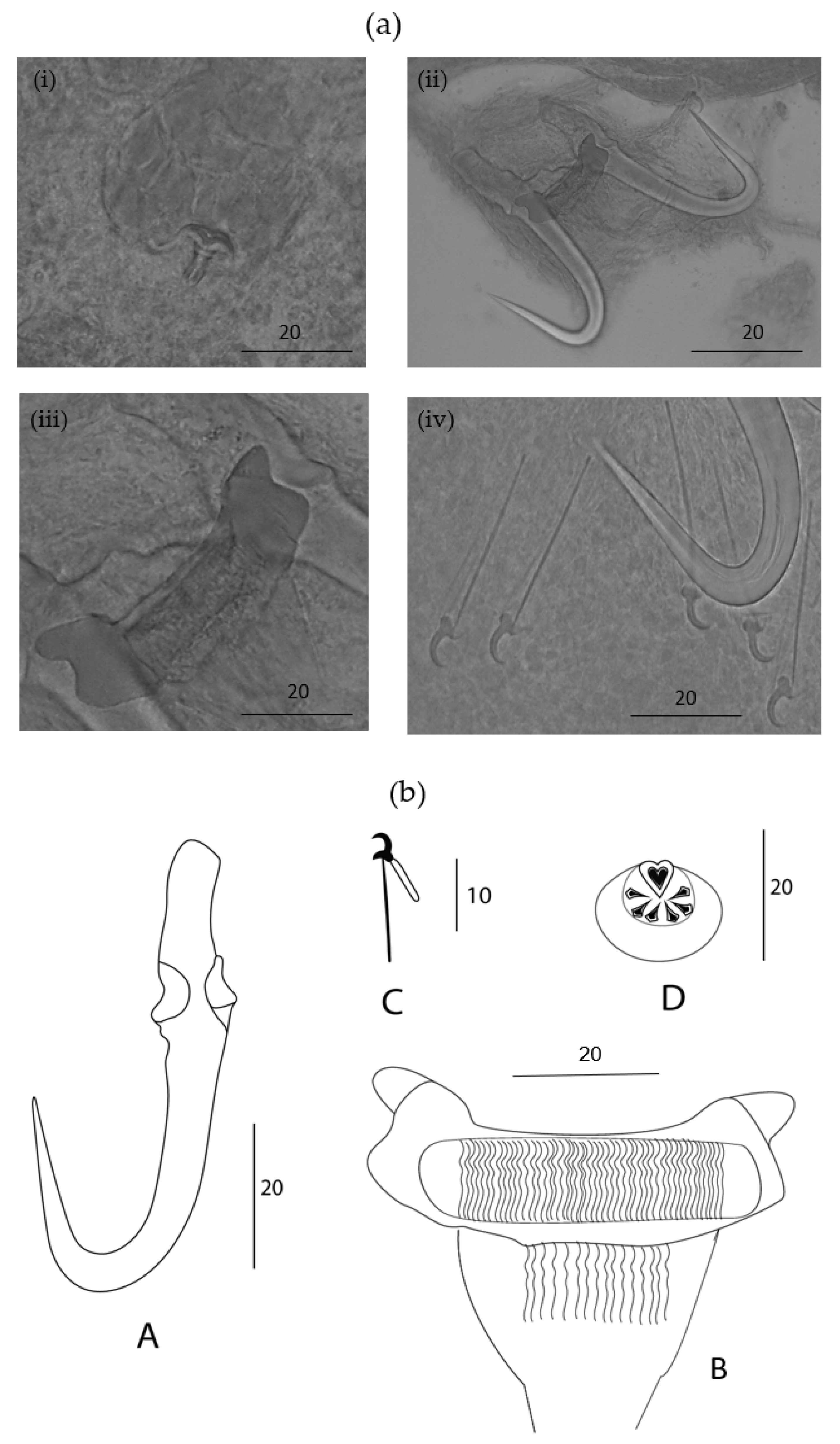
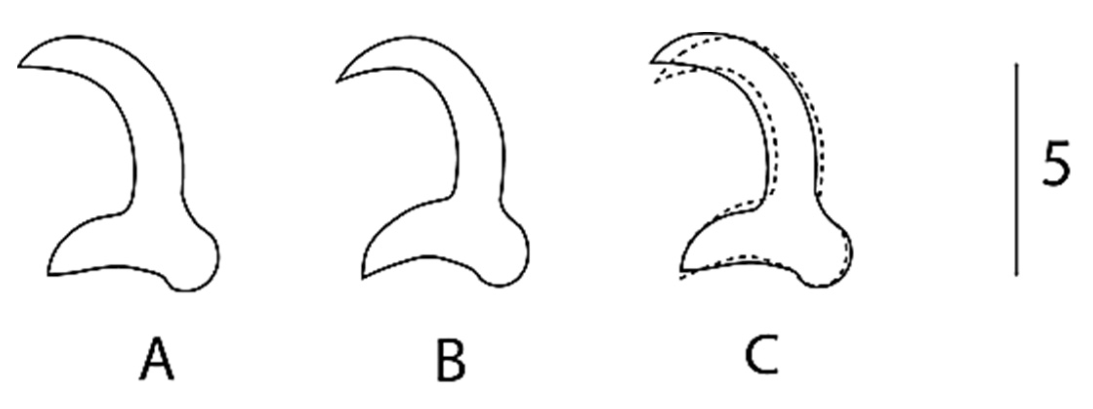
Description: Elongated body. A male copulatory organ (MCO) was observed in five specimens, was spherical (Figure 2a(i) and Figure 3ii), was positioned posteriorly to the pharynx and was armed with one principal spine and a single row of 5–6 smaller spines (Figure 2a(i) and Figure 3ii). Hamuli were slightly slender with a pointed tip with a superficial root (Figure 2a(ii) and Figure 3i,iv). The anterior end where dorsal the bar attaches on the hamulus was prominent, creating a notch between the root and dorsal bar attachment point. The dorsal bar was simple and flexible. The ventral bar had small rounded anterolateral processes with a trapezoid-like membrane having a slightly striated median portion (Figure 2a(iii),b). The marginal hook shaft was approximately perpendicular to the base of the marginal hook sickle (Figure 2a(iv),b(C) and Figure 3iii,v). The sickle point was slightly curved and perpendicular to the base with its tip in line with the distal end of the toe. Overlapping measurements (Table 3) and the similarity in the shape of the marginal hook sickle (Figure 4) suggest that the worms infecting the two host species are conspecific.

Remarks

The comparison with other gyrodactylid species is based on the phenotypic similarities to known parasite species and their occurrence from related hosts. From the three species of Gyrodactylus recorded from cyprinids in Africa, the newly described species of Gyrodactylus can be differentiated by the longer hamuli; G. nyingiae n. sp. 76.5 (65.9–88.2) compared to a hamulus total length in G. ivindoensis of 55 (52–58), 32.1 (23–33) in G. kyogae and 43.3 (35.1–51.5) in G. paludinosus. Like G. paludinosus, G. kyogae has an upward projecting toe, in contrast to that of G. nyingiae n. sp. whose toe points downwards. Additionally, the MCO of G. nyingiae n. sp. has one principal spine and five to six smaller spines arranged in a single row (Figure 2 and Figure 3), in contrast to G. kyogae, which has an unarmed MCO [35]. Gyrodactylus kyogae, in contrast to the other three species, lacks a ventral bar membrane. Gyrodactylus ivindoensis has shorter marginal hooks and a total marginal hook length of 22 (21–24) compared to that of G. nyingiae n. sp., which is 34.8 (31.7–42.1). When comparing the relative length of the root to the hamulus total length respectively, G. nyingiae n. sp. (26.7 vs. 76.4), G. ivindoensis (19.4 vs. 55) and G. paludinosus (15.4 vs. 43.3) have similar ratios of the root length to the total hamulus length (ca. 1:2.8). Gyrodactylus kyogae (9.2 vs. 33.1) on the other hand has a different ratio of the relative root length to the total hamulus length (1:3.5).
Due to the important biogeographical connections between the Middle East and the Maghreb region during the Cenozoic period in the dispersal of freshwater fish fauna, it is interesting to compare the Gyrodactylus fauna of the Iranian region with the North African ones [4,36]. The freshwater species of Gyrodactylus mentioned by [37] and [38] were either known to be from Europe or Central Asia, or undescribed. It is therefore productive to compare G. nyingiae n. sp. with widespread Palearctic species of Gyrodactylus-infecting cyprinids, several of which are reminiscent of G. nyingiae n. sp. in marginal hook morphology. This includes Gyrodactylus mutabilitas Bychowsky, 1957, and Gyrodactylus scardiniensis Glaeser, 1974, which can both be distinguished from G. nyingiae n. sp. by virtue of their shorter hamulus root (max. of 20 in G. mutabilitas and max. of 23 in G. scardiniensis versus min. of 24 in G. nyingiae n. sp.), and Gyrodactylus schulmani Ling, 1962, which has a hamulus of a total length of a max. of 44, shorter than the minimum of 66 of G. nyingiae n. sp. A Gyrodactylus species described from a fish species endemic to Iran is Gyrodactylus jalalii Vanhove, Boeger, Muterezi Bukinga, Volckaert, Huyse and Pariselle, 2012, a parasite of the cichlid host Iranocichla hormuzensis Coad, 1982. It can easily be distinguished from G. nyingiae n. sp. by its more pronounced ventral bar auricles and the sub rectangular ventral bar membrane, which contrast the properties of G. nyingiae n. sp. including its small rounded anterolateral processes and trapezium-shaped ventral bar membrane. Following [39], Gyrodactylus molnari Ergens, 1978, infecting Cyprinus carpio Linnaeus, 1758, in Iraq has a shorter hamulus length (55–65) compared to G. nyingiae n. sp. (65.9–88.2). Additionally, G. molnari has a longer dorsal bar (15–18), compared to G. nyingiae n. sp. (9.9–13.4), and an entirely different shape of the marginal hook sickle. Following [40,41], Gyrodactylus sprostonae Ling, 1962, was found on Cyprinus carpio in Iran. It has a longer dorsal bar (17.4–20) compared to G. nyingiae n. sp. (9.9–13.4) and shorter total hamulus length (48.47–54.23) compared to G. nyingiae n. sp. (65.9–88.2). Due to the fact that the goldfish (Carassius auratus) has been widely introduced in many countries including Morocco, it is also interesting to compare G. nyingiae n. sp. with Gyrodactylus kobayashii Hukuda, 1940, previously isolated from goldfish in central China [42]. G. nyingiae n. sp. has a MCO which resembles that of G. kobayashii with both possessing one principal spine and 5–6 smaller spines. Both species also have slightly curved marginal hook sickles. However, the marginal hook sickle in G. kobayashii has a tip that terminates beyond the limits of its toe while that of G. nyingiae n. sp. has its tip in line with the distal end of the toe. Additionally, G. nyingiae n. sp. has a longer hamulus (76.4 vs. 59.3), a longer sickle (47.4 vs. 40.5), a longer hamulus root (26.7 vs. 21.6), and a longer marginal hook shaft (28.7 vs. 23.3) than G. kobayashii has. Therefore, G. nyingiae n. sp. can be distinguished from the aforementioned Gyrodactylus species by virtue of its longer total hamulus length, longer hamulus root, small rounded anterolateral process and trapezium-shaped ventral bar membrane.

3.4. Multivariate Statistics

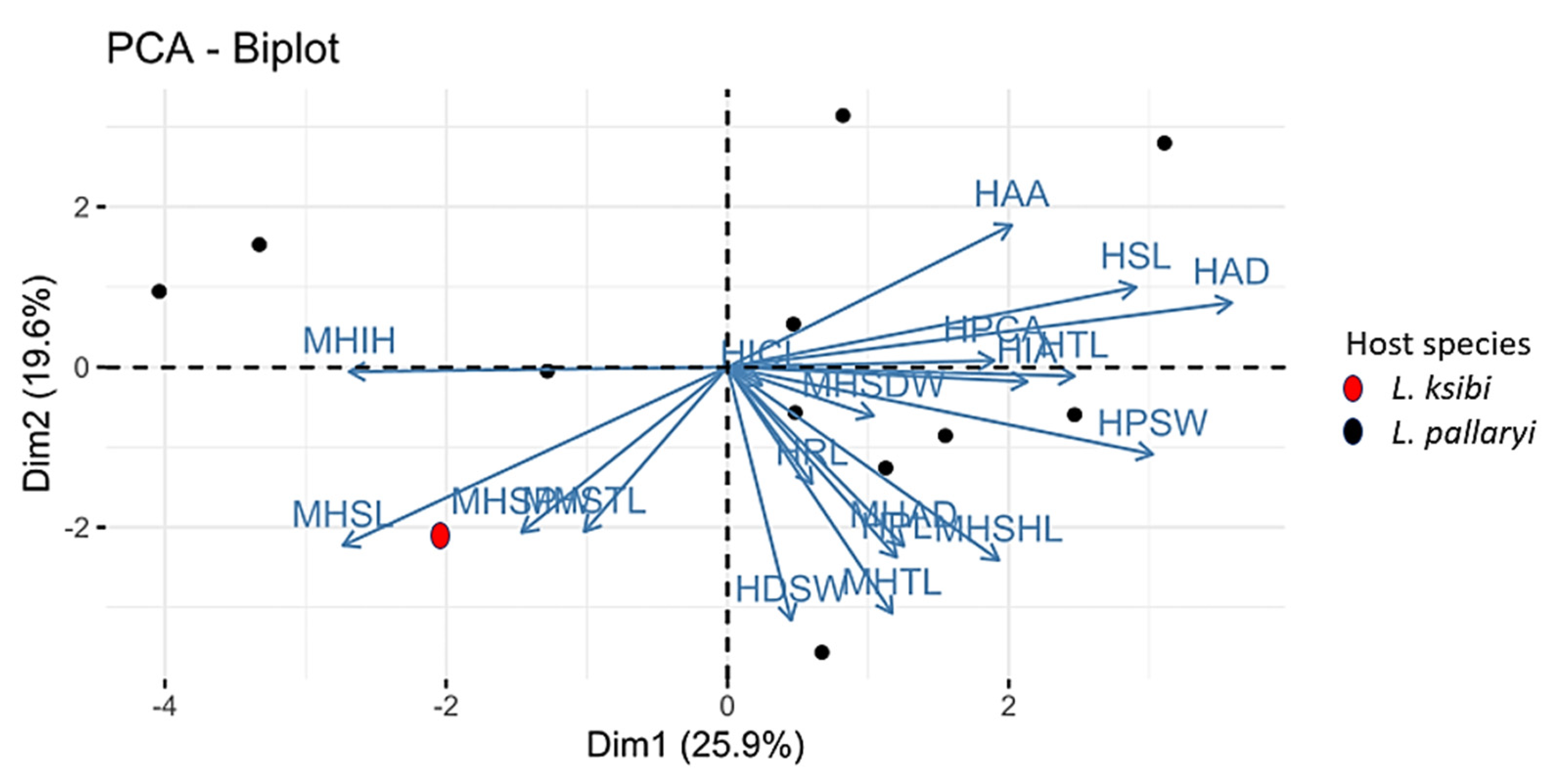
The morphological variation of the 12 specimens of Gyrodactylus was visualized based on a PCA performed on 19 standardized haptoral morphometric characters. The first two principal component axes contributed to 25.9% and 19.6% of the variation, respectively (Figure 5).
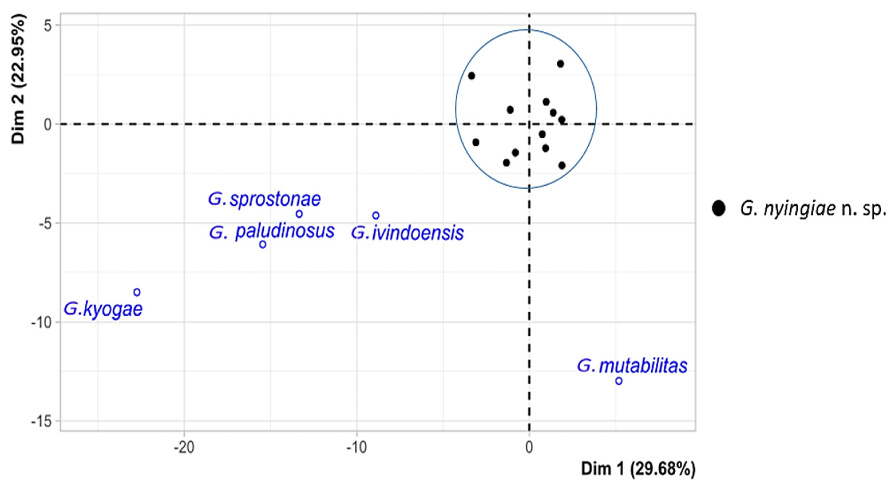
The biplot shows no clear separation which includes all the 12 specimens belonging to Gyrodactylus. From the PCA biplot, we can confirm that we are dealing with a single species described herein as Gyrodactylus nyingiae n. sp. To better illustrate how the newly identified Gyrodactylus nyingiae n. sp. compares to other previously described species of Gyrodactylus, we performed a PCA analysis based on the mean values of 11 haptoral morphometric parameters, i.e., HPL, HSL, HRL, HTL, MHTL, MHSHL, MHSL, MHSPW, MHSDW, MHAD and VBTL (Table 4).
The first two principal components explained 29.7% and 23.0% of the total variation, respectively (Figure 6).
With the average values from the supplementary individuals, the results of the PCA indicate a distinct difference between the Gyrodactylus species that were previously described and the newly described species mentioned in this study.

4. Discussion

Gyrodactylus nyingiae n. sp. is the first described species of Gyrodactylus in Morocco and to the best of our knowledge is the first member of this genus to be identified on a species level in the Maghreb region. Monogenean parasites have been reported in Tunisian aquatic vertebrates, including both marine and freshwater hosts. However, not even a single Gyrodactylus species has been recorded from the examined hosts [43]. The species reported in the current study is also the first gyrodactylid to be described from Luciobarbus in Africa, as previous studies on gyrodactylids infecting cyprinids in Africa have focused on the small barbs belonging to Enteromius, with only three Gyrodactylus species having being described so far [14]. Since Gyrodactylus is a genus of monogeneans with high diversity and minimal morphological variation, it has become a common practice to use a combination of morphological and molecular information for describing and delimiting new species [44,45]. However, due to the limited number of specimens in our study, we opted to use all of the individuals for phenotypic characterization. This decision was based on the fact that our study aimed to unravel the diversity of branchial monogeneans in the hosts, which had not been previously documented.
The current study’s low number of gyrodactylids isolated from cyprinid hosts could be due to the fact that only the gills were examined for parasites. Similarly, Louizi et al. [46] found a species depauperate fauna and low abundances of gill-infecting monogeneans on native cichlid fishes in Morocco. In addition to the low prevalence and possible seasonality of members of Gyrodactylus, less research, a lack of reports on infections, a lack of understanding of relationships between these monogeneans and cyprinid hosts [14], and environmental conditions in Morocco’s freshwater ecosystems might limit the species richness and abundance of certain monogenean taxa. On the other hand, Dactylogyrus reaches higher species richness and higher infection intensities in Moroccan cyprinid–monogenean systems [5,47]. It is also worth noting that the low sample size of the present study is only an indicative value of the population size of the new species of Gyrodactylus in the host.
More research is needed on the African continent to understand the relationship, evolutionary history, and development of gyrodactylids and their hosts, as it is endowed with a diverse endemic fish fauna that undoubtedly possess undiscovered parasite diversity [48,49].

5. Conclusion

Based on the morphoanatomical observation of opisthaptoral parts of 12 individuals of Gyrodactylus in the current study, we describe a new species infecting two cyprinid hosts for the first time in Morocco and the Maghreb region. The new species is different from previously described gyrodactylids infecting cyprinid hosts because it has a longer total hamulus length, a longer hamulus root, a downward projecting toe, trapezium-shaped ventral bar membrane with slightly striated median portion and small rounded anterolateral processes.

Author Contributions

Conceptualization, M.I.S. and A.P.; methodology, I.R.; investigation, M.I.S., H.L. and I.R.; resources, I.R.; data curation, M.I.S. and M.P.M.V.; writing—original draft preparation, M.I.S.; writing—review and editing, M.I.S., A.P. and M.P.M.V.; supervision, A.P. All authors have read and agreed to the published version of the manuscript.

Funding

This research was funded by the Special Research Fund (BOF) of Hasselt University, grant number BOF22DOCLI04, to M.I.S., BOF20TT06 and BOF21INCENT09 to M.P.M.V and by research grant 1513419N of the Research Foundation—Flanders (FWO-Vlaanderen).

Institutional Review Board Statement

The Ethical Committee for Animal Experimentation of Hasselt University, Belgium does not require ethical clearance when animals are euthanized abroad. Fishes were dissected in the field in Morocco. Under Moroccan legislation, no ethical approval for this type of research was required.

Informed Consent Statement

Not applicable.

Data Availability Statement

Type material was deposited in the collection of the research group Zoology: Biodiversity and Toxicology of Hasselt University (Diepenbeek, Belgium) (HU 838-841) and the Institut Scientifique of the Mohammed V University in Rabat (Rabat, Morocco) (ZA PPM 0101).

Acknowledgments

The authors would like to sincerely thank Abdelaziz Benhoussa for allowing us to work in the Biodiversity, Ecology and Genome Laboratory (Mohammed V University in Rabat). Bouabid Badaoui and Hocein Bazairi are thanked for their statistical advice. Nikol Kmentová, Walter Boeger, Daniel Mungai Ndegwa, Kelly Thys, Tiziana Gobbin and Hervé Mandtoumbi are thanked for their laboratory assistance and help with statistical analysis.

Conflicts of Interest

The authors declare no conflict of interest.

References

  1. FAO. The State of World Fisheries and Aquaculture 2018. In Meeting the Sustainable Development Goals; FAO: Rome, Italy, 2018. [Google Scholar]
  2. Dahani, S.; Bouchriti, N.; Benabbes, I.; Boudakkou, A.; Chiaar, A. Occurrence Des Parasites Dans Les Poissons Collectés Au Niveau Du Littoral Marocain. Eur. Sci. J. ESJ 2019, 15, 497–506. [Google Scholar] [CrossRef]
  3. Mohammed, B.; Salwa, O.; Yahyaoui, A. Perceived Barriers to Consumption of Freshwater Fish in Morocco. Br. Food J. 2015, 117, 274–285. [Google Scholar] [CrossRef]
  4. Doadrio, I. Freshwater Fish Fauna of North Africa and Its Biogeography. Ann. Mus. Afr. Centr. Zool. 1994, 275, 21–34. [Google Scholar]
  5. Rahmouni, I.; Řehulková, E.; Pariselle, A.; Rkhami, O.B.; Šimková, A. Four New Species of Dactylogyrus Diesing, 1850 (Monogenea: Dactylogyridae) Parasitising the Gills of Northern Moroccan Luciobarbus Heckel (Cyprinidae): Morphological and Molecular Characterisation. Syst. Parasitol. 2017, 94, 575–591. [Google Scholar] [CrossRef] [PubMed]
  6. Cowling, R.M.; Rundel, P.W.; Lamont, B.B.; Arroyo, M.K.; Arianoutsou, M. Plant Diversity in Mediterranean-Climate Regions. Trends Ecol. Evol. 1996, 11, 362–366. [Google Scholar] [CrossRef] [PubMed]
  7. Sauquet, H.; Weston, P.H.; Anderson, C.L.; Barker, N.P.; Cantrill, D.J.; Mast, A.R.; Savolainen, V. Contrasted Patterns of Hyperdiversification in Mediterranean Hotspots. Proc. Natl. Acad. Sci. USA 2009, 106, 221–225. [Google Scholar] [CrossRef]
  8. Scholz, T.; Smit, N.; Jayasundera, Z.; Gelnar, M. A Guide to the Parasites of African Freshwater Fishes; Royal Belgian Institute of Natural Sciences: Brussels, Belgium, 2018; Volume 18, ISBN 9789073242388. [Google Scholar]
  9. FAO. The State of World Fisheries and Aquaculture 2020. In Sustainability in Action; FAO: Rome, Italy, 2020. [Google Scholar]
  10. Cable, J.; Harris, P.D. Gyrodactylid Developmental Biology: Historical Review, Current Status and Future Trends. Int. J. Parasitol. 2002, 32, 255–280. [Google Scholar] [CrossRef]
  11. Bakke, T.A.; Cable, J.; Harris, P.D. The Biology of Gyrodactylid Monogeneans: The “Russian-Doll Killers”. Adv. Parasitol. 2007, 64, 162–220. [Google Scholar] [CrossRef]
  12. Allalgua, A.; Guerfi, S.; Kaouachi, N.; Boualleg, C.; Boucenna, I.; Barour, C. L’infestation de Cyprinus carpio (Cyprinides) Peuplant Le Barrage Foum El-Khanga (Souk Ahras, Algerie) Par Les Monogenes Parasites. Bull. Soc. Zool. Fr. 2015, 140, 217–232. [Google Scholar]
  13. Bakke, T.A.; Harris, P.D.; Cable, J. Host Specificity Dynamics: Observations on Gyrodactylid Monogeneans. Int. J. Parasitol. 2002, 32, 281–308. [Google Scholar] [CrossRef]
  14. Truter, M.; Smit, N.J.; Malherbe, W.; Přikrylová, I. Description of Gyrodactylus paludinosus sp. nov. (Monogenea: Gyrodactylidae) from the Straightfin Barb, Enteromius paludinosus (Peters, 1852), in South Africa. Acta Parasitol. 2022, 67, 446–453. [Google Scholar] [CrossRef] [PubMed]
  15. Dos Santos, Q.M.; Maina, J.N.; Avenant-Oldewage, A. Gyrodactylus magadiensis n. sp. (Monogenea, Gyrodactylidae), Parasitising the Gills of Alcolapia grahami (Perciformes, Cichlidae), a Fish Inhabiting the Extreme Environment of Lake Magadi, Kenya. Parasite 2019, 26, 76. [Google Scholar] [CrossRef] [PubMed]
  16. Rubio-Godoy, M.; Muñoz-Córdova, G.; Garduño-Lugo, M.; Salazar-Ulloa, M.; Mercado-Vidal, G. Microhabitat Use, Not Temperature, Regulates Intensity of Gyrodactylus cichlidarum Long-Term Infection on Farmed Tilapia-Are Parasites Evading Competition or Immunity? Vet. Parasitol. 2012, 183, 305–316. [Google Scholar] [CrossRef] [PubMed]
  17. Chen, J.; Zhi, T.; Xu, X.; Zhang, S.; Zheng, Y.; Yang, T. Molecular Characterization and Dynamic Expressions of Three Nile Tilapia (Oreochromis niloticus) Complement Genes after Gyrodactylus cichlidarum (Monogenea) Infection. Aquaculture 2019, 502, 176–188. [Google Scholar] [CrossRef]
  18. Buchmann, K.; Bresciani, J. Microenvironment of Gyrodactylus derjavini on Rainbow Trout Oncorhynchus mykiss: Association between Mucous Cell Density in Skin and Site Selection. Parasitol. Res. 1997, 84, 17–24. [Google Scholar] [CrossRef]
  19. Bakke, T.A.; Harris, P.D.; Hansen, H.; Cable, J.; Hansen, L.P. Susceptibility of Baltic and East Atlantic Salmon Salmo salar Stocks to Gyrodactylus salaris (Monogenea). Dis. Aquat. Organ. 2004, 58, 171–177. [Google Scholar] [CrossRef]
  20. Kerr, S.J.; Grant, R.E. Ecological Impacts of Fish Introductions: Evaluating the Risk; Fish and Wildlife Branch, Ontario Ministry of Natural Resources: Peterborough, ON, Canada, 2000; ISBN 0777893169. [Google Scholar]
  21. Hickley, P.; Muchiri, S.M. Management and Ecological Note Discovery of Carp, Cyprinus carpio, in the Already Stressed Fishery of Lake Naivasha, Kenya. Mar. Freshw. Res. 2004, 11, 139–142. [Google Scholar]
  22. Otachi, E.O.; Magana, A.E.M.; Jirsa, F.; Fellner-Frank, C. Parasites of Commercially Important Fish from Lake Naivasha, Rift Valley, Kenya. Parasitol. Res. 2014, 113, 1057–1067. [Google Scholar] [CrossRef]
  23. Ormerod, S.J. Current Issues with Fish and Fisheries: Editor’s Overview and Introduction. J. Appl. Ecol. 2003, 40, 204–213. [Google Scholar] [CrossRef]
  24. Gherardi, F.; Robert Britton, J.; Mavuti, K.M.; Pacini, N.; Grey, J.; Tricarico, E.; Harper, D.M. A Review of Allodiversity in Lake Naivasha, Kenya: Developing Conservation Actions to Protect East African Lakes from the Negative Impacts of Alien Species. Biol. Conserv. 2011, 144, 2585–2596. [Google Scholar] [CrossRef]
  25. Pellegrin, J. Poissons Du Gribingui Par M. Baudon. Description de Sept Especes Nouvelles. Bull. Société Zool. Fr. 1919, 44, 201–214. [Google Scholar]
  26. Justine, J.L.; Briand, M.J.; Bray, R.A. A Quick and Simple Method, Usable in the Field, for Collecting Parasites in Suitable Condition for Both Morphological and Molecular Studies. Parasitol. Res. 2012, 111, 341–351. [Google Scholar] [CrossRef] [PubMed]
  27. Fricke, R.; Eschmeyer, W.; Van der Laan, R. Eschmeyer’s Catalog of Fishes: Genera, Species, References. Available online: https://researcharchive.calacademy.org/research/ichthyology/catalog/fishcatmain.asp (accessed on 23 July 2021).
  28. Pariselle, A.; Euzet, L. Three New Species of Cichlidogyrus Paperna, 1960 (Monogenea, Ancyrocephalidae) Parasitic on Tylochromis jentinki (Steindachner, 1895) (Pisces, Cichlidae) in West Africa. Syst. Parasitol. 1994, 29, 229–234. [Google Scholar] [CrossRef]
  29. Humason, G.L. Animal Tissue Techniques; Beadle, G.W., Emerson, R., Whitaker, D.M., Eds.; W. H. Freeman and Company: New York, NY, USA, 1979. [Google Scholar]
  30. Malmberg, G. The Excretory Systems and the Marginal Hooks as a Basis for the Systematics of Gyrodactylus (Trematoda, Monogenea). Ark. Zool. 1970, 23, 1–235. [Google Scholar]
  31. Bates, J.W. The Slide-Sealing Compound “Glyceel”. J. Nematol. 1997, 29, 565–566. [Google Scholar] [PubMed]
  32. Pugachev, O.N.; Gerasev, P.; Gussev, A.; Ergens, R.; Khotenowsky, I. Guide to Monogenoidea of Freshwater Fish of Palaeartic and Amur Regions; Ledizoni-Ledipublishing: Lombardy, Italy, 2009. [Google Scholar]
  33. Bush, A.O.; Lattertyf, K.D.; Lotz, J.M.; Shostakll, A.W.; Brandon, Z.; Lafferty, K.D.; Lotz, J.M.; Shostak, A.W. Parasitology Meets Ecology on Its Own Terms: Margolis et Al. Revisited. J. Parasitol. 1997, 83, 575–583. [Google Scholar] [CrossRef]
  34. Shinn, A.P.; Hansen, H.; Olstad, K.; Bachmann, L.; Bakke, T.A. The Use of Morphometric Characters to Discriminate Specimens of Laboratory-Reared and Wild Populations of Gyrodactylus salaris and G. thymalli (Monogenea). Folia Parasitol. 2004, 51, 239–252. [Google Scholar] [CrossRef]
  35. Paperna, I. New Species of Monogenea (Vermes) from African Freshwater Fish. A Preliminary Report. Rev. Zool. Bot. Africaines 1973, 87, 505–518. [Google Scholar]
  36. Benovics, M.; Nejat, F.; Abdoli, A.; Šimková, A. Molecular and Morphological Phylogeny of Host-Specific Dactylogyrus Parasites (Monogenea) Sheds New Light on the Puzzling Middle Eastern Origin of European and African Lineages. Parasites and Vectors 2021, 14, 372. [Google Scholar] [CrossRef]
  37. Jalali, B.; Shamsi, S.; Barzegar, M. Occurrence of Gyrodactylus spp. (Monogenea: Gyrodactylidae) from Iranian Freshwater Fishes. Iran. J. Fish. Sci. 2005, 4, 19–30. [Google Scholar]
  38. Barzegar, M.; Ebrahimzadeh Mousavi, H.; Rahmati-holasoo, H.; Taheri Mirghaed, A.; Bozorgnia, A. Gyrodactylus (Monogenea, Gyrodactylidae) Parasite Fauna of Fishes in Some Rivers of the Southern Caspian Sea Basin in Mazandaran Province. Iran. J. Vet. Med. 2017, 12, 35–44. [Google Scholar] [CrossRef]
  39. Younis, S.A.; Shamall, M.A.A. Monogenean Infections on Fishes from Darbandikhan Lake in Kurdistan Region, Iraq. Basrah J. Agric. Sci. 2013, 26, 117–131. [Google Scholar] [CrossRef]
  40. Roohi, J.D.; Dalimi Asl, A.; Pourkazemi, M.; Shamsi, S. Occurrence of Dactylogyrid and Gyrodactylid Monogenea on Common Carp, Cyprinus carpio, in the Southern Caspian Sea Basin. Parasitol. Int. 2019, 73, 101977. [Google Scholar] [CrossRef] [PubMed]
  41. Roohi, J.D.; Asl, A.D.; Mohammad, P.; Ghasemi, M.; Shamsi, S. Morphometric and Molecular Identification of Gyrodactylus sprostonae in Guilan Province Warm Water Fishes with an Attitude of Intensity and Prevalence in Selected Farms. Iran. Vet. J. 2018, 15, 50–59. [Google Scholar] [CrossRef]
  42. Tu, X.; Ling, F.; Huang, A.; Wang, G. An Infection of Gyrodactylus kobayashii Hukuda, 1940 (Monogenea) Associated with the Mortality of Goldfish (Carassius auratus) from Central China. Parasitol. Res. 2015, 114, 737–745. [Google Scholar] [CrossRef]
  43. Derbel, H.; Châari, M.; Neifar, L. Checklist of the Monogenea (Platyhelminthes) Parasitic in Tunisian Aquatic Vertebrates. Helminthologia 2022, 59, 179–199. [Google Scholar] [CrossRef]
  44. Pinacho-Pinacho, C.D.; Calixto-Rojas, M.; García-Vásquez, A.; Guzmán-Valdivieso, I.; Barrios-Gutiérrez, J.J.; Rubio-Godoy, M. Species Delimitation of Gyrodactylus (Monogenea: Gyrodactylidae) Infecting the Southernmost Cyprinids (Actinopterygii: Cyprinidae) in the New World. Parasitol. Res. 2021, 120, 831–848. [Google Scholar] [CrossRef]
  45. Razo-Mendivil, U.; García-Vásquez, A.; Rubio-Godoy, M. Spot the Difference: Two Cryptic Species of Gyrodactylus von Nordmann, 1832 (Platyhelminthes: Monogenea) Infecting Astyanax aeneus (Actinopterygii, Characidae) in Mexico. Parasitol. Int. 2016, 65, 389–400. [Google Scholar] [CrossRef]
  46. Louizi, H.; Vanhove, M.P.M.; Rahmouni, I.; Berrada Rkhami, O.; Benhoussa, A.; Van Steenberge, M.; Pariselle, A. Species Depauperate Communities and Low Abundances of Monogenean Gill Parasites at the Edge of the Natural Distribution Range of Their Cichlid Hosts in Northern Africa. Hydrobiologia 2022. [Google Scholar] [CrossRef]
  47. Gharbi, S.E.; Birgi, E.; Lambert, A. Monogènes Dactylogyridae Parasites de Cyprinidae Du Genre Barbus d’Afrique Du Nord. Syst. Parasitol. 1994, 27, 45–70. [Google Scholar] [CrossRef]
  48. Přikrylová, I.; Blažek, R.; Vanhove, M.P.M. An Overview of the Gyrodactylus (Monogenea: Gyrodactylidae) Species Parasitizing African Catfishes, and Their Morphological and Molecular Diversity. Parasitol. Res. 2012, 110, 1185–1200. [Google Scholar] [CrossRef]
  49. Vanhove, M.P.M.; Snoeks, J.; Volckaert, F.A.M.; Huyse, T. First Description of Monogenean Parasites in Lake Tanganyika: The Cichlid Simochromis diagramma (Teleostei, Cichlidae) Harbours a High Diversity of Gyrodactylus Species (Platyhelminthes, Monogenea). Parasitology 2011, 138, 364–380. [Google Scholar] [CrossRef]
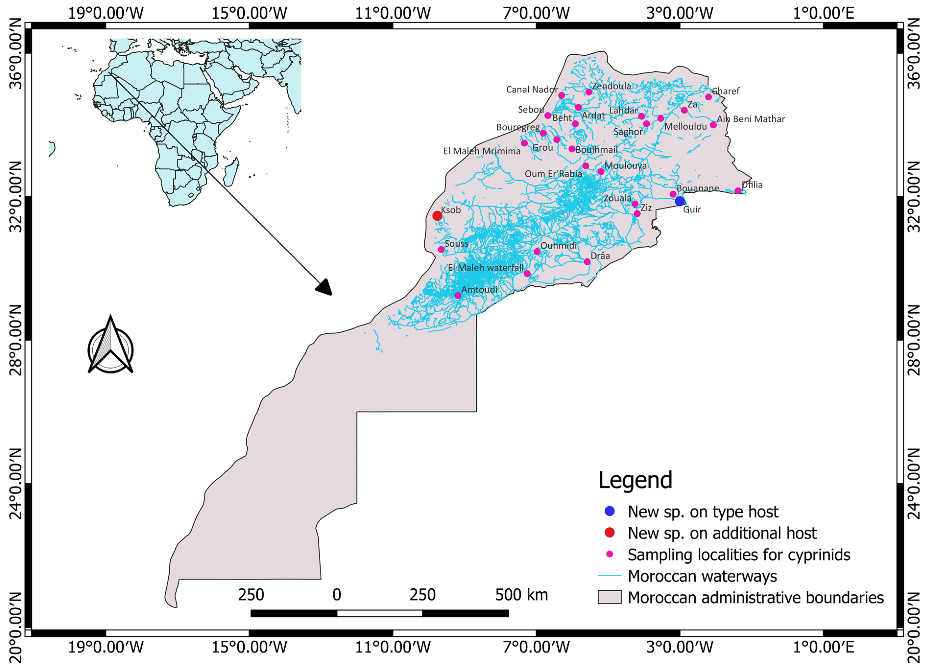
Figure 1. An overview of the sampling locations of cyprinid hosts examined for the presence of members of Gyrodactylus. The type host and additional host localities for the newly described Gyrodactylus species in the present study are depicted by blue and red circles, respectively.
Animals 13 01624 g001
Figure 2. Gyrodactylus nyingiae n. sp. isolated from Luciobarbus pallaryi. (a) Micrograph of (i) male copulatory organ (MCO), (ii) hamuli, (iii) ventral bar, and (iv) marginal hooks (b) Drawings of sclerotized structures of the haptor with (A) hamulus, (B) ventral bar, (C) marginal hook, and (D) male copulatory organ. Scale bars are in µm.
Animals 13 01624 g002
Figure 3. Micrograph and drawings of sclerotized structures (i,iv) hamuli, (ii) male copulatory organ, and (iii,v) marginal hooks of Gyrodactylus nyingiae n. sp. isolated from Luciobarbus ksibi. Scale bar represents 20 µm.
Animals 13 01624 g003
Figure 4. Overlay of marginal hook sickles (C) of G. nyingiae n. sp. from L. ksibi (A) and L. pallaryi (B) (dotted outline). Scale bar represents 5 µm.
Animals 13 01624 g004
Figure 5. Biplot of PCA (first two axes) of all the 12 specimens of Gyrodactylus nyingiae n. sp. (For abbreviations of variables, please refer to Table 3).
Animals 13 01624 g005
Figure 6. PCA (first two axes) of the newly described Gyrodactylus (in blue circle) and previously described species (supplementary individuals) based on average measurements of haptoral morphometric characters.
Animals 13 01624 g006
Table 1. Cyprinid specimens collected, their localities and number of individuals belonging to Gyrodactylus infecting the hosts (note that the number of parasites collected might be an underestimation because only the right gill arches were screened for parasites).
Coordinates
HostLocalityWatershedLatitudeLongitudeNo. of Hosts SampledNo. of Specimens of Gyrodactylus Isolated from the Hosts
Luciobarbus pallaryi (Pellegrin, 1919)Oued GuirZiz31°52′12″ N003°0′00″ W15714
Oued BouananeZiz32°04′04″ N003°11′23.9″ W
Oued DfiliaZiz32°9′48.892″ N001°22′37.4″ W
Luciobarbus rabatensis Doadrio, Perea and Yahyaoui, 2015Oued GrouBouregreg33°35′28.0″ N006°25′49.6″ W243
Oued BouregregBouregreg33°46′18.0″ N006°48′16.6″ W
Oued BoulhmailBouregreg33°19′49.6″ N006°00′15.1″ W
Luciobarbus maghrebensis Doadrio, Perea and Yahyaoui, 2015Oued LahdarSebou34°14′32.7″ N004°03′53.9″ W551
Oued SaghorSebou34°02′4.0″ N003°55′45.5′W
Oued ArdatSebou34°29′26.8″ N005°49′49.2″ W
Oued BehtSebou34°01′55.5″ N005°54′43.2″ W
Oued SebouSebou34°15′48.0″ N006°40′42.0″ W
Canal NadorSebou34°49′19.7″ N006°17′36.7″ W
Luciobarbus rifensis Doadrio, Casal-López and Perea 2015Oued ZendoulaLoukkos34°54′57.6″ N005°32′17.2″ W193
Luciobarbus guercifensis Doadrio, Perea and Yahyaoui, 2016Oued MelloulouMoulouya34°10′51.7″ N003°31′59.6″ W40
Oued ZaMoulouya34°24′38.9″ N002°52′28.1″ W
Luciobarbus yahyaouii Doadrio, Casal-López and Perea 2016Oued ZaMoulouya34°24′38.9″ N002°52′28.1″ W620
Oued CharefMoulouya34°46′44.0″ N002°11′56.0″ W
Oued MelloulouMoulouya34°10′51.7″ N003°31′59.6″ W
Ain Beni MatharMoulouya34°00′00.3″ N002°03′58.6″ W
Luciobarbus zayanensis Doadrio, Casal-López and Yahyaoui, 2016Oued Oum Er’RabiaOum Er’Rabia32°51′32.8″ N005°37′18.9″ W251
Oued MoulouyaMoulouya32°41′55.4″ N005°11′51.2″ W
Luciobarbus lepineyi (Pellegrin, 1939)Oued ZizZiz31°31′34.7″ N004°11′10.0″ W1270
Oued ZoualaZiz31°47′31.9″ N004°14′43.5″ W
Oued DfiliaZiz32°9′48.892″ N001°22′37.4″ W
Oued DrâaDraa30°11′12.24″ N005°34′47.34″ W
Oued OuhmidiDraa30°28′5.64″ N006°58′36.12″ W
Oued El Maleh MrimimaDraa33°29’34.8’’N007°19′58.1″ W
Oued El Maleh WaterfallDraa29°51′108″ N007°15′23″ W
Oued AmtoudiDraa29°14′32.42″ N009°11′8.71″ W
Carasobarbus moulouyensis (Pellegrin, 1924)Oued MoulouyaMoulouya32°41′55.4″ N005°11′51.2″ W441
Luciobarbus ksibi (Boulenger, 1905)Oued Oum Er’RabiaOum Er’Rabia32°51′32.8″ N005°37′18.9″ W401
Oued KsobKsob31°27′50.7″ N009°45′25.3″ W
Luciobarbus massaensis (Pellegrin, 1922)Oued SoussSouss-Massa30°31′33.6″ N009°38′53.6″ W211
Carasobarbus fritschii (Günther, 1874)Oued GrouBouregreg33°35′28.0″ N006°25′49.6″ W1570
Oued BoulhmailBouregreg33°19′49.6″ N006°00′15.1″ W
Oued LahdarSebou34°14′32.7″ N004°03′53.9″ W
Oued Oum Er’RabiaOum Er’Rabia32°41′03.8″ N005°13′00.3″ W
Oued ZaMoulouya34°24′38.9″ N002°52′28.1″ W
Oued CharefMoulouya34°46′44.0″ N002°11′56.0″ W
Oued KsobKsob31°27′50.7″ N009°45′25.3″ W
Oued ArdatSebou34°29′26.8″ N005°49′49.2″ W
Oued BehtSebou34°01′55.5″ N005°54′43.2″ W
Oued SebouSebou34°15′48.0″ N006°40′42.0″ W
Pterocapoeta maroccana Günther, 1902Oued Oum Er’RabiaOum Er’Rabia32°51′32.8″ N005°37′18.9″ W31
Total73826
Table 2. Prevalence, mean intensity and mean abundance of Gyrodactylus infecting the Moroccan cyprinids collected, based on examination of the right-side gill arches only (note that only localities where these parasites were found are retained in this table).
LocalitySpeciesHNnP = (N/H) × 100M.I = n/NM.A = n/H
Oued GuirLuciobarbus
pallaryi
1571140.64140.09
Oued BouregregLuciobarbus
rabatensis
24134.1730.13
Oued SebouLuciobarbus
maghrebensis
55111.8210.02
Oued ZendoulaLuciobarbus
rifensis
19135.2630.16
Oued MoulouyaLuciobarbus
zayanensis
25114.0010.04
Oued MoulouyaCarasobarbus
moulouyensis
44112.2710.02
Oued KsobLuciobarbus ksibi40112.5010.03
Oued SoussLuciobarbus
massaensis
21114.7610.05
Oued Oum
Er’Rabia
Pterocapoeta
maroccana
31133.3310.33
H, number of examined hosts; N, number of infected hosts; n, number of individuals of Gyrodactylus in infected host; P, prevalence; M.I, mean infection intensity; M.A, mean abundance.
Table 3. Morphometric measurements of sclerotized parts of Gyrodactylus nyingiae n. sp. The number of structures measured is given in superscript.
HostLuciobarbus pallaryi (n = 1)Luciobarbus ksibi (n = 1)Both Host Species Combined
Total body length (TBL)386.8 (278.3–456) 5443.7396.3 (278.3–456) 6
Total body width (TBW)133 (115.8–145.9) 6158136.6 (115.8–158.4) 7
Hamulus total length (HTL)76.5 (65.9–88.2) 1075.376.4 (65.9–88.2) 11
Hamulus sickle length (HSL)47.6 (42.5–54.8) 845.147.4 (42.5–54.8) 9
Hamulus aperture distance (HAD)27.5 (21.1–30.2) 922.626.7 (21.1–30.2) 10
Hamulus point length (HPL)36.9 (31.7–41.3) 936.336.2 (31.7–41.3) 10
Hamulus inner curve length (HICL)1.7 (1.4–2.7) 642.1 (1.4–4) 7
Hamulus distal shaft width (HDSW)5.5 (4.6–6.7) 1065.7 (4.6–7.3) 11
Hamulus root length (HRL)26.7 (24.2–28.3) 726.7 (24.2–28.3) 7
Hamulus aperture angle (HAA) (in degrees)36.9 (31.5–45.4) 732.436.4 (31.5–45.4) 8
Hamulus point curve angle (HPCA) (in degrees)4.4 (3.4–5.4) 44.4 (3.4–5.4) 4
Hamulus inner angle (HIA) (in degrees)40.4 (36–45.4) 737.240 (36–45.4) 8
Hamulus proximal shaft width (HPSW)10.4 (8.2–12.1) 1010.310.2 (8.2–12.1) 11
Marginal hook total length (MHTL)34.4 (31.7–42.1) 835.234.8 (31.7–42.1) 9
Marginal hook shaft length (MHSHL)28.6 (26.1–33.4) 929.228.7 (26.1–33.4) 10
Marginal hook sickle length (MHSL)6.2 (5.5–6.5) 96.66.3 (5.5–6.6) 10
Marginal hook sickle proximal width (MHSPW)4.6 (3.9–5) 95.54.7 (3.9–5.5) 10
Marginal hook sickle distal width (MHSDW)4.5 (3.9–5.1) 954.5 (3.9–5.1) 10
Marginal hook sickle toe length (MHSTL)1.9 (1.8–2.1)92.12 (1.8–2.1) 10
Marginal hook aperture distance (MHAD)5.5 (5–5.9) 85.35.4 (5–5.9) 9
Marginal hook in-step height (MHIH)0.6 (0.5–0.9) 80.70.6 (0.5–0.9) 9
Ventral bar total length (VBTL)19.6 (18.6–20.5) 2-19.6 (18.6–20.5) 2
Ventral bar total width (VBTW)25.1 (24.8–25.4) 2-25.1 (24.8–25.4) 2
Ventral bar median length (VBML)6.1 (5.5–6.8) 3-6.1 (5.5–6.8) 3
Ventral bar membrane length (VBMBL)13.6 (12.7–14.5) 3-13.6 (12.7–14.5) 3
Ventral bar process length (VBPL)3.7 (3.6–3.8) 2-3.7 (3.6–3.8) 2
Male copulatory organ diameter (MCO)18.4 (16.5–19.5) 421.218.9 (16.5–21.2) 5
   Principal spine length6.5 (6.3–6.6) 36.56.5 (6.3–6.6) 4
   Small spine length3.3 (3.1–3.5) 35.44.4 (3.1–5.4) 4
Dorsal bar length (DBL)11.9 (9.9–13.4) 3-11.9 (9.9–13.4) 3
Dorsal bar width (DBW)1.6 (1.2–1.9) 3-1.6 (1.2–1.9) 3
Table 4. Table showing the mean values of supplementary individuals included in the PCA.
G. paludinosus Truter, Smit, Malherbe and Přikrylová, 2021G. kyogae Paperna, 1973G. ivindoensis Price & Gery, 1968G. sprostonae Ling, 1962G. mutabilitas Bychowskii, 1957
Reference[14][14][14][40][38]
CountrySouth AfricaUgandaGabonIranIran
HostEnteromius paludinosus (Peters, 1852)Enteromius neumayeri (Fischer, 1884)Enteromius cf. holotaenia (Boulenger, 1904)Cyprinus carpio Linnaeus, 1758Vimba vimba (Linnaeus, 1758)
HPL19.11421.422.823.8
HRL15.49.219.416.6-
HTL43.333.1555267.5
HSL33.828.237.639.825
MHTL18.514.82223.133.7
MHSHL14.211.324.719.4-
MHSL4.43.15.54.510.3
MHSPW2.52.43.33.2-
MHSDW1.92.12.53.1-
MHAD4.13.75.3--
VBTL17.44.81819.728.5
Disclaimer/Publisher’s Note: The statements, opinions and data contained in all publications are solely those of the individual author(s) and contributor(s) and not of MDPI and/or the editor(s). MDPI and/or the editor(s) disclaim responsibility for any injury to people or property resulting from any ideas, methods, instructions or products referred to in the content.