Article

Staphylococcus microti Strains Isolated from an Italian Mediterranean Buffalo Herd

by
Monica Ambrosio
1,
Francesca Paola Nocera
1,*,
Francesca Garofalo
2,
Pasquale De Luca
3,
Alex Grinberg
4 and
Luisa De Martino
1,5
1
Department of Veterinary Medicine and Animal Production, University of Naples ‘Federico II’, Via Delpino 1, 80137 Naples, Italy
2
Istituto Zooprofilattico Sperimentale del Mezzogiorno, 80055 Portici, Italy
3
Stazione Zoologica Anton Dohrn of Naples, Villa Comunale, 80121 Naples, Italy
4
School of Veterinary Science, Massey University, Palmerston North 4442, New Zealand
5
Task Force on Microbiome Studies, University of Naples ‘Federico II’, 80131 Naples, Italy
*
Author to whom correspondence should be addressed.
Animals 2023, 13(1), 182; https://doi.org/10.3390/ani13010182
Submission received: 28 November 2022 / Revised: 20 December 2022 / Accepted: 30 December 2022 / Published: 3 January 2023
(This article belongs to the Section Cattle)

Abstract

:

Simple Summary

Non-aureus staphylococci (NAS) are frequently found in bovine milk samples as well as on teat skin associated with subclinical mastitis (SCM). Staphylococcus microti is a new species among NAS resulted to recently be the predominant species in the milk of water buffaloes (Bubalus bubalis) affected by intramammary infection (IMI). The aim of this study was to assess the occurrence of S. microti in milk and milking parlor equipment samples of an Italian buffalo farm. No clinical mastitis was noticed, but S. microti-associated SCM and IMI were detected. The presence of S. microti was also revealed on milking parlor surfaces. The results of the antimicrobial susceptibility tests highlighted a complete resistance to tetracyclines, and 20.4% of the isolated S. microti exhibited a multidrug-resistant (MDR) profile. In conclusion, the presence of this bacterium in the milk of buffaloes in the absence of evident mastitis clinical signs underlines the need for further studies, and its finding on milking parlor surfaces suggests that the environmental quality of the milking parlor plays an important role in influencing both S. microti’s spread and the microbial communities of the milk.

Abstract

S. microti is a new species among non-aureus staphylococci (NAS) frequently found in bovine milk samples and associated with subclinical mastitis (SCM). The aim of this study was to analyze the presence of S. microti in 200 composite milk samples and 104 milking parlor surface swabs collected at a buffalo farm in Southern Italy to define its presence in milk and a milking parlor environment. The samples were inoculated onto different agar plates, and the isolates were identified by MALDI-TOF MS. The strains identified as S. microti (54/304 samples, 17.8%) were collected, and their purified genomic DNA was subjected to PCR amplification and whole 16S rRNA gene sequencing. Furthermore, their phenotypic resistance profiles were evaluated by a disk diffusion method, and the genotypic characterization of the tetracycline resistance was performed for the tetM and tetK genes by multiplex PCR. Four and forty-seven S. microti isolates from milk samples of lactating animals with subclinical mastitis (SCM) and intramammary infection (IMI), respectively, and three isolates from milking parlor surfaces were recovered. The genomic DNA was purified from the bacterial isolates, and the amplification and sequencing of the 16S gene further supported the proteomic identification as S. microti. No clinical mastitis was detected in the herd during the study period. The antimicrobial susceptibility testing revealed a worrisome 100% resistance to tetracyclines, genotypically mediated by the tetM gene for all strains. This study highlights that S. microti may be commonly isolated from dairy buffalo milk and milking parlor equipment. Its association with SCM or IMI remains to be established.

1. Introduction

The Mediterranean buffalo (Bubalis bubalis) is a large bovid widely distributed throughout Southern Italy. Buffalo milk has a high economic and social value in the Campania Region, as it is the raw material used to produce Mozzarella di Bufala Campana Protected Denomination of Origin (PDO), a worldwide renowned cheese.
Mastitis is one of the most economically important diseases affecting the dairy industry worldwide, and it is almost always caused by bacteria [1]. Thus, the identification of bacterial pathogens associated with clinical and subclinical mastitis is essential to understand the etiology of this disease, reduce animal culling and develop suitable therapy and preventative measures. According to the National Mastitis Council, the diagnosis of mastitis should be based on an interpretation of the somatic cell count (SCC) and bacteriological milk culture results [2].
The milk somatic cell count (SCC) is adopted as an indicator of udder health in single milk samples and is also important for monitoring farm hygiene in bulk milk. Clinically, cow’s milk samples with an SCC < 200 × 103 cells/mL and negative to bacteriological culture are indicative of a healthy status. An SCC < 200 × 103 cells/mL and a positive bacteriological culture is indicative of an intramammary infection (IMI), and an SCC > 200 × 103 cells/mL and a positive bacteriological culture is indicative of a subclinical mastitis (SCM) or clinical mastitis, according to the absence or presence of clinical signs, respectively [3].
Staphylococci, especially the coagulase-positive Staphylococcus aureus, are among the pathogens associated with mastitis in farmed ruminants. The causative role of non-aureus staphylococci (NAS) has grown over the years, and today they are considered as etiological agents of mastitis in cows, goats, and sheep [4] and, in addition, they are more frequently isolated from milk [5]
More than 45 recognized NAS species have been described so far, and S. microti is a new species firstly described in 2010 [6]. Precisely, the first two strains of S. microti were isolated between 1999 and 2002 from the liver and kidneys of common voles (Microtus arvalis Pallas) in the Czech Republic [6]. Another strain was recovered from the skin of a small mammal (belonging to rodents or insectivores) in northeastern Poland [7]. Phylogenetic studies based on the sequence analysis of the 16S rRNA gene and several housekeeping genes revealed that S. microti is closely related to S. rostri and S. muscae [8]. Subsequently, eleven S. microti strains were identified in milk samples collected from dairy cows affected with clinical and subclinical mastitis characterized by a high self-curing rate [9]. The complete genomic sequence of the S. microti DSM 22,147 strain, isolated from the viscera of common voles, has been reported by Hu et al. [10], which described a genome without plasmids and with a GC content of 38% in a 2,381,859 bp long sequence.
Data on the presence and causal role of S. microti in cattle are scarce, but recently the species was found in a buffalo herd and was associated with cases of subclinical mastitis and alterations in milk protein composition [11].
In this study, we investigated the occurrence, supplemented by two identification analyses, of S. microti in buffalo milk and milking parlor surface samples of a buffalo farm in the Campania Region (Salerno, Italy). The strains’ in vitro susceptibility to commonly used antimicrobials was investigated, and a genotypic tetracycline resistance analysis was also performed. In addition, S. microti’s association with different clinical statuses of udders was evaluated.

2. Materials and Methods

2.1. Ethical Statement

This study was approved by the Institutional Animal Ethics Committee of the University of Naples Federico II (Italy) PG/2020/0092228 of 11/06/2020. All of the dairy buffaloes sampled in this study were from a single herd and were subjected to routine milk sampling for diagnostic purposes. Milk and milking parlor surface samples were collected with the owner’s permission, and no written informed consent was required.

2.2. Collection of Samples and Somatic Cell Count

The milk samples were collected from 3 to 5 year old lactating Mediterranean buffaloes in a herd of approximately 100 animals maintained in free-stall barns. The lactating buffaloes were milked twice a day in a tandem milking parlor. A total of 200 composite milk samples were collected aseptically over seven sampling periods from June to December 2020. For each monthly sampling, approximately 20 buffaloes in different stages of lactation were haphazardly chosen for the sampling. Only 23 animals were sampled two times with different sampling intervals in a range of 1–6 months. No clinical signs of mastitis were observed by the breeder for any of the sampled buffalo, and no antibiotic therapy was administered during the study period.
For the milk sampling, the first stream of milk was discharged, and two composite milk samples were collected from each lactating buffalo after the routine pre-milking cleaning during the evening milking and placed in sterile tubes (Thermo Fisher Scientific, Str. Rivoltana, Km 4-20090 Rodano, Milan, Italy). A total of 50 mL was collected for the somatic cell count (SCC) and 15 mL for the bacterial culture in two different tubes. The milk samples were kept at 4 °C and transported to the laboratory within 24 h. The SCC was performed using a NucleoCounter® SCC-100™ (ChemoMetec Inc. 8950 Villa La Jolla Drive-Suite A127 La Jolla, CA, USA) at the Istituto Zooprofilattico Sperimentale del Mezzogiorno (Portici, Naples, Italy), with a measurement range of 1 × 10⁴ to 200 × 104 SCC/mL, and the results are expressed as the number of cells per milliliter of milk. The milk composition was analyzed using a Milkoscan FT120 (Foss Electric A/S, Denmark), based on Fourier transform infrared (FTIR) spectra, to measure the fat, protein, and lactose content; a possible correlation between composition and SCC increase was determined.
Bacteriologically negative milk samples with <200 × 103 cells/mL were considered healthy (H). Differently, bacteriologically positive milk samples were categorized in three groups considered diseased: (1) samples with intramammary infection (IMI) presenting SCCs < 200 × 103 cells/mL; (2) samples with subclinical mastitis (SCM) presenting SCC values > 200 × 103 cells/mL in the absence of clinical signs; (3) samples with clinical mastitis (CM) presenting SCC values > 200 × 103 cells/mL in the presence of clinical signs.
In addition, 104 swabs were collected from the surfaces of the milking parlor, such as the milking boxes and milking unit. Each swab was placed in Stuart W/O CH transport medium (Aptaca Spa, Asti, Italy) and transferred within 24 h to the laboratory for bacteriological examination.

2.3. Bacterial Isolation and Identification

All bacteriological examinations were performed at the Microbiology Laboratory of the Department of Veterinary Medicine and Animal Production of the University of Naples “Federico II” following an internal protocol. Precisely, the microbiological count method was the standard plate count agar method, and 50 μL of each milk sample was diluted in 1 mL of buffered peptone water, and 200 μL of inoculum was spread on each of the following plates: Mac Conkey Agar, 5% Columbia Sheep Blood agar, Mannitol Salt Agar, and Saboraud Dextrose Agar (Oxoid, Milan, Italy). The same plates were used for the surface swabs. All plates were incubated for 24 h at 37 °C in aerobic conditions, and then the phenotypically equal colonies from the milk samples were counted to perform an enumeration of the CFUs.
The isolation of 1 to 3 different colony types and ≥500 CFU/mL for each colony type was considered as a positive sample, whereas milk samples for which more than 3 colony types and/or <500 CFU/mL colonies of any bacterial colony was considered as a contaminated sample. To obtain pure cultures, single colonies were subcultured on Columbia Sheep Blood agar and incubated as above; then, they were identified by matrix-assisted laser desorption/ionization-time of flight mass spectrometry (MALDI-TOF MS) (Bruker Daltonics Inc., Bremen, Germany). Score values below 1.7 indicated a nonreliable identification, between 1.7 and 1.99 a probable genus identification and equal or above 2.0 a certain genus identification and probable or highly probable species identification. A bacterial test standard (BTS) (Bruker Daltonics, Germany) was used as a calibrator for quality control.

2.4. Sequencing of 16S rRNA Gene

The 16S ribosomal gene was amplified from each DNA and then entirely sequenced. The genomic DNA was extracted from the overnight solid cultures of each identified S. microti strain using the commercial Isolate II Genomic DNA kit (Bioline, London, UK) according to the manufacturer’s instructions. The quantity and quality of the DNA were assessed by spectrophotometric reading of the A260/A280 ratio (Eppendorf BioPhotometer 6131). The DNA samples were stored at −20 °C until use. Table 1 shows all of the primers used in this study. We designed the primers Smi16-11F, Smi16-750F, Smi16Fdown, Smi16-337R, Smi16Rup, Smi16-1072R and Smi16-1527R based on the preliminary sequencing data and on the reference sequence MF678892.1 found in GenBank. The other primers were retrieved from the cited literature. We first tested the presence of S. microti in our samples by amplifying the genomic DNA with the primers Smi16F and Smi16R (20 ng each DNA, 2 mM each dNTP and 0.3 υM each primer, 5U Takara PrimeSTAR GXL DNA polymerase, Takara Bio, Inc., San Jose, CA, USA). Amplification protocol: initial denaturation at 98 °C 3 min, 35 cycles at 98 °C 10 s, 60 °C 15 s, 68 °C 1 min, obtaining a fragment of 370 bp, which sequence matched the S. microti and S. rostri 16S rRNA genes. We attempted to identify the whole 16S gene using 2 generic bacterial primers, B27F and B1492R (20 ng each DNA, 2 mM each dNTP and 0.7 µM each primer, 5U Takara PrimeSTAR GXL DNA polymerase, Takara Bio, Inc., San Jose, CA, USA). Amplification protocol: initial denaturation at 98 °C 3 min, 35 cycles at 98 °C 10 s, 57 °C 15 s, 68 °C 1.5 min and a final step at 68 °C 5 min. The amplified product of approximately 1400 bp was then sequenced from its terminal regions using internal primers: Smi16F, Smi16Fdown, Smi16Rup and Smi16R. The sequence obtained showed that we amplified a single fragment that matched exactly the S. microti 16S rRNA gene. Based on our sequencing data and on the reference sequence in GenBank, MF678892.1, we designed the primers Smi16-11F and Smi16-1527R, which encompassed almost the whole rDNA 16S sequence, except for the first 10 bp. With these two primers, we were able to amplify the whole 16S gene from each DNA sample (20 ng each DNA, 2 mM each dNTP and 0.3 uM each primer, 5U Takara PrimeSTAR GXL DNA polymerase, Takara Bio, Inc., San Jose, CA, USA). Amplification protocol: initial denaturation at 98 °C 3 min, 35 cycles at 98 °C 10 s, 60 °C 15 s, 68 °C 1.5 min and a final step at 68 °C 5 min, obtaining a 1516 bp product in each case. Each PCR product was sequenced with all of the primers listed in Table 1 using the Brilliant Dye Terminator kit v3.1 (Nimagen, The Netherlands) and run on the 3700 xl DNA analyzer (Thermo Scientific, Waltham, MA, USA). The obtained sequences were assembled with SeqMan II sequence analysis software (DNASTAR Inc., Madison, WI, USA) independently for each of the 1511 bp amplicons.

2.5. Antimicrobial Susceptibility Testing of S. microti Isolates

All of the S. microti isolates were assessed for the following panel of antibiotics: amoxicillin–clavulanate (AMC, disk content: 20/10 µg), amikacin (AK, disk content: 30 µg), cefoxitin (FOX, disk content: 30 µg), clindamycin (CD, disk content: 2 µg), ciprofloxacin (CIP, disk content: 5 µg), erythromycin (E, disk content: 15 µg), gentamicin (CN, disk content: 10 µg), kanamycin (K, disk content: 30 µg), oxacillin (OX, disk content: 1 µg), oxytetracycline (T, disk content: 30 µg), penicillin (P, disk content: 10 IU), streptomycin (S, disk content: 10 µg), sulfamethoxazole–trimethoprim (SXT, disk content: 25 μg) and tetracycline (TE, disk content: 30 µg). As reported in Table 2, the chosen antibiotics belonged to eight classes. Based on the Kirby–Bauer method, inoculated Muller–Hinton agar plates were incubated at 37 °C for 24 h in an aerobic atmosphere. The isolates were classified as susceptible (S), intermediate (I) or resistant (R) according to the Clinical and Laboratory Standards Institute [15] and to the European Committee on Antimicrobial Susceptibility Testing [16] guidelines. S. microti isolates nonsusceptible to at least one antibiotic in more than two families were considered multidrug-resistant strains, according to Magiorakos et al. [17].

2.6. Genotypic Characterization of Tetracycline Resistance

Resistance to tetracyclines was further investigated in S. microti-isolated strains by performing multiplex PCR for the tetM and tetK genes. It has been reported that tetracycline resistance is mediated above all by these two genes in NAS isolated from bovine subclinical or clinical mastitis cases [18,19]. The TetM gene encodes for ribosomal protection proteins, which reduce the affinity of tetracycline for the ribosome, and the tetK gene encodes for efflux proteins, which prevent tetracycline from accumulating inside the cell. Multiplex PCR was performed for all 54 S. microti isolates, as already described by Ullah et al. [20]. The list of primers and the multiplex PCR conditions used for amplification of tetracycline resistance genes are described in Table 3. For all of the multiplex PCR reactions, there was always a positive (DNA from a clinical S. aureus strain positive to tetM and tetK genes) and a negative (without bacterial DNA) control. Multiplex PCR was performed using Biorad T100TM Thermo cycler (BioRad, Hercules, CA, USA).

2.7. Data Analysis

The interest ratios among the variable categories were compared statistically using the two-tailed Fisher’s exact test (GraphPad Software Inc., Avenida De La Playa La Jolla, CA, USA).

3. Results

3.1. Somatic Cell Count (SCC) Results

The results of the bacteriological analysis in relation to the SCCs of the milk samples are shown in Table 4. Among all of the samples, 54 (27%) were defined as ‘healthy’, having an SCC < 200 × 103 cells/mL and no bacterial growth, while 124 (62%) and 22 (11%) exhibited bacterial growth and variable SCC values. Precisely, among the milk samples obtained from animals without clinical signs, 62% had an SCC < 200 × 103 cells/mL and bacterial growth, classified as IMI, whereas 11% of the samples presented SCC values > 200 × 103 cells/mL and bacterial growth compatible with the definition of SCM. No animals with clinical signs of mastitis were observed during the study period.
Furthermore, the two-tailed Fisher’s exact test evidenced a value statistically significant with p < 0.05 between the two dependent variables: bacterial growth (presence or absence) and values of SCC (≤200 × 103 cells/mL or >200 × 103 cells/mL) in milk samples, as reported in Table 4.

3.2. S. microti and Somatic Cell Count (SCC) Correlation

White and weakly hemolytic, catalase-positive, coagulase-negative and oxidase-negative colonies were identified as S. microti by MALDI-TOF analysis, with a log(score) ≥ 2.0 in all identifications, indicating a reliable identification at the species level.
The S. microti isolation was revealed in 51/200 (25.5%) milk samples. Only 23 animals were sampled two times with different sampling intervals, and precisely 10 showed positivity to S. microti in both samplings, with a maximum interval of 6 months, and 13 animals resulted positive to S. microti in one of the two samplings. Furthermore, 18 animals positive to S. microti were sampled only once.
The evaluation of the SCC for each milk sample revealed the presence of S. microti in four milk samples (4/22, 18.2%) with an SCC value > 200 × 103 cells/mL, and 47 strains were isolated from the milk samples (47/124, 37.9%) with an SCC value < 200 × 103 cells/mL, compatible with SCM and IMI, respectively. S. microti were isolated alone or in combination with other bacteria, as described for the S. microti-associated SCM cases (Table 5). As demonstrated in Table 5, the MALDI-TOF identification of S. microti always presented an optimal score > 2.0, only 11/38 (28.9%) identifications of other bacterial species showed a slightly <2.0 score. The supplementary data (Table S1) show that all 47 S. microti-associated IMI cases exhibited a high level of genus and species identification. Moreover, 3/104 (2.9%) S. microti were isolated from surface swabs in two different samplings, as well as precisely two strains from a milking unit and one from a milking box, which presented a good score of identification (>2.0), as reported in Table S2.

3.3. Sequencing of 16S rRNA Gene

The 16S rRNA genes were amplified and sequenced from genomic DNA purified from the bacterial isolates with the primers listed in Table 1. The sequences were analyzed with the BLAST algorithm (National Center for Biotechnology Information, NCBI, Bethesda, MD, USA) against the Nucleotide Collection database, retrieving the S. microti 16S gene (accession MF678892.1, 99.28% identity).

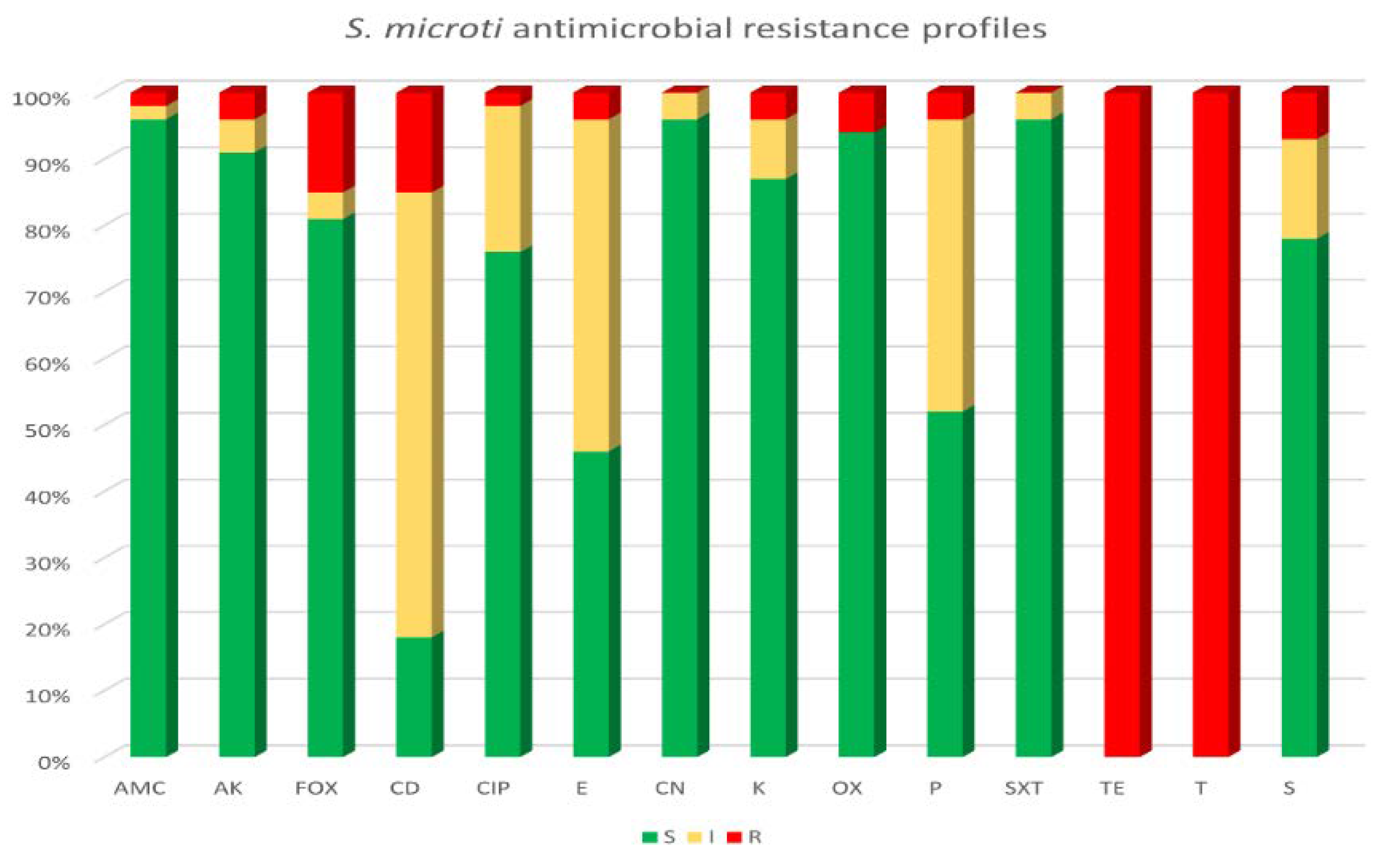
3.4. Antimicrobial Susceptibility Testing and Genotyping Characterization of Tetracycline Resistance in S. microti Strains

The antimicrobial susceptibility testing results highlighted a complete resistance to both tetracycline and oxytetracycline (100%) for all 54 S. microti isolated strains, while the majority of the strains (52/54, 96.5%) were susceptible to amoxicillin–clavulanate, gentamicin and sulfamethoxazole–trimethoprim, followed by amikacin (90.7%, 49/54), kanamycin (87%, 47/54), streptomycin (77.8%, 42/54) and ciprofloxacin (76%, 41/54). Referring to beta-lactams antibiotics, high levels of susceptibility were observed for oxacillin and cefoxitin, being 94.4% (51/54) and 81.5% (44/54), respectively; but nearly half of the isolated strains were resistant to penicillin (51.9%, 28/54). Clindamycin and erythromycin resulted to be the antibiotics with the highest number of strains showing intermediate susceptibility (66.7%, 36/54 and 50%, 27/54, respectively). Figure 1 shows the antimicrobial susceptibility pattern of all bacterial isolates obtained in this study, classifying them as sensitive (S), intermediate (I) or resistance (R).
In addition, an important finding concerns the prevalence of S. microti strains with a multidrug-resistance (MDR) profile, as described in Table 6. In fact, 20.4% (11/54) of the total isolates showed resistance to at least three classes of antibiotics, and all were isolated from the milk samples.
In addition, the genotypic characterization of tetracycline resistance revealed the presence of the tetM gene in all collected S. microti strains. No tetK gene was found.

3.5. Analysis of the Major Constituents of Milk

Data on the nutritional composition of almost all of the milk samples evidenced optimal values as fat (ranging between 7.0 and 8.0 g/100 g), protein (ranging between 3.0 and 4.0 g/100 g) and lactose (4.0 and 5.0 g/100 g) (data not shown). Only one milk sample, exhibiting a positive bacteriological examination with the growth of S. microti and Citrobacter freundii and an SCC > 200 × 103 cells/mL, showed a slight decrease in the value of the tested milk constituents (i.e., fat, protein and lactose).

4. Discussion

Bacteria of the genus Staphylococcus have been considered the main etiological agents associated with cases of SCM [21,22] and CM [23] in buffaloes. However, overall, non-aureus staphylococci (NAS) species are currently the most prevalent pathogens causing SCM, IMI and, rarely, CM in dairy buffaloes [24,25]. The predominance of NAS in milk samples is remarkable [26,27], and their ability to cause inflammatory processes should not be underestimated. Together with microbial analysis, the somatic cell counts (SCCs) contained in milk can provide an accurate evaluation of the milk quality. Based on this principle, in this study, we considered milk samples with an SCC < 200 × 103 cells/mL and negative to bacteriological culture as a healthy status of the buffaloes, and milk samples with an SCC < 200 × 103 cells/mL and a positive bacteriological culture indicative of IMI, and an SCC > 200 × 103 cells/mL with a positive bacteriological culture as SCM cases, as already reported by Puggioni et al. [3]. Moreover, animals with clinical signs, SCC values > 200 × 103 and positive for bacteriological examination were defined as CM cases [3].
The SCC level changed over time through the course of the cases, and the lack of this information represents a limitation of this study; in fact, only 23 animals were sampled twice, and relevant changes were not observed in the SCC values between the two sampling periods.
Our study performed in a buffalo farm determined a statistically significant higher level of IMI (62%) than SCM (11%) cases, with no cases of CM. Several studies have analyzed the prevalence of SCM in buffalo populations around the world, reporting a prevalence of approximately 26.2% and 37.6% in India and Bangladesh, respectively [28,29]. The meta-analysis work conducted by Krishnamoorthy et al. [30] on numerous studies published worldwide during the period 1967–2019 has identified values of 46% and 32% for SCM cases in buffaloes worldwide and in India, respectively. In addition, it has been reported that in SCM mastitis, the percentage of culture-negative milk samples ranged from 28.6% [31] to 38.6% [32], whereas in our study, all milk samples with a high SCC value (>200 × 103 cells/mL) were bacteriologically positive. Our rate of 11% SCM agrees with values between 5 and 20% that were reported by Joshi and Gokhale [33]. In contrast, the percentage of IMI cases that we detected was 62%, a percentage higher than the 40.4% reported by Singha et al. [34] but closer to the value of 63% reported by Moroni et al. [5].
Values of 7.1% or 19.7% have been reported for healthy milk samples with an SCC < 200 × 103 cells/mL and a negative bacteriological examination [3,35], whereas in our study healthy samples were 27%.
The bacterial identification performed in this study, among the isolated NAS, highlighted a new species known as S. microti, a bacterial species that is still not well known on buffalo farms. In fact, the identification of S. microti has been only once reported as the predominant species in the milk of water buffaloes with SCM [11]. Previously, a first case of IMI in cattle was reported in Poland [9] and more recently in a bovine herd in Germany, where S. microti was detected as the causative pathogen of IMI in combination with other NAS [36]. To the best of our knowledge, there are no published studies investigating the presence of S. microti at the same time in milk and milking parlors of buffalo farming. Thus, for the first time, we isolated 54 strains of S. microti over seven samplings, with an average of eight strains per sampling, and no prevalence of isolation between the summer and autumn periods was observed. Moreover, we obtained in 10/41 animals the isolation of S. microti in two different samplings. Precisely, 51 strains were isolated from milk samples and 3 from the milking parlor surfaces, highlighting the predominant presence of this strain in the milk samples.
It is known that sequencing and taxonomic resolution based on 16S rRNA gene alone are limited [37], and a cost-effective alternative are taxonomic analyses using MALDI-TOF. Ribosomal RNA and ribosomal proteins can be used for phylogenetic analysis, being universally conserved in both eukaryotic and prokaryotic cells [38]. The identification of S. microti, here obtained by MALDI-TOF analysis, was perfectly confirmed by the classification method based on the complete sequence analysis of the 16S rRNA gene. However, MALDI-TOF analysis is a time-saving method for the routine identification of bacteria and can achieve 96% accuracy [39].
In addition, S. microti isolates were evaluated for an antibiotic susceptibility profile and all bacteria isolated both from the SCM and IMI cases were found to be 100% resistant to tetracycline and oxytetracycline. The susceptibility to other antibiotics agreed with a previous study that reported a high susceptibility rate to a panel of antibiotics, not including tetracyclines, tested for S. microti strains isolated from bovine milk samples [9]. In our study, 20.4% (11/54) of the S. microti isolates, showing resistance to three or more classes of antibiotics, were defined as MDR. Further molecular investigations are needed for four strains, which resulted to be resistant to cefoxitin but susceptible to penicillins, while two strains were oxacillin-resistant but susceptible to penicillin and cefoxitin. The MDR profile was not detected in the S. microti isolates from the milking parlor surfaces, which exhibited resistance to the tetracycline class and one of them also resistance to lincosamides. Currently, to our knowledge, there are no other studies on the genotypic characterization of tetracycline resistance in S. microti strains from buffalo. In our study, all 54 isolates showed the presence of the tetM gene, justifiable by their isolation performed at the same farm. Previously, the detection of both the tetK and tetM genes in NAS strains has been reported [40,41], as well as in S. aureus isolated from cows with mastitis [42].
The hygienic status of animals is very important to ensure healthy and quality milk, and both microorganisms and SCC should be minimized in raw milk [43]. The monitoring of IMI, SCM, CM and cleaning milking parlor routines represent an important aspect to profitable livestock economics worldwide.
From this study, we can conclude that the pathological and epidemiological roles of S. microti need further investigation as well as its association with IMI or SCM. Furthermore, new information on the presence of this bacterium in Italian buffalo-breeding herds, especially in the Campania Region where buffalo herds represent 80% of the national buffalo assets and is an important zootechnical and economic reality, is very important. In the context of the surveillance of NAS strains and of studies on antibiotic resistance profiles, including resistance gene identification, future work should also be focused on the detection and monitoring of MDR S. microti in wider areas of the Campania Region.

5. Conclusions

The present study revealed the occurrence and antibiotic resistance of S. microti strains isolated from buffalo milk samples and milking parlor surfaces. A limitation of this study is represented by the research performed only in one buffalo herd; thus, further surveillance studies are required to better understand the spread of this bacterial species in buffalo farms and particularly in lactating buffalo and milking parlors. Furthermore, the role of this bacterium and its pathological and epidemiological aspects need further investigation, as well as its association with IMI or SCM.

Supplementary Materials

The following are available online at https://www.mdpi.com/article/10.3390/ani13010182/s1, Table S1: Bacterial species identification by MALDI-TOF of 47 milk samples positive to S. microti growth with or without other bacterial species. All milk samples presented somatic cell count (SCC) < 200 × 103 cells/mL indicative of intramammary infection. Table S2: S. microti and other bacterial species isolated from 3 milking surface samples.

Author Contributions

Conceptualization, M.A. and L.D.M.; methodology, F.G., P.D.L., M.A. and F.P.N.; software, M.A. and P.D.L.; validation, F.P.N., A.G., P.D.L. and L.D.M.; investigation, M.A., F.G. and P.D.L.; data curation, A.G., F.P.N. and L.D.M.; writing—original draft preparation, M.A., F.P.N. and L.D.M.; writing—review and editing, A.G., F.P.N., P.D.L., M.A. and L.D.M. All authors have read and agreed to the published version of the manuscript.

Funding

The study was part of a research project concerning the funding of scholarships financed by the Ministry of Education, University and Scientific Research (MIUR) (D.D. n.1090 of 05.04.2018) under the FSE-ERDF Research and Innovation PON 2014-2020, Action l.1 “Innovative doctorates with industrial characterization”, Department of Veterinary Medicine and Animal Production, University of Naples “Federico II”.

Institutional Review Board Statement

This study was approved by the Institutional Animal Ethics Committee of the University of Naples Federico II (Italy) PG/2020/0092228 of 11.06.2020.

Informed Consent Statement

Not applicable.

Data Availability Statement

Not applicable.

Acknowledgments

The authors would like to thank the owner of the farm and the working staff for their availability.

Conflicts of Interest

The authors declare no conflict of interest. The funder had no role in the design of the study, in the collection, analyses, or interpretation of data, in the writing of the manuscript, or in the decision to publish the results.

References

  1. Nagasawa, Y.; Kiku, Y.; Sugawara, K.; Yabusaki, T.; Oono, K.; Fujii, K.; Suzuki, T.; Maehana, K.; Hayashi, T. The bacterial load in milk is associated with clinical severity in cases of bovine coliform mastitis. J. Vet. Med. Sci. 2019, 81, 107–112. [Google Scholar] [CrossRef] [Green Version]
  2. Middleton, J.R.; Fox, L.K.; Pighetti, G. Laboratory Handbook on Bovine Mastitis (National Mastitis Council), 3rd ed.; National Mastitis Council: New Prague, MN, USA, 2017. [Google Scholar]
  3. Puggioni, G.M.G.; Tedde, V.; Uzzau, S.; Guccione, J.; Ciaramella, P.; Pollera, C.; Moroni, P.; Bronzo, V.; Addis, M.F. Evaluation of a bovine cathelicidin ELISA for detecting mastitis in the dairy buffalo: Comparison with milk somatic cell count and bacteriological culture. Res. Vet. Sci. 2020, 128, 129–134. [Google Scholar] [CrossRef] [PubMed]
  4. Lange, C.C.; Brito, M.A.; Reis, D.R.; Machado, M.A.; Guimarães, A.S.; Azevedo, A.L.; Salles, B.; Alvim, M.C.; Silva, F.S.; Meurer, I.R. Species-level identification of staphylococci isolated from bovine mastitis in Brazil using partial 16S rRNA sequencing. Vet. Microbiol. 2015, 176, 382–388. [Google Scholar] [CrossRef] [PubMed]
  5. Moroni, P.; Rossi, C.S.; Pisoni, G.; Bronzo, V.; Castiglioni, B.; Boettcher, P.J. Relationships between Somatic Cell Count and Intramammary Infection in Buffaloes. J. Dairy Sci. 2006, 89, 998–1003. [Google Scholar] [CrossRef] [PubMed] [Green Version]
  6. Nováková, D.; Pantůček, R.; Hubálek, Z.; Falsen, E.; Busse, H.-J.; Schumann, P.; Sedláček, I. Staphylococcus microti sp. nov., isolated from the common vole (Microtus arvalis). Int. J. Syst. Evol. Microbiol. 2010, 60, 566–573. [Google Scholar] [CrossRef] [PubMed] [Green Version]
  7. Hauschild, T.; Śliżewski, P.; Masiewicz, P. Species distribution of staphylococci from small wild mammals. Syst. Appl. Microbiol. 2010, 33, 457–460. [Google Scholar] [CrossRef] [PubMed]
  8. Riesen, A.; Perreten, V. Staphylococcus rostri sp. nov., a haemolytic bacterium isolated from the noses of healthy pigs. Int. J. Syst. Evol. Microbiol. 2010, 60, 2042–2047. [Google Scholar] [CrossRef] [Green Version]
  9. Król, J.; Wanecka, A.; Twardoń, J.; Mrowiec, J.; Dropińska, A.; Bania, J.; Podkowik, M.; Korzeniowska-Kowal, A.; Paściak, M. Isolation of Staphylococcus microti from milk of dairy cows with mastitis. Vet. Microbiol. 2016, 182, 163–169. [Google Scholar] [CrossRef]
  10. Hu, X.; Shang, Y.; Guo, J.; Zhang, H.; Liang, Y.; Sun, J.; Yue, F. Draft genome sequence of Staphylococcus microti DSM 22147, isolated from the common vole. Genome Announc. 2018, 6, e00420-18. [Google Scholar] [CrossRef] [Green Version]
  11. Addis, M.F.; Maffioli, E.M.; Penati, M.; Albertini, M.; Bronzo, V.; Piccinini, R.; Tangorra, F.; Tedeschi, G.; Cappelli, G.; Di Vuolo, G.; et al. Peptidomic changes in the milk of water buffaloes (Bubalus bubalis) with intramammary infection by non-aureus staphylococci. Sci. Rep. 2022, 12, 8371. [Google Scholar] [CrossRef]
  12. Bannoehr, J.; Ben Zakour, N.L.; Waller, A.S.; Guardabassi, L.; Thoday, K.L.; van den Broek, A.H.M.; Fitzgerald, J.R. Population genetic structure of the Staphylococcus intermedius group: Insights into agr diversification and the emergence of methicillin-resistant strains. J. Bacteriol. 2007, 189, 8685–8692. [Google Scholar] [CrossRef] [PubMed] [Green Version]
  13. Hongoh, Y.; Ohkuma, M.; Kudo, T. Molecular analysis of bacterial microbiota in the gut of the termite Reticulitermes speratus (Isoptera; Rhinotermitidae). FEMS Microbiol. Ecol. 2003, 44, 231–242. [Google Scholar] [CrossRef] [PubMed] [Green Version]
  14. McAllister, S.M.; Davis, R.E.; McBeth, J.M.; Tebo, B.M.; Emerson, D.; Moyer, C.L. Biodiversity and emerging biogeography of the neutrophilic iron-oxidizing Zetaproteobacteria. Appl. Environ. Microbiol. 2011, 77, 5445–5457. [Google Scholar] [CrossRef] [Green Version]
  15. Clinical and Laboratory Standards Institute. Performance Standards for Antimicrobial Disk and Diluition Susceptibility Tests for Bacteria Isolated From Animals. VET01S; Clinical and Laboratory Standards Institute: Wayne, IL, USA, 2015. [Google Scholar]
  16. European Committee on Antimicrobial Susceptibility Testing. Breakpoint Tables for Interpretation of MICs and Zone Diameters; European Committee on Antimicrobial Susceptibility Testing: Växjö, Sweden, 2018; Available online: http://www.eucast.org. (accessed on 20 March 2018).
  17. Magiorakos, A.-P.; Srinivasan, A.; Carey, R.B.; Carmeli, Y.; Falagas, M.E.; Giske, C.G.; Harbarth, S.; Hindler, J.F.; Kahlmeter, G.; Olsson-Liljequist, B.; et al. Multidrug-resistant, extensively drug-resistant and pandrug-resistant bacteria: An international expert proposal for interim standard definitions for acquired resistance. Clin. Microbiol. Infect. 2012, 18, 268–281. [Google Scholar] [CrossRef] [Green Version]
  18. Schwarz, S.; Chaslus-Dancla, E. Use of antimicrobials in veterinary medicine and mechanisms of resistance. Vet. Res. 2001, 32, 201–225. [Google Scholar] [CrossRef] [PubMed] [Green Version]
  19. Qu, Y.; Zhao, H.; Nobrega, D.B.; Cobo, E.R.; Han, B.; Zhao, Z.; Li, S.; Li, M.; Barkema, H.W.; Gao, J. Molecular epidemiology and distribution of antimicrobial resistance genes of Staphylococcus species isolated from Chinese dairy cows with clinical mastitis. J. Dairy Sci. 2019, 102, 1571–1583. [Google Scholar] [CrossRef] [PubMed] [Green Version]
  20. Ullah, F.; Malik, S.A.; Ahmed, J.; Ullah, F.; Shah, S.M.; Ayaz, M.; Hussain, S.; Khatoon, L. Investigation of the genetic basis of tetracycline resistance in Staphylococcus aureus from Pakistan. Trop. J. Pharm. Res. 2012, 11, 925–931. [Google Scholar] [CrossRef] [Green Version]
  21. Dhakal, I.P.; Neupane, M.; Nagahata, H. Evaluation of direct and indirect measures of quarter milk from crossbred buffaloes. Anim. Sci. J. 2008, 79, 628–633. [Google Scholar] [CrossRef]
  22. Vásquez-Garcia, A.; dos Santos Silva, T.; de Alemida-Queiroz, S.R.; Godoy, S.H.S.; Fernandes, A.M.; Sousa, R.L.M.; Franzolin, R. Species identification and antimicrobial susceptibility profile of bacteria causing subclinical mastitis in buffalo. Pesq. Vet. Bras. 2017, 37, 447–452. [Google Scholar] [CrossRef] [Green Version]
  23. Pizauro, L.J.L.; Silva, D.G.; Santana, A.M.; Clemente, V.; Lara, G.H.B.; Listoni, F.J.P.; Vaz, A.C.N.; Vidal-Martins, A.M.C.; Ribeiro, M.G.; Fagliari, J.J. Prevalence and etiology of buffalo mastitis and milk somatic cell count in dry and rainy seasons in a buffalo herd from Analândia, São Paulo State, Brazil. Arq. Bras. Med. Vet. Zootec. 2014, 66, 1703–1710. [Google Scholar] [CrossRef]
  24. Condas, L.A.Z.; De Buck, J.; Nobrega, D.B.; Carson, D.A.; Naushad, S.; De Vliegher, S.; Zadoks, R.N.; Middleton, J.R.; Dufour, S.; Kastelic, J.P.; et al. Prevalence of non-aureus staphylococci species causing intramammary infections in Canadian dairy herds. J. Dairy Sci. 2017, 100, 5592–5612. [Google Scholar] [CrossRef] [Green Version]
  25. Thorberg, B.-M.; Danielsson-Tham, M.-L.; Emanuelson, U.; Persson Waller, K. Bovine subclinical mastitis caused by different types of coagulase-negative staphylococci. J. Dairy Sci. 2009, 92, 4962–4970. [Google Scholar] [CrossRef] [PubMed] [Green Version]
  26. Supré, K.; Haesebrouck, F.; Zadoks, R.N.; Vaneechoutte, M.; Piepers, S.; De Vliegher, S. Some coagulase-negative Staphylococcus species affect udder health more than others. J. Dairy Sci. 2011, 94, 2329–2340. [Google Scholar] [CrossRef] [Green Version]
  27. Hosseinzadeh, S.; Saei, H.D. Staphylococcal species associated with bovine mastitis in the North West of Iran: Emerging of coagulase-negative staphylococci. Int. J. Vet. Sci. Med. 2014, 2, 27–34. [Google Scholar] [CrossRef] [Green Version]
  28. Srinivasan, P.; Jagadeswaran, D.; Manoharan, R.; Giri, T.; Balasubramaniam, G.A.; Balachandran, P. Prevalence and etiology of subclinical mastitis among buffaloes (Bubalus bubalus) in Namakkal, India. Pak. J. Biol. Sci. 2013, 16, 1776–1780. [Google Scholar] [CrossRef] [PubMed] [Green Version]
  29. Hoque, M.N.; Talukder, A.K.; Saha, O.; Hasan, M.M.; Sultana, M.; Rahman, A.A.; Das, Z.C. Antibiogram and virulence profiling reveals multidrug resistant Staphylococcus aureus as the predominant aetiology of subclinical mastitis in riverine buffaloes. Vet. Med. Sci. 2022, 8, 2631–2645. [Google Scholar] [CrossRef] [PubMed]
  30. Krishnamoorthy, P.; Goudar, A.L.; Suresh, K.P.; Roy, P. Global and countrywide prevalence of subclinical and clinical mastitis in dairy cattle and buffaloes by systematic review and meta-analysis. Res. Vet. Sci. 2021, 136, 561–586. [Google Scholar] [CrossRef] [PubMed]
  31. Koivula, M.; Pitkälä, A.; Pyörälä, S.; Mäntysaari, E.A. Distribution of bacteria and seasonal and regional effects in a new database for mastitis pathogens in Finland. Acta Agric. Scand. Sect. A Anim. Sci. 2007, 57, 89–96. [Google Scholar] [CrossRef]
  32. Bradley, A.J.; Leach, K.A.; Breen, J.E.; Green, L.E.; Green, M.J. Survey of the incidence and aetiology of mastitis on dairy farms in England and Wales. Vet. Rec. 2007, 160, 253–258. [Google Scholar] [CrossRef] [Green Version]
  33. Joshi, S.; Gokhale, S. Status of mastitis as an emerging disease in improved and periurban dairy farms in India. Ann. N. Y. Acad. Sci. 2006, 1081, 74–83. [Google Scholar] [CrossRef]
  34. Singha, S.; Ericsson, C.D.; Chowdhury, S.; Nath, S.C.; Paul, O.B.; Hoque, M.A.; Boqvist, S.; Persson, Y.; Rahman, M.M. Occurrence and aetiology of subclinical mastitis in water buffalo in Bangladesh. J. Dairy Res. 2021, 88, 314–320. [Google Scholar] [CrossRef] [PubMed]
  35. Catozzi, C.; Sanchez Bonastre, A.; Francino, O.; Lecchi, C.; De Carlo, E.; Vecchio, D.; Martucciello, A.; Fraulo, P.; Bronzo, V.; Cuscó, A.; et al. The microbiota of water buffalo milk during mastitis. PLoS ONE 2017, 12, e0184710. [Google Scholar] [CrossRef] [PubMed] [Green Version]
  36. Hamel, J.; Zhang, Y.; Wente, N.; Krömker, V. Non-S. aureus staphylococci (NAS) in milk samples: Infection or contamination? Vet. Microbiol. 2020, 242, 108594. [Google Scholar] [CrossRef] [PubMed]
  37. Rajendhran, J.; Gunasekaran, P. Microbial phylogeny and diversity: Small subunit ribosomal RNA sequence analysis and beyond. Microbiol. Res. 2011, 166, 99–110. [Google Scholar] [CrossRef]
  38. Yutin, N.; Puigbò, P.; Koonin, E.V.; Wolf, Y.I. Phylogenomics of prokaryotic ribosomal proteins. PLoS ONE 2012, 7, e36972. [Google Scholar] [CrossRef] [Green Version]
  39. Cameron, M.; Barkema, H.W.; De Buck, J.; De Vliegher, S.; Chaffer, M.; Lewis, J.; Keefe, G.P. Identification of bovine-associated coagulase-negative staphylococci by matrix-assisted laser desorption/ionization time-of-flight mass spectrometry using a direct transfer protocol. J. Dairy Sci. 2017, 100, 2137–2147. [Google Scholar] [CrossRef] [PubMed] [Green Version]
  40. Ruegg, P.L.; Oliveira, L.; Jin, W.; Okwumabua, O. Phenotypic antimicrobial susceptibility and occurrence of selected resistance genes in gram-positive mastitis pathogens isolated from Wisconsin dairy cows. J. Dairy Sci. 2015, 98, 4521–4534. [Google Scholar] [CrossRef] [PubMed] [Green Version]
  41. Mbindyo, C.M.; Gitao, G.C.; Plummer, P.J.; Kulohoma, B.W.; Mulei, C.M.; Bett, R. Antimicrobial Resistance Profiles and Genes of Staphylococci Isolated from Mastitic Cow’s Milk in Kenya. Antibiotics 2021, 10, 772. [Google Scholar] [CrossRef] [PubMed]
  42. Guzmán-Rodríguez, J.J.; León-Galván, M.F.; Barboza-Corona, J.E.; Valencia-Posadas, M.; Loeza-Lara, P.D.; Sánchez-Ceja, M.; Ochoa-Zarzosa, A.; López-Meza, J.E.; Gutiérrez-Chávez, A.J. Analysis of virulence traits of Staphylococcus aureus isolated from bovine mastitis in semi-intensive and family dairy farms. J. Vet. Sci. 2020, 21, e77. [Google Scholar] [CrossRef]
  43. Erdem, H.; Okuyucu, İ.C. Influence of hygiene status of cows on somatic cell count and milk components during summer season. Large Anim. Rev. 2019, 25, 7–10. [Google Scholar]
Figure 1. Antibiotic resistance profiles of 54 S. microti isolates. Tested antibiotics: amoxicillin–clavulanate (AMC), amikacin (AK), cefoxitin (FOX), clindamycin (CD), ciprofloxacin (CIP), erythromycin (E), gentamicin (CN), kanamycin (K), oxacillin (OX), penicillin (P), sulfamethoxazole–trimethoprim (SXT), tetracycline (TE), oxytetracycline (T) and streptomycin (S).
Animals 13 00182 g001
Table 1. Primers used for the PCR sequencing of the 16S ribosomal gene.
PrimerSequenceReference
Smi16-11FGGCGGCGTGCCTAATACATGThis study
Smi16-750FGTGGGGATCAAACAGGATThis study
Smi16FCCTCTTCGGAGGACAAAGTGA[12]
Smi16FdownGAATACGTTCCCGGGTCTTGThis study
Smi16-337RCTGCTGCCTCCCGTAGGThis study
Smi16RupATCCTGTTTGATCCCCACThis study
Smi16-1027RTCACTTTGTCCTCCGAAGAGGThis study
Smi16RGACCCGGGAACGTATTCACC[12]
Smi16-1527RTAGAAAGGAGGTGATCCAGCThis study
B27FAGAGTTTGATCMTGGCTCAG[13]
B1492RTACCTTGTTACGACTT[14]
Table 2. Antibiotic classes tested to define the antimicrobial susceptibility profiles of the isolates.
AntibioticsDisk ContentAntibiotic ClassReference for Breakpoints
Amoxicillin–clavulanate (AMC)20/10 µgPenicillins[15]
Penicillin (P)10 IU
Oxacillin (OX)1 µg
Amikacin (AK)30 µgAminoglycosides[15]
Kanamycin (K)30 µg
Gentamicin (CN)10 µg
Streptomycin (S)10 µg
Cefoxitin (FOX)30 µgCephalosporins[16]
Ciprofloxacin (CIP)5 µgQuinolones[15]
Clindamycin (DA)2 µgLincosamides[15]
Erythromycin (E)15 µgMacrolides[15]
Tetracycline (TE)30 µgTetracyclines[15]
Oxytetracycline (T)30 µg
Sulfamethoxazole–trimethoprim (SXT)25 µgSulfonamides[16]
Table 3. Primer sequences, amplicon size and amplicon program of tetM and tetK genes.
GenePrimer Sequences
(5’-3’ Sense and Antisense)
Amplicon Size (bp)Amplifications
Program
tetMF: AGTTTTAGCTCATGTTGATG
R: TCCGACTATTTAGACGACGG
186294 °C 15 s; 94 °C 1 min, 52 °C 1 min, 72 °C 90 s,
for 30 cycles;
72 °C 5 min
tetKF: GTAGCGACAATAGGTAATAGT
R: GTAGTGACAATAAACCTCCTA
360
Table 4. Bacterial culture results and somatic cell counts (SCCs) in buffalo milk samples.
Bacterial CultureSCC Values (cell/mL)Status *No. of Samples/
200 Samples
%Fisher’s
Two-Tailed
No bacterial growthSCC ≤ 200.000H5427.0%
p < 0.05
SCC > 200.000SCM00%
Bacterial growthSCC ≤ 200.000IMI12462%
p < 0.05
SCC > 200.000SCM2211%
* H (healthy), SCM (subclinical mastitis) and IMI (intramammary infection).
Table 5. Bacterial species and MALDI-TOF scores in 22 buffalo milk samples showing high somatic cells counts (SCC) compatible with the definition of subclinical mastitis (SCM).
Sample SCC/mLIdentified Bacterial Strains
(Colony Forming Unit, CFU)
MALDI-TOF Score
12 × 105 Aeromonas hydrophila (3000 CFU/mL)2.11
22.9 × 10⁵ Acinetobacter johnsonii (5000 CFU/mL)
Staphylococcus simulans (1000 CFU/mL)
2.26
2.20
32.5 × 10⁵Staphylococcus microti (10,000 CFU/mL)
Citrobacter freundii (10,000 CFU/mL)
2.07
1.96
42.8 × 10⁵
Escherichia coli (5000 CFU/mL)
Staphylococcus simulans (1000 CFU/mL)
Staphylococcus sciuri (500 CFU/mL)
2.09
2.20
2.09
52 × 106
Streptococcus agalactiae (6000 CFU/mL)
Lactococcus lactis (1100 CFU/mL)
Aeromonas hydrophila (5000 CFU/mL)
2.10
2.17
1.88
62.7 × 10⁵Aeromonas hydrophila (3000 CFU/mL)2.12
75.3 × 10⁵ Streptococcus agalactiae (10,000 CFU/mL)
Corynebacterium xerosis (1000 CFU/mL)
2.27
2.04
83.6 × 10⁵
Staphylococcus microti (3000 CFU/mL)
Aerococcus viridans (2000 CFU/mL)
Rothia endophytica (200 CFU/mL)
2.04
1.79
1.91
92.9 × 10⁵Aerococcus viridans (2000 CFU/mL)1.81
102.7 × 10⁵Staphylococcus microti (4000 CFU/mL)2.04
112.3 × 10⁵Escherichia coli (4000 CFU/mL)2.16
122.9 × 10⁵Pseudomonas aeruginosa (4000 CFU/mL)2.34
132.2 × 10⁵ Streptococcus agalactiae (3000 CFU/mL)2.43
142.5 × 10⁵ Rothia amarae (6000 CFU/mL)2.05
155.1 × 10⁵ Escherichia coli (2800 CFU/mL)
Rothia amarae (500 CFU/mL)
Aerococcus viridans (700 CFU/mL)
2.22
1.94
1.82
162.9 × 10⁵ Escherichia coli (400 CFU/mL)
Streptococcus agalactiae (5400 CFU/mL)
Aerococcus viridans (200 CFU/mL)
2.14
2.29
1.99
171.5 × 10⁶Escherichia coli (2000 CFU/mL)
Aerococcus viridans (1100 CFU/mL)
2.17
2.14
188.3 × 10⁵Streptococcus agalactiae (5000 CFU/mL)2.19
192.1 × 10⁵Staphylococcus microti (200 CFU/mL)
Aerococcus viridans (100 CFU/mL)
Escherichia coli (500 CFU/mL)
2.06
1.89
2.31
204.5 × 10⁵Aerococcus viridans (400 CFU/mL)1.90
218.8 × 10⁵Streptococcus agalactiae (4700 CFU/mL)2.04
222.3 × 10⁵Aerococcus viridans (4800 CFU/mL)1.99
Table 6. Antibiotic resistance profiles of 11 multidrug-resistant S. microti strains.
PenicillinsAminoglycosidesCephalosporinsQuinolonesLincosamidesMacrolidesTetracyclinesSulfonamides
S. microti
Strains
AMCPOXAKKCNSFOXCIPDAETETSXT
7 R RRR
11 RR R RR
12 R R RR
13RR R RR
14 R R RR
15 R R RR
16 R R RR
17 RR RR
19 R R RR
20 R R RR
22 R R RRR
Disclaimer/Publisher’s Note: The statements, opinions and data contained in all publications are solely those of the individual author(s) and contributor(s) and not of MDPI and/or the editor(s). MDPI and/or the editor(s) disclaim responsibility for any injury to people or property resulting from any ideas, methods, instructions or products referred to in the content.

