Article

Characterization of Salmonella spp. and E. coli Strains Isolated from Wild Carnivores in Janos Biosphere Reserve, Mexico

by
Jonathan J. López-Islas
1,
Estela T. Méndez-Olvera
2,*,
Daniel Martínez-Gómez
2,*,
Andrés M. López-Pérez
3,4,
Libertad Orozco
4,
Gerardo Suzan
3 and
Carlos Eslava
5
1
Doctorado en Ciencias Agropecuarias, Universidad Autónoma Metropolitana, Calz. del Hueso1100, Villa Quietud, Coyoacán, Ciudad de México City 04960, Mexico
2
Departamento de Producción Agrícola y Animal, Universidad Autónoma Metropolitana, Calz. del Hueso 1100, Villa Quietud, Coyoacán, Ciudad de México City 04960, Mexico
3
Departamento de Etología, Fauna Silvestre y Animales de Laboratorio, Facultad de Medicina Veterinaria y Zootecnia, Universidad Nacional Autónoma de México, Avenida Universidad 3000, Ciudad de México City 04510, Mexico
4
Fundación para el Manejo y la Conservación de la Vida Silvestre FMCOVIS A.C., Avenida Universidad 3000, Ciudad Universitaria, Ciudad de México City 04510, Mexico
5
Unidad Periférica Investigación Básica y Clínica de Enfermedades Infecciosas-Hospital Infantil de México Federico Gómez, Facultad de Medicina, Universidad Nacional Autónoma de México, Ciudad de México City 04510, Mexico
*
Authors to whom correspondence should be addressed.
Animals 2022, 12(9), 1064; https://doi.org/10.3390/ani12091064
Submission received: 29 March 2022 / Revised: 15 April 2022 / Accepted: 17 April 2022 / Published: 20 April 2022
(This article belongs to the Special Issue Animal Infectious Diseases and Zoonoses)

Abstract

:

Simple Summary

Emerging diseases (EIDs) represent a constant challenge in public health. With the recent emergence of new pathogens, some questions about the mechanisms and sites where they are generated have aroused interest. Natural environments could be the sites where pathogenic microorganisms find the conditions to generate new variants. It has been established that approximately 60.3% of EIDs are caused by potentially zoonotic pathogens, of which more than half are thought to have originated from wild individuals. In this aspect, carnivores can play an important role in the dynamics of various diseases, since there are species that are widely distributed, roam large areas, and can be carriers of a wide range of microorganisms, some of which are zoonotic. The results obtained in this work show that different species of wild carnivores can be carriers of atypical strains of pathogenic microorganisms, which shows that natural environments can represent important sites for the study of EIDs.

Abstract

Enterobacteriaceae are considered one the most important zoonotic pathogens. In this study, we analyzed the characteristics of E. coli and Salmonella spp. strains present in carnivores from Janos Biosphere Reserve, Mexico. These microorganisms had been isolated from a wide range of domestic and free-range animals, including wild carnivores. Fifty-five individuals were sampled, and the presence of Salmonella and E. coli was determined by bacteriological standard methods. Strains isolated were characterized by molecular methods and in vitro infection assays. Eight different species of carnivores were captured, including coyotes (Canis latrans), gray fox (Urocyon cinereoargenteus), desert foxes (Vulpes macrotis), striped skunks (Mephitis mephitis), hooded skunks (Mephitis macroura), lynxes (Lynx rufus), raccoons (Procyon lotor), and badgers (Taxidea taxus). Salmonella spp. and E. coli were isolated from four species of carnivores. Five Salmonella spp. strains were isolated, and their molecular characterization revealed in three of them the presence of fimbrial and virulence genes associated with cell invasion. In vitro evaluation of these strains showed their capability to invade human Hep2 cells. Sixty-one E. coli strains were isolated; different serotypes and phylogroups were observed from these strains. Additionally, the presence of virulence genes showed differently.

1. Introduction

Emerging infectious diseases (EIDs) are defined as diseases that have recently increased in incidence or geographic range, recently moved into new host populations, recently been discovered, or are caused by newly evolved pathogens [1]. EIDs are a significant problem for public health and global economies. Their emergence is thought to be driven largely by environmental, ecological, and anthropogenic factors s [2]. General strategies to reduce the negative effects of emerging diseases are focused on post-emergence outbreak control, through quarantine procedures, antimicrobial or antiviral drug development, and vaccination. However, delays in recognition of new pathogens in natural areas, lack of scientific research about the genetic characteristics, combined with increased urbanization and global connectivity, have resulted in recent EIDs causing extensive worldwide mortality and important economic damage (e.g., SARS, EHEC O104:H4) [3].
Wildlife populations are considered play an important role in pathogen emergence, by forming the reservoirs from which zoonotic pathogens (pathogens transmissible between animals and humans) may emerge [1]. Enteric infections are one of the most important causes of morbidity and mortality throughout the world, particularly in children. In recent years, scientific research, focused on pathogenesis, treatment development, and prevention strategies, has made important progress. Unfortunately, enteric infections remain an important public health worldwide problem [4]. New outbreaks of enteric infections produced by Salmonella spp., Campylobacter spp., and Shiga toxin-producing Escherichia coli (STEC) are reported each year, with new virulence properties and epidemiological characteristics. This phenomenon showed the capability of pathogen agents to evolve and develop mechanisms for spreading to and infecting new hosts, and therefore represent a public health challenge [5].
Escherichia coli is the most abundant bacteria in the intestines of mammals including humans, and diverse microbiome studies showed their abundance in many species of animals. New pathogenic E. coli strains have been isolated and described in recent years; understanding the origin of these pathogenic strains is important for developing control strategies [6,7]. A significant number of acute E. coli infections are known to have zoonotic origins. Diverse studies have shown that diverse pathogenic E. coli strains are present in natural areas and nonhuman hosts, representing reservoirs for pathogenic strains [8]. Salmonella spp. represents an important zoonotic enteropathogen that affects humans, livestock, wildlife, companion, and zoo animals [9]. Although the intestinal tract of production animals is the primary reservoir for non-typhoidal Salmonella, it is also found in many wildlife species [10]. Strains reported in wildlife have been isolated from humans, suggesting that wildlife species can be reservoirs for Salmonella spp. [11]. In this study, different E. coli and Salmonella spp. strains were isolated from a wild population of carnivores from the Janos Biosphere Reserve Chihuahua, Mexico, and the virulence characteristics of strains were described.

2. Materials and Methods

2.1. Study Area

This study was performed in the Janos Biosphere Reserve (RBJ) in Chihuahua Mexico, at the northwest of Chihuahua, located between meridians 108°56′49″ and 108°56′22″ W and parallels 31°11′7″ and 30°11′27″ N. The reserve has an area of 5264 km2 with an altitude range of 1200–2700 m above sea level. Five sampling locations were selected: “El Cuervo” (30°41′19″ N, 108°15′05″ W), “Ejido San Pedro” (30°53′08″ N, 108°25′57″ W), “Monte Verde” (30°58′28″ N, 108°42′40″ W), “Rancho ojitos” (30°46′52″ N, 108°32′14″ W), and “Rancho San Pedro” (30°41′12″ N, 108°32′46″ W) (Supplementary Figure S1).

2.2. Experimental Design and Animal Management

Wild carnivores were captured at each of the five locations over nine consecutive days per season (autumn and spring), with 16 trapping sets placed at intervals of 500–800 m along a 10 km transect. Each set contained one box trap (Tomahawk Live Trap Inc., Hazelhurst, WI, USA, 76 × 76 × 178 cm or 152 × 51 × 71 cm) and one leg-hold trap (#1.75 or #3 Victor Coil Soft Catch) with at least 30 m distance between them. Each wild carnivore was chemically immobilized with a mixture of ketamine hydrochloride and xylazine hydrochloride (Pisa, México) according to standard doses [12]. Samples of feces were taken through rectal swabs; each swab was placed in a transport medium and kept at 4 °C until processing in the laboratory.

2.3. Isolation and Identification of Salmonella spp. from feces

Feces samples collected from carnivores were inoculated on 10 mL of peptone broth (Oxoid, Monterrey, Mexico) and incubated at 37 °C for 18 h. After, 1 mL was transferred to 10 mL of selenite broth (Merck, Branchburg, NJ, USA) and incubated at 37 °C for 20 h. Each selenite broth was seeded on Salmonella-Shigella agar (Merck, Branchburg, NJ, USA) and MacConkey agar (Merck, Branchburg, NJ, USA) and incubated for 24 h at 37 °C. All colonies with characteristic morphology of Salmonella spp. were transferred to Salmonella-Shigella agar and incubated at 37 °C for 24 h. Finally, microorganisms were identified by standard microbiology methods. Salmonella enterica Typhimurium ATCC 14028 was included in all bacteriological procedures, PCR, and invasion assays, as a reference strain.

2.4. Isolation and Identification of E. coli from Feces

Fecal samples were inoculated into 9 mL buffered peptone water and incubated for 24 h at 37 °C; then 0.1 mL was picked and inoculated onto EMB agar (Oxoid, Monterrey, Mexico) and incubated for 24 h at 37 °C. Thirty-six Green metallic sheen-like colonies from each sample were picked and tested in MRVP broth (Oxoid, Monterrey, Mexico), Simmons citrate (Oxoid, Monterrey, Mexico), SIM agar (Oxoid, Monterrey, Mexico), TSI agar (Oxoid, Monterrey, Mexico), and urea agar (Oxoid, Monterrey, Mexico). Isolates that were positive to the methyl red test (Sigma-Aldrich, St. Louis, MO, USA), negative to the Voges–Prokeur test (Merck, Branchburg, NJ, USA), negative to the Simmons citrate test, negative to the H2S test, positive to the indole test (Merck, Branchburg, NJ, USA), positive to the motility test, negative to the urease production test, and produced acid over acid reaction in the TSI test, were subcultured on EMB agar (Oxoid, Monterrey, Mexico) at 37 °C for 24 h. One pure culture colony was picked using a sterile loop and suspended in 0.1 mL of LB broth (Merck, Branchburg, NJ, USA) for PCR identification. Six E. coli strains, EPEC O127:H6 (E2348/69), EHEC O157:H7 (EOL933), ETEC O78:H11 (H10407), EAEC O42: NM, EIEC O143: NM and UPEC O73: NM., were used as controls in this study. The EPEC O127:H6, EHEC O157:H7, and ETEC O78:H11 are ATCC strains. The EAEC, EIEC, and UPEC (CFT) strains came from human patients with enteric processes, provided by Dr. Carlos A. Eslava Campos, from the Federico Gómez Children’s Hospital.

2.5. E. coli and Salmonella spp. Characterization by PCR

All isolates and control strains of Salmonella spp. and E. coli were seeded in 10 mL of Brain-Heart Infusion broth (Merck, Branchburg, NJ, USA) and incubated for 24 h at 37 °C. The biomass was recuperated by centrifugation at 1200× g for 15 min at 4 °C. For DNA extraction, the CTAB-NaCl method was used [13]. The DNA was used for amplification of virulence and genotyping genes by PCR. A commercial kit (PCR Master Mix 2X, Thermo Scientific, Waltham, MA, USA) was used for the PCR assays, following the manufacturers protocol. Primer sequences and PCR conditions are shown in Supplementary Table S1. PCR products were analyzed by 1.5% agarose gel electrophoresis.

2.6. E. coli Serotyping and Genotyping

Strains identified as E. coli were serotyped by agglutination assays using 96-well micro titer plates and rabbit antisera against O1 to O187 somatic (O) antigens and 53 flagellar (H) antigens prepared in rabbits (SERUNAM, registered trademark in Mexico, with number 323158/2015) using a method previously reported [14] with minor modifications. Phylogenetic groups were determined by PCR multiplex using the primers described in supplementary Table S1 and under conditions detailed by a method previously reported [15]. The designation of the phylogenetic groups (A, B1, B2, C, D, E, F, and Escherichia cryptic clade I) was established by the presence or absence of chuA, yjaA, TspE4.C2, and the arpA gene [15].

2.7. Salmonella spp. Intracellular Survival Assays

Three independent trials were performed with three replicates at a time, according to the standard protocol for intracellular survival with gentamicin, with some modifications. First, all Salmonella strains were grown on Luria Bertani broth supplemented with 0.5% of NaCl for 24 h and harvested in PBS. Bacteria growth was quantified by spectrophotometry and diluted in DMEM (Sigma-Aldrich, St. Louis, MO, USA) at 2 × 10⁶ CFU/mL to prepare inoculum. Hep-2 cells were seeded at 40,000 cells per well in a 48-well dish. After washing three times with PBS, epithelial cells were infected at a multiplicity of infection (MOI) of 100 CFU/cell for 2 h at 37 °C. Following incubation, the monolayers were washed three times with PBS and incubated with DMEM supplemented with 10% fetal bovine serum (Sigma-Aldrich, St. Louis, MO, USA), 50 mM HEPES, and 30 µg/mL gentamicinb (Sigma-Aldrich, St. Louis, MO, USA). At 0, 2, 4, 10, and 24 h post-infection (p.i.), media was removed, and intracellular bacteria were recovered by 1 mL of 1% Triton X-100 (Sigma-Aldrich, St. Louis, MO, USA) addition. The cell lysate was homogenized and transferred to microtubes. In total, 50 µL of each well were diluted and seeded on LB agar for 37 °C for 24 h.

2.8. E. coli Adhesion Assays

The adherence assays were carried out using glass coverslips contained in 24-well microplates at 80% cell confluence as previously described [16]. Hep2 cells were grown on DMEM media (Sigma-Aldrich, St. Louis, MO, USA) supplemented with 10% fetal sera (Sigma-Aldrich, St. Louis, MO, USA), and adhesion assays were carried in DMEM media without sera. E. coli were grown overnight to an OD600 of 0.8–1.2. Cells were inoculated with an infection dose of 1000:1 bacterium per cell and incubated at 37 °C for five hours. The DMEM medium was removed, and the cells were washed three times with PBS. After, 70% methanol was added for 7 min and cells were stained with Giemsa (Sigma-Aldrich, St. Louis, MO, USA) (10% solution in ddH2O) for 40 min at room temperature. Slides were evaluated for adhering bacteria by light microscopy. All tests were repeated at least three times in triplicates. Two distinct patterns of adherence were considered: localized adherence, when the bacteria attached to confined areas of the HEp-2 cells in culture, and diffuse adhesion (DA), when bacteria adhered to the entire surface of the HEp-2.

2.9. Statistical Analysis

Descriptive and comparative statistics were performed using R software. Salmonella spp. and E. coli frequencies by species were established. An analysis of variance (ANOVA) was performed to confirm the significant differences between the means of each infection time in invasion assays.

3. Results

3.1. Animals Captured

During autumn and spring, a total of 55 carnivores were captured. In autumn, 24 specimens were captured: 7 coyotes (Canis latrans), 1 gray fox (Urocyon cinereoargenteus), 5 desert foxes (Vulpes macrotis), 2 striped skunks (Mephitis mephitis), 3 hooded skunks (Mephitis macroura), 4 lynxes (Lynx rufus), 1 raccoon (Procyon lotor), and 1 badger (Taxidea taxus). In spring, 31 individuals were captured: 8 coyotes, 9 desert foxes, 5 gray foxes, 1 lynx, 3 raccoons, and 5 badgers. Coyote and desert foxes represented the species with the highest capture frequency, with 27.2% and 22.7%, respectively (Table 1).

3.2. Isolation and Identification of E. coli and Salmonella spp. from Feces

To isolate Salmonella spp. and E. coli in the carnivore population of RBJ, a standard bacteriological method was used. From 55 samples of feces recovered through rectal swabs, five strains of Salmonella spp. were isolated (9.09%). The Salmonella isolates were retrieved from 1 lynx (Lynx rufus), 1 striped skunk (Mephitis mephitis), 1 coyote (Canis latrans), and 2 gray foxes (Urocyon cinereoargenteus) (Table 1).
From the same 55 samples of feces, 1980 strains of E. coli were isolated. From these strains, 61 showed positive PCR results for at least one virulence gene evaluated in this study (Supplementary Table S2). The E. coli isolates were retrieved from 3 coyotes (Canis latrans), 2 desert foxes (Vulpes macrotis), and 2 striped skunks (Mephitis mephitis) (Table 1). It is important to clarify that in this part, only animals with isolations of strains with virulence genes are mentioned. The isolation of commensal E. coli was achieved from all individuals.
All Salmonella isolates were characterized by PCR to describe the presence of virulence genes (Table 2). In this analysis, a 16s-ITS set of primers was included to confirm bacteriological identification. All Salmonella strains showed positive results for 16s-ITS, PCR, but only three strains had positive results for invA, cdtB, and sopE genes. These strains were isolated from a striped skunk (Mephitis mephitis) and 2 gray foxes (Urocyon cinereoargenteus). Considering that invA and sopE genes are essential virulence factors associated with cell invasion [17,18], strains with negative results for these genes were not included in the analysis of fimbrial operons and invasion assays (Table 2). For PCR analysis of fimbrial operons, the strain 1-B-Uro showed positive results for all evaluated genes. On the other hand, strains 1-BR-Uro and Meph 18 lacked fimA and steA genes, and the strain Meph 18 was also deficient in the stfA gene (Table 2).
Serotyping of the 61 isolations identified as E. coli strains indicated that 46 (75.4%) belonged to different, recognized E. coli serogroups and 15 (24.6%) were non-typable (O?). Of the strains with a recognized O group, 17 (36.9%) belonged to O153 pathogenic serogroups, 5 (8.19%) to O139 serogroups, and 4 (6.55%) to O21 serogroup (Table 3 and Supplementary Table S2). Most of the E. coli strains were included in an unknown group (63.9%), with only 31.14% belonging to virulent groups B2, D, and E (Table 3). The analysis showed that only 3 strains (4.9%) belonged to commensal group A (Table 3).
Supplementary Table S2 shows the results obtained from genetic evaluation of pathogenic characteristics of the 61 E. coli strains isolated from carnivores. The presence of stx1 and stx2 genes was observed in 25 (40.98%) E. coli strains and of these, only 5 (8.19%) showed the presence of both genes, while the remaining 20 strains (32.78%) were positive for only one gene; stx1 (3 strains) and stx2 (17 strains). The presence of the escV and eae genes was observed in 36 strains (59.02%) and of these, 7 strains (11.47) were positive for both genes. These strains were evaluated in cell culture to evaluate their capability to produce localized adhesion (Figure 1). Twenty-three E. coli strains were positive for the escV gene, and six for the eae gene. The escV-eae positive E. coli strains belonged to different serotypes, O153H17 and O63H6, and different phylogroups, E, B2, and unknown. For stx1-stx2 positive strains the serotypes observed were O75H38, O17H18, and O110H28, and all belonged to the unknown phylogroup.

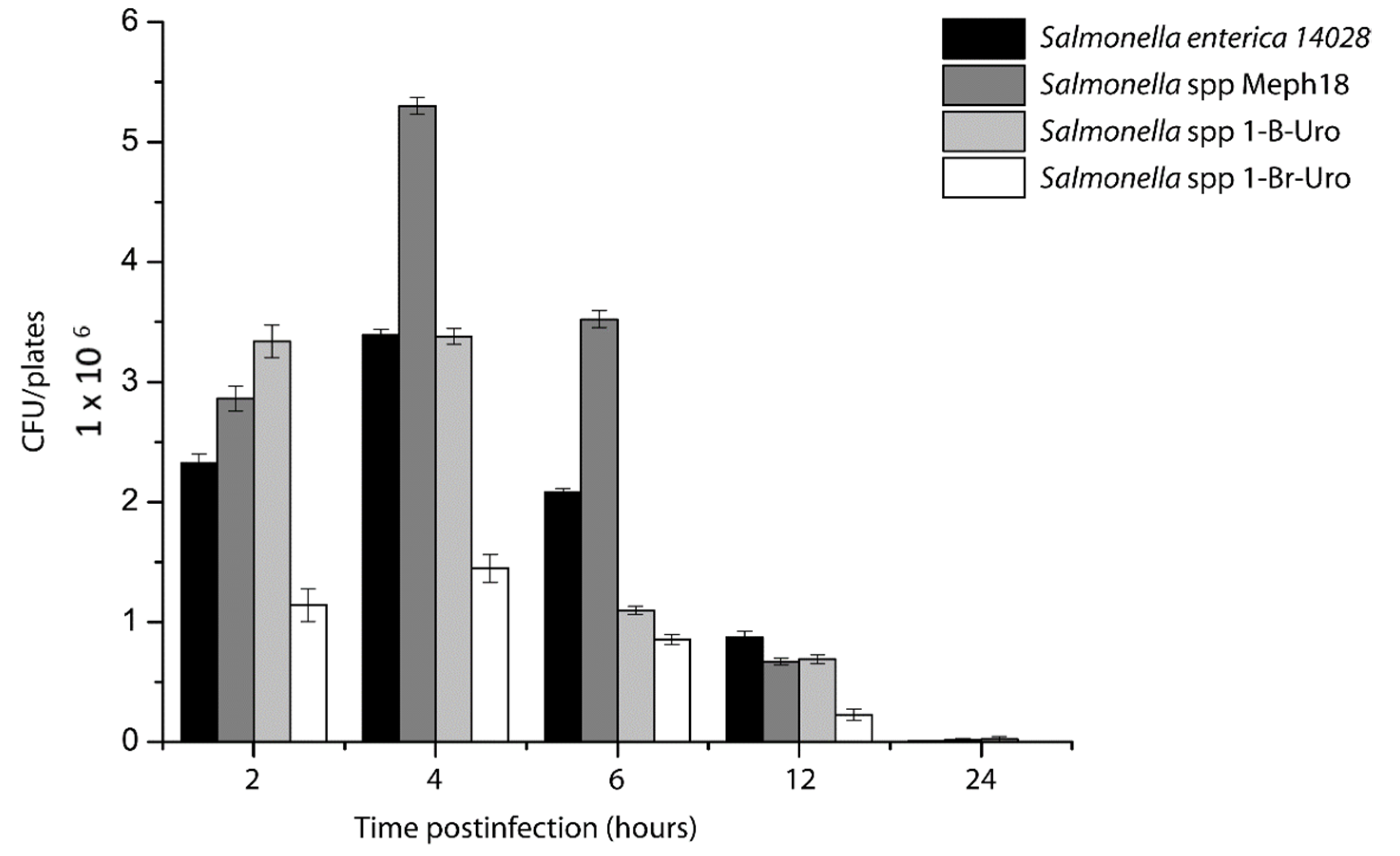
3.3. Salmonella spp. Intracellular Survival Assays

Salmonella strains showed different capabilities to invade and replicate inside of Hep-2 epithelial cells on intracellular survival assays. In these assays, strain Meph 18 showed the highest ability to replicate inside epithelial cells, even more than the reference strain included in this analysis (Figure 2). At 2 h post-infection, the number of intracellular microorganisms from strain Meph 18 was 5.3 × 10⁶ CFU/mL, in comparison with 3.39 × 10⁶ CFU/mL of Salmonella Typhimurium ATCC14028. Meanwhile, for 1-B-Uro and 1-BR-Uro, the number of intracellular microorganisms at this same time were 3.38 and 1.44 × 10⁶ CFU/mL, respectively. The strain 1-BR-Uro showed a reduced ability to replicate inside of epithelial cells (Figure 2). After 4 h post-infection, all strains showed a reduction in the number of CFU/mL, but this could result from loss of cell monolayer. At 24 h post-infection, more than 80% of the cells were lysed by Salmonella, compared with 4 h post-infection, where more than 85% of the monolayer was present (data not shown). The results obtained in intracellular survival assays demonstrated that Salmonella strains isolated from carnivores were virulent for human cells at different levels.

3.4. E. coli Adhesion Assays

Figure 1 shows the occurrence of localized adhesion (LA) in E. coli strains isolated from feces. LA occurred with the seven E. coli strains with positive PCR results for eae and escV evaluated in this study. The results of the adhesion assays showed that at 1:40 h post-infection, all strains showed a LA pattern. Modifications in the cell membrane with a pedestal shape were also observed at 2:30 h postinfection. In the control strain, AL was observed at 1:40 h and the pedestal-like structure at 2:30 h postinfection.

4. Discussion

Identification of animal reservoirs for bacterial pathogens are important for understanding and preventing disease emergence. Salmonella spp. and E. coli are important pathogens for public health, usually associated with foodborne diseases. However, animal sources of these pathogens are also critical in transmission; therefore, broad research has been directed to wildlife reservoirs lately [19,20]. In this study, a community of carnivores in the Biosphere Reserve in the northwest of Mexico was analyzed to describe the genetic and virulent characteristics of Salmonella spp. and E. coli strains present in this area. Previous studies related to the composition of the carnivore population in the Janos Biosphere Reserve had reported 14 species of wild carnivores [21]. In this study, different Salmonella and E. coli strains were isolated from diverse carnivore species in the Janos Biosphere Reserve; the analysis of virulence genes, phylogroups, and serotypes showed an important diversity of these pathogens in this area (Table 2 and Table 3). Interestingly some E. coli serotypes were specific for each carnivore species; for example, the serogroup O153 was only found in Desert fox (Vulpes macrotis) and serogroup O21 in Coyote (Canis latrans). This phenomenon was also observed for phylogroup B2, which was only detected in Hooded skunk (Mephitis macroura).
The results obtained showed the existence of Salmonella and E. coli in only 4 of 55 animals captured. Salmonella spp. strains were recuperated from coyote (Canis latrans), Gray fox (Urocyon cinereoargenteus), Striped skunk (Mephitis mephitis), and Lynx (Linx rufus). There are several reports about the presence of Salmonella spp. in carnivores such as civets, otters, raccoons, foxes, coyotes, and wild felids [22,23,24,25,26,27]. In these studies, the reported frequencies range from 2.1% to 65%. The frequency of 9.09% (5/55 carnivores) in our study is much lower than the 49% reported in free-ranging raccoons in Costa Rica [28] but higher than the prevalence of 7.4% in a study of raccoons in Pennsylvania [26]. Salmonella sp. has been also isolated from other wild carnivores such as red fox (Vulpes vulpes), raccoon (Procyon lotor), badger (Meles meles), coyote (Canis latrans), masked palm civets (Paguma larvata), and otter (Lutra lutra) [22,26,27,29,30,31].
In this study, the Salmonella strains were analyzed for the presence of different virulence genes associated with intracellular invasion, survival, and bacterial adherence. The results showed only three strains positive for invA and sopE, genes associated with the capability to invade cells. From these strains, only one was positive for all fimbrial operons evaluated in this study, and the rest have a deficiency of two operons. Various reports describe that all Salmonella spp. strains (from different serotypes), with positive results for the invA gene, can be associated with invasive strains [17,32]. The results obtained in invasion assays with Salmonella spp. strains isolated from carnivores are congruent with this statement.
Previous report showed that diverse fimbriae provide to Salmonella Typhimurium the capability to adhere to different cell types; for example, the adhesion of this bacteria to HEp-2 cells has been proposed to be mediated by type 1 fimbriae [32,33]. However, in this study, in two Salmonella spp. strains, the type I fimbriae was not present; thus, the invasion of Hep2 cells by these strains should be affected. Interestingly, one of these strains (Meph 18) showed a high capability to invade Hep-2 cells, even more than reference strain S. typhimurium 14028. This result suggested that other fimbriae could participate in bacterial adhesion. Bäumler [33] reported that lpf fimbrial operon was enough to promote S. typhimurium adhesion to HEp-2 cells. In survival assays, only two strains showed a capability to replicate inside Hep2 cells, 1-B-Uro and Mep18A. This last strain was hyperinvasive in contrast to the 1-BR-Uro strain, which did not replicate. In other studies, Salmonella spp. strains isolated from reptiles showed a high capability to invade human cells [34,35]. In our study, we identified only two invasive strains. These data suggest that Salmonella spp. strains recovered from wildlife were able to invade human cells.
Previous studies had recognized red foxes (Vulpes vulpes), European badgers (Meles meles), and coyotes as possible reservoirs of Salmonella spp. [29,36,37,38]. In this study, the species with the highest frequency of Salmonella spp. were desert foxes, coyotes, and badgers. This result could support the hypothesis that these carnivores could be natural reservoirs for Salmonella spp.; however, more studies are necessary to confirm this hypothesis. Finally, cattle had also been described as a reservoir of Salmonella spp. [39,40]. Therefore, their presence in wild areas could result in the possible spread of Salmonella spp. from domestic to wild animals, altering infectious disease dynamics and creating opportunities for cross-species transmission. These phenomena carry implications for wildlife disease management and highlight areas for future work.
The presence of E. coli in different hosts is normal since it is a microorganism that is part of the intestinal microflora [41]. Therefore, it is not unusual to find E. coli in carnivores; however, for this microorganism, diverse pathogenic variants have been described, based on virulence genes, serotypes, and phylogroups. In this study these criteria were used to evaluate E. coli strains isolated from carnivores.
Some of the serogroups found in this study, such as O153:H7, O139:21, and O21:H21, are related to Shiga toxin-producing strains. O153:H7 has been found associated to hemorrhagic enteritis in rabbits, and some strains showed an atypical EPEC profile with the presence of escV and eae genes. Strains O139:21 and O21:21 are also associated with enteric infections in humans and are also related with Shiga toxin-producing strains. Finally, E. coli O17H18 strains have been associated with urinary tract infection [42].
The presence of E. coli strains positive for escV and eae genes, able to produce localized adhesion and pedestal-type structure in human cells, indicates the importance of analyzing E. coli strains isolated from wild animals, like a potential source of virulence strains able to cause human infections. There is no clear explanation for why these pathogenic strains were found in carnivores.
The higher percentage of strains belonging to unknown phylogenetic groups found in the present study is similar to previous findings with strains retrieved from wild animals [43].

5. Conclusions

The distribution and incidence of emerging diseases has increased in recent years, causing economic losses and problems in public and animal health. One of the reasons for this phenomenon is the recent evolution of pathogenic microorganisms present in natural environments, with their subsequent dissemination outside their areas. In this study, the virulence characteristics of Salmonella spp. and E. coli strains isolated from carnivores in the Janos Biosphere Reserve were analyzed. The results obtained show that different species of wild carnivores can carry atypical strains of pathogenic microorganisms, suggesting the presence of potential pathogens in natural environments. On the other hand, the genetic analysis of E. coli strains showed that it is necessary to increase strain classification systems, since in wild animals the variations in E. coli may be different from those described with human strains.

Supplementary Materials

The following supporting information can be downloaded at: https://www.mdpi.com/article/10.3390/ani12091064/s1, Figure S1: Localization of the study area and sampling sites; Table S1: Primer sequences and conditions for Salmonella spp. and E. coli genetic characterization by PCR; Table S2: Characteristics of E. coli strains isolated from carnivores. References [44,45] are cited in the supplementary materials.

Author Contributions

J.J.L.-I., D.M.-G., E.T.M.-O. and A.M.L.-P. designed the research. J.J.L.-I., L.O. and E.T.M.-O. conducted the research. D.M.-G., G.S. and C.E. analyzed the data. J.J.L.-I., D.M.-G., E.T.M.-O., A.M.L.-P. wrote the manuscript. D.M.-G., G.S. and C.E. funding acquisition J.J.L.-I., E.T.M.-O. and D.M.-G. are responsible for the final content of the manuscript. All authors have read and agreed to the published version of the manuscript.

Funding

This research was supported in part by Universidad Autónoma Metropolitana, Unidad Xochimilco (project “Laboratorio de Microbiología Agropecuaria”), by CONACyT (Grants No. 179482 and 2016-01-1851, CONACYT-becario 747804), and in part by Fundación para el Manejo y Conservación de la Vida Silvestre A.C.

Institutional Review Board Statement

The experiment was carried out in accordance with Official Mexican Norm (NOM-059- SEMARNAT-2010) guidelines for environmental protection—Mexican native species of wild flora and fauna. All procedures for trapping and handling carnivores were approved by the Mexican Secretary of Environment and Natural Resources (SEMANART-Permit FAUT-0250) and by the Institutional Animal Care and Use Committee (Comité Institucional para el cuidado y uso de los animales experimentales—CICUAE FMVZUNAM) (Permit FMED/CI/JMO/004/2012).

Informed Consent Statement

Not applicable.

Data Availability Statement

The raw data supporting the conclusions of this manuscript will be made available by the authors, without undue reservation, to any qualified researcher.

Acknowledgments

We are grateful A. Vigueras A. Rubio, and K. Moreno for helping during field sampling. We thank E. Ponce, and R. Sierra (Janos Grassland Biological Station, IE-UNAM), and A. Esquer (Rancho El Uno TNC) for logistical support in the field. This work was supported by Universidad Autónoma Metropolitana, Unidad Xochimilco, CONACyT (Grants No. 179482 and 2016-01-1851, CONACYT-becario 747804) and Fundación para el Manejo y Conservación de la Vida Silvestre A.C.

Conflicts of Interest

The authors declare no conflict of interest.

References

  1. Daszak, P.; Cunningham, A.; Hyatt, A. Anthropogenic environmental change and the emergence of infectious diseases in wildlife. Acta Trop. 2001, 78, 103–116. [Google Scholar] [CrossRef]
  2. Jones, K.E.; Patel, N.G.; Levy, M.A.; Storeygard, A.; Balk, D.; Gittleman, J.L.; Daszak, P. Global trends in emerging infectious diseases. Nature 2008, 451, 990–993. [Google Scholar] [CrossRef] [PubMed]
  3. Allen, T.; Murray, K.; Zambrana-Torrelio, C.; Morse, S.S.; Rondinini, C.; Di Marco, M.; Breit, N.; Olival, K.; Daszak, P. Global hotspots and correlates of emerging zoonotic diseases. Nat. Commun. 2017, 8, 1124. [Google Scholar] [CrossRef] [PubMed]
  4. Wolf, M.K. Bacterial infections of the small intestine and colon. Curr. Opin. Gastroenterol. 2000, 16, 4–11. [Google Scholar] [CrossRef] [PubMed]
  5. Aboutaleb, N.; Kuijper, E.J.; van Dissel, J.T. Emerging infectious colitis. Curr. Opin. Gastroenterol. 2014, 30, 106–115. [Google Scholar] [CrossRef] [PubMed]
  6. Murphy, R.; Palm, M.; Mustonen, V.; Warringer, J.; Farewell, A.; Parts, L.; Moradigaravand, D. Genomic Epidemiology and Evolution of Escherichia coli in Wild Animals in Mexico. mSphere 2021, 6, e00738-20. [Google Scholar] [CrossRef]
  7. García, A.; Fox, J.G.; Besser, T.E. Zoonotic Enterohemorrhagic Escherichia coli: A One Health Perspective. ILAR J. 2010, 51, 221–232. [Google Scholar] [CrossRef]
  8. Tenaillon, O.; Skurnik, D.; Picard, B.; Denamur, E. The population genetics of commensal Escherichia coli. Nat. Rev. Genet. 2010, 8, 207–217. [Google Scholar] [CrossRef]
  9. Forshell, L.P.; Wierup, M. Salmonella contamination: A significant challenge to the global marketing of animal food products. Rev. Sci. Tech. 2006, 25, 541–554. [Google Scholar]
  10. Hilbert, F.; Smulders, F.J.M.; Chopra-Dewasthaly, R.; Paulsen, P. Salmonella in the wildlife-human interface. Food Res. Int. 2012, 45, 603–608. [Google Scholar] [CrossRef]
  11. Ashbolt, R.; Kirk, M.D. Salmonella Mississippi infections in Tasmania: The role of native Australian animals and untreated drinking water. Epidemiol. Infect. 2006, 134, 1257–1265. [Google Scholar] [CrossRef] [PubMed]
  12. Kreeger, T.J.; Arnemo, J.M.; Jacobus, P.R. Handbook of Wildlife Chemical Immobilization; Wildlife Pharmaceuticals: Laramie, WY, USA, 2002. [Google Scholar]
  13. Sambrook, J.; Fritsch, E.F.; Maniatis, T. Molecular Cloning: A Laboratory Manual; Cold Spring Laboratory, Cold Spring Harbor: New York, NY, USA, 1989; Volume 2. [Google Scholar]
  14. Orskov, F.; Orskov, I. Serotyping of Escherichia coli; Bergan, T., Ed.; Academic Press Ltd.: London, UK, 1984; pp. 43–112. [Google Scholar]
  15. Clermont, O.; Christenson, J.; Denamour, E.; Gordon, D. The Clermont Escherichia coli phylotyping method revisited: Improvement of specificity and detection of new phylogroups. Environ. Microbiol. Rep. 2013, 5, 58–65. [Google Scholar] [CrossRef] [PubMed]
  16. Santos, F.F.; Yamamoto, D.; Abe, C.M.; Bryant, J.A.; Hernandes, R.T.; Kitamura, F.C.; Castro, F.S.; Valiatti, T.B.; Piazza, R.M.F.; Elias, W.P.; et al. The Type III Secretion System (T3SS)-Translocon of Atypical Enteropathogenic Escherichia coli (aEPEC) Can Mediate Adherence. Front. Microbiol. 2019, 10, 1527. [Google Scholar] [CrossRef] [PubMed] [Green Version]
  17. Galan, J.E.; Ginocchio, C.; Costeas, P. Molecular and functional characterization of the Salmonella invasion gene invA: Homology of InvA to members of a new protein family. J. Bacteriol. 1992, 174, 4338–4349. [Google Scholar] [CrossRef] [PubMed] [Green Version]
  18. Deleu, S.; Choi, K.; Reece, J.M.; Shears, S.B. Pathogenicity of Salmonella: SopE-mediated membrane ruffling is independent of inositol phosphate signals. FEBS Lett. 2006, 580, 1709–1715. [Google Scholar] [CrossRef] [Green Version]
  19. Ashida, H.; Ogawa, M.; Kim, M.; Mimuro, H.; Sasakawa, C. Bacteria and host interactions in the gut epithelial barrier. Nat. Chem. Biol. 2011, 8, 36–45. [Google Scholar] [CrossRef]
  20. LaRock, D.; Chaudhary, A.; Miller, S.I. Salmonellae interactions with host processes. Nat. Rev. Genet. 2015, 13, 191–205. [Google Scholar] [CrossRef]
  21. Pacheco, J.; Ceballos, G.; List, R. Los mamíferos de la región de Janos-Casas Grandes, Chihuahua, México. Rev. Mex. Mastozoología. 2000, 4, 69–83. [Google Scholar]
  22. Millan, J.; Aduriz, G.; Moreno, B.; Juste, R.; Barral, M. Salmonella isolates from wild birds and mammals in the Basque Country (Spain). Rev. Sci. Tech. 2004, 23, 905–911. [Google Scholar] [CrossRef]
  23. Miller, M.A.; Byrne, B.A.; Jang, S.S.; Dodd, E.M.; Dorfmeier, E.; Harris, M.D.; Ames, J.; Paradies, D.; Worcester, K.; Jessup, D.A.; et al. Enteric bacterial pathogen detection in southern sea otters (Enhydra lutris nereis) is associated with coastal urbanization and freshwater runoff. Vet. Res. 2009, 41, 1. [Google Scholar] [CrossRef] [Green Version]
  24. Oliveira, M.; Pedroso, N.M.; Sales-Luís, T.; Santos-Reis, M.; Tavares, L.; Vilela, C.L. Antimicrobial-Resistant Salmonella Isolated from Eurasian Otters (Lutra lutra Linnaeus, 1758) in Portugal. J. Wildl. Dis. 2010, 46, 1257–1261. [Google Scholar] [CrossRef] [Green Version]
  25. Jardine, C.; Reid-Smith, R.J.; Janecko, N.; Allan, M.; McEwen, S.A. Salmonella in Raccoons (Procyon Lotor) in Southern Ontario, Canada. J. Wildl. Dis. 2011, 47, 344–351. [Google Scholar] [CrossRef] [Green Version]
  26. Compton, J.A.; Baney, J.A.; Donaldson, S.C.; Houser, B.A.; San Julian, G.J.; Yahner, R.H.; Chmielecki, W.; Reynolds, S.; Jayarao, B.M. Salmonella infections in the common raccoon (Procyon lotor) in western Pennsylvania. J. Clin. Microbiol. 2008, 46, 3084–3086. [Google Scholar] [CrossRef] [Green Version]
  27. Lee, K.; Iwata, T.; Nakadai, A.; Kato, T.; Hayama, S.; Taniguchi, T.; Hayashidani, H. Prevalence of Salmonella, Yersinia and Campylobacter spp. in Feral Raccoons (Procyon lotor) and Masked Palm Civets (Paguma larvata) in Japan. Zoonoses Public Health 2011, 58, 424–431. [Google Scholar] [CrossRef]
  28. Baldi, M.; Calvo, E.B.; Hutter, S.E.; Walzer, C. Salmonellosis detection and evidence of antibiotic resistance in an urban raccoon population in a highly populated area, Costa Rica. Zoonoses Public Health 2019, 66, 852–860. [Google Scholar] [CrossRef] [PubMed] [Green Version]
  29. Handeland, K.; Nesse, L.L.; Lillehaug, A.; Vikøren, T.; Djønne, B.; Bergsjø, B. Natural and experimental Salmonella Typhimurium infections in foxes (Vulpes vulpes). Vet. Microbiol. 2008, 132, 129–134. [Google Scholar] [CrossRef] [PubMed]
  30. Jay-Russell, M.T.; Hake, A.F.; Bengson, Y.; Thiptara, A.; Nguyen, T. Prevalence and Characterization of Escherichia coli and Salmonella Strains Isolated from Stray Dog and Coyote Feces in a Major Leafy Greens Production Region at the United States-Mexico Border. PLoS ONE 2014, 9, e113433. [Google Scholar] [CrossRef]
  31. Very, K.J.; Kirchner, M.K.; Shariat, N.; Cottrell, W.; Sandt, C.H.; Dudley, E.G.; Kariyawasam, S.; Jayarao, B.M. Prevalence and Spatial Distribution of Salmonella Infections in the Pennsylvania Raccoon (Procyon lotor). Zoonoses Public Health 2015, 63, 223–233. [Google Scholar] [CrossRef] [PubMed]
  32. Douce, G.R.; Amin, I.I.; Stephen, J. Invasion of HEp-2 cells by strains of Salmonella Typhimurium of different virulence in relation to gastroenteritis. J. Med. Microbiol. 1991, 35, 349–357. [Google Scholar] [CrossRef] [PubMed] [Green Version]
  33. Bäumler, A.J.; Tsolis, R.M.; Heffron, F. The lpf fimbrial operon mediates adhesion of Salmonella typhimurium to murine Peyer’s patches. Proc. Natl. Acad. Sci. USA 1996, 93, 279–283. [Google Scholar] [CrossRef] [PubMed] [Green Version]
  34. Mokracka, J.; Krzymińska, S.; Ałtunin, D.; Wasyl, D.; Koczura, R.; Dudek, K.; Dudek, M.; Chyleńska, Z.A.; Ekner-Grzyb, A. In Vitro virulence characteristics of rare serovars of Salmonella enterica isolated from sand lizards (Lacerta agilis L.). Int. J. Gen. Mol. Microbiol. 2018, 111, 1863–1870. [Google Scholar] [CrossRef] [PubMed] [Green Version]
  35. McWhorter, A.; Owens, J.; Valcanis, M.; Olds, L.; Myers, C.; Smith, I.; Trott, D.; McLelland, D. In vitro invasiveness and antimicrobial resistance of Salmonella enterica subspecies isolated from wild and captive reptiles. Zoonoses Public Health 2021, 68, 402–412. [Google Scholar] [CrossRef] [PubMed]
  36. Gese, E.M.; Schultz, R.D.; Johnson, M.R.; Williams, E.S.; Crabtree, R.L.; Ruff, R.L. Serological survey for disease in free-ranging coyotes (Canis latrans) in Yellowstone National Park, Wyoming. J. Wildl. Dis. 1997, 33, 47–56. [Google Scholar] [CrossRef] [PubMed]
  37. Wilson, J.S.; Hazel, S.M.; Williams, N.J.; Phiri, A.; French, N.; Hart, C.A. Nontyphoidal Salmonellae in United Kingdom Badgers: Prevalence and Spatial Distribution. Appl. Environ. Microbiol. 2003, 69, 4312–4315. [Google Scholar] [CrossRef] [Green Version]
  38. Chiari, M.; Ferrari, N.; Giardiello, D.; Lanfranchi, P.; Zanoni, M.; Lavazza, A.; Alborali, L.G. Isolation and identification of Salmonella spp. from red foxes (Vulpes vulpes) and badgers (Meles meles) in northern Italy. Acta Vet Scand 2014, 56, 1–4. [Google Scholar] [CrossRef] [Green Version]
  39. Miranda, J.M.; Mondragón, A.C.; Martinez, B.; Guarddon, M.; A Rodriguez, J. Prevalence and antimicrobial resistance patterns of Salmonella from different raw foods in Mexico. J. Food Prot. 2009, 72, 966–971. [Google Scholar] [CrossRef]
  40. Glawischnig, W.; Wallner, A.; Lazar, J.; Kornschober, C. Cattle-derived Salmonella enterica serovar Dublin infections in red foxes (Vulpes vulpes) in Tyrol, Austria. J. Wildl. Dis. 2017, 53, 361–363. [Google Scholar] [CrossRef]
  41. Krause, G.; Zimmermann, S.; Beutin, L. Investigation of domestic animals and pets as a reservoir for intimin- (eae) gene positive Escherichia coli types. Vet. Microbiol. 2005, 106, 87–95. [Google Scholar] [CrossRef]
  42. Manges, A.R.; Tabor, H.; Tellis, P.; Vincent, C.; Tellier, P.P. Endemic and epidemic lineages of Escherichia coli that cause urinary tract infections. Emerg. Infect. Dis. 2008, 10, 1575–1583. [Google Scholar] [CrossRef]
  43. Clermont, O.; Olier, M.; Hoede, C.; Diancourt, L.; Brisse, S.; Keroudean, M.; Glodt, J.; Picard, B.; Oswald, E.; Denamur, E. Animal and human pathogenic Escherichia coli strains share common genetic backgrounds. Infect Genet Evol. 2011, 11, 654–662. [Google Scholar] [CrossRef]
  44. Chiu, T.H.; Chen, T.R.; Hwang, W.Z.; Tsen, H.Y. Sequencing of an internal transcribed spacer region of 16S-23S rRNA gene and designing of PCR primers for the detection of Salmonella spp. in food. Int. J. Food Microbiol. 2005, 97, 259–265. [Google Scholar] [CrossRef] [PubMed]
  45. Müller, D.; Hagedorn, P.; Brast, S.; Heusipp, G.; Bielaszewska, M.; Friedrich, A.W.; Karch, H.; Schmidt, M.A. Rapid identification and differentiation of clinical isolates of enteropathogenic Escherichia coli (EPEC), atypical EPEC, and Shiga toxin-producing Escherichia coli by a one-step multiplex PCR method. J. Clin. Microbiol. 2006, 44, 2626–2629. [Google Scholar] [CrossRef] [PubMed] [Green Version]
Figure 1. E. coli adhesion assays in Hep-2 cells. (A) Uninfected Hep-2 cells, (B) Hep-2 cells infected with control strain EPEC O127:H6, (CF) E. coli strains escV+, eae+ isolated from carnivores. Localized adhesions are observed, also lesions suggestive of pedestals.
Animals 12 01064 g001
Figure 2. Intracellular invasion assays. Invasion capability of Salmonella spp. strains isolated from carnivores was determined by gentamicin-protection assays. The reference strain S. enterica Typhimurium 14028 and Salmonella spp. strains isolated from carnivores are shown.
Animals 12 01064 g002
Table 1. Salmonella and E. coli isolates by species.
SpeciesNumber of Animals CapturedSalmonella spp.
Isolation
E. coli
Isolation *
Coyote (Canis latrans)151/153/15
Gray fox (Urocyon cinereoargenteus)62/60/6
Desert fox (Vulpes macrotis)140/142/14
Striped skunk (Mephitis mephitis)21/20/2
Hooded skunk (Mephitis macroura) 30/32/3
Lynx (Linx rufus)51/50/5
Raccoon (Procyon lotor)40/40/4
Badger (Taxidea taxus)60/60/6
Total animals555/55
(9.09%)
7/55 (12.72%)
* Only animals with virulent E. coli isolations (strains with positive PCR results to virulence genes evaluated in this study) were reported. Commensal E. coli were found in all animals.
Table 2. Characteristics of Salmonella spp. strains isolated from carnivores.
SpeciesGenotyping
Gray fox
(Urocyon cinereoargenteus)
2 Salmonella spp. (invA+, sopE+, cdtB+ pegA+, stdA+, lpfA+, stfA+, bcfA+, stiA+, stbA+, steA+, fimA+, sefA+)
Striped skunk
(Mephitis mephitis)
1 Salmonella spp. (invA+, sopE+, cdtB+ pegA+, stdA+, lpfA+, stfA+, bcfA+, stiA+, stbA+, steA+, fimA+, sefA+)
Coyote (Canis latrans)1 Salmonella spp. (invA, sopE, cdtB)
Lynx (Linx rufus)1 Salmonella spp. (invA, sopE, cdtB)
Table 3. E. coli strains isolated from carnivores.
SpeciesSerotypeGenotyping
Desert fox (Vulpes macrotis)8 E. coli O153H21
9 E. coli O153H7
1 E. coli O175H16
4 E. coli O139H21
1 E. coli O139 H **
6 E. coli (eae+)
11 E. coli (escV+)
6 E. coli (escV+, eae+)
Coyote (Canis latrans)2 E. coli O75H38
1 E. coli O17H18
4 E. coli O21H21
2 E. coli O110H28
13 E. coli O ** H11
17 E. coli (stx2+)
5 E. coli (stx1+, stx2+)
Hooded skunk (Mephitis macroura) 7 E. coli O63H6
4 E. coli O96H49
3 E. coli O93H **
2 E. coli nd
12 E. coli (escV+)
3 E. coli (stx1+)
1 E. coli (escV+, eae+)
** For these strains, agglutination did not allow assigning a specific group.
Publisher’s Note: MDPI stays neutral with regard to jurisdictional claims in published maps and institutional affiliations.

