Article

Effect of Intramuscularly Administered Oxytetracycline or Enrofloxacin on Vancomycin-Resistant Enterococci, Extended Spectrum Beta-Lactamase- and Carbapenemase-Producing Enterobacteriaceae in Pigs

1
Department of Food Technology, CIVA Research Center, University of La Rioja, 26006 Logrono, Spain
2
AgriFood Research Institute of Aragón-IA2, University of Zaragoza-CITA, 50013 Zaragoza, Spain
*
Author to whom correspondence should be addressed.
These authors contributed equally to this work.
Animals 2022, 12(5), 622; https://doi.org/10.3390/ani12050622
Submission received: 14 January 2022 / Revised: 25 February 2022 / Accepted: 28 February 2022 / Published: 1 March 2022
(This article belongs to the Special Issue The Impact of Emerging Hazards in Animal Production)

Abstract

:

Simple Summary

Nowadays, there is great concern about the prevalence of multidrug resistant bacteria in food-producing animals since they are potential sources of transmission to humans. The aim of this work was to evaluate the effect of two antibiotics (oxytetracycline and enrofloxacin) treatments in pigs on resistant bacteria that are considered a threat to public health. This study highlights that the use of oxytetracycline or enrofloxacin in food-producing animals could select resistant bacteria in pig faeces. Special care should be taken to avoid faecal contamination of carcasses during slaughter.

Abstract

Nowadays, there is a great concern about the prevalence of multidrug resistant Enterococcus spp. and Enterobacteriaceae in food-producing animals. The aim of this work was to evaluate the effect of oxytetracycline or enrofloxacin treatment on vancomycin-resistant enterococci (VRE), extended spectrum β-lactamase (ESBL) and carbapenemase-producing Enterobacteriaceae in pigs. A total of 26 piglets were received and distributed in three groups. Group 1 was treated with enrofloxacin (N = 12), group 2 with oxytetracycline (N = 10) and group 3 did not receive any treatment (control group) (N = 4). A higher number of vancomycin-resistant E. faecium were recovered compared to E. faecalis. In the pigs treated with enrofloxacin, vancomycin resistant E. faecium was found in a higher percentage of animals than in the control group. ESBL-producing E. coli was not detected in rectal samples from control animals. However, it was detected in 17–20% of animals treated with oxytetracycline on days 6 to 17 and in 17–50% of the animals treated with enrofloxacin. Carbapenemase-producing E. coli was isolated in animals treated with oxytetracycline, but not in animals treated with enrofloxacin or in the control group. This study highlights that the use of oxytetracycline or enrofloxacin in food-producing animals could select ESBL and carbapenemase-producing E. coli. Further studies shall be needed to validate the results obtained, considering a more robust and extended experimental design.

1. Introduction

The increase of antimicrobial resistance is considered a great threat to animal and human health, being responsible for a large number of human deaths annually [1,2,3,4]. Antimicrobials are necessary for the treatment of bacterial infections in food-producing animals, but they can contribute to the expansion of antimicrobial resistance [2,5]. The antibiotic resistance problem should be approached in a “One Health” perspective considering human medicine, veterinary medicine and the environment since all living species and the environment are interconnected [6]. Thus, reducing the transmission and dissemination of multidrug resistance in one of these sectors might influence the others [7].
The antimicrobial resistance of bacterial species from food-producing animals could influence human health since they are potential sources of transmission to humans [8,9]. There is a special concern on enteric bacteria from animals, such as Enterococcus spp. and Enterobacteriaceae [10,11]. Nowadays, the worldwide spread of vancomycin-resistant-enterococci (VRE) along with extended-spectrum β-lactamase (ESBL) and carbapenemase-producing Enterobacteriaceae is considered a threat to public health [12,13,14,15].
Enterococci are part of the natural intestinal microbiota of animals and humans. These bacteria are ubiquitous and can be found in water, soil and plants [16]. Enterococci can be spread by indirect contact (faeces, contaminated food) as well as direct contact with animals [16,17]. The Enterococcus species of concern to human health are Enterococcus faecalis and Enterococcus faecium. On the one hand, one of the primary factors contributing to the morbidity related to infections caused by Enterococcus is their antimicrobial resistance [18]. Moreover, it is of concern the ability of enterococci to acquire resistance to antibiotics and their possible role as a reservoir of antibiotic resistance genes that could be transferred to other bacteria. Since vancomycin is one of the treatments of choice for severe infections caused by multi-drug-resistant enterococci in humans, it is of special worry the presence of VRE in animals [19]. Vancomycin is not used in food-producing animals as it pertains to group A of the EMA (European Medicines Agency) categorisation of antibiotics used in Veterinary Medicine [20], and resistance prevalence to this antimicrobial in animal isolates is low [21], although some authors considered this finding worrying [22].
Enterobacteriaceae are common inhabitants of the intestinal tract of animals and humans. One of the key mechanisms in the resistance of Enterobacteriaceae to antibiotics is the production of β-lactamases [23]. Especially of concern are the extended-spectrum β-lactamases (ESBL), which can inactivate the majority of the β-lactams except for carbapenems and cephamycins [24]. Moreover, resistance determinants of ESBLs are usually located on plasmids [25], and thus can easily be responsible for horizontal gene transfer. On their behalf, extended-spectrum β-lactamases producing Escherichia coli have been detected in farm animals, including swine [26]. As food producing animals could be a reservoir of ESBL producers for humans, there is special concern about their prevalence [14].
Furthermore, carbapenemases are the most powerful β-lactamases. These enzymes can hydrolyse almost all available β-lactam antimicrobials including carbapenems [27]. Carbapenemase-producing genes are primarily plasmid-mediated and co-resistance is an important issue to consider [28]. The emerging carbapenem resistance is considered critical since carbapenems are used in human medicine to treat severe infections caused by Gram-negative bacteria [28,29]. Although the use of carbapenems is not allowed in farm animals [29], carbapenem-resistant Enterobacteriaceae have been found in food-producing animals [14,24,30].
Moreover, the degree of antibiotic resistance in food-producing animals has been correlated with antibiotic usage, since antibiotic administration can act as a selective pressure for resistant bacteria [31,32]. Tetracyclines and enrofloxacin are antimicrobials used for the treatment of infectious diseases in livestock. Tetracyclines are used to a great extent in animal infections treatment [33]. These antibiotics can play a role in the selection of resistant enterococci since tetracycline-resistant enterococci are often resistant to other antimicrobial agents, such as vancomycin [19]. Enterococci can acquire resistance to quinolones and tetracyclines [19]. Enrofloxacin is a fluoroquinolone used for the treatment of a varied range of diseases in veterinary medicine, such as respiratory and gastrointestinal infections in pigs [34]. Since enrofloxacin can affect intestinal commensal microbiota, there is a special concern about their influence on the selection of resistant bacteria [34].
Thus, the aim of the present work was to evaluate the impact of oxytetracycline or enrofloxacin treatment on vancomycin-resistant enterococci, extended spectrum β-lactamase and carbapenemase-producing Enterobacteriaceae in pigs.

2. Materials and Methods

2.1. Experimental Design

A total of 26 piglets were received from a swine farm on the 5th of September of 2018 in the experimental facilities of the Faculty of Veterinary (Zaragoza, Spain). They were already vaccinated with Mycoplasma hyopneumoniae and porcine circovirus type 2 upon arrival. Preventive treatment for coccidia consisting of 0.4 mL/kg of toltrazuril was administered orally to piglets at 3 days of age. No antibiotic treatment was given to the piglets in this period. They were housed in a box that had previously been disinfected and received a sanitary break. They were kept isolated from other animals. The average age of piglets was 28–30 days and mean weight was 43.00 ± 12.79 kg at treatment onset. Each animal was identified with a numbered ear tag. In addition, they were ad-libitum administered an antibiotic-free feed (ARS Alendi, S.A., Huesca, Spain), and water was provided from a separated, controlled water circuit.
The experiment started after an acclimatization period of 40 days. After this time, piglets were divided into three groups and assigned to different pens depending on the antimicrobial treatment given. Group 1 was treated with enrofloxacin (12 piglets: six females and six males), group 2 with oxytetracycline (10 piglets: five females and five males) and group 3 did not receive any treatment (control group) (four piglets: two females and two males).
The following treatments were given to healthy animals under the supervision of qualified veterinarians: Enrofloxacin (100 mg/mL solution) was administered following the treatment guidelines normally used in swine, consisting of a dose of 7.5 mL/kg by intramuscular injection into neck muscles. This treatment consisted of two doses in 48 h, administered alternately on both neck parts of each animal according to its weight. Samples were obtained over 7 days after the administration of the antibiotic. Oxytetracycline was administered according to the guidelines recommended for pigs, consisting of a single dose of 30 mg/kg by intramuscular injection into the neck muscles. Samples were obtained 7 and 19 days after the administration of enrofloxacin and oxytetracycline, respectively. Time 0 of the experiment matched with the last administration of the antibiotic. Samples were collected from the control group on days 0 and 14.

2.2. Sampling

Samples were collected from the genital system (vaginal or preputial mucosa) after cleaning the area with a povidone-iodine solution at 10%, aided by a plastic speculum and using a sterile swab. For rectal samples, the area was cleaned in the same way and the swab was introduced in the rectum, using rotational movement to take the sample. All samples were immediately transported to the laboratory and kept at −80 °C until analysis.
The number of animals sampled for each group and day is shown in Table 1. This study was carried out with the animals used in a previous study to evaluate the detection of antibiotics administered in meat and blood [35]. In that study, after antibiotic administration, animals were slaughtered at pre-set intervals within the withdrawal period. For that reason, the number of animals sampled was higher on day 0 compared to the other sampling days. On other hand, in that study, the initial number of animals treated with enrofloxacin was higher than those treated with oxytetracycline. The number of treated animals sampled was six, except on day 0. On day 6, only data of five animals treated with oxytetracycline were shown due to technical problems with one of the samples. The number of animals in the control group was four and samples were only taken on days 0 and 14, due to the complexity of handling such a high number of animals in the veterinary facilities for the previous work [35]. A total of 84 samples were taken from animals treated with enrofloxacin (12 piglets: 42 samples from rectum and 42 samples from the genital system) and 78 from animals treated with oxytetracycline (10 piglets: 39 samples from rectum and 39 samples from the genital system). A total of eight samples were taken from the control group (two piglets: four samples from the rectum and four samples from the genital system).

2.3. Bacterial Isolation and Identification

Before microbiological analysis, samples were defrosted. Pre-enrichment was carried out in tubes containing 5.0 mL of Brain Heart Infusion (BHI) broth (Oxoid, Thermo Fisher Scientific, Basingstoke, UK), and incubated at 37 °C for 24 h. After the incubation period, the samples were plated with the streak plate method in three selective chromogenic media: CHROMID® VRE, CHROMID® ESBL and CHROMID® CARBA (BioMérieux, Marcy l’Etoile, France). These media were used to select vancomycin resistant enterococci, ESBL-producing Enterobacteriaceae and carbapenemase-producing Enterobacteriaceae, respectively. Plates were incubated at 37 °C for 24 h under aerobic conditions.
CHROMID® VRE medium contains a mixture of antibiotics, including vancomycin, which allows the growth of E. faecium and E. faecalis resistant to this antibiotic. In addition, the chromogenic components provide a preliminary rapid identification of E. faecium and E. faecalis by the coloration of colonies. CHROMID® ESBL medium contains a mixture of antibiotics, including cefpodoxime, which is the marker of choice for the ESBL resistance mechanism. In addition, the chromogenic components provide a preliminary rapid identification of suspicious ESBL-producing Enterobacteriaceae strains by the coloration of colonies including E. coli, Klebsiella, among others. CHROMID® CARBA medium contains a mixture of antibiotics, which allows the selective growth of carbapenemase-producing Enterobacteriaceae. In addition, the chromogenic components provide a preliminary rapid identification of suspicious carbapenemase-producing Enterobacteriaceae strains, including E. coli and Klebsiella, among others.
Isolates were identified by the colour, according to manufacturer instructions. Bacterial species were confirmed by VITEK®2 compact (BioMérieux, Marcy l’Etoile, France) in the case of enrofloxacin treated animal isolates and MALDITOF® Biotyper (Bruker, Billerica, MA, USA) in the case of control and tetracycline treated animal isolates. These identification methods are considered reliable for bacteria identification, including E. faecalis, E. faecium, E. coli and K. pneumoniae [36,37].
The percentage of animals in which vancomycin resistant enterococci, (ESBL)-producing Enterobacteriaceae and carbapenemase-producing Enterobacteriaceae was calculated considering the animals harbouring at least one isolate of vancomycin resistant enterococci, ESBL, or carbapenemase-producers over the total number of animals studied.

2.4. Statistical Analysis

Data obtained were analysed and submitted to Chi-Square test for comparison of frequencies using SPSS version 26 software (IBM SPSS Statistics). Differences were considered significant if p < 0.05.

2.5. Ethical Considerations
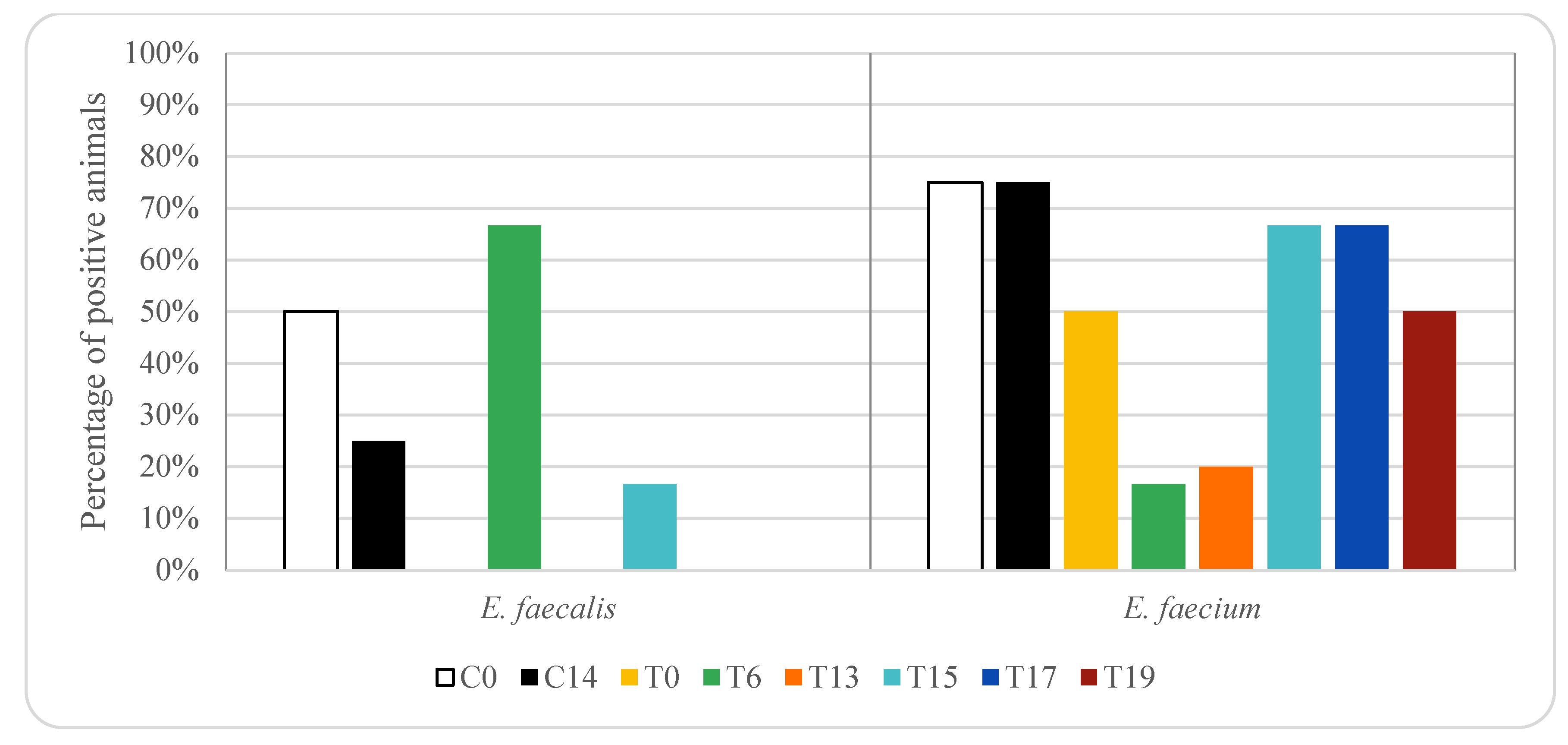
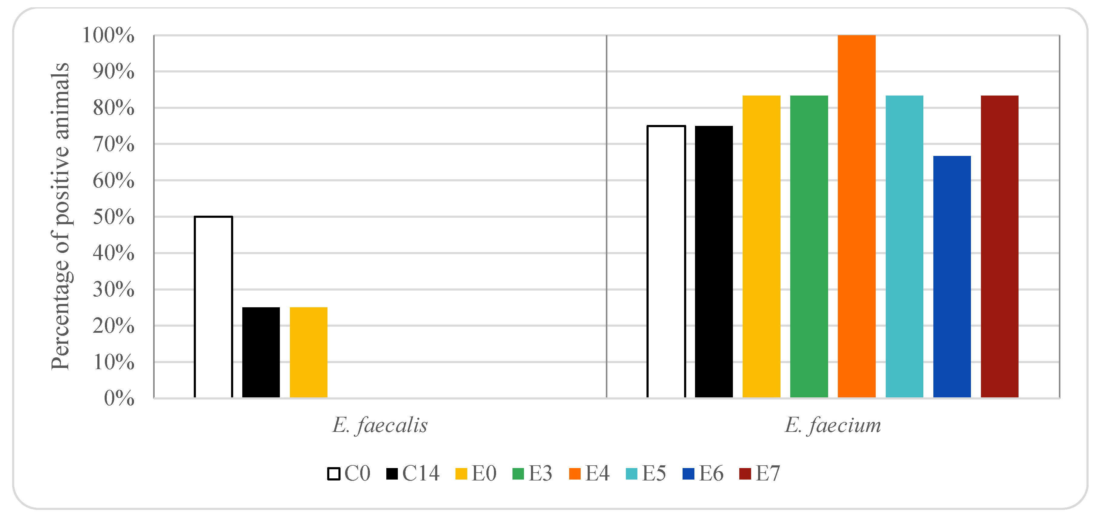
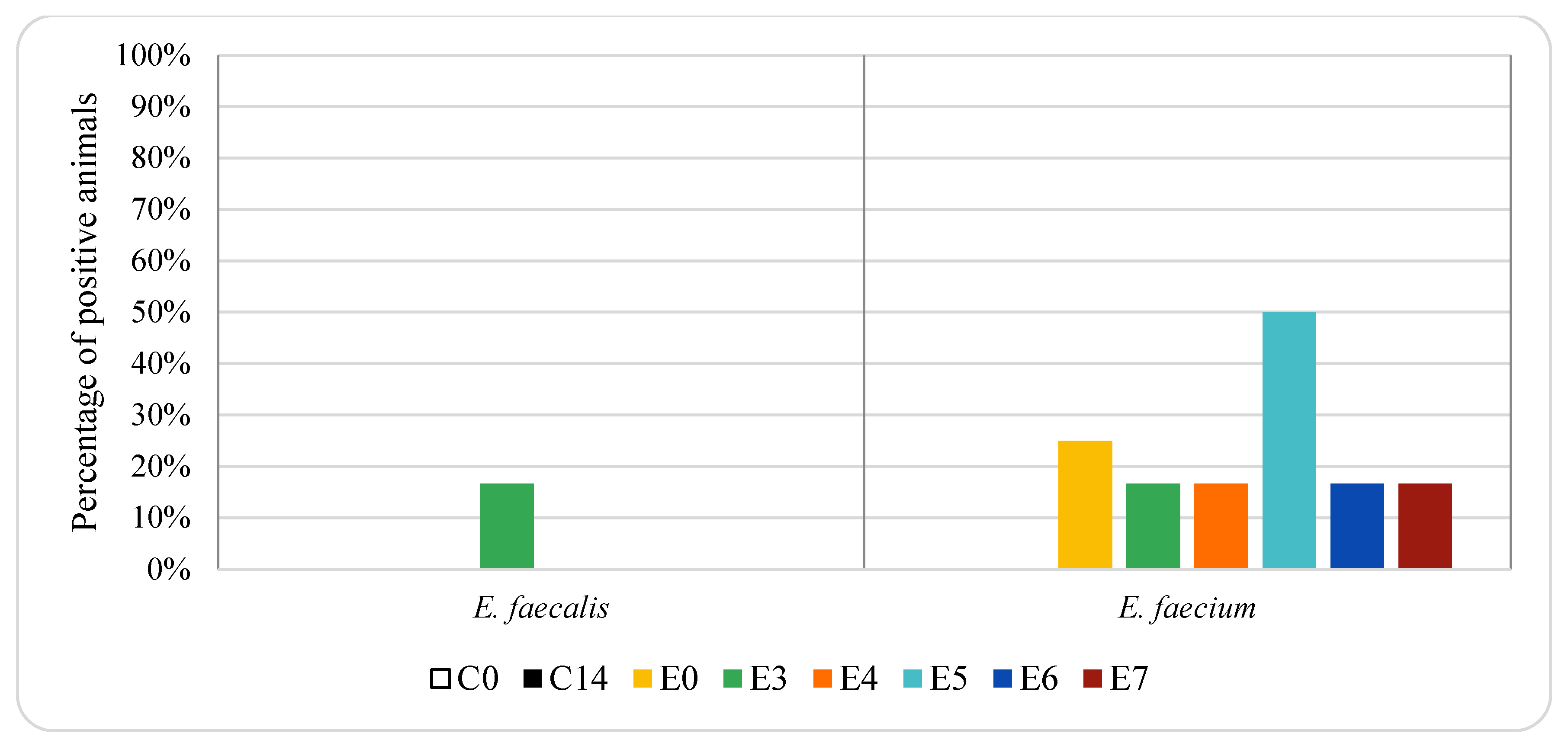
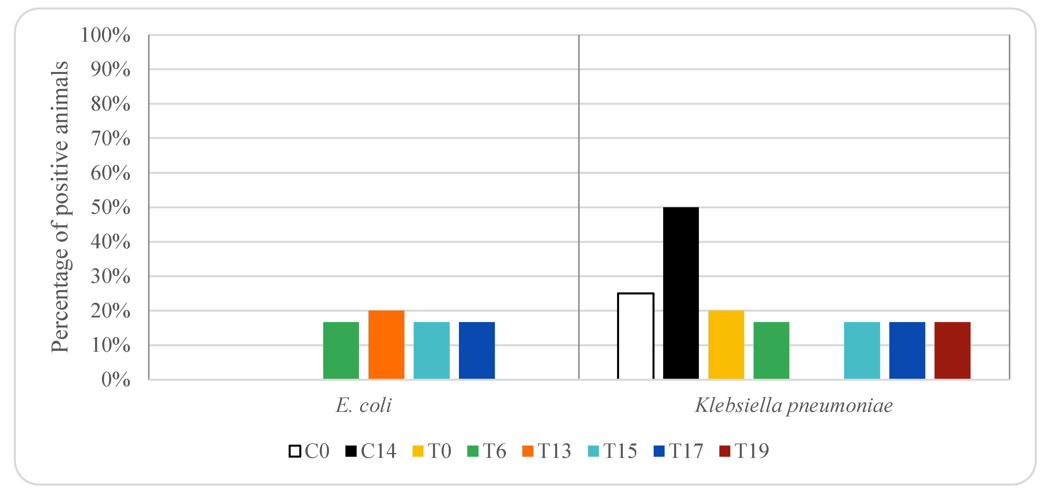
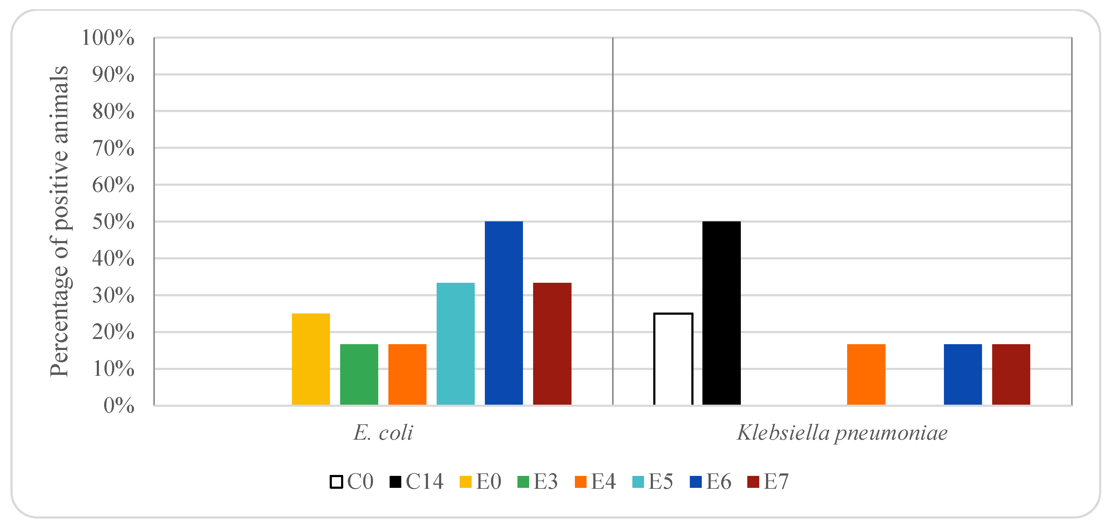
This work was included in the project “Development of a pioneering self-control solution in live animals to minimize the presence of antibiotic residues in the food chain of the Spain-France cross-border area (POCTEFA-TESTACOS)”, approved by the Ethical Advisory Commission for Animal Experimentation of the University of Zaragoza, reference number PI58/17. The study was carried out in accordance with the ARRIVE (Animal Research: Reporting of In Vivo Experiments) initiative and was handled and used in accordance with the Spanish Animal Protection Policy RD 53/2013 [38], which complies with the European Union Directive 2010/63 [39] on the protection of animals used for experimental and other scientific purposes.

3. Results

Figure 1 and Figure 2 show the percentage of animals in which vancomycin-resistant enterococci (VRE) isolates were found in rectal samples from pigs treated with oxytetracycline and enrofloxacin, respectively. In the control group, without any antibiotic treatment, vancomycin-resistant E. faecium was detected in 75% of the animals on days 0 and 14 (three piglets), while E. faecalis was detected in 50% (two piglets) and 25% (one piglet) on days 0 and 14, respectively. In animals treated with oxytetracycline, vancomycin-resistant E. faecium was detected in 50% of the animals on day 0 (5 piglets), while E. faecalis was not detected in any animal (0%). On days 15, 17 and 19 vancomycin-resistant E. faecium was detected in 50–67% of the animals (between three and four piglets). On days 17 and 19 vancomycin-resistant E. faecalis was not detected in any sample. However, on days 6 and 15 E. faecalis was detected in 67% (four piglets) and 17% (one piglet) of the animals, respectively.
In animals treated with enrofloxacin, vancomycin resistant E. faecium was detected in 83% of the animals on days 0 and 7 and in 100% on day 4 (10, five and six piglets, respectively). Vancomycin-resistant E. faecalis was only detected in 25% of the animals on day 0 (three piglets), corresponding to animals in which E. faecium was also isolated. On days 3 to 7 Vancomycin resistant E. faecalis was not detected in any animal.
Significant differences (p < 0.05) in the prevalence of vancomycin resistant E. faecium and E. faecalis were found among the three groups of animals studied, and thus, depended on antibiotic treatment.
Figure 3 and Figure 4 show the percentage of animals in which VRE isolates were found in genital samples from pigs treated with oxytetracycline and enrofloxacin, respectively. In the genital samples, E. faecalis and E. faecium resistant to vancomycin were not detected in control animals. In the animals treated with oxytetracycline, E. faecalis and E. faecium resistant to vancomycin were only detected on day 6 after treatment (17% E. faecalis and 17% E. faecium, one piglet). In the animals treated with enrofloxacin, E. faecium resistant to vancomycin was detected in 17–50% of the animals (between one and three piglets), depending on the sampling day. No E. faecalis resistant to vancomycin was detected in genital samples from animals treated with enrofloxacin, except on day 3, when both E. faecalis and E. faecium were isolated from 17% of the animals (one piglet). Significant differences (p < 0.05) in the prevalence of vancomycin resistant E. faecium were found in genital samples among the three groups of animals studied, and thus depended on antibiotic treatment.
Figure 5 and Figure 6 show the percentage of animals in which ESBL-producing E. coli and K. pneumoniae isolates were found in rectal samples from pigs treated with oxytetracycline and enrofloxacin, respectively. ESBL-producing E. coli was not detected in rectal samples taken from animals treated with oxytetracycline on days 0 and 19. However, on days 6 to 17, it was isolated among 17–20% of the animals treated with oxytetracycline (1 piglet). In animals treated with enrofloxacin, ESBL producing E. coli was detected in 25% of the animals on day 0 (three piglets), on days 3 and 4 was observed in 17% of the animals (1 piglet) and on days 5–7 was among 33 and 50% (between 2 and 3 piglets). ESBL-producing E. coli was not detected in rectal samples taken from control animals, not receiving any antimicrobial treatment. Significant differences (p < 0.05) in the prevalence of ESBL-producing E. coli were found in rectal samples among the three groups of animals studied, and thus depended on antibiotic treatment.
ESBL-producing K. pneumoniae was detected in 17–20% of the rectal samples taken from animals treated with oxytetracycline (between one and two piglets), except on day 13 (0%). In animals treated with enrofloxacin, ESBL producing K. pneumoniae was 0% on days 0, 3 and 5, while on days 4, 6 and 7 the percentage was 17% (one piglet). ESBL-producing K. pneumoniae was detected in control animals, animals that did not receive any treatment, 25% on day 0 (1 piglet) and 50% on day 14 (2 piglets).
Figure 7 and Figure 8 show the percentage of animals in which ESBL-producing E. coli and K. pneumoniae isolates were found in genital samples from pigs treated with oxytetracycline and enrofloxacin, respectively. ESBL-producing E. coli was not detected either in genital samples taken from animals treated with oxytetracycline or in samples taken from the control group. However, it was detected in 17% and 33% of the animals treated with enrofloxacin on days 5 and 6, respectively (one and two piglets, respectively).
ESBL-producing K. pneumoniae was only detected in the 17% of genital samples collected from animals treated with oxytetracycline on day 19 (one piglet), while it was not detected in any animal treated with enrofloxacin. In the control group, it was not detected any positive animal on day 0, while 25% were positive on day 14 (one piglet).
In the animals treated with enrofloxacin, no carbapenemase producers were detected in any sample. However, in animals treated with oxytetracycline, carbapenemase E. coli producers were detected in rectum samples on days 0, 6, 13 and 17, with percentages between 10 and 33% (between one and two piglets) (Figure 9), while carbapenemase K. pneumoniae producers were not detected in any animal treated with oxytetracycline. Carbapenemase producers were detected neither in rectal samples nor in genital samples from control animals, not treated with antibiotics. Significant differences (p < 0.05) in the prevalence of carbapenemase E. coli producers were found in rectal samples among the three groups of animals studied, and thus depended on antibiotic treatment.

4. Discussion

Some studies carried out in pigs show that Enterococcus spp. isolated are susceptible to vancomycin [31,40,41,42,43]. However, other authors have reported the presence of VRE in pigs [22,44,45,46]. Aarestrup et al. [44] observed that 17% of E. faecium isolates from pigs in Denmark were vancomycin resistant. The occurrence of VRE in pigs has been associated with the use of avoparcin for growth promotion since avoparcin is a vancomycin analog that confers cross-resistance to vancomycin [47]. Although avoparcin was banned in the European Union in 1997 [48] and later in other countries [49,50,51], the presence of VRE in pigs has still been detected in the last decades [22,45,46]. This fact could be related to co-selection with other antimicrobials [17,22,49].
It should be noted that the occurrence of resistant Enterococcus strains in food-producing animals depends on the geographical region since in some areas a high use of antibacterial agents has been observed [31,52,53]. In the present study, vancomycin-resistant enterococci were isolated both in rectal samples from control animals and those treated with oxytetracycline or enrofloxacin. Several authors have reported that a high percentage of enterococci isolated from pigs are resistant to tetracyclines and fluoroquinolones [31,43]. A higher number of vancomycin-resistant E. faecium were recovered compared to E. faecalis. This finding could be explained since E. faecium is more prevalent in pig than E. faecalis [32,41,42,43,44]. Prevalence of 45.7% and 12.9% of E. faecium and E. faecalis have been reported in pigs, respectively [42]. In the control group, the percentage of rectal samples with vancomycin-resistant E. faecium was higher than E. faecalis. In the pigs treated with enrofloxacin vancomycin resistant E. faecium was found in a higher percentage of animals than in the control group and remained above 80% on day 7 (above five piglets). These results can be explained since the persistence of VRE is suggested to be maintained by co-selection, by the use of other antibiotics [17,45,54]. However, in animals treated with oxytetracycline, vancomycin-resistant E. faecium was found in a lower percentage of animals compared to the control group. In the case of vancomycin-resistant E. faecalis higher percentage of positive animals was found in the control group than in those treated with enrofloxacin or oxytetracycline, except on day 6 after treatment with oxytetracycline, when a percentage above 60% was observed (above 4 piglets). In contrast, Nowakiewicz et al. [43] observed that in a farm in which only oxytetracycline was used, a high percentage of isolated enterococci were susceptible to most of the antimicrobial agents including vancomycin. The high percentage of VRE found in the present work could be explained by the use of specific media to isolate VRE, since VRE could be at a lower level than susceptible enterococci and could be not isolated if a non-selective media is used [42]. In fact, some authors did not detect VRE in pig faecal samples when no selective media were used, but when media were supplemented with vancomycin, VRE were detected [42]. Thus, the isolation media used could explain some of the discrepancies in the prevalence of VRE found in the bibliography [22].
In the present work, a higher percentage of vancomycin-resistant E. faecium were found in rectal samples from animals treated with enrofloxacin compared to those treated with oxytetracycline, while a higher percentage of vancomycin-resistance E. faecalis was found in rectal samples from animals treated with oxytetracycline compared to those treated with enrofloxacin. These results could be explained by the different susceptibility of enterococci species to these antimicrobials. In fact, Novais et al. [16] reported that E. faecium was more often resistant to fluoroquinolones, and E. faecalis to tetracyclines. Even more, some authors have only found resistance to fluoroquinolones in E. faecalis strains [43].
VRE was not isolated from genital samples in the control group. A lower percentage of VRE was observed in the genital samples in those animals treated with enrofloxacin or oxytetracycline compared to the respective faecal samples. This finding could be explained since enterococci are common inhabitants of the pig intestinal bacteria [17,51]. According to Novais et al. [16], the pig farm environment has an underestimated potential role in the transmission of multidrug resistant Enterococcus spp. to animals and, probably, to humans. The contact of pigs with multidrug resistant Enterococcus spp. by different routes (air, rooms, feed, dust, etc.) could decrease the impact of restrictive antibiotic use and underline the need of adopting additional control measures. Moreover, oxytetracycline is eliminated in urine (60%) and faeces (40%) [55]. After intramuscular administration of oxytetracycline, an exponential decay has been reported, with its half-life in blood being 3.59 days [35]. Enrofloxacin is mainly excreted via urine and small amounts in faeces [56]. Faster depletion of enrofloxacin compared to oxytetracycline, with a half-life of 1.90 days in blood, has been described [35]. These differences between enrofloxacin and oxytetracycline could affect the surviving bacteria, including VRE.
Moreover, there is a great concern about ESBL and carbapenemase-producing Enterobacteriaceae occurring in food-producing animals, since they may constitute a public-health risk [14,57]. Some studies have suggested that ESBL-producing E. coli can spread from livestock to humans [58,59]. Besides, the presence of ESBL-producing E. coli in swine has been documented by several studies worldwide [8,9,22,24,26,60,61,62,63,64]. High rates of ESBL-producing E. coli (up to 70%) have been reported in swine [24,58,59]. Additionally, Graesboll et al. [57] found ESBL-producing coliforms in all the farms evaluated, with 20% of the pigs positive. In the present work, ESBL-producing E. coli was not detected in rectal samples from control animals. However, it was detected in 17–20% of animals treated with oxytetracycline on days 6 to 17 (one piglet) and 17–50% of the animals treated with enrofloxacin (between one and three piglets). The percentage of positive animals was higher in pigs treated with enrofloxacin than in those treated with oxytetracycline. The higher prevalence of ESBL-producing Enterobacteriaceae when antimicrobials are used has been also reported by other authors. Fournier et al. [65] studied the presence of ESBL-producing Enterobacteriaceae in pig rectal samples from two farms, one using antibiotics and the other without antibiotics. These authors reported that the ESBL-producing Enterobacteriaceae prevalence was 86% in the farm using antibiotics, while the prevalence in the farm in which antibiotics were not used was 55%. These authors pointed out that there is a link between selective antibiotic pressure and the corresponding resistance rate. They also reported that in the farm using antibiotics, 92% of ESBL producers were resistant to tetracycline, while in the farm where antibiotics were not used co-resistances were lower, with only 44% of ESBL producers resistant to tetracyclines. [65]. It should be noted that some studies show that in pig farms with low antibiotics usage a high percentage of animals are colonised by ESBL-producing E. coli [9]. As it is shown in the present study, other authors observed that not all the animals from the same farm were colonised with ESBL producers [22].
Moreover, ESBL producers have been associated with resistance to non-β-lactam antimicrobials, such as fluoroquinolones and tetracyclines, which are often used to treat diseases on pigs [22,24,66,67,68]. Galler et al. [22] isolated ESBL-producing E. coli in 46.6% of the swine Austrian farms studied (7 of 15). All isolates were susceptible to fluoroquinolones, while high resistance rates were observed to tetracyclines (73.3%). Additionally, Fournier et al. [9] found low rates of co-resistance to fluoroquinolones among ESBL-producing isolates from pigs. In contrast, Picozzi et al. [66] pointed out that ESBL-producing strains from pigs often presented cross-resistance to quinolones. On their behalf, Liu et al. [67] reported that all ESBL producers isolated from swine exhibited a multidrug resistance phenotype, and more than 90% of them were resistant to tetracycline and enrofloxacin.
Some studies have associated the presence of ESBL-producing E. coli with the selective pressure induced by the use of antibiotics in animals [68]. This finding could be linked to the one of De Koster et al. [64], who isolated ESBL-producing E. coli in pig faeces from farms with high antibiotic use. However, other studies have shown a high prevalence of ESBL-producing E. coli in farm animals with low use of antibiotics (up to 50%) [69,70]. In consequence, other factors apart from antibiotic usages, such as farm conditions, farm environment, farm hygiene and contact with humans could be affecting the presence of ESBL-producing E. coli in livestock [24,64,71,72,73]. For instance, Tamta et al. [24] reported that piglets and pig farm workers were a potential source of ESBL-producing E. coli. These authors associated the high prevalence of ESBL producing E. coli isolates in piglets (44.4%,) and farmworkers (90.5%) to the use of the selective medium for detecting resistant E. coli isolates or the use of β-lactam and cephalosporin antibiotics on the farms studied. Other authors have also reported a high percentage of ESBL-producing E. coli using selective media [74,75]. In the present work, CHROMID ESBL medium was used to isolate ESBL-producing E. coli. This medium allows detecting ESBL producers when they are present at low levels, especially E. coli, which is one of the most frequent ESBL producers [76,77,78,79].
As stated above, the use of antibiotics in food-producing animals may select bacteria resistant to them. Moreover, the antimicrobial treatment affects both the targeted pathogen and the commensal bacteria. Since some amount of administrated antibiotics end up in the intestines [80], the intestinal tract of animals acts as an important reservoir for the selection of antibiotic resistance [64,81]. Thus, the formation and selection of resistant strains in the intestinal commensal bacteria could play an important role in the spread of resistant bacteria [82]. Several studies have shown that tetracycline resistant commensal E. coli bacteria from livestock are often resistant to other antimicrobials, indicating co-selection [83,84,85]. Jensen et al. [85] reported that the usage of tetracycline in pig farms can promote resistance to critically important antimicrobials. Tetracycline resistance is often found in ESBL-producing isolates and transmitted with ESBL containing plasmids [86]. In contrast, Gruel et al. [33] reported that the use of tetracycline is not correlated to ESBL-producing E. coli. Although tetracycline is not listed as critically important for human treatment [20], there is a great concern since it can promote resistance to other antimicrobials.
Furthermore, the way of administration of antimicrobials might also be a critical factor to consider when talking about selective pressure for resistant bacteria. Intramuscular administration of antibiotics is considered to have lower influence on intestinal microbiota than oral administration, since it does not require absorption from the gut [34]. Nevertheless, although renal excretion has been reported as the main excretion mechanism of enrofloxacin after intramuscular administration of enrofloxacin [86], high faecal concentrations of this antibiotic have been found in pigs [34]. These high enrofloxacin faecal levels could affect the faecal microbiota, especially Gram-negative bacteria including E. coli, as it has been documented that enrofloxacin is effective against these bacteria [87]. On the other hand enrofloxacins can reach high concentrations in the gut because they are partially excreted in the bile acid [88]. Moreover, intestinal efflux transporters may transport enrofloxacin into the gut lumen [89]. Thus, enrofloxacin can disrupt the gut commensal bacteria, even when treatment is by intramuscular injection [90]. Enrofloxacin can influence the population dynamics of enteric bacteria and may select for resistance [91]. Besides, it has been observed that intramuscular enrofloxacin treatment reduces the faecal E. coli wild type population [34,92], and some authors have observed that at the end of enrofloxacin treatment only non-wild type E. coli isolates are found in faeces [34]. Moreover, Béraud et al. [92] reported that the intramuscular administration of enrofloxacin reduced the faecal E. coli counts from 3.79 log cfu/g to counts below 2 log cfu/g. Afterward, these authors observed a regrowth of E. coli, and these E. coli isolates recovered from pigs with intramuscular administration of enrofloxacin in the regrowth stage, were resistant to enrofloxacin and other antibiotics. Römer et al. [93] also observed that intramuscular administration of enrofloxacin in pigs caused an important reduction of the susceptible intestinal E. coli population, in favour of resistant E. coli. These authors pointed out that the intramuscular administration of enrofloxacin reduced the susceptible intestinal E. coli population, which was replaced by enrofloxacin resistant strains, but also control pigs were affected, maybe due to the transferability of strains through the environment. Wiuff et al. [94] also observed an increase of resistance in E. coli from the gut of pigs after intramuscular administration of enrofloxacin. Since intramuscularly administered enrofloxacin may exert selective pressure on the intestinal microbiota, including E. coli, there is a risk of resistance selection [92]. Besides, some of the studies found in the bibliography administered ciprofloxacin instead of enrofloxacin. It should be taken into account that after administration in pigs, enrofloxacin partially metabolizes into ciprofloxacin [95]. The metabolic conversion of enrofloxacin to ciprofloxacin is 51.5% in healthy pigs [95]. It should be noted that fluoroquinolones are important antibiotics for the treatment of infections in humans and they have been categorised as “highest priority critically important antimicrobials” [20].
On the other hand, despite rational use of antimicrobials, the rate of ESBL-producing E. coli in livestock, although moderate, is of concern [33]. Fewer data are available on ESBL-producing K. pneumoniae, since most of the studies are related to ESBL-producing E. coli. On the other hand, K. pneumoniae is present in lower numbers compared to E. coli [64]. De Koster et al. [64] isolated resistant Enterobacteriaceae from poultry and pig, 91.4% were identified as E. coli, whereas only 1.78% were identified as K. pneumoniae. ESBL-producing K. pneumoniae has been isolated in meat [96] and in pigs [97]. Since colonisation with E. coli and other Enterobacteriaceae occurs in the digestive tract, a higher percentage of positive animals were found in rectal samples than in genital samples [10]. Moreover, the presence of antibiotics in faeces could select ESBL and carbapenemase-producing E. coli [34].
Several authors have not detected carbapenemase-producing Enterobacteriaceae in pigs [9,65]. However, some studies have found carbapenemase-producing E. coli in pigs [14,24,75,98,99]. Carbapenemase producing E. coli have been isolated from the environment of swine farms in the USA [75]. Additionally, carbapenemase-producing Enterobacteriaceae have been reported in pig farms in Germany [100]. According to Köck et al. [30] the prevalence of carbapenemase-producing Enterobacteriaceae in farm animals is low in Europe (<1%), whereas a higher prevalence has been observed in China, India and Algeria. Specifically, the prevalence of carbapenemase-producing E. coli in pigs is low, since carbapenems are not used in food-producing animal treatment [98]. Indeed, Fournier et al. [9,65] did not detect carbapenemase-producing Enterobacteriaceae in pigs from farms using or not using antibiotics. Related to this varying occurrence, it has been suggested that carbapenem resistant E. coli in pigs could originate from the human contact environment [24]. According to Dandachi et al. [10], the emergence of carbapenemase-producers in livestock is related to the co-selective pressure by the usage of non-β-lactams antibiotics. It should be noted that the spread of carbapenemase-producing Enterobacteriaceae is of great concern since they are multidrug-resistant [27] and their presence in animals could constitute a reservoir and be a risk for human health [14]. As an example, carbapenemase producers are often co-resistant to non-β-lactam antibiotics including tetracyclines and fluoroquinolones [101].
Data presented in this work show that carbapenemase-producing Enterobacteriaceae were not detected in any rectal sample taken from control animals or those treated with enrofloxacin. However, carbapenemase E. coli producers were detected on days 0, 6, 13 and 17, with percentages between 10 and 33% in animals treated with oxytetracycline (between one and two piglets). These results suggest that tetracycline exposure could influence the occurrence of carbapenemase-producing E. coli. A selective medium, CHROMID CARBA, was used for the detection of carbapenemase-producing Enterobacteriaceae; since these bacteria could be in low numbers, it is possible that the use of non-specific media fails to detect them [102]. Moreover, carbapenemase-producing E. coli were not isolated in genital samples, and carbapenemase-producing K. pneumoniae were not isolated either in faecal samples or genital samples. There are few data available on carbapenemase-producing K. pneumoniae. Only some studies have found carbapenemase-producing K. pneumoniae in poultry [103,104].
It should be noted that in the present study samples were collected and immediately frozen at −80 °C. As pointed out by other authors, the immediate processing of faecal samples after collection is not always technically or economically feasible with freezing being the most common method of preservation [105,106]. The survival and diversity of microbial populations in faecal samples after freezing and storage at −80 °C have been evaluated by several studies [105,107,108]. Masters et al. [105] studied the survival and diversity of E. coli and enterococci populations in faecal samples of animal origin (including pig) after storage at either −20 or −80 °C for 30 days. These authors reported that the numbers of enterococci were similar in fresh and frozen faecal pig samples. The number and the distribution of E. coli and enterococci assigned to different biochemical phenotypes in fresh samples did not vary significantly from those stored at −80 °C [105]. Furthermore, the population structure of E. coli and enterococci did not change significantly after storage at −80 °C [105]. Similar findings were reported by Tedjo et al. [107], who did not observe significant changes in the overall microbiota composition between frozen faecal samples at −80 °C and samples stored at room temperature or 4 °C for 24 h. According to Deschamps et al. [108], freezing at −80 °C without cryoprotectant was the most efficient method for faecal preservation considering both stabilization time of microbial profiles and metabolic activities.
This study was carried out with three groups of animals: group 1 treated with enrofloxacin (N = 12), group 2 treated with oxytetracycline (N = 10) and group 3 that did not receive any treatment (control group) (N = 4). The three groups were kept separated in the experimental facilities of the Faculty of Veterinary. They were maintained in the same conditions, then the effect of the farm environment, farm hygiene, and personal hygiene of farmworkers were the same in the three groups. The effect of the antibiotic treatments has been compared with the control group. The results obtained show that there are differences between animals treated with antibiotics and those non-treated on VRE, ESBL and carbapenemase-producing Enterobacteriaceae. Further works are needed to know the role of antibiotic treatments on antimicrobial resistance. The differences found with other studies could be due to the influence of farm environment, farm hygiene and personal hygiene of farmworkers.
Thus, the degree of antibiotic resistance in food-producing animals has been correlated with antibiotic usage, since antibiotic administration can act as a selective pressure for resistant bacteria [32,33]. Data presented prove that special care should be taken in the slaughter process to avoid the faecal contamination of pig carcasses since Enterobacteriaceae and enterococci are normally present in the intestinal tract and could be multi-resistant bacteria [23], resistance tightly linked to selective pressure triggered by antimicrobial treatment. This cross-contamination of animal carcasses may be a food safety risk [42,94] as it might be enhancing the already serious problem of antimicrobial resistance dissemination. On the other hand, dissemination could occur if the environment is contaminated by pig faeces.

5. Conclusions

This study highlighted that the use of tetracycline in food-production animals could select ESBL and carbapenemase-producing E. coli in the intestinal tract as suggested by the results found in rectal samples; while the use of enrofloxacin could select ESBL-producing E. coli in the intestinal tract and in a lesser extent in the genital system. Thus, special care should be taken to avoid faecal contamination of carcasses during slaughter. Additional studies are needed on ESBL-producing K. pneumoniae, since data presented in this study pointed to this bacterium being present in pigs. Vancomycin resistant E. faecium can be present in faeces from pigs; the treatment with enrofloxacin could increase the percentage of positive animals. The high percentage of animals with the presence of VRE found underlines the relevance of using selective media for the isolation of VRE. It should be noted that in the present study only chromogenic media were used and further works are needed to confirm the results obtained. On the other hand, further research is needed to estimate the magnitude of the effect of antibiotic treatments on antimicrobial resistance and to know the mechanisms involved. Further studies shall be needed to validate the results obtained, considering a more robust and extended experimental design.

Author Contributions

Conceptualization and methodology, E.G.-F., A.M.-L., A.A.-F., E.S., R.B.; M.J.S., O.M., C.B., A.L., M.V.F., R.P.; formal analysis, E.G.-F., A.M.-L., A.A.-F., E.S., R.B.; investigation, E.G.-F., A.M.-L., A.A.-F., E.S., R.B., M.J.S., O.M., C.B., A.L., M.V.F., R.P.; resources, E.G.-F., A.M.-L., A.A.-F., E.S., R.B., M.J.S., O.M., C.B., A.L., M.V.F., R.P.; data curation, E.G.-F., A.M.-L., A.A.-F., R.B.; writing—original draft preparation, E.G.-F.; writing-review and editing, E.G.-F., A.M.-L., A.A.-F., E.S., R.B., M.J.S., O.M., C.B., A.L., M.V.F., R.P.; project administration, E.G.-F., M.J.S., R.P.; funding acquisition, E.G.-F., R.P. All authors have read and agreed to the published version of the manuscript.

Funding

This project was 65% cofinanced by the European Regional Development Fund (ERDF) through the Interreg V-A Spain-France-Andorra programme (POCTEFA (Programa INTERREG V-A España-Francia-Andorra) 2014–2020) (EFA (España-Francia-Andorra) 152/16). POCTEFA aims to reinforce the economic and social integration of the French–Spanish–Andorran border. Its support is focused on developing economic, social, and environmental cross-border activities through joint strategies favouring sustainable territorial development.

Institutional Review Board Statement

This study was approved by the Ethical Advisory Commission for Animal Experimentation of the University of Zaragoza, reference number PI58/17. The study was carried out in accordance with the ARRIVE (Animal Research: Reporting of In Vivo Experiments) initiative, and were handled and used in accordance with the Spanish Animal Protection Policy RD 53/2013 (Real Decreto 53/2013), which complies with the European Union Directive 2010/63 (Directive 2010/63) on the protection of animals used for experimental and other scientific purposes.

Data Availability Statement

Not applicable.

Acknowledgments

AML acknowledges the University of La Rioja for her predoctoral fellowship (UR-CAR-2019).

Conflicts of Interest

The authors declare no conflict of interest.

References

  1. O’Neill, J. Antimicrobial Resistance: Tackling a Crisis for the Health and Wealth of Nations. London: Review on Antimicrobial Resistance. 2014. Available online: https://amr-review.org/sites/default/files/AMR%20Review%20Paper%20-%20Tacklng%20a%20crisis%20for%20the%20health%20and%20wealth%20of%20nations_1.pdf (accessed on 2 November 2021).
  2. Wall, B.A.; Mateus, A.; Marshall, L.; Pfeiffer, D.; Lubroth, J.; Ormel, H.J.; Otto, P.; Patriarchi, A.; Food and Agriculture Organization of the United Nations. Drivers, Dynamics and Epidemiology of Antimicrobial Resistance in Animal Production; Food and Agriculture Organization (FAO): Rome, Italy, 2016. [Google Scholar]
  3. World Health Organization. Antibacterial Agents in Clinical Development: An Analysis of The Antibacterial Clinical Development Pipeline; World Health Organization: Geneva, Switzerland, 2019. [Google Scholar]
  4. World Health Organization. Global Antimicrobial Resistance and Use Surveillance System (GLASS) Report; World Health Organization: Geneva, Switzerland, 2021. [Google Scholar]
  5. World Organization for Animal Health (OIE): OIE List of Antimicrobials of Veterinary Importance. 2019. Available online: http://www.oie.int (accessed on 18 November 2021).
  6. Amusi, J.; Tamara, L.; Horton, R.; Winkler, A.S. Reconnecting for our future: The Lancet One Health commission. Lancet 2020, 395, 1469–1471. [Google Scholar] [CrossRef]
  7. Colligon, P.J.; McEwen, S.A. One Health. Its importance in helping to better control antimicrobial resistance. Trop. Med. Infect. Dis. 2019, 4, 22. [Google Scholar] [CrossRef] [PubMed] [Green Version]
  8. Bergšpica, I.; Kaprou, G.; Alexa, E.A.; Prieto, M.; Alvarez-Ordóñez, A. Extended spectrum β-lactamase (ESBL) Escherichia coli in pigs and pork meat in European Union. Antibiotics 2020, 9, 678. [Google Scholar] [CrossRef]
  9. Fournier, C.; Nordmann, P.; Pittet, O.; Poirel, L. Does an antibiotic stewardship applied in a pig farm lead to low ESBL prevalence? Antibiotics 2021, 10, 574. [Google Scholar] [CrossRef] [PubMed]
  10. Dandachi, I.; Chabou, S.; Daoud, Z.; Rolain, J.M. Prevalence and emergence of extended-spectrum cephalosporin, carbapenem and colistin-resistant Gram negative bacteria of animal origin in the Mediterranean basin. Front. Microbiol. 2018, 9, 2299. [Google Scholar] [CrossRef] [PubMed]
  11. Unal, N.; Bal, E.; Kapagoz, A.; ALtun, B.; Kozag, N. Detection of vancomycin-resistant enterococci in samples from broiler flocks and houses. Acta Vet. Hung. 2020, 68, 117–122. [Google Scholar] [CrossRef]
  12. Fournier, S.; Brun-Buisson, C.; Jarlier, V. Twenty years of antimicrobial resistance control programme in a regional multi hospital institution, with focus on emerging bacteria (VRE and CPE). Antimicrob. Resist. Infect. Control 2012, 1, 9. [Google Scholar] [CrossRef] [Green Version]
  13. Adler, A.; Katz, D.E.; Marchaim, D. The continuing plague of extended-spectrum beta-lactamase-producing Enterobacteriaceae infections. Infect. Dis. Clin. North Am. 2016, 30, 347–375. [Google Scholar] [CrossRef]
  14. Madec, J.Y.; Haenni, M.; Nordmann, P.; Poirel, L. Extended-spectrum beta-lactamase/AmpC- and carbapenemase-producing Enterobacteriaceae in animals: A threat for humans? Clin. Microbiol. Infect. 2017, 23, 826–833. [Google Scholar] [CrossRef] [PubMed] [Green Version]
  15. European Food Safety Authority; European Centre for Disease Prevention and Control. The European Union Summary Report on Antimicrobial Resistance in Zoonotic and Indicator Bacteria from Humans, Animals and Food in 2018/2019. EFSA J. 2021, 19, e06490. [Google Scholar]
  16. Novais, C.; Freitas, A.R.; Silveira, E.; Antunes, P.; Silva, R.; Coque, T.M.; Peixe, L. Spread of multidrug-resistant Enterococcus to animals and humans: An underestimated role for the pig farm environment. J. Antimicrob. Chemother. 2013, 68, 2746–2754. [Google Scholar] [CrossRef] [PubMed]
  17. Nilsson, O. Vancomycin resistant enterococci in farm animals. Occurrence and importance. Infect. Ecol. Epidemiol. 2012, 2, 16959. [Google Scholar] [CrossRef] [PubMed] [Green Version]
  18. Gastmeier, P.; Schröder, C.; Behnke, M.; Meyer, E.; Geffers, C. Dramatic increase in vancomycin-resistant enterococci in Germany. J. Antimicrob. Chemother. 2014, 69, 1660–1664. [Google Scholar] [CrossRef] [Green Version]
  19. Hammerum, A.M. Enterococci of animal origin and their significance for public health. Clin. Microbiol. Infect. 2012, 18, 619–625. [Google Scholar] [CrossRef] [PubMed]
  20. EMA (European Medicine Agency). Categorisation of Antibiotics for Use in Animals for Prudent and Responsible Use. 2020. Available online: https://www.ema.europa.eu/en/documents/report/infographic-categorisation-antibiotics-use-animals-prudent-responsible-use_en.pdf (accessed on 27 April 2021).
  21. Tyson, G.H.; Nyirabahizi, E.; Crarey, E.; Kabera, C.; Lam, C.; Rice-Trujillo, C.; McDermott, P.F.; Tate, H. Prevalence and antimicrobial resistance of enterococci isolated from world retail meats in the United States, 2002 to 2014. Appl. Environ. Microbiol. 2018, 84, e01902-17. [Google Scholar] [CrossRef] [PubMed] [Green Version]
  22. Galler, H.; Luxner, J.; Petternel, C.; Reinthaler, F.F.; Habib, J.; Haas, D.; Kittinger, C.; Pless, P.; Feierl, G.; Zarfel, G. Multiresistant bacteria isolated from intestinal faeces of farm animals in Austria. Antibiotics 2021, 10, 466. [Google Scholar] [CrossRef]
  23. Doi, Y.; Iovleva, A.; Bonomo, R.A. The ecology of extended-spectrum B-lactamases (ESBLs) in the developed world. J. Travel Med. 2017, 24, S44–S51. [Google Scholar] [CrossRef]
  24. Tamta, S.; Vonodh-Kumar, O.R.; Pruthvishree, B.S.; Karthikeyan, R.; Ramkumar, R.R.; Chethan, G.E.; Dubal, Z.B.; Sinha, D.K.; Singh, B.R. Faecal carriage of extended spectrum beta-lactamase (ESBL) and New Delhi metallobeta-lactamase(NDM) producing Escherichia coli between piglets and pig farmworkers. Comp. Immunol. Microbiol. Infect. Dis. 2020, 73, 101564. [Google Scholar] [CrossRef]
  25. Cantas, L.; Suer, K.; Guler, E.; Imir, T. High emergence of ESBL producing E. coli cystitis: Time to get smarter in Cyprus. Front. Microbiol. 2015, 6, 1446. [Google Scholar] [CrossRef]
  26. Stefani, S.; Giovanelli, I.; Anacarso, I.; Condo, C.; Messi, P.; de Niederhausern, S.; Bondi, M.; Iseppi, R.; Sabia, C. Prevalence and characterization of extended-spectrum beta-lactamase-producing Enterobacteriaceae in food-producing animals in Northern Italy. New Microbiol. 2014, 37, 551–555. [Google Scholar]
  27. Nordmann, P. Carbapenemase-producing Enterobacteriaceae: Overview of a major public health challenge. Méd. Mal. Infect. 2014, 44, 51–56. [Google Scholar] [CrossRef] [PubMed] [Green Version]
  28. European Food Safety Authority. Scientific Opinion on Carbapenem resistance in food animal ecosystems. EFSA J. 2013, 11, 3501. [Google Scholar]
  29. World Health Organization. Antimicrobial Resistance: Global Report on Surveillance; World Health Organization: Geneva, Switzerland, 2014. [Google Scholar]
  30. Köck, R.; Daniels-Haardt, I.; Becker, K.; Mellmann, A.; Friedrich, A.W.; Mevius, D.; Schwarz, S.; Jurke, A. Carbapen Carbapenem-resistant Enterobacterales in wildlife, food-producing, and companion animals: A systematic review. Clin. Microbiol. Infect. 2018, 24, 1241–1250. [Google Scholar] [CrossRef] [PubMed] [Green Version]
  31. Tan, S.C.; Chong, C.W.; Teh, C.S.J.; Ooi, P.T.; Thong, K.L. Occurrence of virulent multidrug resistant Enterococcus faecalis and Enterococcus faecium in the pigs, farmers and farm environments in Malaysia. PeerJ 2018, 6, e5353. [Google Scholar] [CrossRef] [PubMed] [Green Version]
  32. Aasmäe, B.; Häkkinen, L.; Kaartt, T.; Kalmus, P. Antimicrobial resistance of Escherichia coli and Enterococcus spp. isolated from Estonian cattle and swine from 2010 to 2015. Acta Vet. Scand. 2019, 61, 5. [Google Scholar] [CrossRef] [PubMed]
  33. Gruel, G.; Sellin, A.; Riveiro, H.; Pot, M.; Breurec, S.; Guyomard-Rabenirina, S.; Talarmin, A.; Ferdinand, S. Antimicrobial use and resistance in Escherichia coli from healthy food-producing animals in Guadeloupe. BMC Vet. Res. 2021, 17, 116. [Google Scholar] [CrossRef]
  34. De Smet, J.; Boyen, F.; Croubels, S.; Rasschaert, G.; Haesebrouck, F.; Temmerman, R.; Rutjens, S.; De Backer, P.; Devreese, M. The impact of therapeutic-dose induced intestinal enrofloxacin concentrations in healthy pigs on fecal Escherichia coli populations. BMC Vet. Res. 2020, 16, 382. [Google Scholar] [CrossRef]
  35. Serrano, M.J.; Mitjana, O.; Bonastre, C.; Laborda, A.; Falceto, M.F.; García-Gonzalo, D.; Abilleira, E.; Elorduy, J.; Bousquet-Melou, A.; Mata, L.; et al. Is Blood a Good Indicator for Detecting Antimicrobials in Meat? Evidence for the Development of In Vivo Surveillance Methods. Antibiotics 2020, 9, 175. [Google Scholar] [CrossRef] [Green Version]
  36. Fang, H.; Ohlsson, A.K.; Ullberg, M.; Özenci, V. Evaluation of species-specific PCR, Bruker MS, VITEK MS and the VITEK 2 system for the identification of clinical Enterococcus isolates. Eur. J. Clin. Microbiol. Infect. Dis. 2012, 31, 3073–3077. [Google Scholar] [CrossRef]
  37. Bilecen, K.; Yaman, I.G.; Ciftci, U.; Laleli, Y.R. Performances and reliability of Bruker Microflex LT and VITEK MS MALDI-TOF mass spectrometry systems for the identification of clinical microorganisms. BioMed Res. Int. 2015, 2015, 516410. [Google Scholar] [CrossRef] [Green Version]
  38. Real Decreto 53/2013, de 1 de febrero, por el que se establecen las normas básicas aplicables para la protección de los animales utilizados en experimentación y otros fines científicos, incluyendo la docencia. Boletín Of. Estado 2013, 34, 11370–11421.
  39. Directive 2010/63/EU of the European Parliament and of the Council of 22 September 2010 on the protection of animals used for scientific purposes. Off. J. Eur. Union 2010, 276, 33–79.
  40. Hershberger, E.; Oprea, S.F.; Donabedian, S.M.; Perri, M.; Bozigar, P.; Bartlett, P.; Zervos, M. Epidemiology of antimicrobial resistance in enterococci of animal origin. J. Antimicrob. Chemother. 2005, 55, 127–130. [Google Scholar] [CrossRef] [PubMed] [Green Version]
  41. Liu, Y.; Liu, K.; Lai, J.; Wu, C.; Shen, J.; Wang, Y. Prevalence and antimicrobial resistance of Enterococcus species of food animal origin from Beijing and Shandong Province, China. J. Appl. Microbiol. 2012, 114, 555–563. [Google Scholar] [CrossRef] [PubMed]
  42. Ramos, S.; Igrejas, G.; Capelo-Martinez, J.L.; Poeta, P. Antibiotic resistance and mechanisms implicated in fecal enterococci recovered from pigs, cattle and sheep in a Portuguese slaughterhouse. Ann. Microbiol. 2012, 62, 1485–1494. [Google Scholar] [CrossRef]
  43. Nowakiewicz, A.; Ziółkowska, G.; Troscianczyk, A.; Zięba, P.; Gnat, S. Determination of antimicrobial resistance of Enterococcus strains isolated from pigs and their genotypic characterization by method of amplification of DNA fragments surrounding rare restriction sites (ADSRRS fingerprinting). J. Med. Microbiol. 2017, 66, 175–183. [Google Scholar] [CrossRef] [PubMed]
  44. Aarestrup, F.M.; Agerso, Y.; Gerner-Smidt, P.; Madsen, M.; Jensen, L.B. Comparison of antimicrobial resistance phenotypes and resistance genes in Enterococcus faecalis and Enterococcus faecium from humans in the community, broilers, and pigs in Denmark. Diagn. Microbiol. Infect. Dis. 2000, 37, 127–137. [Google Scholar] [CrossRef]
  45. Wist, V.; Morach, M.; Schneeberger, M.; Cernela, N.; Stevens, M.J.A.; Zurfluh, K.; Stephan, R.; Nüesch-Inderbinen, M. Phenotypic and genotypic traits of vancomycin-resistant enterococci from healthy food-producing animals. Microorganisms 2020, 8, 261. [Google Scholar] [CrossRef] [PubMed] [Green Version]
  46. Pruksakorn, C.; Pimarn, C.; Boonsoongnern, A.; Narongsak, W. Detection and phenotypic characterization of vancomycin-resistant enterococci in pigs in Thailand. Agric. Nat. Resour. 2016, 50, 199–203. [Google Scholar]
  47. Bager, F.; Madsen, M.; Christensen, J.; Aarestrup, F.M. Avoparcin used as a growth promoter is associated with the occurrence of vancomycin-resistant Enterococcus faecium on Danish poultry and pig farms. Prev. Vet. Med. 1997, 31, 95–112. [Google Scholar] [CrossRef]
  48. Directive 97/6/EC. of 30 January amending Council Directive 70/524/EEC concerning additives in feeding stuffs. Off. J. Eur. Commun. 1997, 35, 11–13.
  49. Yoshimura, H.; Ishimaru, M.; Endoh, Y.S.; Suginaka, M.; Yamatani, S. Isolation of glycopeptide-resistant enterococci from chicken in Japan. Antimicrob. Agents Chemother. 1998, 42, 3333. [Google Scholar] [CrossRef] [Green Version]
  50. Lauderdale, T.L.; Shiau, Y.R.; Wang, H.Y.; Lai, J.F.; Huang, I.W.; Chen, P.C.; Chen, H.Y.; Lai, S.S.; Liu, Y.F.; Ho, M. Effect of banning vancomycin analogue avoparcin on vancomycin-resistant enterococci in chicken farms in Taiwan. Environ. Microbiol. 2007, 9, 819–823. [Google Scholar] [CrossRef]
  51. Hammerum, A.M.; Lester, C.H.; Heuer, O.E. Antimicrobial resistant enterococci in animals and meat: A human health hazard? Foodborne Pathog. Dis. 2010, 7, 1137–1146. [Google Scholar] [CrossRef] [PubMed]
  52. Frye, J.G.; Jackson, C.R. Genetic mechanisms of antimicrobial resistance identified in Salmonella enterica, Escherichia coli, and Enteroccocus spp. isolated from U.S. food animals. Front. Microbiol. 2013, 4, 135. [Google Scholar] [CrossRef] [PubMed] [Green Version]
  53. Garrido, A.M.; Galvez, A.; Pulido, R.P. Antimicrobial resistance in enterococci. J. Infect. Dis. Ther. 2014, 2, 4. [Google Scholar] [CrossRef] [Green Version]
  54. Ahmed, M.O.; Baptiste, K.E. Vancomycin-resistant enterococci: A review of antimicrobial resistance mechanisms and perspectives of human and animal health. Microb. Drug Resist. 2018, 24, 590–606. [Google Scholar] [CrossRef] [Green Version]
  55. Riviere, J.E.; Spoo, J.W. Chapter 42. Tetracycline Antibiotics. In Veterinary Pharmacology and Therapeutics, 8th ed.; Adams, R.H., Ed.; Iowa State University Press: Ames, IA, USA, 2001; pp. 828–840. [Google Scholar]
  56. Papich, M.G.; Riviere, J.E. Chapter 45. Fluoroquinolone Antimicrobial Drugs. In Veterinary Pharmacology and Therapeutics, 8th ed.; Adams, R.H., Ed.; Iowa State University Press: Ames, IA, USA, 2001; pp. 898–917. [Google Scholar]
  57. Græsbøll, K.; Damborg, P.; Mellerup, A.; Herrero-Fresno, A.; Larsen, I.; Holm, A.; Nielsen, J.P.; Christiansen, L.E.; Angen, Ø.; Ahmed, S.; et al. Effect of tetracycline dose and treatment mode on selection of resistant coliform bacteria in nursery pigs. Appl. Environ. Microbiol. 2017, 83, 00538-17. [Google Scholar] [CrossRef] [PubMed] [Green Version]
  58. Geser, N.; Stephan, R.; Hächler, H. Occurrence and characteristics of extended spectrum β-lactamase (ESBL) producing Enterobacteriaceae in food producing animals, minced meat and raw milk. BMC Vet. Res. 2012, 8, 21. [Google Scholar] [CrossRef] [Green Version]
  59. Loayza, F.; Graham, J.P.; Trueba, G. Factors obscuring the role of E. coli from Domestic animals in the global antimicrobial resistance crisis: An evidence-based review. Int. J. Environ. Res. Public Health 2020, 17, 3061. [Google Scholar] [CrossRef] [PubMed]
  60. Escudero, E.; Vinue, L.; Teshager, T.; Torres, C.; Moreno, M.A. Resistance mechanisms and farm-level distribution of fecal Escherichia coli isolates resistant to extended-spectrum cephalosporins in pigs in Spain. Res. Vet. Sci. 2010, 88, 83–87. [Google Scholar] [CrossRef]
  61. Ewers, C.; Bethe, A.; Semmler, T.; Guenther, S.; Wieler, L.H. Extended spectrum beta-lactamase-producing and AmpC-producing Escherichia coli from livestock and companion animals, and their putative impact on public health: A global perspective. Clin. Microbiol. Infect. 2012, 18, 646–655. [Google Scholar] [CrossRef] [PubMed] [Green Version]
  62. Fischer, J.; Hille, K.; Ruddat, I.; Mellmann, A.; Köck, R.; Kreienbrock, L. Simultaneous occurrence of MRSA and ESBL-producing Enterobacterales on pig farms and in nasal and stool samples from farmers. Vet. Microbiol. 2017, 200, 107–113. [Google Scholar] [CrossRef] [PubMed]
  63. Van Damme, I.; Garcia-Graells, C.; Biasino, W.; Gowda, T.; Botteldoorn, N.; De Zutter, L. High Abundance and diversity of extended-spectrum beta-lactamase (ESBL)-producing Escherichia coli in faeces and tonsils of pigs at slaughter. Vet. Microbiol. 2017, 208, 190–194. [Google Scholar] [CrossRef] [PubMed]
  64. De Koster, S.; Ringenier, M.; Lammens, C.; Stegeman, A.; Tobias, T.; Velkers, F.; Vernooij, H.; Kluytmans-Van Den Bergh, M.; Kluytmans, J.; Dewulf, J.; et al. ESBL-producing, carbapenem and ciprofloxacin-resistant Escherichia coli in belgian and dutch broiler and pig farms: A cross-sectional and cross-border study. Antibiotics 2021, 10, 945. [Google Scholar] [CrossRef] [PubMed]
  65. Fournier, C.; Aires de Sousa, M.; Nordmann, P.; Poirel, L. Occurrence of CTX-M-15- and MCR-1-producing Enterobacterales in pigs in Portugal: Evidence of direct links with antibiotic selective pressure. Int. J. Antimicrob. Agents 2020, 55, 105802. [Google Scholar] [CrossRef]
  66. Picozzi, S.C.M.; Casellato, S.; Rossini, M.; Paola, G.; Tejada, M.; Costa, E.; Carmignani, L. Extended-spectrum beta-lactamase-positive Escherichia coli causing complicated upper urinary tract infection: Urologist should act in time. Urol. Ann. 2014, 6, 107–112. [Google Scholar] [CrossRef] [PubMed]
  67. Liu, X.; Liu, H.; Wang, L.; Peng, Q.; Li, Y.; Zhou, H.; Li, Q. Molecular characterization of extended-spectrum β-lactamase-producing multidrug resistant Escherichia coli from swine in Northwest China. Front. Microbiol. 2018, 9, 1756. [Google Scholar] [CrossRef] [Green Version]
  68. Benavides, J.A.; Salgado-Caxito, M.; Opazo-Capurro, A.; Muñoz, P.G.; Piñeiro, A.; Medina, M.O.; Rivas, L.; Munita, J.; Millán, J. ESBL-producing Escherichia coli carrying CTX-M genes circulating among livestock, dogs, and wild mammals in small-scale farms of central Chile. Antibiotics 2021, 10, 510. [Google Scholar] [CrossRef]
  69. Benavides, J.A.; Shiva, C.; Virhuez, M.; Tello, C.; Appelgren, A.; Vendrell, J.; Solassol, J.; Godreuil, S.; Streicker, D.G. Extended spectrum beta-lactamase-producing Escherichia coli in common vampire bats Desmodus rotundus and livestock in Peru. Zoonoses Public Health 2018, 65, 454–458. [Google Scholar] [CrossRef] [Green Version]
  70. Benavides, J.A.; Streicker, D.G.; Gonzales, M.S.; Rojas-Paniagua, E.; Shiva, C. Knowledge and use of antibiotics among low-income small-scale farmers of Peru. Prev. Vet. Med. 2021, 189, 105287. [Google Scholar] [CrossRef] [PubMed]
  71. Snow, L.; Warner, R.; Cheney, T.; Wearing, H.; Stokes, M.; Harris, K.; Teale, C.; Coldham, N. Risk factors associated with extended spectrum beta-lactamase Escherichia coli (CTX-M) on dairy farms in NorthWest England and NorthWales. Prev. Vet. Med. 2012, 106, 225–234. [Google Scholar] [CrossRef] [PubMed]
  72. Hille, K.; Felski, M.; Ruddat, I.; Woydt, J.; Schmid, A.; Friese, A.; Fischer, J.; Sharp, H.; Valentin, L.; Michael, G.B.; et al. Association of farm-related factors with characteristics profiles of extended-spectrum β-lactamase-/plasmid-mediated AmpC -lactamase-producing Escherichia coli isolates from German livestock farms. Vet. Microbiol. 2018, 223, 93–99. [Google Scholar] [CrossRef]
  73. Gay, N.; LeClaire, A.; Laval, M.; Miltgen, G.; Jégo, M.; Stéphane, R.; Jaubert, J.; Belmonte, O.; Cardinale, E. Risk factors of extended-spectrum β-lactamase producing Enterobacteriaceae occurrence in farms in Reunion, Madagascar and Mayotte Islands, 2016–2017. Vet. Sci. 2018, 5, 22. [Google Scholar] [CrossRef] [PubMed] [Green Version]
  74. Roschanski, N.; Friese, A.; von Salviati-Claudius, C.; Hering, J.; Kaesbohrer, A.; Kreienbrock, L.; Roesler, U. Prevalence of carbapenemase producing Enterobacterales isolated from German pig-fattening farms during the years 2011–2013. Vet. Microbiol. 2017, 200, 124–129. [Google Scholar] [CrossRef]
  75. Mollenkopf, D.F.; Stull, J.W.; Mathys, D.A.; Bowman, A.S.; Feicht, S.M.; Grooters, S.V.; Daniels, J.B.; Wittuma, T.E. Carbapenemase-producing Enterobacterales recovered from the environment of a swine farrow-to-finish operation in the United States. Antimicrob. Agents Chemother. 2017, 61, e01298-16. [Google Scholar] [CrossRef] [Green Version]
  76. Canton, R.; Coque, T.M. The CTX-M b-lactamase pandemic. Curr. Opin. Microbiol. 2006, 9, 466–475. [Google Scholar] [CrossRef] [PubMed]
  77. Naas, T.; Oxacelay, C.; Nordmann, P. Identification of CTXM-type extended-spectrum-b-lactamase genes using real-time PCR and pyrosequencing. Antimicrob. Agents Chemother. 2007, 51, 223–230. [Google Scholar] [CrossRef] [PubMed] [Green Version]
  78. Paterson, D.L. Resistance in Gram-negative bacteria: Enterobacteriaceae. Am. J. Med. 2006, 119, S20–S28. [Google Scholar] [CrossRef] [PubMed]
  79. Réglier-Poupet, H.; Naas, T.; Carrer, A.; Cady, A.; Adam, J.M.; Fortineau, N.; Poyart, C. Performance of ChromID ESBL, a chromogenic medium for detection of Enterobacteriaceae producing extended-spectrum b-lactamases. J. Med. Microbiol. 2008, 573, 310–315. [Google Scholar] [CrossRef] [Green Version]
  80. Burow, E.; Rostalski, A.; Harlizius, J.; Gangl, A.; Simoneit, C.; Grobbel, M.; Kollas, C.; Tenhagen, B.A.; Käsbohrer, A. Antibiotic resistance in Escherichia coli from pigs from birth to slaughter and its association with antibiotic treatment. Prev. Vet. Med. 2019, 165, 52–62. [Google Scholar] [CrossRef]
  81. Modi, S.R.; Collins, J.J.; Relman, D.A. Antibiotics and the gut microbiota. J. Clin. Investig. 2014, 124, 4212–4218. [Google Scholar] [CrossRef] [Green Version]
  82. Wei, R.; Ge, F.; Huang, S.; Chen, M.; Wang, R. Occurrence of veterinary antibiotics in animal wastewater and surface water around farms in Jiangsu Province, China. Chemosphere 2011, 82, 1408–1414. [Google Scholar] [CrossRef]
  83. Lay, K.K.; Koowattananukul, C.; Chansong, N.; Chuanchuen, R. Antimicrobial resistance, virulence, and phylogenetic characteristics of Escherichia coli isolates from clinically healthy swine. Foodborne Pathog. Dis. 2012, 9, 992–1001. [Google Scholar] [CrossRef]
  84. Kanwar, N.; Scott, H.M.; Norby, B.; Loneragan, G.H.; Vinasco, J.; Cottell, J.L.; Chalmers, G.; Chengappa, M.M.; Bai, J.; Boerlin, P. Impact of treatment strategies on cephalosporin and tetracycline resistance gene quantities in the bovine fecal metagenome. Sci. Rep. 2014, 4, 5100. [Google Scholar] [CrossRef] [Green Version]
  85. Jensen, L.B.; Birk, T.; Borck Høg, B.; Stehr, L.; Aabo, S.; Korsgaard, H. Cross and coresistance among Danish porcine E. coli isolates. Res. Vet. Sci. 2018, 119, 247–249. [Google Scholar] [CrossRef] [Green Version]
  86. Zhou, X.J.; Chen, C.X.; Yue, L.; Sun, Y.X.; Ding, H.Z.; Liu, Y.H. Excretion of enrofloxacin in pigs and its effect on ecological environment. Environ. Toxicol. Pharmacol. 2008, 26, 272–277. [Google Scholar] [CrossRef]
  87. Edlund, C.; Nord, C.E. Effect of quinolones on intestinal ecology. Drugs 1999, 58, 65–70. [Google Scholar] [CrossRef]
  88. Giguère, S.; Prescott, J.; Dowling, P. Antimicrobial Therapy in Veterinary Medicine, 5th ed.; John Wiley & Sons: Hoboken, NJ, USA, 2013. [Google Scholar]
  89. Alvarez, A.; Pérez, M.; Prieto, J.; Molina, A.; Real, R.; Merino, G. Fluoroquinolone efflux mediated by ABC transporters. J. Pharm. Sci. 2008, 97, 3483–3493. [Google Scholar] [CrossRef]
  90. Donskey, C.J.; Helfand, M.S.; Pultz, N.J.; Rice, L.B. Effect of parenteral fluoroquinolone administration on persistence of vancomycin-resistant Enterococcus faecium in the mouse gastrointestinal tract. Antimicrob. Agents Chemother. 2004, 48, 326–328. [Google Scholar] [CrossRef] [Green Version]
  91. Erwin, S.; Foster, D.M.; Jacob, M.E.; Papich, M.G.; Lanzas, C. The effect of enrofloxacin on enteric Escherichia coli: Fitting a mathematical model to in vivo data. PLoS ONE 2020, 15, e0228138. [Google Scholar] [CrossRef]
  92. Béraud, R.; Huneault, L.; Bernier, D.; Beaudry, F.; Letellier, A.; del Castillo, J.R.E. Comparison of the selection of antimicrobial resistance in fecal Escherichia coli during enrofloxacin administration with a local drug delivery system or with intramuscular injections in a swine model. Can. J. Vet. Res. 2008, 72, 311–319. [Google Scholar]
  93. Römer, A.; Scherz, G.; Reupke, S.; Meißner, J.; Wallmann, J.; Kietzmann, M.; Kaspar, H. Effects of intramuscularly administered enrofloxacin on the susceptibility of commensal intestinal Escherichia coli in pigs (Sus scrofa domestica). BMC Vet. Res. 2017, 13, 378. [Google Scholar] [CrossRef] [Green Version]
  94. Wiuff, C.; Lykkesfeldt, J.; Aarestrup, F.M.; Svendsen, O. Distribution of enrofloxacin in intestinal tissue and contents of healthy pigs after oral and intramuscular administrations. J. Vet. Pharmacol. Ther. 2002, 25, 335–342. [Google Scholar] [CrossRef]
  95. Anadon, A.; Martinez-Larranaga, M.R.; Diaz, M.J.; Fernandez-Cruz, M.L.; Martinez, M.A.; Frejo, M.T.; Martínez, M.; Iturbe, J.; Tafur, M. Pharmacokinetic variables and tissue residues of enrofloxacin and ciprofloxacin in healthy pigs. Am. J. Vet. Res. 1999, 60, 1377–1382. [Google Scholar]
  96. Ojer-Usoz, E.; Gonzalez, D.; Vitas, A.I.; Leiva, J.; Garcia-Jalon, I.; Febles-Casquero, A.; Escolano, M.S. Prevalence of extended-spectrum beta-lactamase-producing Enterobacteriaceae in meat products sold in Navarra, Spain. Meat Sci. 2013, 93, 316–321. [Google Scholar] [CrossRef]
  97. Leangapichart, T.; Lunha, K.; Jiwakanon, J.; Angkititrakul, S.; Järhult, J.D.; Magnusson, U.; Sunde, M. Characterization of Klebsiella pneumoniae complex isolates from pigs and humans in farms in Thailand: Population genomic structure, antibiotic resistance and virulence genes. J. Antimicrob. Chemother. 2021, 76, 2012–2016. [Google Scholar] [CrossRef]
  98. Pulss, S.; Semmler, T.; Prenger-Berninghoff, E.; Bauerfeind, R.; Ewers, C. First report of an Escherichia coli strain from swine carrying an OXA- 181 carbapenemase and the colistin resistance determinant MCR-1. Int. J. Antimicrob. Agents 2017, 50, 232–236. [Google Scholar] [CrossRef]
  99. Pruthvishree, B.S.; Vinodh Kumar, O.R.; Sinha, D.K.; Malik, Y.P.S.; Dubal, Z.B.; Desingu, P.A.; Shivakumar, M.; Krishnaswamy, N.; Singh, B.R. Spatial molecular epidemiology of carbapenem-resistant and New Delhi metallo beta-lactamase (blaNDM) producing Escherichia coli in the piglets of organized farms in India. J. Appl. Microbiol. 2017, 122, 1537–1546. [Google Scholar] [CrossRef]
  100. Fischer, J.; Rodríguez, I.; Schmoger, S.; Friese, A.; Roesler, U.; Helmuth, R.; Guerra, B. Escherichia coli producing VIM-1 carbapenemase isolated on a pig farm. J. Antimicrob. Chemother. 2012, 67, 1793–1795. [Google Scholar] [CrossRef]
  101. Collignon, P.C.; Conly, J.M.; Andremont, A.; McEwen, S.A.; Aidara-Kane, A. world health organization ranking of antimicrobials according to their importance in human medicine: A critical step for developing risk management strategies to control antimicrobial resistance from food animal production. Clin. Infect. Dis. 2016, 63, 1087–1093. [Google Scholar] [CrossRef] [PubMed] [Green Version]
  102. Samra, Z.; Bahar, J.; Madar-Shapiro, L.; Aziz, N.; Israel, S.; Bishara, J. Evaluation of CHROMagar KPC for rapid detection of carbapenem-resistant Enterobacteriaceae. J. Clin. Microbiol. 2008, 46, 3110–3111. [Google Scholar] [CrossRef] [PubMed] [Green Version]
  103. Abdallah, H.M.; Reuland, E.A.; Wintermans, B.B.; Al Naiemi, N.; Koek, A.; Abdelwahab, A.M.; Ammar, A.M.; Mohamed, A.A.; Vandenbroucke-Grauls, C.M.J.E. Extended-spectrum beta lactamases and/or carbapenemases-producing Enterobacteriaceae isolated from retail chicken meat in Zagazig, Egypt. PLoS ONE 2015, 10, e0136052. [Google Scholar]
  104. Hamza, E.; Dorgham, S.M.; Hamza, D.A. Carbapenemase-producing Klebsiella pneumoniae in broiler poultry farming in Egypt. J. Glob. Antimicrob. Resist. 2016, 7, 8–10. [Google Scholar] [CrossRef]
  105. Masters, N.; Christie, M.; Stratton, H.; Katouli, M. Viability and stability of Escherichia coli and enterococci populations in fecal samples upon freezing. Can. J. Microbiol. 2015, 61, 495–501. [Google Scholar] [CrossRef]
  106. Saliba, R.; Zahar, J.R.; El Allaoui, F.; Carbonnelle, E.; Lescat, M. Impact of freeze/thaw cycles and single freezing at −80 °C on the viability of aerobic bacteria from rectal swabs performed with the Eswab™ system. Diagn. Microbiol. Infect. Dis. 2020, 96, 114895. [Google Scholar] [CrossRef] [PubMed]
  107. Tedjo, D.I.; Jonkers, D.M.A.E.; Savelkoul, P.H.; Masclee, A.A.; van Best, N.; Pierik, M.J.; Penders, J. The effect of sampling and storage on the fecal microbiota composition in healthy and diseased subjects. PLoS ONE 2015, 10, e0126685. [Google Scholar] [CrossRef]
  108. Deschamps, C.; Fournier, E.; Uriot, O.; Lajoie, F.; Verdierm, C.; Comtet-Marre, S.; Thomas, M.; Kapel, N.; Cherbuy, C.; Alric, M.; et al. Comparative methods for fecal sample storage to preserve gut microbial structure and function in an in vitro model of the human colon. Appl. Microbiol. Biotechnol. 2020, 104, 10233–10247. [Google Scholar] [CrossRef]
Figure 1. Effect of oxytetracycline treatment on vancomycin-resistant enterococci in pig rectal samples. For group descriptions C0, C14, T0, T6, T13, T15, T17 and T19: see Table 1.
Animals 12 00622 g001
Figure 2. Effect of enrofloxacin treatment on vancomycin-resistant enterococci in pig rectal samples. For group descriptions C0, C14, E0, E3, E4, E5, E6, E7: see Table 1.
Animals 12 00622 g002
Figure 3. Effect of oxytetracycline treatment on vancomycin-resistant enterococci in pig genital samples. For group descriptions C0, C14, T0, T6, T13, T15, T17 and T19: see Table 1.
Animals 12 00622 g003
Figure 4. Effect of enrofloxacin treatment on vancomycin-resistant enterococci in pig genital samples. For group descriptions C0, C14, E0, E3, E4, E5, E6, E7: see Table 1.
Animals 12 00622 g004
Figure 5. Effect of oxytetracycline treatment on ESBL-producing E. coli and K. pneumoniae in pig rectal samples. For group descriptions, C0, C14, T0, T6, T13, T15, T17 and T19: see Table 1.
Animals 12 00622 g005
Figure 6. Effect of enrofloxacin treatment on ESBL-producing E. coli and K. pneumoniae in pig rectal samples. For group descriptions C0, C14, E0, E3, E4, E5, E6, E7: see Table 1.
Animals 12 00622 g006
Figure 7. Effect of oxytetracycline treatment on ESBL-producing K. pneumoniae in pig genital samples. For group descriptions, C0, C14, T0, T6, T13, T15, T17 and T19: see Table 1.
Animals 12 00622 g007
Figure 8. Effect of enrofloxacin treatment on ESBL-producing E. coli and K. pneumoniae in pig genital samples. For group descriptions C0, C14, E0, E3, E4, E5, E6, E7: see Table 1.
Animals 12 00622 g008
Figure 9. Effect of oxytetracycline treatment on carbapenemase-producing E. coli in pig rectal samples. For group descriptions, C0, C14, T0, T6, T13, T15, T17 and T19: see Table 1.
Animals 12 00622 g009
Table 1. Number of animals sampled for each treatment day.
TreatmentDaySample CodeNumber of Animals
Control0C04
14C144
Oxytetracycline0T010
6T66
13T135
15T156
17T176
19T196
Enrofloxacin0E012
3E36
4E46
5E56
6E66
7E76
Publisher’s Note: MDPI stays neutral with regard to jurisdictional claims in published maps and institutional affiliations.

