Article

Role of Guinea Pigs (Cavia porcellus) Raised as Livestock in Ecuadorian Andes as Reservoirs of Zoonotic Yeasts

1
Carrera de Bioquímica y Farmacia, Unidad Académica de Salud y Bienestar, Universidad Católica de Cuenca, Av. Las Américas, Cuenca 010101, Ecuador
2
Carrera de Medicina Veterinaria, Unidad Académica de Ciencias Agropecuarias, Universidad Católica de Cuenca, Av. Las Américas, Cuenca 010101, Ecuador
3
Carrera de Odontología, Unidad Académica de Salud y Bienestar, Universidad Católica de Cuenca, Av. Las Américas, Cuenca 010101, Ecuador
4
Centro de Investigación, Innovación y Transferencia de Tecnología (CIITT), Universidad Católica de Cuenca, Ricaurte 010162, Ecuador
*
Author to whom correspondence should be addressed.
Animals 2022, 12(24), 3449; https://doi.org/10.3390/ani12243449
Submission received: 12 September 2022 / Revised: 10 November 2022 / Accepted: 14 November 2022 / Published: 7 December 2022
(This article belongs to the Collection Veterinary Microbiology in Farm Animals)

Abstract

:

Simple Summary

Guinea pigs are reared not only to be kept as pets, but also for human consumption. This happens mostly in the Andean countries (Ecuador, Bolivia, and Peru), where guinea pig meat is one important source of animal protein. In this region, animal husbandry is performed usually by small farmers, who enter into frequent and close contact with guinea pigs. This poses a potential threat to human health because these (and other domestic) animals carry opportunistic human pathogens in their tissues and organs. Using traditional microbiological procedures and molecular biology techniques, we show here that the nasal mucosa of guinea pigs may contain up to 11 species of potentially pathogenic yeasts. Several of these yeasts are resistant to compounds used to treat fungal infections, which warns against their virulence potential if acquired by humans or other animals. We suggest that more attention should be given to this situation to prevent the risk of infectious diseases caused by microbes that are transmitted from animals to humans (=zoonoses).

Abstract

Guinea pigs (Cavia porcellus) have been reared for centuries in the Andean region for ceremonial purposes or as the main ingredient of traditional foods. The animals are kept in close proximity of households and interact closely with humans; this also occurs in western countries, where guinea pigs are considered pets. Even though it is acknowledged that domestic animals carry pathogenic yeasts in their tissues and organs that can cause human diseases, almost nothing is known in the case of guinea pigs. In this work we used traditional microbiological approaches and molecular biology techniques to isolate, identify, and characterize potentially zoonotic yeasts colonizing the nasal duct of guinea pigs raised as livestock in Southern Ecuador (Cañar Province). Our results show that 44% of the 100 animals studied were colonized in their nasal mucosa by at least eleven yeast species, belonging to eight genera: Wickerhamomyces, Diutina, Meyerozyma, Candida, Pichia, Rhodotorula, Galactomyces, and Cryptococcus. Noticeably, several isolates were insensitive toward several antifungal drugs of therapeutic use, including fluconazole, voriconazole, itraconazole, and caspofungin. Together, our results emphasize the threat posed by these potentially zoonotic yeasts to the farmers, their families, the final consumers, and, in general, to public and animal health.

1. Introduction

For millennia, guinea pigs (Cavia porcellus) have been raised in the Andean region either for ceremonial purposes or as the main ingredient of traditional foods. In countries such as Bolivia, Peru and Ecuador, these rodents are reared by small farmers for family subsistence or are mass-produced as livestock to be sold in markets and supermarkets. Guinea pigs can be raised in small numbers in artisanal facilities located in the close vicinity of households, being nurtured and treated with care; they can also be raised in huge barns under intensive feeding regimes for meat production [1,2]. On the contrary, in many countries of the western hemisphere, guinea pigs are considered easy-care pets and, thus, they are reared indoors and in intimate contact with their owners (mostly children).
According to the Food and Agriculture Organization (FAO) of the United Nations, in 2009, the stable population of guinea pigs in the Andean region reached approximately 36 million animals for human consumption [3]. In Ecuador, more than 700.00 families depend on guinea pig production and commercialization for their subsistence [4]. Unfortunately, almost no measures of sanitary control are taken by small farmers to avoid health problems related to careless handling of these animals.
Yet, in the last 20 years, a few reports have warned about the identification of guinea pigs as being either accidental hosts or as bona fide reservoirs for human pathogens [5,6,7,8,9]. In the particular context of the Tropical Andes, guinea pigs have been shown to be infected by Fasciola hepatica [10], Yersinia pestis [11], Trypanosoa cruzi [12], Campilobacter jejuni, Shiga toxin-producing Escherichia coli [2,13], and methicillin-resistant Staphylococcus aureus [13]. Guinea pigs have also been shown to carry influenza virus antibodies in their blood [14].
Among opportunistic human pathogens, yeasts—especially Candida species—stand out. However, these are far from being the only worrying species; instead, new emerging fungal pathogens include members of other lesser-known genera, such as Malassezia, Trichosporon, Rhodotorula, and Wickerhamomyces [15]. Animals, either wild or domesticated, may act as natural or accidental reservoirs for these emerging pathogens, playing an essential role in their transmission to human populations [16,17]. Noticeably, it has been recently established that almost 37% of the 202 zoonotic events registered in the period of 1940–2004 were related to animals kept in captivity for food production [18]. These animals were considered as reservoirs of the pathogens in 64 of these events, and as intermediates or amplifying hosts in another 8 events.
Some domestic animals, such as pigeons and horses, have already been shown to carry opportunistic pathogenic yeasts in their tissues and organs [19,20,21]. However, in the case of guinea pigs raised for human consumption—and despite the abovementioned proximity and close interaction with humans—almost nothing is known. The absence of these kinds of studies is striking, since zoonotic fungi have been known for centuries and are considered as threats to human and animal health [17,22,23]. Thus, the initial aim of the present study was to isolate and identify Candida spp. species colonizing the nasal mucosa of healthy guinea pigs raised in Southern Ecuador as livestock, and to test their susceptibility against several antifungal drugs. Later on, we also aimed to detect other potentially zoonotic yeast species colonizing the same environment.

2. Materials and Methods

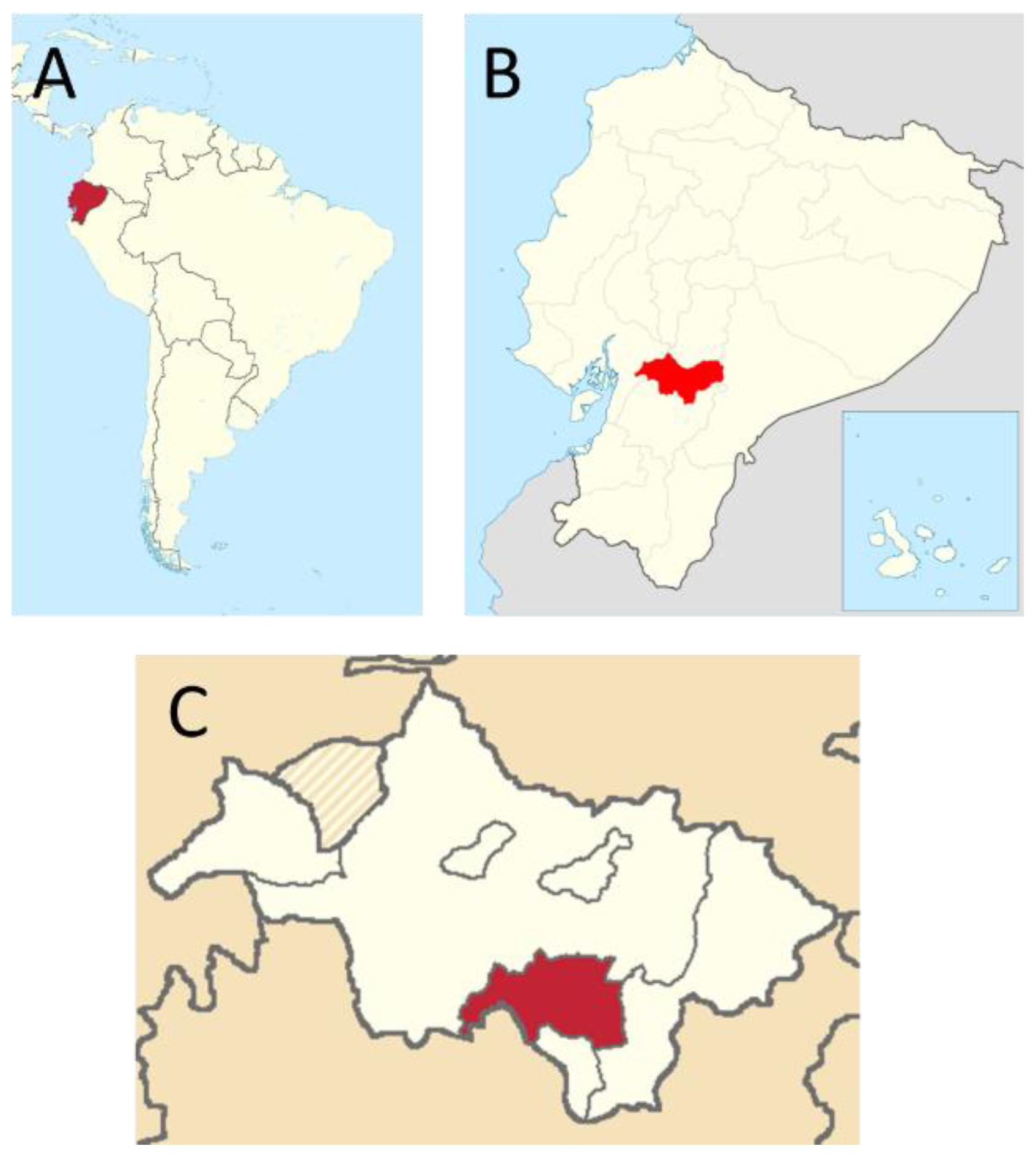
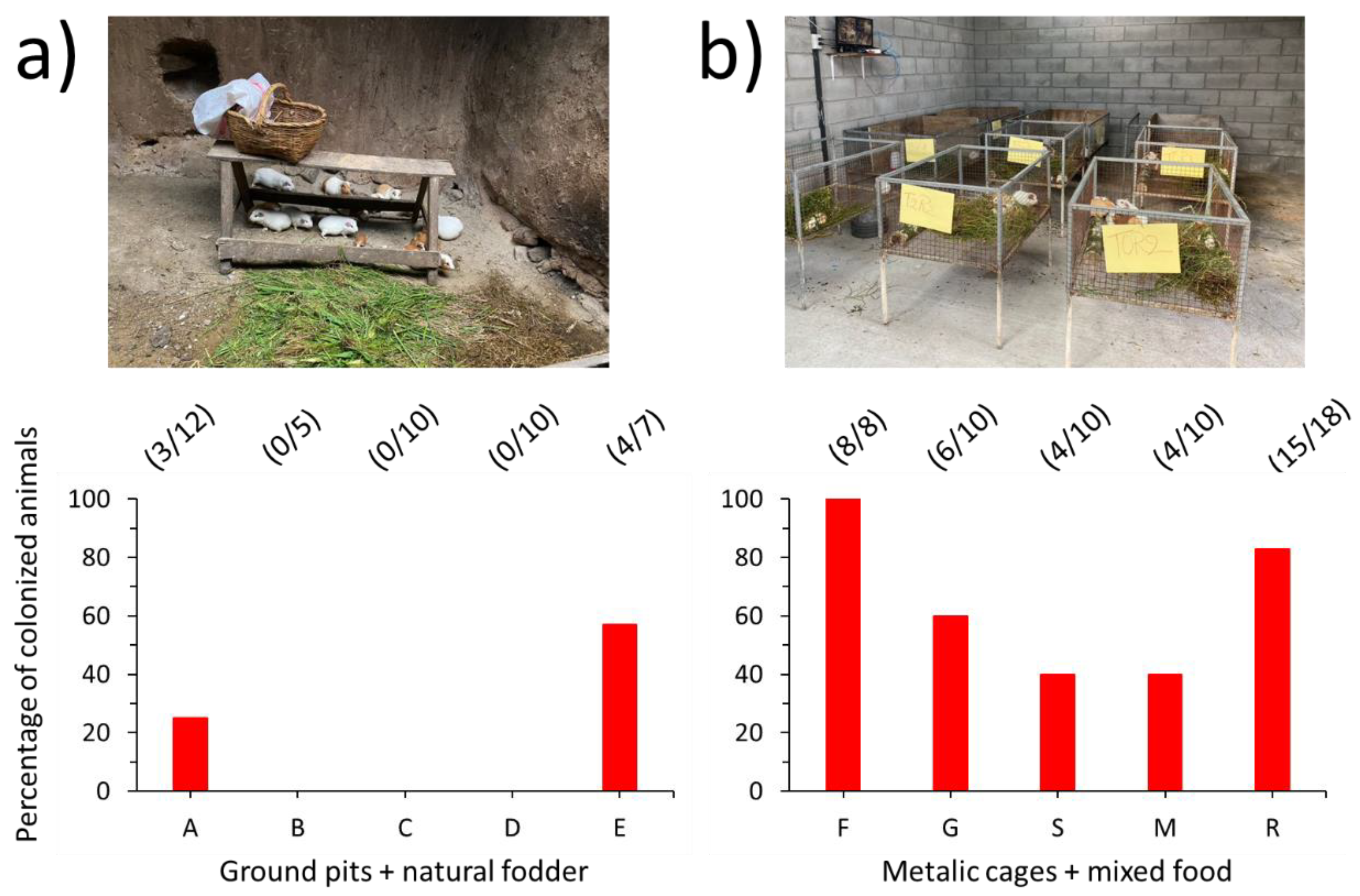
2.1. Sampling Area and Animals

A total of 100 guinea pigs from 10 farms in the municipality of Biblián (2°42′36″S, 78°52′48″W), Cañar (Southern Ecuadorian Andes) (Figure 1), were included in this survey, which was performed from March to November 2021.
Samples were collected indistinctly from clinically healthy adult animals. According to national regulations in Ecuador, sample collections for the diagnosis in farm animals do not require approval from an ethics committee (“Ley Orgánica de Sanidad Agropecuaria” 2017, Asamblea Nacional, República del Ecuador) [24]. Nevertheless, the farm owners were asked to sign a written consent and were present during the collection of samples.

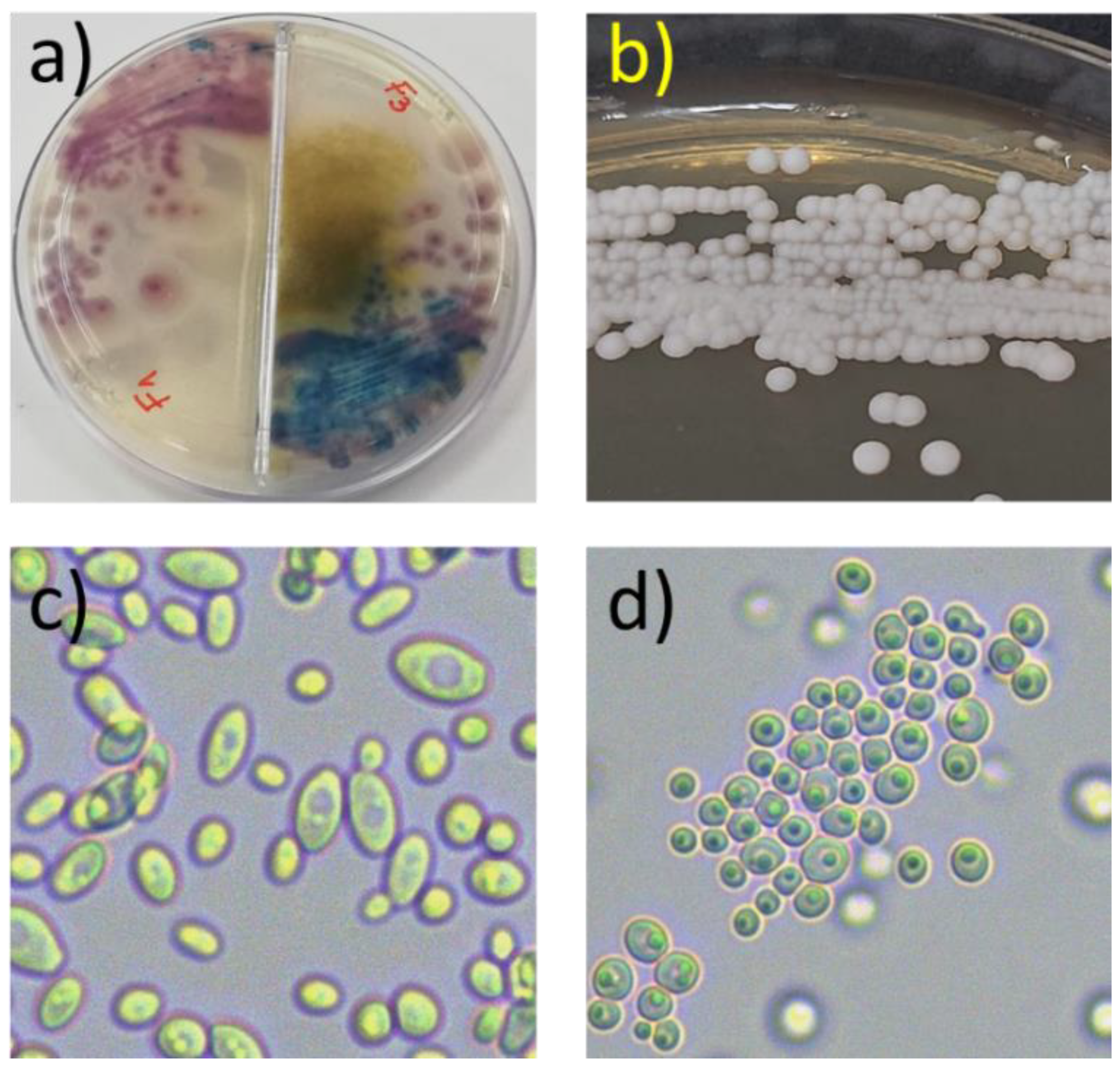
2.2. Sample Collection and Isolation Procedures

Samples were collected from the nasal cavity of healthy adult guinea pigs by using sterile cotton swabs. The swabs were moistened with sterile saline solution before collecting the samples and were gently rubbed inside the nose cavity. Once finished, the swab tips were introduced into sterile glass tubes containing Trypticase soy broth (Oxoid) and stored in coolers to be transported to the laboratory. Petri dishes containing Hi-Crome Candida Differential Agar (HiMedia Laboratories, Mumbai, India) were inoculated by gently spreading the sample in one side, and then streaked with an inoculating loop to obtain single colonies. Incubation was carried out at 30 °C for up to six days, with daily observations to detect yeast colonies. All colonies with a microscopic morphology consistent with yeasts were re-streaked several times until purification and were further identified by molecular methods (see below).
Isolates were stored in distilled water at room temperature, in the dark, and were recovered from storage by plating onto Potato Dextrose Agar (Difco Laboratories, Bergen, NJ, USA) for further experimentation.

2.3. Antifungal Susceptibility Tests

Susceptibility to four antifungal drugs of a subgroup of 22 selected pure isolates was ascertained by the disk diffusion technique, according to the Clinical and Laboratory Standards Institute (CLSI) [25]. The assay was performed on Mueller–Hinton agar (MHA) plates, supplemented with 2% glucose and 0.5 µg ml−1 methylene blue dye. The antifungals tested were fluconazole (25 µg), voriconazole (1 µg), caspofungin (5 µg) (Liofilchem S.R.L., Rosetto degli Abruzzi, Italy), and itraconazole (10 µg) (Bioanalyse, Ankara, Turkey). The diameter of the zone of inhibition of yeast growth was measured and registered after 24 h at 30 °C. The Candida albicans ATCC 90028 strain was used as reference.

2.4. DNA Extraction and Molecular Identification of the Isolates

Yeast DNA extraction was performed according to the rapid-boiling method described by Silva et al. [26]. The species identification was performed by PCR amplification of the rDNA internal transcribed spacer (ITS), nucleotide sequencing of the amplicons, and further analysis. For this, forward (ITS1 5′-TCC GTA GGT GAA CCT GCG G-3′) and reverse (ITS4 5′-TCC TCC GCT TAT TGA TAT GC-3′) primers were used [27,28]. The PCR mixture contained 25 μL premix (DreamTaq Green PCR Mastermix, Thermoscientific), 19 μL deionized sterile water, 2 μL from each forward and reverse primer, and 2 μL of genomic DNA, which served as the DNA template in a final volume of 50 μL. The PCR cycling conditions were: an initial denaturation phase at 95 °C for 5 min, followed by 35 cycles of denaturation at 94 °C for 1 min, annealing at 58 °C for 1 min, and extension at 72 °C for 1 min, with a final extension phase at 72 °C for 10 min.
Amplicons were sequenced by Macrogen (Seoul, Republic of Korea) and the nucleotidic sequences were subjected to BLAST searches against fungal sequences existing in DNA databases (http://blast.ncbi.nlm.nih.gov/Blast.cgi accessed on 7 June 2022). The sequences were compared using nucleotide–nucleotide BLAST (blastn) with default settings, except that the sequences were not filtered for low complexity. Species were identified based on the highest similarity score (100%) with the reference database sequence. All nucleotide sequences were deposited in GenBank.

3. Results

One or several yeast colony morphotypes were retrieved from forty-four out of one hundred healthy guinea pigs (44%) reared in ten farms in the Cañar Province (Figure 2a,b).
All isolates exhibited a characteristic yeast morphology at the microscopic level (Figure 2c,d). The number of colony morphotypes retrieved in Hi-Crome Candida Differential Agar from each animal ranged from one (13 animals) to five (1 animal).
Incidentally, fewer animals were colonized in their nasal mucosa by yeasts when raised in ground pits and fed with natural fodder (five farms: A, B, C, D, and E) (Figure 3a), as compared to animals raised intensively in cages (five farms: F, G, S, M, and R) and fed with a mixture of fodder and cereal concentrates (Figure 3b).
In order to determine the diversity of yeast species colonizing the nasal duct of guinea pigs, we selected a subgroup of 30 isolates showing conspicuous morphological differences (or showing similar phenotypes but derived from different animals) at the colony level for molecular identification. The results obtained by nucleotide sequencing and BLAST analysis of the chromosomal ITS1-ITS4 region allowed us to identify these isolates as members of eleven yeast species, belonging to eight different genera: Wickerhamomyces, Diutina, Meyerozyma, Candida, Pichia, Rhodotorula, Galactomyces, and Cryptococcus (Table 1).
Several of these isolates were subsequently tested for their susceptibility to four antifungal drugs (Table 2). The results show that 15 out of 22 yeast isolates were insensitive to at least one of the antifungal drugs tested. Some isolates were insensitive to two or three of these antifungals. Itraconazole (at the dose tested) was shown to be ineffective against 14 (63.6%) of the isolates tested, followed by caspofungin and fluconazole (4/22, 18.2%), and, finally, voriconazole (3/22, 13.6%). On the other hand, three Pichia spp. were insensitive to three of the drugs tested (namely fluconazole, itraconazole, and voriconazole), while three Meyerozyma spp. were insensitive to two of them (itraconazole and caspofungin).

4. Discussion

Interactions between humans and farm animals are at the origin of several zoonotic diseases which may threaten the health of farmers, their families, the final consumers, and—in general—the whole community. Considering that (i) guinea pigs are among the most important sources of animal protein for human consumption in the Andean region; (ii) these rodents are frequently handled by breeders, distributors, sellers, and consumers; and (iii) almost no safety measures are taken during breeding, slaughter, sale, and preparation, a better understanding of their role as potential reservoirs of opportunistic pathogens is necessary. In the present work, our initial aim was to prospect the nasal mucosa of guinea pigs for opportunistic Candida spp. strains. Surprisingly, as we have shown here, at least 11 yeast species colonized the nasal mucosa of guinea pigs raised for human consumption, some of which having already been identified as opportunistic human pathogens. As far as we know, this is the first report showing that raised guinea pigs are an important reservoir of these kinds of potentially pathogenic non-Candida yeasts.
The presence of yeasts in the nasal mucosa of guinea pigs was not totally unexpected. In fact, it is well known that, due to its constant humidity, this particular environment is colonized by all kinds of microbes, including saprophytic or pathogenic fungi. Opportunistic yeast species have also been shown to persist outside of clinical settings, mainly in soils and plants, which can also act as natural reservoirs. This suggests that the ecology of these fungi may be more complex than previously thought [29].
Non-albicans Candida and other rare yeast species have emerged in recent decades as agents of serious diseases in humans, especially targeting immunocompromised individuals, children, and the elderly [30]. Among the virulence factors expressed by many of these pathogens, resistance against antifungal drugs is of utmost importance [31]. Since no specific clinical breakpoints have been established to date, by either the European Committee on Antimicrobial Susceptibility Testing (EUCAST) or the CLSI, for environmental or rare opportunistic yeasts, we decided to present here only the descriptive results (inhibition halo diameter, expressed in mm values), with no attempt to categorize the isolates according to the results of the antifungal susceptibility tests (except when the inhibition of growth was complete). Nevertheless, we also considered that the benefits and advantages of the disk diffusion tests, as tools for antifungal resistance surveillance, have been previously shown, as in the case of the ARTEMIS DISK Global Antifungal Surveillance Study [32]. In the case of fluconazole and voriconazole, disk testing is highly accurate and can be routinely performed in clinical laboratories.
Wickerhamomyces anomalus (formerly Pichia anomala) belongs to this group of non-Candida, non-Cryptococcus yeasts of clinical interest. It is considered an environmental yeast, usually found in soils, plants, and fruits, but also in the feces of bats and birds [21,33,34]. W. anomalus is increasingly being considered as an emerging pathogen of clinical importance, often being associated with a wide range of fungal infections, from ocular keratitis to meningitis and fatal candidemia [35]. Even though it is believed to be ubiquitous in the environment, the knowledge concerning W. anomalus ecology is still limited.
Our results show that more than half of the W. anomalus isolates tested were completely insensitive to itraconazole. From a public health perspective, this finding is of relevance since previous studies have revealed that some nosocomial W. anomalus isolates can, in fact, tolerate in vitro high doses of azoles, including flucytosine, itraconazole, and fluconazole [35,36,37].
Diutina catenulata (formerly Candida catenulata), an ascomycete closely related to Saccharomyces cerevisiae and often used for bioremediation purposes [38,39], was also present in the nasal mucosa of the healthy guinea pigs studied. This species has been previously isolated from the gut of birds [20,40,41] and the nasolacrimal duct of healthy horses [18]. More importantly, a few reports have documented human infections with D. catenulata and increasingly recognize its importance as an emerging pathogen [42].
D. catenulata animal isolates produce and secrete potent hydrolases (e.g., proteases and phospholipases), as previously shown by Brilhante et al. [19] and Rhimi et al. [43]. These enzymes are considered as virulence factors, which play fundamental roles in the pathogenesis of yeast infections [44]. In addition, D. catenulata isolates generally exhibit antifungal sensitivity, particularly to azoles and echinocandins [20,42,45,46]. However, it has been shown that some nosocomial isolates can tolerate high concentrations of fluconazole or caspofungin [47]. Thus, it is important to explore their susceptibility to these antifungals. In the present report, we show that one D. catenulata isolate was insensitive to caspofungin, while another two were slightly inhibited by this antifungal drug. This opens the question regarding their virulence potential if spread to human hosts, since echinocandins represent the first choice of treatment against invasive candidiasis in patients [48].
On the other hand, three fungal isolates were identified as Meyerozyma caribbica (anamorph Candida fermentati), and a fourth as Meyerozyma carpophila (formerly Candida carpophila). These phenotypically indistinguishable species are phylogenetically related and belong to the M. guilliermondii species complex [49,50]. Even though yeasts of this complex are ubiquitous in the environment and can be isolated from a variety of sources—including fermented foods, plants, and arthropods—some isolates can also cause human infections [51,52]. Furthermore, species within this complex are increasingly regarded as emerging infectious yeasts of the non-albicans Candida species group, which are considered as opportunistic pathogens in immunocompromised patients and are responsible for 1–5% of nosocomial bloodstream infections worldwide [23].
In a recent report, Chaves and coworkers isolated M. caribbica and M. guillermondii strains from patients suffering from bloodstream infections in an oncology reference center in Brazil [52]. The isolates were poorly susceptible to antifungals and exhibited high minimum inhibitory concentrations (MICs) for fluconazole and echinocandins. Strikingly, all four tested Meyerozyma spp. isolates studied here were insensitive to itraconazole, and three of them to caspofungin (an echinocandin). Again, these results raise concerns about their potential as virulent zoonotic pathogens.
We also identified five isolates belonging to Candida railenensis or C. parapsilosis. Both species belong to the non-albicans Candida group, some of which cause important infections in humans and whose incidence has been increasing in recent years [22]. Most of these species are generally considered commensal microorganisms, frequently found in the skin and mucosae (respiratory, genital, and gastrointestinal) of animals such as dogs, horses, and birds [53,54]. However, they are also considered emerging pathogens and must be considered with care, particularly in the case of C. parapsilopsis isolates, since it is the second most frequently isolated opportunistic Candida species [55].
The identification of an R. glutinis isolate is of upmost importance. This pink yeast belongs to a very heterogeneous group, commonly found in the tissues and organs of animals; it can also be easily isolated from environmental samples [56]. Although infections caused in humans by R. glutinis are less frequent than those caused by R. mucilaginosa, it is an opportunistic pathogen, which can infect the blood, the central nervous system, the eyes, and the heart, among other organs and tissues [57]. Rhodotorula species appear to be intrinsically resistant to fluconazole, which has been predicted to be ineffective to treat infections caused by this fungus [58]. In line with this information, the only R. glutinis isolate tested here was insensitive to fluconazole and itraconazole, a result that adds to its potential as a virulent zoonotic pathogen.
As said above, the presence of commensal or opportunistic pathogenic fungi in the nasal cavity of animals was far from unexpected. In fact, it is known that the nasal resident bacteria interact with the immune system of the mammal host, and this dynamic interaction favors the colonization of the mucosa by other microbes, such as Candida spp. and other yeasts [58,59]. In this particular environment, microbes—either bona fide commensals or opportunistic pathogens—are sheltered and well nurtured, being able to proliferate depending on the topography of the cavity [60].
Many of these nasal-resident microorganisms reach the nasal cavity from the soil or through the food used to feed the rodents. It is well acknowledged that several yeasts are ubiquitous in the environment and can be easily isolated from the soil or some plants where the fungi survive, especially when the environment is humid [61]. On the other hand, the high density of animals in more sophisticated settlements favors the dissemination of yeasts and other pathogens by frequent animal-to-animal contact and the use of contaminated water and stored manure. Both circumstances have been already highlighted by Graham et al. [4] while studying the infection of guinea pigs with Campylobacter jejuni in rural Ecuador. As we have shown here, guinea pigs raised intensively in cages and fed with a mixture of fodder and concentrates are colonized by more yeast species/strains in their nasal mucosa than their counterparts raised in ground pits and fed only with natural fodder. This very preliminary observation deserves to be addressed in more depth in the future.
Among the limitations of our work, the most obvious is the limited scope of the study, in geographical terms. This circumstance obliges us to consider the presented results very carefully, and to be cautious in order to not extrapolate them to other regions. Similarly, since the study was transversal, we did not consider fluctuations during different periods of the year. Finally, since we followed a classical microbiological approach—using culture methods—we did not investigate the actual diversity of the mycobiome residing in the nasal cavity of guinea pigs. In order to shed light on the actual composition and structure of this microbial community, metagenomic techniques should be used.
Nevertheless, and to the best of our knowledge, this is the first attempt to describe the diversity of culturable yeasts found in the nasal mucosa of such an important animal resource in the Andean region of Ecuador. Furthermore, our results emphasize the threat posed to human health by some of these potentially pathogenic zoonotic yeast species.

5. Conclusions

In summary, we isolated potentially zoonotic yeasts from the nasal duct of guinea pigs raised as livestock in Southern Ecuador. At least 11 species, belonging to 8 genera, were identified. Furthermore, the resistance exhibited by 22 of these isolates toward four antifungal drugs of therapeutic use was also revealed. This particular aspect is of fundamental importance since several of these yeast species are considered as emergent pathogens. Considering the frequent manipulation of guinea pigs without any safety measures by breeders, distributors, sellers, and consumers, the presence of pathogenic or potentially pathogenic microorganisms in their tissues and organs represents a risk from a public health point of view.

Author Contributions

Conceptualization, L.A.Y.; methodology, L.A.Y., L.B., M.C. and J.S.; investigation, L.B., D.P., J.S., A.Y.M. and E.J.C.; resources, L.A.Y.; data curation, L.A.Y. and L.B.; writing—original draft preparation, L.A.Y.; writing—review and editing, L.A.Y. and L.B.; visualization, L.A.Y.; project administration L.A.Y.; funding acquisition, L.A.Y. All authors have read and agreed to the published version of the manuscript.

Funding

This research was funded by the Catholic University of Cuenca, grant number PICCIITT19-37.

Institutional Review Board Statement

Not applicable.

Informed Consent Statement

Written informed consent was obtained from the farm owners to proceed with the sampling procedure in their presence.

Data Availability Statement

Not applicable.

Acknowledgments

The authors would like to thank Dayana Mishell Escobar Narváez and Arianna Michelle González Cabrera for technical support and preliminary surveys. We also express our deepest gratitude to the farm owners and cuy producers of the Cantón Biblián (Azuay, Ecuador).

Conflicts of Interest

The authors declare no conflict of interest. The funders had no role in the design of the study; in the collection, analyses, or interpretation of data; in the writing of the manuscript; or in the decision to publish the results.

References

  1. Avilés, D.; Martínez, A.; Landi, V.; Delgado, J. The guinea pig (Cavia porcellus): An Andean resource of interest as an agricultural food source. Anim. Genet. Res. 2014, 55, 87–91. [Google Scholar] [CrossRef]
  2. Vasco, K.; Graham, J.P.; Trueba, G. Detection of zoonotic enteropathogens in children and domestic animals in a semirural community in Ecuador. Appl. Environ. Microbiol. 2016, 82, 4218–4224. [Google Scholar] [CrossRef] [PubMed] [Green Version]
  3. De Zaldívar, L.C. Producción de Cuyes (Cavia porcellus) en los Países Andinos. Food and Agriculture Organization Document. Available online: https://www.fao.org/3/V6200T/v6200T05.htm (accessed on 2 October 2022).
  4. Graham, J.P.; Vasco, K.; Trueba, G. Hyperendemic Campylobacter jejuni in guinea pigs (Cavia porcellus) raised for food in a semi-rural community of Quito, Ecuador. Environ. Microbiol. Rep. 2016, 8, 382–387. [Google Scholar] [CrossRef] [PubMed] [Green Version]
  5. Amman, B.R.; Pavlin, B.I.; Albarino, C.G.; Comer, J.A.; Erickson, B.R.; Oliver, J.B.; Sealy, T.K.; Vincent, M.J.; Nichol, S.T.; Paddock, C.D.; et al. Pet rodents and fatal lymphocytic choriomeningitis in transplant patients. Emerg. Infect. Dis. 2007, 13, 719–725. [Google Scholar] [CrossRef]
  6. Lutz-Wohlgroth, L.; Becker, A.; Brugnera, E.; Huat, Z.L.; Zimmermann, D.; Grimm, F.; Haessig, M.; Greub, G.; Kaps, S.; Spiess, B.; et al. Chlamydiales in guinea-pigs and their zoonotic potential. J. Vet. Med. A Physiol. Pathol. Clin. Med. 2006, 53, 185–193. [Google Scholar] [CrossRef]
  7. Fredriksson-Ahomaa, M. Yersinia enterocolitica and Yersinia pseudotuberculosis. In Foodborne Diseases; Simjee, S., Ed.; Humana Press: New York, NY, USA, 2007; pp. 79–113. [Google Scholar]
  8. Gruszynski, K.; Young, A.; Levine, S.J.; Garvin, J.P.; Brown, S.; Turner, L.; Fritzinger, A.; Gertz, R.E., Jr.; Murphy, J.M.; Vogt, M.; et al. Streptococcus equi subsp zooepidemicus infections associated with guinea pigs. Emerg. Infect. Dis. 2015, 21, 156–158. [Google Scholar] [CrossRef]
  9. Walther, B.; Wieler, L.H.; Vincze, S.; Antão, E.M.; Brandenburg, A.; Stamm, I.; Kopp, P.A.; Kohn, B.; Semmler, T.; Lübke-Becker, A. MRSA variantin companion animals. Emerg. Infect. Dis. 2012, 18, 2017–2020. [Google Scholar] [CrossRef]
  10. González, L.C.; Esteban, J.G.; Bargues, M.D.; Valero, M.A.; Ortiz, P.; Naquira, C.; Mas-Coma, S. Hyper-endemic human fascioliasis in Andean valleys: An altitudinal transect analysis in children of Cajamarca province, Peru. Acta Trop. 2011, 120, 119–129. [Google Scholar] [CrossRef]
  11. Gabastou, J.M.; Proano, J.; Vimos, A.; Jaramillo, G.; Hayes, E.; Gage, K.; Chu, M.; Guarner, J.; Zaki, S.; Bowers, J.; et al. An outbreak of plague including cases with probable pneumonic infection, Ecuador, 1998. Trans. R. Soc. Trop. Med. Hyg. 2000, 94, 387–391. [Google Scholar] [CrossRef]
  12. Levy, M.Z.; Bowman, N.M.; Kawai, V.; Waller, L.A.; Cornejo del Carpio, J.G.; Cordova Benzaquen, E.; Gilman, R.H.; Bern, C. Periurban Trypanosoma cruzi-infected Triatoma infestans, Arequipa, Peru. Emerg. Infect. Dis. 2006, 12, 1345–1352. [Google Scholar] [CrossRef]
  13. Zambrano-Mila, M.; Rodriguez, A.S.; Rivera-Olivero, I.A.; Salas-Rueda, M.; Caceres-Orellana, M.V.; de Waard, J.H.; Garcia-Bereguiain, M.A. Methicillin resistant Staphylococcus aureus carriage among guinea pigs raised as livestock in Ecuador. One Health 2020, 9, 100118. [Google Scholar] [CrossRef] [PubMed]
  14. Leyva-Grado, V.H.; Mubareka, S.; Krammer, F.; Cárdenas, W.B.; Palese, P. Influenza virus infection in Guinea pigs raised as livestock, Ecuador. Emerg. Infect. Dis. 2012, 18, 1135–1137. [Google Scholar] [CrossRef] [Green Version]
  15. Miceli, M.H.; Díaz, J.A.; Lee, S.A. Emerging opportunistic yeast infections. Lancet Infect. Dis. 2011, 1, 142–151. [Google Scholar] [CrossRef]
  16. WHO. Managing Zoonotic Public Health Risks at the Human Animal–Ecosystem Interface; WHO: Geneva, Switzerland, 2011. [Google Scholar]
  17. Seyedmousavi, S.; Guillot, J.; Tolooe, A.; Verweij, P.E.; de Hoog, G.S. Neglected fungal zoonoses: Hidden threats to man and animals. Clin. Microbiol. Infect. 2015, 21, 416–425. [Google Scholar] [CrossRef] [PubMed] [Green Version]
  18. Otte, J.; Pica-Ciamarra, U. Emerging infectious zoonotic diseases: The neglected role of food animals. One Health 2021, 13, 100323. [Google Scholar] [CrossRef] [PubMed]
  19. Brilhante, R.S.; Bittencourt, P.V.; Castelo-Branco, D.; de Oliveira, J.S.; Alencar, L.P.; Cordeiro, R.; Pinheiro, M.; Nogueira-Filho, E.F.; Pereira-Neto, W.; Sidrim, J.J.; et al. Trends in antifungal susceptibility and virulence of Candida spp. from the nasolacrimal duct of horses. Med. Mycol. 2016, 54, 147–154. [Google Scholar] [CrossRef] [PubMed] [Green Version]
  20. Glushakova, A.M.; Rodionova, E.N.; Kachalkin, A.V. Yeasts in feces of pigeons (Columba Livia) in the city of Moscow. Curr. Microbiol. 2021, 78, 238–243. [Google Scholar] [CrossRef] [PubMed]
  21. Rosario Medina, I.; Román Fuentes, L.; Batista Arteaga, M.; Real Valcárcel, F.; Acosta Arbelo, F.; Padilla Del Castillo, D.; Déniz Suárez, S.; Ferrer Quintana, O.; Vega Gutiérrez, B.; Silva Sergent, F.; et al. Pigeons and their droppings as reservoirs of Candida and other zoonotic yeasts. Rev. Iberoam. Micol. 2017, 34, 211–214. [Google Scholar] [CrossRef] [Green Version]
  22. Giacobbe, D.R.; Maraolo, A.E.; Simeon, V.; Magnè, F.; Pace, M.C.; Gentile, I.; Chiodini, P.; Viscoli, C.; Sanguinetti, M.; Mikulska, M.; et al. Changes in the relative prevalence of candidaemia due to non-albicans Candida species in adult in-patients: A systematic review, meta-analysis and meta-regression. Mycoses 2020, 63, 334–342. [Google Scholar] [CrossRef]
  23. Papon, N.; Savini, V.; Lanoue, A.; Simkin, A.J.; Crèche, J.; Giglioli-Guivarc’h, N.; Clastre, M.; Courdavault, V.; Sibirny, A.A. Candida guilliermondii: Biotechnological applications, perspectives for biological control, emerging clinical importance and recent advances in genetics. Curr. Genet. 2013, 59, 73–90. [Google Scholar] [CrossRef]
  24. Ley Orgánica de Sanidad Agropecuaria. Asamblea Nacional, República del Ecuador. Available online: https://www.gob.ec/sites/default/files/regulations/2018-09/Documento_Ley%20Org%C3%A1nica%20de%20Sanidad%20Agropecuaria.pdf (accessed on 11 November 2022).
  25. Clinical and Laboratory Standards Institute (CLSI). Method for Antifungal Disk Diffusion Susceptibility Testing of Yeasts, 3rd ed.; CLSI Guideline M44; CLSI: Wayne, PA, USA, 2018. [Google Scholar]
  26. Almeida da Silva, G.; Bernardi, T.L.; Carvalho Schaker, P.D.; Menegotto, M.; Valente, P. Rapid yeast DNA extraction by boiling and freeze-thawing without using chemical reagents and DNA purification. Braz. Arch. Biol. Technol. 2012, 55, 319–327. [Google Scholar] [CrossRef] [Green Version]
  27. Gardes, M.; Bruns, T.D. ITS primers with enhanced specificity for Basidiomycetes: Application to the identification of mycorrhizae and rusts. Mol. Ecol. 1993, 2, 113–118. [Google Scholar] [CrossRef] [PubMed]
  28. White, T.J.; Bruns, T.; Lee, S.; Taylor, J. Amplification and direct sequencing of fungal ribosomal RNA genes for phylogenetics. In PCR Protocols a Guide to Methods and Applications; Academic Press: San Diego, CA, USA, 1990; pp. 315–322. [Google Scholar]
  29. Opulente, D.A.; Langdon, Q.K.; Buh, K.V.; Haase, M.A.B.; Sylvester, K.; Moriarty, R.V.; Jarzyna, M.; Considine, S.L.; Schneider, R.M.; Hittinger, C.T. Pathogenic budding yeasts isolated outside of clinical settings. FEMS Yeast Res. 2019, 19, foz032. [Google Scholar] [CrossRef] [PubMed]
  30. Deorukhkar, S.C.; Saini, S.; Mathew, S. Non-albicans Candida infection: An emerging threat. Interdiscip. Perspect. Infect. Dis. 2014, 2014, 615958. [Google Scholar] [CrossRef] [Green Version]
  31. Farmakiotis, D.; Kontoyiannis, D.P. Epidemiology of antifungal resistance in human pathogenic yeasts: Current viewpoint and practical recommendations for management. Int. J. Antimicrob. Agents 2017, 50, 318–324. [Google Scholar] [CrossRef]
  32. Pfaller, M.A.; Diekema, D.J.; Gibbs, D.L.; Newell, V.A.; Ellis, D.; Tullio, V.; Rodloff, A.; Fu, W.; Ling, T.A. Global Antifungal Surveillance Group. Results from the ARTEMIS DISK Global Antifungal Surveillance Study, 1997 to 2007: A 10.5-year analysis of susceptibilities of Candida Species to fluconazole and voriconazole as determined by CLSI standardized disk diffusion. J. Clin. Microbiol. 2010, 48, 1366–1377. [Google Scholar] [CrossRef] [Green Version]
  33. Botelho, N.S.; Paula, S.B.; Panagio, L.A.; Pinge-Filho, P.; Yamauchi, L.M.; Yamada-Ogatta, S.F. Candida species isolated from urban bats of Londrina-Paraná, Brazil and their potential virulence. Zoonoses Public Health 2012, 59, 16–22. [Google Scholar] [CrossRef]
  34. Mancianti, F.; Nardoni, S.; Ceccherelli, R. Occurrence of yeasts in psittacines droppings from captive birds in Italy. Mycopathologia 2002, 153, 121–124. [Google Scholar] [CrossRef]
  35. Zhang, L.; Xiao, M.; Arastehfar, A.; Ilkit, M.; Zou, J.; Deng, Y.; Xu, Y.; Liao, W.; Zhao, J.; Fang, W.; et al. Investigation of the emerging nosocomial Wickerhamomyces anomalus infections at a Chinese tertiary teaching hospital and a systemic review: Clinical manifestations, risk factors, treatment, outcomes, and anti-fungal susceptibility. Front. Microbiol. 2021, 12, 744502. [Google Scholar] [CrossRef]
  36. Lin, H.C.; Lin, H.Y.; Su, B.H.; Ho, M.W.; Ho, C.M.; Lee, C.Y.; Lin, M.H.; Hsieh, H.Y.; Lin, H.C.; Li, T.C.; et al. Reporting an outbreak of Candida pelliculosa fungemia in a neonatal intensive care unit. J. Microbiol. Immunol. Infect. 2013, 46, 456–462. [Google Scholar] [CrossRef]
  37. Pfaller, M.A.; Castanheira, M.; Messer, S.A.; Jones, R.N. In vitro antifungal susceptibilities of isolates of Candida spp. and Aspergillus spp. From China to nine systemically active antifungal agents: Data from the SENTRY antifungal surveillance program, 2010 through 2012. Mycoses 2015, 58, 209–214. [Google Scholar] [CrossRef]
  38. Joo, H.S.; Ndegwa, P.M.; Shoda, M.; Phae, C.G. Bioremediation of oil-contaminated soil using Candida catenulata and food waste. Environ. Pollut. 2008, 156, 891–896. [Google Scholar] [CrossRef]
  39. Babaei, F.; Habibi, A. Fast biodegradation of diesel hydrocarbons at high concentration by the sophorolipid-producing yeast Candida catenulata KP324968. J. Mol. Microbiol. Biotechnol. 2018, 28, 240–253. [Google Scholar] [CrossRef] [PubMed]
  40. Cafarchia, C.; Iatta, R.; Danesi, P.; Camarda, A.; Capelli, G.; Otranto, D. Yeasts isolated from cloacal swabs, feces, and eggs of laying hens. Med. Mycol. 2019, 57, 340–345. [Google Scholar] [CrossRef] [PubMed]
  41. Subramanya, S.H.; Sharan, N.K.; Baral, B.P.; Hamal, D.; Nayak, N.; Prakash, P.Y.; Sathian, B.; Bairy, I.; Gokhale, S. Diversity, in-vitro virulence traits and antifungal susceptibility pattern of gastrointestinal yeast flora of healthy poultry, Gallus domesticus. BMC Microbiol. 2017, 17, 113. [Google Scholar] [CrossRef] [Green Version]
  42. Boden, S.; Morio, F.; Zhou, M.; Gerrits van den Ende, B.; Hagen, F. De novo Nanopore genome sequencing of the clinical Diutina catenulata type-strain CBS565. Mycopathologia 2022, 187, 417–420. [Google Scholar] [CrossRef] [PubMed]
  43. Rhimi, W.; Aneke, C.I.; Annoscia, G.; Camarda, A.; Mosca, A.; Cantacessi, C.; Otranto, D.; Cafarchia, C. Virulence and in vitro antifungal susceptibility of Candida albicans and Candida catenulata from laying hens. Int. Microbiol. 2021, 24, 57–63. [Google Scholar] [CrossRef]
  44. GunTang, W.; Kamonvoradej, N.; Chomchat, C.; Suriyakan, S.; Sanit, S.; Wongwigkarn, J.; Lamlertthon, S. Prevalence and virulence factors of Candida spp. associated with blow flies. Asian Pac. J. Trop. Biomed. 2017, 7, 428–431. [Google Scholar] [CrossRef]
  45. Radosavljevic, M.; Koenig, H.; Letscher-Bru, V.; Waller, J.; Maloisel, F.; Lioure, B.; Herbrecht, R. Candida catenulata fungemia in a cancer Patient. J. Clin. Microbiol. 1999, 37, 475–477. [Google Scholar] [CrossRef] [Green Version]
  46. Ha, M.V.; Choy, M.S.; McCoy, D.; Fernandez, N.; Suh, J.S. Candida catenulata candidaemia and possible endocarditis in a cirrhotic patient successfully de-escalated to oral fluconazole. J. Clin. Pharm. Ther. 2018, 43, 910–913. [Google Scholar] [CrossRef]
  47. Chen, X.-F.; Zhang, W.; Fan, X.; Hou, X.; Liu, X.-Y.; Huang, J.-J.; Kang, W.; Zhang, G.; Zhang, H.; Yang, W.-H.; et al. Antifungal susceptibility profiles and resistance mechanisms of clinical Diutina catenulata isolates with high MIC values. Front. Cell. Infect. Microbiol. 2021, 11, 739496. [Google Scholar] [CrossRef]
  48. Lamoth, F.; Lewis, R.E.; Kontoyiannis, D.P. Role and interpretation of antifungal susceptibility testing for the management of invasive fungal infections. J. Fungi 2021, 7, 17. [Google Scholar] [CrossRef] [PubMed]
  49. Bai, F.Y.; Liang, H.Y.; Jia, J.H. Taxonomic relationships among the taxa in the Candida guilliermondii complex, as revealed by comparative electrophoretic karyotyping. Int. J. Syst. Evol. Microbiol. 2000, 50, 417–422. [Google Scholar] [CrossRef]
  50. Vaughan-Martini, A.; Kurtzman, C.P.; Meyer, S.A.; O’Neill, E.B. Two new species in the Pichia guilliermondii clade: Pichia caribbica sp. nov., the ascosporic state of Candida fermentati, and Candida carpophila comb. nov. FEMS Yeast Res. 2005, 5, 463–469. [Google Scholar] [CrossRef] [PubMed] [Green Version]
  51. De Marco, L.; Epis, S.; Capone, A.; Martin, E.; Bozic, J.; Crotti, E.; Ricci, I.; Sassera, D. The genomes of four Meyerozyma caribbica isolates and novel insights into the Meyerozyma guilliermondii species complex. G3 2018, 8, 755–759. [Google Scholar] [CrossRef] [PubMed] [Green Version]
  52. Chaves, A.; Trilles, L.; Alves, G.M.; Figueiredo-Carvalho, M.; Brito-Santos, F.; Coelho, R.A.; Martins, I.S.; Almeida-Paes, R. A case-series of bloodstream infections caused by the Meyerozyma guilliermondii species complex at a reference center of oncology in Brazil. Med. Mycol. 2021, 4, 235–243. [Google Scholar] [CrossRef] [PubMed]
  53. Brilhante, R.; Castelo-Branco, D.; Soares, G.; Astete-Medrano, D.J.; Monteiro, A.J.; Cordeiro, R.A.; Sidrim, J.; Rocha, M. Characterization of the gastrointestinal yeast microbiota of cockatiels (Nymphicus hollandicus): A potential hazard to human health. J. Med. Microbiol. 2010, 59, 718–723. [Google Scholar] [CrossRef] [Green Version]
  54. Brito, E.H.; Fontenelle, R.O.; Brilhante, R.S.; Cordeiro, R.A.; Monteiro, A.J.; Sidrim, J.J.; Rocha, M.F. The anatomical distribution and antimicrobial susceptibility of yeast species isolated from healthy dogs. Vet. J. 2009, 182, 320–326. [Google Scholar] [CrossRef]
  55. Bliss, J.M. Candida parapsilosis: An emerging pathogen developing its own identity. Virulence 2015, 6, 109–111. [Google Scholar] [CrossRef] [Green Version]
  56. Wirth, F.; Goldani, L.Z. Epidemiology of Rhodotorula: An emerging pathogen. Interdiscip. Perspect. Infect. Dis. 2012, 2012, 465717. [Google Scholar] [CrossRef]
  57. Ioannou, P.; Vamvoukaki, R.; Samonis, G. Rhodotorula species infections in humans: A systematic review. Mycoses 2019, 62, 90–100. [Google Scholar] [CrossRef] [PubMed]
  58. Rasmussen, T.T.; Kirkeby, L.P.; Poulsen, K.; Reinholdt, J.; Kilian, M. Resident aerobic microbiota of the adult human nasal cavity. APMIS 2000, 108, 663–675. [Google Scholar] [CrossRef] [PubMed]
  59. Noverr, M.C.; Huffnagle, G.B. Does the microbiota regulate immune outside the gut? Trends Microbiol. 2004, 12, 562–568. [Google Scholar] [CrossRef] [PubMed]
  60. Schenck, L.P.; McGrath, J.; Lamarche, D.; Stämpfli, M.R.; Bowdish, D.; Surette, M.G. Nasal tissue extraction is essential for characterization of the murine upper respiratory tract microbiota. mSphere 2020, 5, e00562-20. [Google Scholar] [CrossRef] [PubMed]
  61. Banerjee, S.; van der Heijden, M. Soil microbiomes and one health. Nat. Rev. Microbiol. 2022; 1–15, epub ahead of print. [Google Scholar] [CrossRef]
Figure 1. Location and map of Biblián Municipality (Cañar Province, Ecuador). (A) Ecuador in the South American context (in red); (B) Location of Cañar Province in Ecuador (in red). (C) Biblián Municipality (in red) (reproduced from Wikimedia Commons under Creative Commons licenses 3.0 and 4.0).
Animals 12 03449 g001
Figure 2. Isolation of yeasts from the nasal mucosa of guinea pigs. (a) Primary isolation cultures, showing pigmented colonies after 48–72 h growth at 30 °C on Hi-Crome Candida Differential Agar. (b) Pure isolates after re-streaking. (c,d) Microscopic view at 400× magnification of two pure isolates (isolates F2.3 and G3.1, respectively).
Animals 12 03449 g002
Figure 3. Colonization of guinea pigs’ nasal mucosa by potentially zoonotic yeasts. Up: Photographs of animals reared in ground pits and fed with natural fodder (a), or in metallic cages and fed with a mixture of fodder and cereal concentrate (b). Down: Percentage of animals colonized by yeasts in their nasal mucosa in each one of the ten farms included in this study. The number of animals actually colonized by yeasts in their nasal mucosa (as shown by cultivation in Hi-Crome Candida Differential Agar) versus the number of animals tested in each farm and of the animals is presented on top of each bar, between parentheses.
Animals 12 03449 g003
Table 1. Molecular identification of yeast isolates by ITS1-ITS4 region sequencing and analysis.
Isolate IDGenBank Accession NumberClosest Phylogenetic Neighbor% Identity% QuerySequence Length
E4.1ON706026Diutina catenulata CBS 565 99100390
E6.2ON706027Diutina catenulata CBS 5659999397
E6.3ON706028Cryptococcus aspenensis DS71299.886512
E7ON706029Wickerhamomyces anomalus L428/15100100590
F2.1ON706030Diutina catenulata CBS 56599.596405
F2.2ON706031Wickerhamomyces anomalus L428/15100100591
F2.3ON706032Pichia kluyveri E22599.897434
F2.4ON706033Candida parapsilosis LMICRO180100100500
F4.1ON706034Wickerhamomyces anomalus CBS 113100100590
F4.2ON706035Diutina catenulata CBS 56599100398
F5.3ON706036Galactomyces geotrichum SPPRISTMF198100350
F6ON706037Candida parapsilosis IFM 6356499.8100490
F9.1ON706038Wickerhamomyces anomalus CBS:261100100592
G2.1ON706039Wickerhamomyces anomalus CBS 575999.8100590
G3.1ON706040Wickerhamomyces anomalus T12100100422
G5.1ON706041(Candida) railenensis R9730899.8100600
G6.2ON706042Wickerhamomyces anomalus 3Y66100100580
G6.3ON706043(Candida) railenensis R97308 99.8100600
G7.2ON706044Wickerhamomyces anomalus SSL32100100420
G8.1ON706045Wickerhamomyces anomalus SSL32100100422
G8.2ON706046Pichia fermentans strain E224100100424
G9.1ON706047Wickerhamomyces anomalus TTG-100100100425
G9.2ON706048(Candida) railenensis R9730899.8100602
G9.4ON706049Pichia kluyveri E22599.8100422
MC.2ON706050Meyerozyma caribbica E12 + 4 9998590
MC.3ON706051Meyerozyma (Candida) carpophila FF398.899590
MC.10ON706052Meyerozyma caribbica CBS 9966 9999595
PCE.3ON706054Rhodotorula glutinis H199.597592
PCA8ON706053Diutina catenulata CBS 56599.5100372
SP.8ON706055Meyerozyma caribbica CBS 996699.899582
Table 2. Antifungal susceptibility of yeast isolates obtained from the nasal mucosa of guinea pigs.
Isolate IDMolecular IdentificationAntifungals
FLU-25 *ITC-10VO-1CAS-5
ATCC90028Candida albicans28122920
E4.1Diutina catenulata19122111
E7Wickerhamomyces anomalus26NGI1824
F2.1Diutina catenulata171731NGI
F2.2Wickerhamomyces anomalus26NGI2223
F2.3Pichia kluyveriNGINGINGI23
F4.1Wickerhamomyces anomalus22101722
F4.2Diutina catenulata15151127
G2.1Wickerhamomyces anomalus28122223
G3.1Wickerhamomyces anomalus32NGI2124
G6.2Wickerhamomyces anomalus26NGI1425
G7.2Wickerhamomyces anomalus31NGI2022
G8.1Wickerhamomyces anomalus27123023
G8.2Pichia fermentansNGINGINGI23
G9.1Wickerhamomyces anomalus29NGI2022
G9.2(Candida) railenensis1571030
G9.4Pichia kluyveriNGINGINGI21
MC10Meyerozyma caribbica27NGI39NGI
MC2Meyerozyma caribbica30NGI31NGI
MC3Meyerozyma carpophila24NGI31NGI
PCA.8Diutina catenulata16112210
SP8Meyerozyma caribbica29NGI3012
PCE3Rhodotorula glutinisNGINGINDND
Pure isolates were spread on the surface of Mueller–Hinton Agar plates supplemented with 2% glucose and 0.5 µg mL−1 methylene blue dye. The test was performed according to the Clinical and Laboratory Standards Institute (CLSI) approved protocol [25], using paper discs loaded with the respective antifungal drug. * The diameter of the zone of inhibition for each isolate was measured after incubation for 24 h at 30 °C and is expressed in mm. NGI: No growth inhibition (inhibition halo absent); ND: Not determined. FLU-25: Fluconazole (25 mcg); ITC-50: Itraconazole (50 μg); VO-1: Voriconazole (1 μg); CAS-5: Caspofungin (5 μg).
Publisher’s Note: MDPI stays neutral with regard to jurisdictional claims in published maps and institutional affiliations.

