Article

A Nonsense Variant in the DMD Gene Causes X-Linked Muscular Dystrophy in the Maine Coon Cat

1
Department of Veterinary and Biosciences, Faculty of Veterinary Medicine, Ghent University, B-9820 Merelbeke, Belgium
2
Small Animal Department, Faculty of Veterinary Medicine, Ghent University, B-9820 Merelbeke, Belgium
3
Department of Pathology, School of Medicine, University of California San Diego, La Jolla, CA 92093-0709, USA
*
Author to whom correspondence should be addressed.
These authors contributed equally to this work.
Animals 2022, 12(21), 2928; https://doi.org/10.3390/ani12212928
Submission received: 19 September 2022 / Revised: 18 October 2022 / Accepted: 22 October 2022 / Published: 25 October 2022
(This article belongs to the Special Issue (Epi) Genetic Disorders in Companion Animals)

Abstract

:

Simple Summary

Muscular dystrophy (MD) in cats is a muscular disease that can have a fatal outcome. It is often caused by variants in the DMD gene and shows an X-linked recessive mode of inheritance. Therefore, females can carry such a variant without showing clinical signs of disease and can spread the variant to their offspring. These carriers can only be identified through DNA tests. Here, we identified the variant causing MD in two Maine coon cats. Maine coons can now be screened for this variant through a DNA test.

Abstract

(1) Feline dystrophin-deficient muscular dystrophy (ddMD) is a fatal disease characterized by progressive weakness and degeneration of skeletal muscles and is caused by variants in the DMD gene. To date, only two feline causal variants have been identified. This study reports two cases of male Maine coon siblings that presented with muscular hypertrophy, growth retardation, weight loss, and vomiting. (2) Both cats were clinically examined and histopathology and immunofluorescent staining of the affected muscle was performed. DMD mRNA was sequenced to identify putative causal variants. (3) Both cats showed a significant increase in serum creatine kinase activity. Electromyography and histopathological examination of the muscle samples revealed abnormalities consistent with a dystrophic phenotype. Immunohistochemical testing revealed the absence of dystrophin, confirming the diagnosis of dystrophin-deficient muscular dystrophy. mRNA sequencing revealed a nonsense variant in exon 11 of the feline DMD gene, NC_058386.1 (XM_045050794.1): c.1180C > T (p.(Arg394*)), which results in the loss of the majority of the dystrophin protein. Perfect X-linked segregation of the variant was established in the pedigree. (4) ddMD was described for the first time in the Maine coon and the c.1180C>T variant was confirmed as the causal variant.

1. Introduction

Duchenne muscular dystrophy (DMD, MIM #310200) is an early-onset, rapidly progressive neuromuscular disease in humans. Primarily, male individuals are affected by DMD because of the X-linked recessive inheritance, and the prevalence ranges between 15.9 and 19.5 in 10,000 births [1]. It is a dystrophin-deficient muscular dystrophy (ddMD) caused by variants in the DMD gene. Symptoms are often attributed to the loss or significant shortening of the C-terminal domain, a region containing functionally important binding sites [2]. Generally, DMD-causing variants disrupt the translational ORF and lead to a premature termination codon, resulting in absent or non-functional dystrophin [3]. The most commonly reported variants are large deletions and duplications (66−72% and 9−14% of all cases, respectively), followed by nonsense variants (about 10% of all cases) [4,5]. Other types of variants that cause DMD are small indels, splice site, and missense variants (about 4%, 2%, and 1% of all cases, respectively) [4].
ddMDs, similar to DMD, exist in mice [6], rats [7], rabbits [8], pigs [9], dogs [10,11,12], cats [13,14], and monkeys [15]. The mdx mouse and golden retriever MD are commonly used as animal models for pre-clinical trials [16]. In dogs, ddMDs have been described in several dog breeds other than the golden retriever and, so far, 12 causal variants have been identified in this species. Comparable to humans, a wide variety of variant types and positions exists in dogs [17].
Because muscle hypertrophy is a prominent clinical sign in cats with ddMD, particularly of the tongue and diaphragm, the term hypertrophic feline MD has been used. However, selective muscle hypertrophy and pseudohypertrophy have also been described in humans with DMD [18] and in dogs with ddMD [19]. Therefore, the differentiation of a hypertrophic feline MD may not be appropriate in keeping with the one medicine approach. Similar to other animals, cats with ddMD display an X-linked recessive mode of inheritance, so feline X-linked MD (FXMD) would be a better term. So far, only two causal variants have been reported to cause FXMD in domestic shorthair cats, both large deletions involving one or more DMD promoters and one or more first exons [13,14]. Here, we describe the clinical and histopathological findings in two male Maine coon siblings with FXMD. Further molecular characterization led to the discovery of a new underlying causal variant.

2. Materials and Methods

2.1. Clinical Examination

Two Maine coon brothers were presented independently to the Small Animal Department of the Faculty of Veterinary Medicine of Ghent University with growth retardation, weight loss, hypersalivation, and vomiting. A general physical and complete neurological examination was performed on both cats. Complete blood counts and serum biochemistry including serum creatine kinase (sCK) activity were obtained in one cat and only sCK activity in the other cat. sCK activities were also obtained from the affected cats’ mother and two unaffected half-siblings. Electromyography and motor nerve conduction velocity studies were performed under general anesthesia in both affected cats. Electromyography recordings were taken from the facial, truncal, and appendicular muscles of the front and hind limbs. A commercially available electrophysiological unit (Natus Synergy UltraPro, Acertys Healthcare NV, Aartselaar, Belgium) was used for electrodiagnostic recordings. During the same anesthesia, echocardiography was performed in both cats and thoracic and abdominal radiographs were performed on one cat. Furthermore, skeletal muscle biopsies from the vastus lateralis and triceps muscles were obtained in both cats during the same anesthesia (standard biopsies as proposed by Dickinson and LeCouteur [20]).

2.2. Sample Collection

Blood was collected in EDTA-laced tubes from both cases, their mother, an unaffected half-brother and -sister, and five unrelated unaffected Maine coon cats and stored at −20 °C. Biopsies from the vastus lateralis and triceps brachii muscles were obtained from both affected cats. About 1 cm3 of each sample was either prepared for histopathological and histochemical processing or gathered in a 1.5 mL test tube (Greiner Bio-One, Vilvoorde, Belgium), snap-frozen in liquid nitrogen, and stored at −80 °C for subsequent RNA-extraction (see below). A pedigree of the affected family with an overview of the available samples is depicted in Figure 1c.

2.3. Histopathology and Immunofluorescent Staining

To screen for morphological abnormalities in the affected cat muscle and verify the presence of dystrophin protein, unfixed chilled and formalin-fixed muscle biopsies from case 1 were shipped to the Comparative Neuromuscular Laboratory, School of Medicine, University of California San Diego. Subsequently, chilled muscle samples were flash-frozen in isopentane precooled in liquid nitrogen and processed by a standard panel of histochemical stains and reactions [21]. Formalin-fixed samples were routinely processed into paraffin. Cryosections were further processed by indirect immunofluorescence using monoclonal antibodies listed in Table S1. Immunofluorescent staining of cryosections from the triceps muscle of case 1 was compared to an archived control muscle (limb muscle of a domestic short-haired cat of unknown age).

2.4. Muscle mRNA

Total RNA was isolated from 50 mg muscle tissue (triceps brachii) of case 2 and an unrelated Maine coon cat without signs of muscular dystrophy using the AurumTM Total RNA Fatty and Fibrous Tissue Kit (Bio-Rad Laboratories, Hercules, CA, USA), according to the manufacturer’s spin protocol instructions. Subsequent cDNA synthesis was performed as described by Van Poucke et al. [22] and 2 µL was used as a PCR template.

2.4.1. mRNA Expression

To inspect dystrophin mRNA expression in the affected muscle, five primer pairs were designed with NCBI primer-blast based on F.catus_Fca126_mat1.0, the latest feline reference assembly (Acc. No.: GCF_018350175.1; DMD location: NC_058386.1:g.26741260-29120869), taking into account secondary structures (mFold [23]), known SNPs (employ SNP handling in primer-blast), and repeat sequences (repeat filter set to automatic). For each primer pair, two identical PCR reactions were performed. The whole PCR product (10 µl) of one reaction was used to analyze the amplicons via gel electrophoresis, as described by Van Poucke et al. [22], whereas 2 µl of PCR product of the other reaction was loaded on gel electrophoresis, after which sequencing was performed with the remaining 8 µl, as described by Van Poucke et al. [22]. Primer sequences, amplicon length, and corresponding exon numbers for each amplicon are listed in Table S2 and details of PCR mixes and programs are presented in Table S3.

2.4.2. Variant Identification

Because the dystrophin muscle isoform is not annotated in F.catus_Fca126_mat1.0, the homology of the 20 described feline dystrophin protein isoforms was compared to the human dystrophin muscle isoform Dp427m (Acc. No.: NP_003997.2) using NCBI blast to identify the feline muscle isoform. Subsequently, the whole length of the respective transcript variant X8 (Acc. No.: XM_045050794.1) was sequenced, including the 5’- and 3’-UTR. Primer pairs were designed on the cDNA level, as described above. The 5’ and 3’ ends were sequenced using primers designed on gDNA. A 340 bp fragment in the 3’-UTR (XM_045050794.1:r.12596_12935) could not be sequenced because of the presence of long T-repeats. Primer sequences, amplicon length, PCR/sequencing mixes, and programs are listed in Tables S4 and S5.

2.5. Variant Confirmation and MYBPC3 Genotyping

To confirm the identified variant at the genomic level, DNA was isolated from EDTA blood, as previously described [24]. Five individuals from the family and five unrelated, unaffected Maine coon cats (controls) were genotyped for the variant through PCR with primer pair gDNA-3 (gDNA-F3 and gDNA-R3, Table S4) and subsequent sequencing with gDNA-F3 (Table S5). Variant segregation in the pedigree was inspected. The potential association between genotype and phenotype was determined by calculating the odds ratio (OR) with a 95% confidence interval (95% CI) and using a Fisher exact test. The effect of the variant on protein level was estimated with the PROVEAN online prediction tool [25] and MutPred-LOF web application [26].
Because the cardiac involvement was variable between both cases, they were genotyped for the A31P variant in MYBPC3 that causes hypertrophic cardiomyopathy in the Maine coon [27]. The essay information for this genotyping can be found in Table S6.

2.6. Population Screening

The variant allele frequency in the Belgian Maine coon population was estimated by genotyping 95 EDTA blood samples (unrelated samples brought in by cat owners and veterinarians for routine genetic testing) from the biobank at the Animal Genetics Laboratory of the Faculty of Veterinary Medicine of Ghent University, as described above. Additionally, the variant allele frequency was determined in the general cat population based on the 99-Lives Consortium database [28] and the European Variation Archive (EVA) database [29].

2.7. Variant Classification

The standards and guidelines for the interpretation of sequence variants by the American College of Medical Genetics and Genomics (ACMG) were used to combine all of the aforementioned findings to classify the variant as (likely) pathogenic, (likely) benign, or of uncertain significance [30].

3. Results

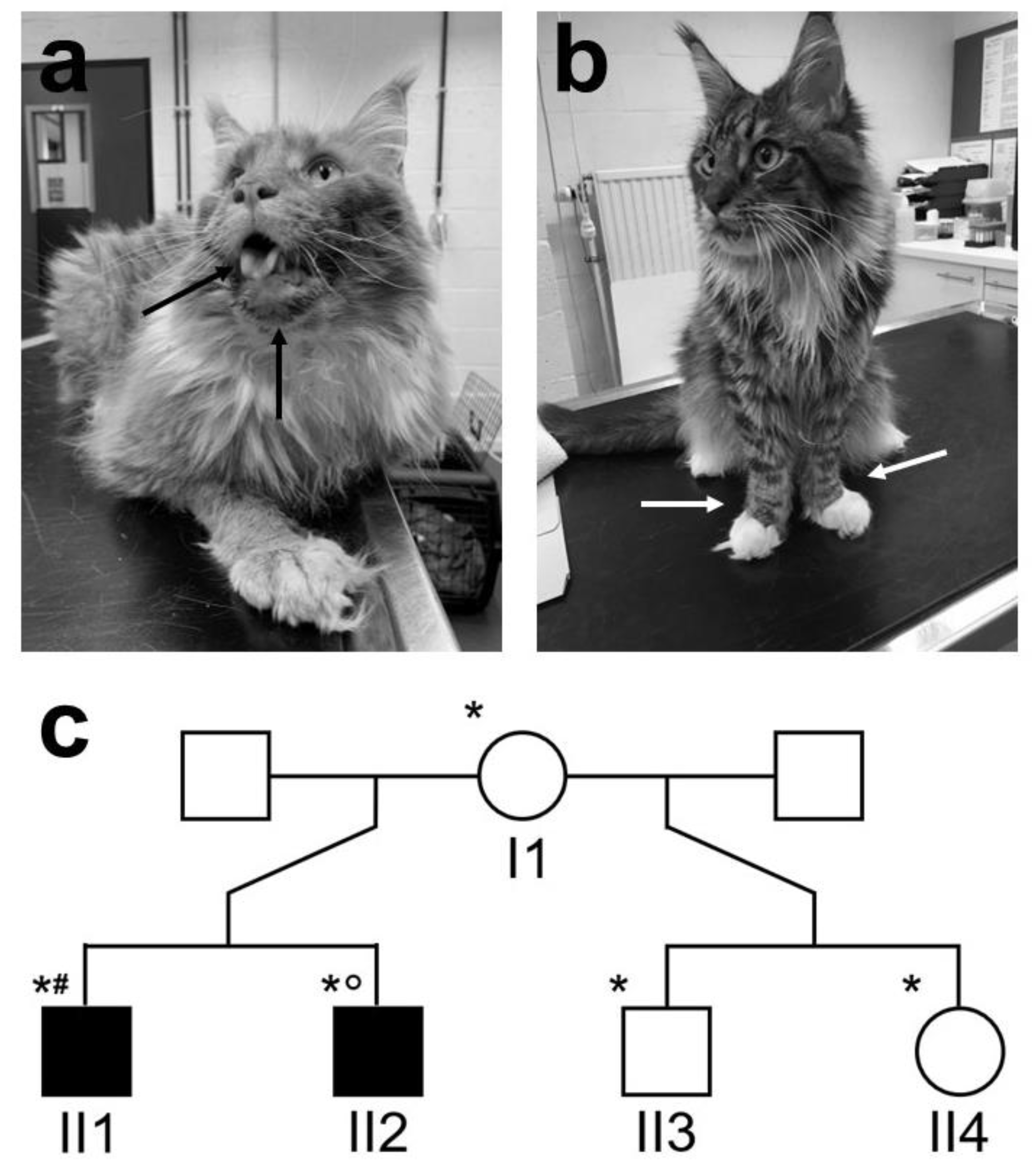
3.1. Clinical Findings

Case 1 (Figure 1a,c: individual II1), a neutered male Maine coon, was presented at 5 months of age with growth retardation, weight loss, hypersalivation, and vomiting. No abnormal behavior was noticed by the owners. General physical examination revealed a mild systolic heart murmur grade 2/6 and prominent skeletal muscles (mainly gastrocnemius, caudal thigh, and triceps muscles) that appeared painful on palpation. An abnormal gait was present, consisting of a narrow stance with internal hock rotation in the pelvic limbs and a valgus stance in the thoracic limbs. Neurological examination was within normal limits. Spontaneous electrical activity consisting primarily of pseudomyotonic discharges with a typical “dive bomber” sound was present in all appendicular muscles (including masticatory and tongue muscles). Electroneurography and repetitive nerve stimulation were normal in both examined limbs. Echocardiography did not show any abnormalities besides a dynamic right ventricular outflow tract obstruction, causing an innocent heart murmur.
Case 2 (Figure 1b,c: individual II2), a male brother of case 1, was presented at 9 months of age with similar clinical signs. General physical and neurological examinations were within normal limits, apart from the same abnormal limb stance and prominent skeletal muscles described in case 1. Radiographs revealed cardiomegaly, marked hepatomegaly, and bilaterally increased volume of the caudal thigh muscles. Echocardiography revealed a trivial mitral and tricuspid insufficiency and no other abnormalities.
Complete blood count was normal for both cats. Biochemistry profiles showed a significant increase in sCK activities (13225 (case 1)–30850 U/l (case 2)), indicating a muscle disease. The unaffected mother (6 years old) and both unaffected half-siblings (4 years old) showed normal sCK activity (individuals I1, II3, and II4 in Figure 1b, respectively).

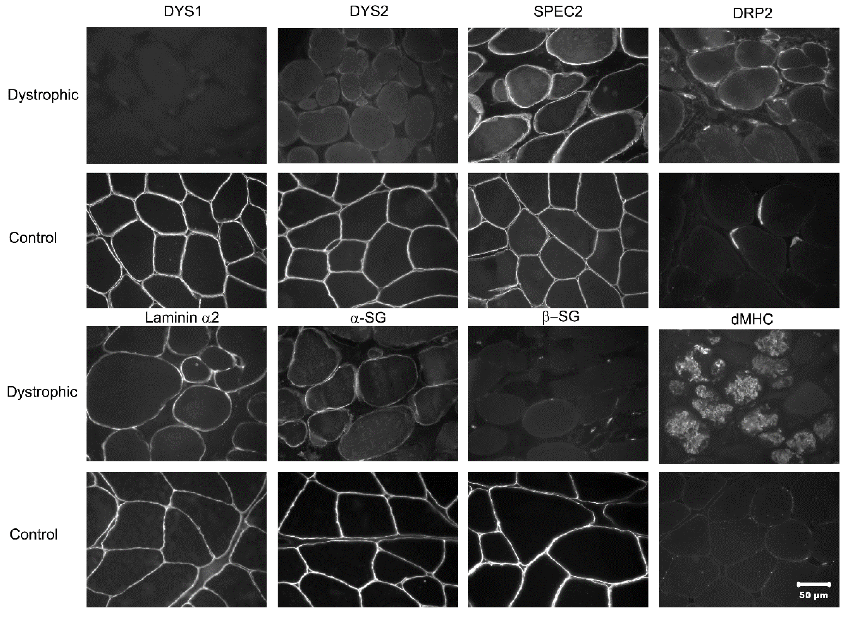
3.2. Histopathology and Immunofluorescent Staining

H&E stained cryosections of the triceps and vastus lateralis muscles (Figure S1) showed a dystrophic phenotype with variable myofiber sizes (diameters range: 13–114 µm), clusters of degenerating and regenerating fibers, and scattered calcific deposits that were highlighted by the alizarin stain (1), indicating a form of MD.
Immunofluorescence staining was used to determine a specific form of MD and is shown in Figure 2. Compared with control staining, sarcolemmal staining in the dystrophic muscle was not detected using antibodies against the rod (DYS1) and C-terminus (DYS2) of dystrophin. Staining for spectrin (SPEC2) and laminin α2 confirmed good tissue quality. Utrophin protein was increased in the dystrophic muscle’s sarcolemma (DRP2). Staining of α- and β-sarcoglycan was markedly reduced or absent. Groups of regenerating fibers were highlighted by the antibody against developmental myosin heavy chain (dMHC). These findings supported the diagnosis of ddMD.

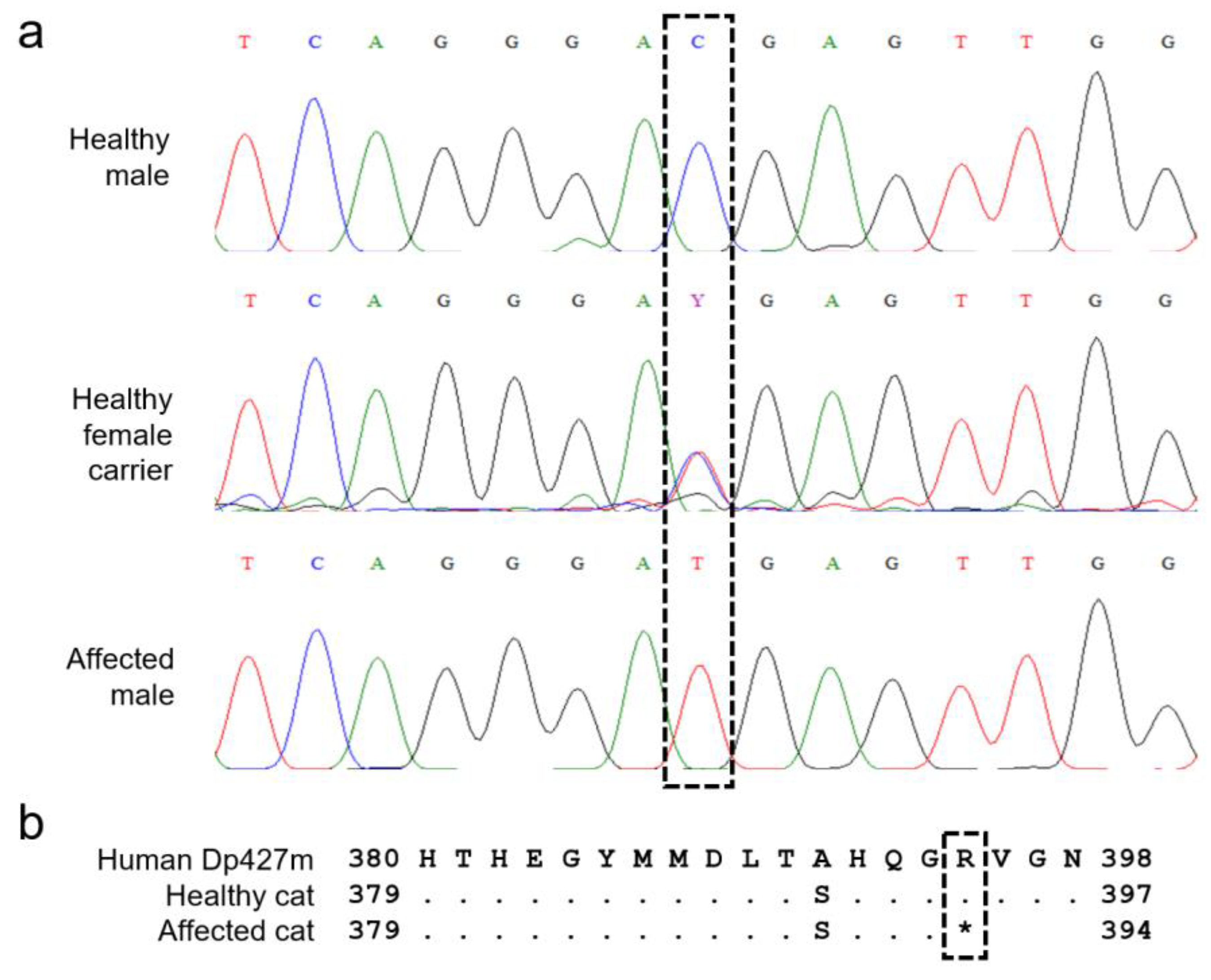
3.3. Muscle mRNA Expression and Variant Identification

The presence of dystrophin mRNA was inspected by reverse transcription PCR, followed by the amplification of five dystrophin mRNA segments and gel electrophoresis. All five amplicons were found in the unaffected and affected muscle, confirming mRNA expression in the affected cats. The result for the affected muscle is shown in Figure S2 and similar results were found for the unaffected muscle (not shown).
Dystrophin isoform X8 was identified as the feline muscle isoform. Sequencing of the respective mRNA transcript variant X8 revealed eight variants. Of those variants, one is located in the 3’UTR (c.11088 + 408G > C) and six are synonymous variants (c.618A > G, c.2565G > A, c.2931G > A, c.6708A > G, c.8316T > C, and c.8949T > C). The remaining variant, NC_058386.1 (XM_045050794.1): c.1180C > T (p.(Arg394*)), is a nonsense variant in exon 11, leading to a truncated protein (Figure 3). This variant was predicted to be deleterious by PROVEAN, with a PROVEAN score of −5779.508; deleterious cutoff = −2.5) and pathogenic by MutPred-LOF, with a score of 0.45586 (pathogenicity threshold = 0.4 at a 10% false-positive rate).

3.4. Variant Confirmation, Population Screening, and MYBPC3 Genotyping

The c.1180C > T variant was confirmed at the gDNA level in both affected brothers. Pedigree analysis revealed perfect X-linked segregation after testing the healthy mother (heterozygous), half-brother (homozygous wildtype), and -sister (homozygous wildtype). The variant was absent in the five unrelated controls without signs of MD and diagnosed with disorders unrelated to MD. Because of perfect segregation, the OR was infinite. A significant association between genotype and phenotype (p = 0.02) was found using the Fisher exact test. The variant was not found in the 99-Lives Consortium database, EVA database, or Belgian Maine coon population.
Both cases were genotyped for the A31P variant in MYBPC3 to discern if this could explain the difference in cardiac involvement between them. The variant was absent in both cats.

3.5. ACMG Classification

Altogether, six relevant criteria supporting classification as a pathogenic variant are fulfilled, whereas none of the criteria supporting a benign role are met (Table 1).

4. Discussion

This study reports two castrated male Maine coon littermates that were presented at a young age (5–9 months) with clinical signs of an early-onset inherited myopathy and markedly elevated sCK activities. Only a small number of congenital and dystrophic myopathies have been reported in cats including nemaline rod myopathy [31], X-linked myotubular myopathy in a Maine coon cat [32], laminin α2 deficient muscular dystrophy [33], muscular dystrophy associated with β-sarcoglycan deficiency [34], and X-linked ddMD [13,14]. Usually, sCK activities are normal or slightly elevated in congenital myopathies, making these disorders unlikely in the cats in this report. Immunofluorescent staining ruled out laminin α2 deficiency, but a β-sarcoglycan variant could not be ruled out. However, sarcoglycan variants result in autosomal recessive MDs and do not fit the X-linked pattern of inheritance seen here. As sarcolemmal staining was not detected using antibodies against dystrophin, the diagnosis of dystrophin-deficient FXMD was confirmed.
Clinical signs in both FXMD-affected cats of this study were very similar to those in previously described domestic shorthair cases, with dystrophin deficiency including muscle hypertrophy as a prominent clinical feature and a markedly increased sCK activity as a clear indicator [13,14]. However, no hypertrophy of the diaphragm was noticed here, which is often seen in FXMD [35]. Case 1 showed progression of clinical signs, but was still alive at 3 years old at the time of this report. Case 2 died suddenly at home. Though this cat could have developed cardiomyopathy or fatal cardiac arrhythmia, clinical heart disease is rarely encountered in FXMD [35] and the underlying cause of death was not investigated in this cat. Of note, case 1 did display cardiac involvement and did not carry the MYBPC3 A31P variant causing hypertrophic cardiomyopathy in this breed. While another (genetic) cause of hypertrophic cardiomyopathy cannot be excluded [36], this might be a case of FXMD with clinical heart disease. The variability in cardiac involvement makes the cat an interesting model to investigate MD modifier genes.
In humans, female carriers of DMD variants are usually asymptomatic, but some do present with (mild) symptoms. About 8% of female carriers present with dilated cardiomyopathy [37]. Similarly, cardiac abnormalities, including enlargement and hyperechogenicity of the papillary musculature after the age of 2 years, were reported in obligate carrier females in a cat breeding colony [35]. Therefore, the owner of the cases’ mother was advised to present for regular echocardiographical monitoring. To date, no abnormalities have been found but, to be on the safe side, we advise monitoring other carriers of the c.1180C > T variant. Besides myocardial changes, many human female carriers also have increased sCK activity [38]. Similarly, a study in cats reported a mild increase in 2 out of 10 healthy female carriers, though the difference between the carrier and control group was not significant [39]. Here, the mother was the only healthy carrier available and her sCK activities were within normal limits.
To date, only two disease-causing variants have been identified for FXMD, both large deletions in the DMD gene of domestic shorthair cats. The first deletion involves the muscle promotor and muscle and Purkinje neuronal first exon, whereas the second includes the cortical and skeletal neuronal promotors and their respective first exons. Both studies reported markedly reduced or absent levels of dystrophin in affected skeletal muscle, as was the case in the current study [13,14]. Contrary to reports of Winand et al. [14], the whole length of the muscle transcript was present in the dystrophic cat muscle of case 2.
The mRNA expression in the affected muscle is not surprising, as this has been reported before in DMD patients and mdx mice with premature termination codons, albeit greatly reduced [40,41]. Here, no (semi)quantitative analysis was performed on dystrophin mRNA expression because of sample size limitations. Of note, visual inspection of the amplicon fragments after loading 10 µl of PCR product from the affected muscle onto a gel (Figure S2) revealed a comparable intensity to that of unaffected muscle, suggesting that mRNA expression is not extremely reduced in the FXMD cats from this study.
To confirm the differential diagnosis of FXMD and rule out any other congenital myopathies, a histopathological evaluation of muscle biopsies was performed. Immunohistochemical staining showed absent protein localization for the dystrophin rod-domain and C-terminus in dystrophic muscle. Because commercially available antibodies for humans were used and the antibody against the N-terminus is human-specific, no staining was performed against this region. The presence of the N-terminal domain cannot be excluded as the c.1180C > T variant is positioned just downstream of this region. A small amount of dystrophin has already been reported in the affected muscle of a cavalier king Charles spaniel with a 7 bp deletion, resulting in a premature termination codon in exon 42 [42]. Likewise, multiple human dystrophinopathy patients showed normal or reduced immunohistochemical staining for one or more dystrophin domains, though staining for the C-terminus is typically negative in DMD [43].
Staining for α-, β-, and γ-sarcoglycans was markedly reduced or absent in dystrophic muscle from case 1. The sarcoglycan subcomplex is part of the dystrophin-associated protein complex, allowing dystrophin to interact with the sarcolemma, cytoskeleton, channel proteins, and signaling or scaffolding proteins [44]. The reduced sarcoglycan staining is likely secondary to dystrophin deficiency [43]. Likewise, an increased utrophin staining of the FXMD muscle is also secondary to dystrophy and is often seen in DMD [43,44]. Utrophin is homologous to dystrophin and can recruit many proteins involved in the dystrophin-associated protein complex. Normally, the protein’s expression is downregulated by dystrophin but, as dystrophin is downregulated in ddMD individuals, utrophin expression is upregulated [44].
The absence of dystrophin protein by immunofluorescence staining in FXMD muscle made DMD an obvious candidate gene for genetic analysis. Because of the expense of whole-genome sequencing, the enormous size of the DMD gene, and the availability of mRNA in the affected muscle samples, variant identification was performed at the mRNA level. After performing RT-PCR and sequencing the entire dystrophin cDNA, the most likely disease-causing variant was identified as NC_058386.1 (XM_045050794.1): c.1180C > T (p.(Arg394*)), a nonsense variant in exon 11 leading to a truncated protein. Combining the aforementioned results, we evaluated whether the newly identified variant could be classified as pathogenic based on the ACMG guidelines. The first important criterion (PVS1) is a nonsense variant where loss-of-function is a known mechanism of disease [30]. Nonsense variants are known to cause 10% of DMD cases in humans [4,5] and the causal variant in the mdx mouse is also a nonsense variant [45]. A recent study looked at the impact of the position of nonsense variants in the DMD gene on the phenotype. The authors found that nonsense variants in 51 exons, including exon 11 (which corresponds to exon 11 in feline muscle transcript variant X8), were associated with DMD 100% of the time [46]. In evaluating the evidence level of a nonsense variant, the presence of multiple gene transcripts must be considered. The latest feline reference assembly predicts 20 transcript variants. The c.1180C > T variant would not affect transcript variants X12–X20, which are predicted to create shorter protein isoforms. These shorter isoforms are comparable to human Dp260, Dp140, Dp116, and Dp71, none of which play important roles in muscle tissue [47]. Therefore, they are not biologically relevant to the disease phenotype. Altogether, the nonsense nature of the c.1180C > T variant is very strong evidence (PVS1) of pathogenicity [30].
Strong evidence of this variant’s pathogenicity is provided by the abundance of functional studies (PS3) on the human DMD gene [44]. Additional strong evidence is delivered by the variant’s increased prevalence in affected cats over controls (PS4), measured by the infinite OR. As a CI cannot be calculated with an infinite OR (because of perfect segregation), a p value (p = 0.02) was calculated using a Fisher exact test to demonstrate a significant association between genotype and phenotype. The absence of the variant in multiple population cohorts yields further moderate evidence (PM2). While the cases’ mothers’ importation from Italy might explain the absence of the variant in the Belgian Maine coon population, the variant was also absent from the 99-Lives Consortium database, which contains samples from all over the world. Whether the variant is absent from the Italian Maine coon population could not be determined in this study. Finally, supporting evidence is provided by perfect segregation of the variant with the FXMD phenotype demonstrated by the pedigree analysis (PP1) as well as two in silico predictive programs estimating a deleterious effect (PP3). When one very strong (PVS1) and one or more strong (PS1–PS4) criteria are fulfilled, a variant can be classified as pathogenic, as proposed by the ACMG [30]. Not only did we provide very strong (PVS1) and strong evidence (PS3 and PS4), but additional moderate (PM2) and supporting evidence (PP1 and PP3) is also available. Furthermore, none of the criteria supporting a benign role are met. Consequently, we can conclude there is abundant evidence that the c.1180C > T variant is pathogenic.

5. Conclusions

Overall, this study is the first to report ddMD in the Maine coon. It describes the dystrophic phenotype, absence of dystrophin by immunofluorescence, and the disease-causing variant, with ample evidence of pathogenicity and the effect at the transcript and protein level. Furthermore, X-linked segregation was confirmed based on pedigree analysis and the variant was absent in three databases.

Supplementary Materials

The following supporting information can be downloaded at https://www.mdpi.com/article/10.3390/ani12212928/s1. Table S1: Monoclonal antibodies used for indirect immunofluorescence [48]; Table S2: mRNA expression—primers; Table S3: mRNA expression–PCR/sequencing information; Table S4: Transcript variant X8—primers; Table S5: Transcript variant X8—PCR/sequencing information; Table S6: Primer, probe, and qPCR information for MYBPC3 genotyping. Figure S1: Histopathology of muscle biopsies; Figure S2: mRNA gel electrophoresis results.

Author Contributions

Conceptualization, E.B., L.P. and B.J.G.B.; methodology, E.B.; validation, E.B.; formal analysis, E.B.; investigation, E.B., I.C., S.F.M.B., P.S., G.D.S. and L.T.G.; resources, I.C., S.F.M.B. and P.S.; data curation, E.B.; writing—original draft preparation, E.B.; writing—review and editing, E.B., I.C., S.F.M.B., P.S., G.D.S., L.T.G., L.P. and B.J.G.B.; visualization, E.B. and G.D.S.; supervision, L.P. and B.J.G.B.; project administration, L.P. and B.J.G.B. All authors have read and agreed to the published version of the manuscript.

Funding

This research received no external funding.

Institutional Review Board Statement

The animal study protocol was approved by the Ethics Committee of the Faculties of Veterinary Medicine and Bioscience Engineering at Ghent University (EC2017-86).

Informed Consent Statement

Informed consent was obtained from all subjects’ owners involved in the study.

Data Availability Statement

The data generated during this study can be found within the published article and its supporting information. The variant data for this study have been deposited in the European Variation Archive (EVA) at EMBL-EBI under accession number PRJEB52511. Additional data are available from the corresponding author upon reasonable request.

Acknowledgments

The authors would like to thank the 99-Lives sequencing consortium for their support. We also thank Linda Impe, Dominique Vander Donckt, Carolien Rogiers, and Ruben Van Gansbeke for excellent technical assistance, as well as the breeders and owners who collaborated on this study.

Conflicts of Interest

The authors declare no conflict of interest.

References

  1. Ryder, S.; Leadley, R.M.; Armstrong, N.; Westwood, M.; De Kock, S.; Butt, T.; Jain, M.; Kleijnen, J. The burden, epidemiology, costs and treatment for Duchenne muscular dystrophy: An evidence review. Orphanet J. Rare Dis. 2017, 12, 79. [Google Scholar] [CrossRef] [PubMed] [Green Version]
  2. Monaco, A.P.; Bertelson, C.J.; Liechti-Gallati, S.; Moser, H.; Kunkel, L.M. An explanation for the phenotypic differences between patients bearing partial deletions of the DMD locus. Genomics 1988, 2, 90–95. [Google Scholar] [CrossRef]
  3. Douglas, A.G.; Wood, M.J. Splicing therapy for neuromuscular disease. Mol. Cell. Neurosci. 2013, 56, 169–185. [Google Scholar] [CrossRef]
  4. Kong, X.; Zhong, X.; Liu, L.; Cui, S.; Yang, Y.; Kong, L. Genetic analysis of 1051 Chinese families with Duchenne/Becker Muscular Dystrophy. BMC Med. Genet. 2019, 20, 139. [Google Scholar] [CrossRef] [PubMed] [Green Version]
  5. Magri, F.; Govoni, A.; D’Angelo, M.G.; Del Bo, R.; Ghezzi, S.; Sandra, G.; Turconi, A.C.; Sciacco, M.; Ciscato, P.; Bordoni, A.; et al. Genotype and phenotype characterization in a large dystrophinopathic cohort with extended follow-up. J. Neurol. 2011, 258, 1610–1623. [Google Scholar] [CrossRef] [PubMed]
  6. Yucel, N.; Chang, A.C.; Day, J.W.; Rosenthal, N.; Blau, H.M. Humanizing the mdx mouse model of DMD: The long and the short of it. NPJ Regen. Med. 2018, 3, 4. [Google Scholar] [CrossRef] [Green Version]
  7. Taglietti, V.; Kefi, K.; Bronisz-Budzyńska, I.; Mirciloglu, B.; Rodrigues, M.; Cardone, N.; Coulpier, F.; Periou, B.; Gentil, C.; Goddard, M.; et al. Duchenne muscular dystrophy trajectory in R-DMDdel52 preclinical rat model identifies COMP as biomarker of fibrosis. Acta Neuropathol. Commun. 2022, 10, 60. [Google Scholar] [CrossRef]
  8. Sui, T.; Lau, Y.S.; Liu, D.; Liu, T.; Xu, L.; Gao, Y.; Lai, L.; Li, Z.; Han, R. A novel rabbit model of Duchenne muscular dystrophy generated by CRISPR/Cas9. Dis. Model. Mech. 2018, 11, dmm032201. [Google Scholar] [CrossRef] [Green Version]
  9. Hollinger, K.; Yang, C.X.; Montz, R.E.; Nonneman, D.; Ross, J.W.; Selsby, J.T. Dystrophin insufficiency causes selective muscle histopathology and loss of dystrophin-glycoprotein complex assembly in pig skeletal muscle. FASEB J. 2013, 28, 1600–1609. [Google Scholar] [CrossRef] [Green Version]
  10. Barthélémy, I.; Calmels, N.; Weiss, R.B.; Tiret, L.; Vulin, A.; Wein, N.; Peccate, C.; Drougard, C.; Beroud, C.; Deburgrave, N.; et al. X-linked muscular dystrophy in a Labrador Retriever strain: Phenotypic and molecular characterisation. Skelet. Muscle 2020, 10, 23. [Google Scholar] [CrossRef]
  11. Brunetti, B.; Muscatello, L.V.; Letko, A.; Papa, V.; Cenacchi, G.; Grillini, M.; Murgiano, L.; Jagannathan, V.; Drögemüller, C. X-Linked Duchenne-Type Muscular Dystrophy in Jack Russell Terrier Associated with a Partial Deletion of the Canine DMD Gene. Genes 2020, 11, 1175. [Google Scholar] [CrossRef] [PubMed]
  12. Shelton, G.D.; Minor, K.M.; Vieira, N.M.; Kunkel, L.M.; Friedenberg, S.G.; Cullen, J.N.; Guo, L.T.; Zatz, M.; Mickelson, J.R. Tandem Duplication within the DMD gene in Labrador Retrievers with a Mild Clinical Phenotype. Neuromuscul. Disord. 2022, in press. [Google Scholar] [CrossRef] [PubMed]
  13. Gambino, A.N.; Mouser, P.J.; Shelton, G.D.; Winand, N.J. Emergent Presentation of a Cat with Dystrophin-Deficient Muscular Dystrophy. J. Am. Anim. Hosp. Assoc. 2014, 50, 130–135. [Google Scholar] [CrossRef]
  14. Winand, N.J.; Edwards, M.; Pradhan, D.; Berian, C.A.; Cooper, B.J. Deletion of the dystrophin muscle promotor in feline muscular dystrophy. Neuromuscul. Disord. 1994, 4, 433–445. [Google Scholar] [CrossRef]
  15. Chen, Y.; Zheng, Y.; Kang, Y.; Yang, W.; Niu, Y.; Guo, X.; Tu, Z.; Si, C.; Wang, H.; Xing, R.; et al. Functional disruption of the dystrophin gene in rhesus monkey using CRISPR/Cas9. Hum. Mol. Genet. 2015, 24, 3764–3774. [Google Scholar] [CrossRef] [PubMed] [Green Version]
  16. Kornegay, J.N.; Spurney, C.F.; Nghiem, P.P.; Brinkmeyer-Langford, C.L.; Hoffman, E.; Nagaraju, K. Pharmacologic Management of Duchenne Muscular Dystrophy: Target Identification and Preclinical Trials. ILAR J. 2014, 55, 119–149. [Google Scholar] [CrossRef] [Green Version]
  17. Kornegay, J.N.; Bogan, J.R.; Bogan, D.J.; Childers, M.; Li, J.; Nghiem, P.; Detwiler, D.A.; Larsen, C.A.; Grange, R.W.; Bhavaraju-Sanka, R.K.; et al. Canine models of Duchenne muscular dystrophy and their use in therapeutic strategies. Mamm. Genome 2012, 23, 85–108. [Google Scholar] [CrossRef] [Green Version]
  18. Marden, F.A.; Connolly, A.M.; Siegel, M.J.; Rubin, D. Compositional analysis of muscle in boys with Duchenne muscular dystrophy using MR imaging. Skelet. Radiol. 2004, 34, 140–148. [Google Scholar] [CrossRef]
  19. Shieh, P.B. Muscular Dystrophies and Other Genetic Myopathies. Neurol. Clin. 2013, 31, 1009–1029. [Google Scholar] [CrossRef]
  20. Dickinson, P.J.; LeCouteur, R.A. Muscle and nerve biopsy. Vet. Clin. N. Am. Small Anim. Pract. 2002, 32, 63–102. [Google Scholar] [CrossRef]
  21. Dubowitz, V.; Sewry, C.; Oldfors, A. Muscle Biopsy: A practical approach. In Muscle Biopsy: A Practical Approach, 4th ed.; Saunders Elsevier: Philadelphia, PA, USA, 2013; pp. 16–27. [Google Scholar] [CrossRef]
  22. Van Poucke, M.; Martlé, V.; Van Brantegem, L.; Ducatelle, R.; Van Ham, L.; Bhatti, S.; Peelman, L.J. A canine orthologue of the human GFAP c.716G>A (p.Arg239His) variant causes Alexander disease in a Labrador retriever. Eur. J. Hum. Genet. 2015, 24, 852–856. [Google Scholar] [CrossRef] [PubMed] [Green Version]
  23. Zuker, M. Mfold web server for nucleic acid folding and hybridization prediction. Nucleic Acids Res. 2003, 31, 3406–3415. [Google Scholar] [CrossRef] [PubMed]
  24. Van Poucke, M.; Vandesompele, J.; Mattheeuws, M.; Van Zeveren, A.; Peelman, L.J. A dual fluorescent multiprobe assay for prion protein genotyping in sheep. BMC Infect. Dis. 2005, 5, 13. [Google Scholar] [CrossRef] [PubMed] [Green Version]
  25. Choi, Y.; Sims, G.E.; Murphy, S.; Miller, J.R.; Chan, A.P. Predicting the Functional Effect of Amino Acid Substitutions and Indels. PLoS ONE 2012, 7, e46688. [Google Scholar] [CrossRef] [Green Version]
  26. Pagel, K.A.; Pejaver, V.; Lin, G.N.; Nam, H.-J.; Mort, M.; Cooper, D.N.; Sebat, J.; Iakoucheva, L.M.; Mooney, S.D.; Radivojac, P. When loss-of-function is loss of function: Assessing mutational signatures and impact of loss-of-function genetic variants. Bioinformatics 2017, 33, i389–i398. [Google Scholar] [CrossRef] [Green Version]
  27. Meurs, K.M.; Sanchez, X.; David, R.M.; Bowles, N.E.; Towbin, J.A.; Reiser, P.J.; Kittleson, J.A.; Munro, M.J.; Dryburgh, K.; Macdonald, K.A.; et al. A cardiac myosin binding protein C mutation in the Maine Coon cat with familial hypertrophic cardiomyopathy. Hum. Mol. Genet. 2005, 14, 3587–3593. [Google Scholar] [CrossRef]
  28. Lyons, L.A.; Buckley, R.M.; Harvey, R.J.; The 99 Lives Cat Genome Consortium; Abitbol, M.; Aberdein, D.; Alves, P.C.; Andersson, A.O.; Bellone, R.R.; Bergström, T.F.; et al. Mining the 99 Lives Cat Genome Sequencing Consortium database implicates genes and variants for the Ticked locus in domestic cats (Felis catus). Anim. Genet. 2021, 52, 321–332. [Google Scholar] [CrossRef]
  29. Cezard, T.; Cunningham, F.; Hunt, S.E.; Koylass, B.; Kumar, N.; Saunders, G.; Shen, A.; Silva, A.F.; Tsukanov, K.; Venkataraman, S.; et al. The European Variation Archive: A FAIR resource of genomic variation for all species. Nucleic Acids Res. 2021, 50, D1216–D1220. [Google Scholar] [CrossRef]
  30. Richards, S.; Aziz, N.; Bale, S.; Bick, D.; Das, S.; Gastier-Foster, J.; Grody, W.W.; Hegde, M.; Lyon, E.; Spector, E.; et al. Standards and guidelines for the interpretation of sequence variants: A joint consensus recommendation of the American College of Medical Genetics and Genomics and the Association for Molecular Pathology. Genet. Med. 2015, 17, 405–424. [Google Scholar] [CrossRef]
  31. Kube, S.A.; Vernau, K.M.; LeCouteur, R.A.; Mizisin, A.P.; Shelton, G.D. Congenital myopathy with abundant nemaline rods in a cat. Neuromuscul. Disord. 2006, 16, 188–191. [Google Scholar] [CrossRef]
  32. Kopke, M.A.; Shelton, G.D.; Lyons, L.A.; Wall, M.J.; Pemberton, S.; Gedye, K.R.; Owen, R.; Guo, L.T.; Buckley, R.M.; Valencia, J.A.; et al. X-linked myotubular myopathy associated with an MTM1 variant in a Maine coon cat. J. Vet. Intern. Med. 2022, 26, 1800–1805. [Google Scholar] [CrossRef] [PubMed]
  33. Poncelet, L.; Résibois, A.; Engvall, E.; Shelton, G.D. Laminin alpha2 deficiency-associated muscular dystrophy in a Maine coon cat. Proc. Natl. Acad. Sci. USA 2003, 44, 550–552. [Google Scholar]
  34. Bouillon, J.; Taylor, S.M.; Vargo, C.; Lange, M.; Zwicker, L.A.; Sukut, S.L.; Guo, L.T.; Shelton, G.D. Beta-sarcoglycan-deficient muscular dystrophy presenting as chronic bronchopneumonia in a young cat. J. Feline Med. Surg. Open Rep. 2019, 5, 2055116919856457. [Google Scholar] [CrossRef] [PubMed]
  35. Gaschen, L.; Lang, J.; Lin, S.; Adé-Damilano, M.; Busato, A.; Lombard, C.W.; Gaschen, F.P. Cardiomyopathy in Dystrophin-Deficient Hypertrophic Feline Muscular Dystrophy. J. Vet. Intern. Med. 1999, 13, 346–356. [Google Scholar] [CrossRef] [PubMed]
  36. Longeri, M.; Ferrari, P.; Knafelz, P.; Mezzelani, A.; Marabotti, A.; Milanesi, L.; Pertica, G.; Polli, M.; Brambilla, P.; Kittleson, M.; et al. Myosin-Binding Protein C DNA Variants in Domestic Cats (A31P, A74T, R820W) and their Association with Hypertrophic Cardiomyopathy. J. Vet. Intern. Med. 2013, 27, 275–285. [Google Scholar] [CrossRef] [Green Version]
  37. Lim, K.R.Q.; Sheri, N.; Nguyen, Q.; Yokota, T. Cardiac Involvement in Dystrophin-Deficient Females: Current Understanding and Implications for the Treatment of Dystrophinopathies. Genes 2020, 11, 765. [Google Scholar] [CrossRef]
  38. Zhang, J.; Meng, Q.; Zhong, J.; Zhang, M.; Qin, X.; Ni, X.; Ma, J.; He, Y.; Zeng, D.; Lan, D. Serum MyomiRs as Biomarkers for Female Carriers of Duchenne/Becker Muscular Dystrophy. Front. Neurol. 2020, 11, 563609. [Google Scholar] [CrossRef]
  39. Howard, J.; Jaggy, A.; Busato, A.; Gaschen, F. Electrodiagnostic evaluation in feline hypertrophic muscular dystrophy. Vet. J. 2004, 168, 87–92. [Google Scholar] [CrossRef]
  40. Haslett, J.N.; Sanoudou, D.; Kho, A.T.; Bennett, R.R.; Greenberg, S.A.; Kohane, I.S.; Beggs, A.H.; Kunkel, L.M. Gene expression comparison of biopsies from Duchenne muscular dystrophy (DMD) and normal skeletal muscle. Proc. Natl. Acad. Sci. USA 2002, 99, 15000–15005. [Google Scholar] [CrossRef] [Green Version]
  41. Chamberlain, J.S.; Pearlman, J.A.; Muzny, D.M.; Gibbs, R.A.; Ranier, J.E.; Reeves, A.A.; Caskey, C.T. Expression of the Murine Duchenne Muscular Dystrophy Gene in Muscle and Brain. Science 1988, 239, 1416–1418. [Google Scholar] [CrossRef]
  42. Nghiem, P.P.; Bello, L.; Balog-Alvarez, C.; López, S.M.; Bettis, A.; Barnett, H.; Hernandez, B.; Schatzberg, S.J.; Piercy, R.J.; Kornegay, J.N. Whole genome sequencing reveals a 7 base-pair deletion in DMD exon 42 in a dog with muscular dystrophy. Mamm. Genome 2016, 28, 106–113. [Google Scholar] [CrossRef] [PubMed] [Green Version]
  43. Vogel, H.; Zamecnik, J. Diagnostic Immunohistology of Muscle Diseases. J. Neuropathol. Exp. Neurol. 2005, 64, 181–193. [Google Scholar] [CrossRef] [PubMed] [Green Version]
  44. Duan, D.; Goemans, N.; Takeda, S.; Mercuri, E.; Aartsma-Rus, A. Duchenne muscular dystrophy. Nat. Rev. Dis. Primers 2021, 7, 13. [Google Scholar] [CrossRef]
  45. Sicinski, P.; Geng, Y.; Ryder-Cook, A.S.; Barnard, E.A.; Darlison, M.G.; Barnard, P.J. The Molecular Basis of Muscular Dystrophy in the mdx Mouse: A Point Mutation. Science 1989, 244, 1578–1580. [Google Scholar] [CrossRef] [PubMed]
  46. Torella, A.; Zanobio, M.; Zeuli, R.; Del Vecchio Blanco, F.; Savarese, M.; Giugliano, T.; Garofalo, A.; Piluso, G.; Politano, L.; Nigro, V. The position of nonsense mutations can predict the phenotype severity: A survey on the DMD gene. PLoS ONE 2020, 15, e0237803. [Google Scholar] [CrossRef]
  47. Doorenweerd, N.; Mahfouz, A.; Van Putten, M.; Kaliyaperumal, R.; Hoen, P.A.; Hendriksen, J.G.M.; Aartsma-Rus, A.; Verschuuren, J.J.G.M.; Niks, E.; Reinders, M.J.T.; et al. Timing and localization of human dystrophin isoform expression provide insights into the cognitive phenotype of Duchenne muscular dystrophy. Sci. Rep. 2017, 7, 12575. [Google Scholar] [CrossRef] [Green Version]
  48. Liu, L.A.; Engvall, E. Sarcoglycan Isoforms in Skeletal Muscle. J. Biol. Chem. 1999, 274, 38171–38176. [Google Scholar] [CrossRef]
Figure 1. Image of affected cases with pedigree. (a) Case 1 (individual II1 in c) at the age of 3 years old, showing tongue hypertrophy, visible in the mouth and beneath the mandibula (black arrows). (b) Case 2 (Individual II2 in c), showing internally rotated carpi of the thoracic limbs (white arrows). (c) Pedigree with numbering of the individuals. Sample availability is indicated by a * for an EDTA blood sample, a ° for a muscle biopsy for genetic analysis, and a # for a muscle sample for histopathology and immunofluorescent staining.
Animals 12 02928 g001
Figure 2. Immunofluorescent staining of dystrophic and control muscle. Immunofluorescent staining of muscle cryosections showing localization of dystrophin and laminin α2. Staining was performed using monoclonal antibodies against the rod-domain (DYS1) and C-terminus (DYS2) of dystrophin, spectrin (SPEC2), utrophin (DRP2), laminin α2, α- and β-sarcoglycan (α- and β-SG, respectively), and developmental myosin heavy chain (dMHC). The magnification bar in the lower right = 50 µm for all images.
Animals 12 02928 g002
Figure 3. Disease-associated variant. (a) Chromatogram alignment of a healthy male, a healthy female carrier, and an affected male. (b) Protein alignment of the sequence around the variant. A * indicates the end of the protein.
Animals 12 02928 g003
Table 1. Relevant criteria for variant classification.
CriterionResultRemarksConclusion
Null variant in a gene where LOF is a known mechanism of disease (PVS1)LOF confirmed at protein levelPresence of alternate gene transcripts, but the variant affects the biologically relevant transcriptPVS1 fulfilled
Functional studies show deleterious (PS3) or no deleterious (BS3) effectAbundance of functional studies of the human DMD gene show a deleterious effect PS3 fulfilled
Significant OR > 5 (PS4)OR = infinite; p = 0.02CI could not be calculated because of perfect segregation, so p value was calculatedPS4 fulfilled
Variant absent in population databases (PM2)Variant absent in two databases and a Belgian Maine coon cohort PM2 fulfilled
Co-segregation in multiple affected family members (PP1)Perfect variant segregation with FXMD phenotype in the pedigreeLimited sample availability, but segregation confirmed in two affected membersPP1 fulfilled
Multiple lines of computational evidence (PP3)2/2 programs predicted deleterious/pathogenic effects PP3 fulfilled
Publisher’s Note: MDPI stays neutral with regard to jurisdictional claims in published maps and institutional affiliations.

