Article

Transcriptome Analyses Provide Insights into the Auditory Function in Trachemys scripta elegans

Ministry of Education Key Laboratory for Ecology of Tropical Islands, Key Laboratory of Tropical Animal and Plant Ecology of Hainan Province, College of Life Sciences, Hainan Normal University, Haikou 571158, China
*
Author to whom correspondence should be addressed.
Animals 2022, 12(18), 2410; https://doi.org/10.3390/ani12182410
Submission received: 11 August 2022 / Revised: 5 September 2022 / Accepted: 9 September 2022 / Published: 14 September 2022
(This article belongs to the Special Issue Evolution, Diversity, and Conservation of Herpetofauna)

Abstract

:

Simple Summary

Auditory function is an important sensory ability that contributes to the survival and reproduction of vertebrates. Studies have shown that turtles can hear and that sex-related differences exist in the auditory function of Trachemys scripta elegans. However, the associated gene expression characteristics are unknown. Therefore, we performed comparative transcriptomics to identify hub genes related to hearing organs involved in development and signal transduction. Six differentially expressed genes in the GABAergic synapse pathway were identified to explain the differences in hearing sensitivity. These results offer new insights into the genetic mechanisms underlying hearing characteristics and auditory adaptation in turtles.

Abstract

An auditory ability is essential for communication in vertebrates, and considerable attention has been paid to auditory sensitivity in mammals, birds, and frogs. Turtles were thought to be deaf for a long time; however, recent studies have confirmed the presence of an auditory ability in Trachemys scripta elegans as well as sex-related differences in hearing sensitivity. Earlier studies mainly focused on the morphological and physiological functions of the hearing organ in turtles; thus, the gene expression patterns remain unclear. In this study, 36 transcriptomes from six tissues (inner ear, tympanic membrane, brain, eye, lung, and muscle) were sequenced to explore the gene expression patterns of the hearing system in T. scripta elegans. A weighted gene co-expression network analysis revealed that hub genes related to the inner ear and tympanic membrane are involved in development and signal transduction. Moreover, we identified six differently expressed genes (GABRA1, GABRG2, GABBR2, GNAO1, SLC38A1, and SLC12A5) related to the GABAergic synapse pathway as candidate genes to explain the differences in sexually dimorphic hearing sensitivity. Collectively, this study provides a critical foundation for genetic research on auditory functions in turtles.

1. Introduction

An auditory ability is necessary for most vertebrates to detect sound signals in the external environment and for their survival and reproduction, particularly for frogs, birds, and mammals [1,2,3]. Frequency sensitivity is a critical parameter of auditory characteristics that varies among species [4]. The hearing frequency range in humans is approximately 20–20,000 Hz [5]. However, other mammals have broader ranges than those of humans. For example, giraffes and elephants can communicate with infrasound (<20 Hz), whereas dolphins and bats can hear ultrasound (>20,000 Hz) [6,7,8,9]. Birds and anuran amphibians also have a wide range of hearing frequencies, including ultrasound [10,11]. In contrast, turtles and snakes respond only to low-frequency sounds. This led to the misunderstanding that these animals were deaf for a long time. However, the auditory function of turtles and snakes has been confirmed through combined behavioral and electrophysiological methods [12,13]. The electrical potentials in response to sounds have been recorded from the inner ear of many turtle species [14,15,16]. For Chelonia mydas, the ear is a low-frequency receptor with a probable range of 60–1000 Hz, which enables the animals to perceive several vital signals on land and in water [17]. Therefore, turtles are thought to have auditory functions that receive sounds within certain frequency limits.
The basic structures and function of auditory systems in vertebrates are identical [18]. The most well-developed auditory system structures are observed in mammals, for which the hearing apparatus consists of three parts: the outer, middle, and inner ear. The function of the outer ear is to collect and transfer sound energy to the middle ear [19]. The middle ear and functional hearing first appeared in amphibians [20], and some reptiles have a rudimentary external acoustic meatus [21]. The eardrum or tympanic membrane transforms sound into vibrations in the middle ear. The middle ear ossicles then amplify the signal before it is transmitted to the inner ear, where it stimulates the sensory receptors in the cochlea [22]. For turtles, the sound is mechanically conveyed to the inner ear by the tympanic membrane and middle ear ossicles [23]. The tympanic membrane and inner ear are two important sound receivers and processors in turtle auditory systems.
Auditory phenotypic diversity might be related to changes in gene expression. Significant efforts have been made to profile gene expression patterns in mouse inner ears at different developmental stages, as well as following various stimuli, physiological damage, and specific gene knock out [24,25,26,27]. Dong et al. (2013) reported that inner ear gene expression differs between echolocating bats and non-echolocating bats via a transcriptome analysis and identified several candidate genes related to echolocation ability [28]. Chen et al. (2022) compared differential gene expression in the brain transcriptomes of male and female concave-eared torrent frogs, Odorrana tormota, thus revealing the molecular mechanisms of differences in ultrasonic hearing between sexes [29]. Since the ear structure and hearing ability of turtles are different from those of other vertebrates, exploring the molecular architecture of the inner ear and tympanic membrane in turtles is warranted. Wang et al. (2019) measured hearing sensitivity in male and female Trachemys scripta elegans and observed that auditory brainstem response thresholds were significantly lower in females than in males for frequencies in the 0.2–1.1 kHz range, with the exception of 0.9 kHz, indicating that the hearing of females is more sensitive to this frequency [30]. However, the underlying molecular mechanism remains unclear.
Sexual dimorphism in hearing sensitivity is a great model to research the molecular basis of auditory functions in turtles. Therefore, we conducted comparative transcriptomics on six tissues (inner ear, tympanic membrane, brain, eye, lung, and muscle) to explore the molecular function related to hearing organs and to explain the difference in hearing sensitivity between sexes in T. scripta elegans. We believe that our study will provide novel insights into the genetic basis of auditory functions in turtles.

2. Materials and Methods

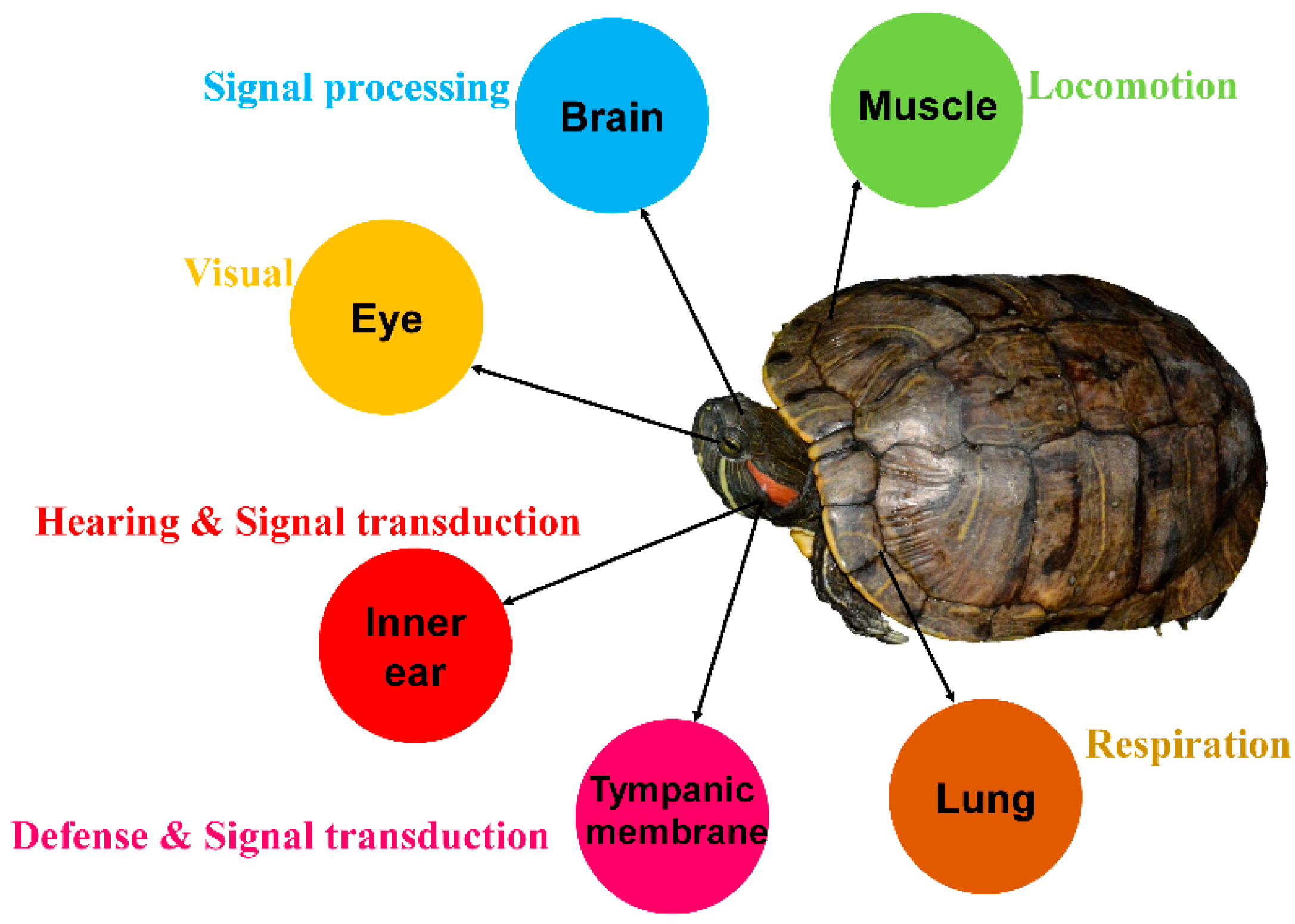
2.1. Collecting Samples

In this study, six 5-year-old T. scripta elegans individuals (three males and three females) were purchased from farms in Hainan Province, China (110.7191700° E, 19.8033100° N, 22 m). These turtles were anesthetized with Tricaine (MS-222) before euthanizing them. Six tissues (inner ear, tympanic membrane, brain, eyes, lung, and muscle) from each turtle were collected for RNA sequencing analysis. Specifically, the brain tissue refers to the whole brain, and the muscle tissue was sampled from the turtles’ forelimbs. All animal treatment procedures were approved by the Animal Research Ethics Committee of the Hainan Provincial Education Centre for Ecology and Environment, Hainan Normal University (HNECEE-2018-001).

2.2. RNA Extraction and Illumina Sequencing

Total RNA from each sample was extracted using TRIzol kit (Invitrogen, Carlsbad, CA, USA) following the manufacturer’s instructions. After RNA quantification, quality assessment, and purification, cDNA libraries were constructed following the method described by Zhu et al. [31]. After cluster generation, the library preparations were sequenced on an Illumina Novaseq 6000 platform, and 150bp paired-end reads were generated.

2.3. Transcriptomic Analyses

Clean reads were obtained by removing the adapter, poly-N, and low-quality reads. The read quality was verified using FastQC software. All clean reads were assembled by mapping them to the reference genome using the RefSeq assembly accession number GCF_013100865.1 [32]. Transcriptome data from this study were submitted to the Genome Sequence Archive (GSA, https://bigd.big.ac.cn/gsa/, accessed on 14 June 2022) under accession number CRA007204.

2.4. Weighted Gene Co-Expression Network Analysis (WGCNA)

WGCNA is a systematic biological method used to construct scale-free networks based on gene expression profiles. The potential tissue-specific gene modules were filtered using WGCNA v1.69 [33], which constructs a co-expression network of all genes based on transcripts per million (TPM). WGCNA can be used to find clusters of highly correlated genes, summarizing such clusters using the module eigengenes (MEs) or the most representative genes; to associate them with other modules and external sample traits; and to calculate module membership (MM) measures. MM was measured using Pearson’s correlation coefficient to analyze the correlation between MEs and traits. Gene significance (GS) and MM represent the correlation between gene expression profiles and MEs. The hub genes were screened to reveal the biological functions of the modules.

2.5. Screening of Differently Expressed Genes (DEGs)

DEGs were identified using DESeq2 (R package) [34] based on a threshold fold change > 2 (|log2Fold Change| > 1) and q-value < 0.05 (after Benjamini–Hochberg correction). We focused on the functional analysis of DEGs in the inner ear and tympanic membrane between males and females.

2.6. Enrichment Analysis of Gene Functions

A pathway enrichment analysis was performed based on the Kyoto Encyclopedia of Genes and Genomes (KEGG) and Gene Ontology (GO) databases using KOBAS 3.0 software [35]. The p-value was adjusted using the Benjamini–Hochberg false discovery rate, and terms with a corrected p-value < 0.05 were recognized as significant. GO enrichment analyses of genes in the inner ear and tympanic membrane-related modules were performed. A KEGG functional enrichment analysis was conducted for DEGs between males and females.

3. Results

3.1. Transcriptome Sequencing Analysis

In this study, we obtained 233.8 Gb of raw data from six tissues of six T. scripta elegans individuals (three males and three females; Table S1 and Figure 1). After quality control, a total of 221.4 Gb of clean reads was obtained for downstream analysis. Clean reads were mapped to the reference genome, and the mapping rates of the 36 samples ranged from 86.3% to 92.0% (Table S2 and Figure S1).

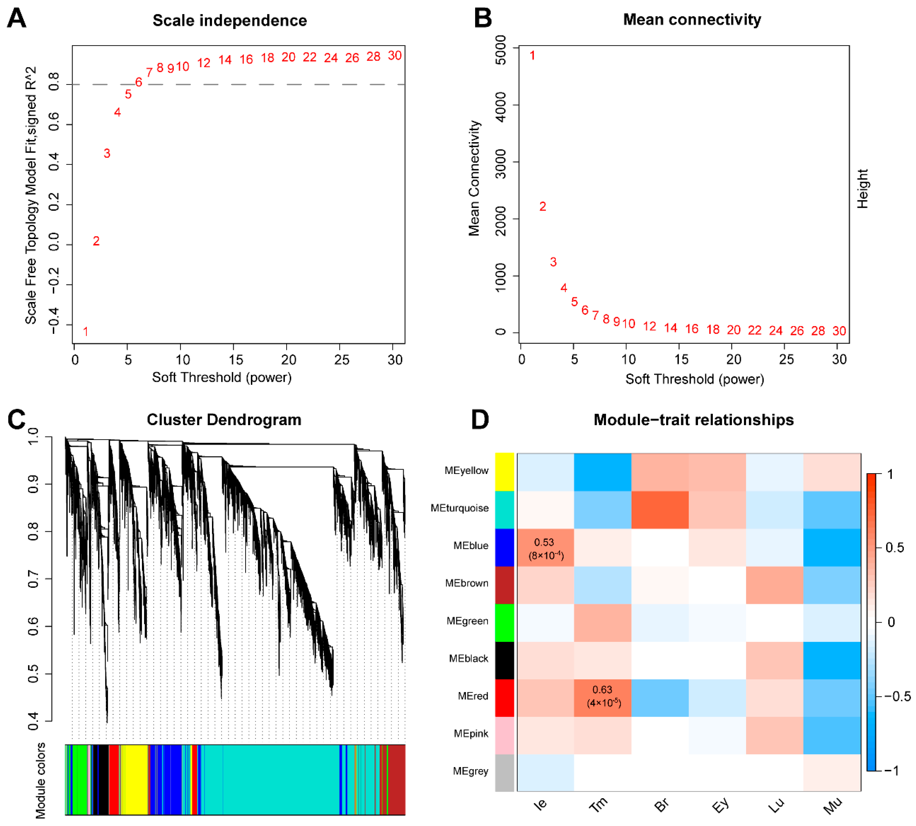
3.2. Co-Expression Network Construction

WGCNA was used to screen the characteristic gene groups in the tissues. According to the model, connectivity among genes in the network indicated a scale-free network distribution; as the correlation coefficient threshold was set to 0.9, the best soft-thresholding power was seven (Figure 2A,B), and modules with high ME correlations (R2 > 0.8) were merged (Figure 2C). We conducted a correlation analysis between the modules and tissues to identify the modules most relevant to auditory tissues. The results revealed that 16,398 genes could be divided into nine co-expression gene modules, and the blue (R2 = 0.53, p = 8 × 10⁻⁴, gene number = 2057) and red (R2 = 0.63, p = 4 × 10⁻⁵, gene number = 729) modules were significantly correlated with the inner ear and tympanic membrane, respectively (Figure 2D).

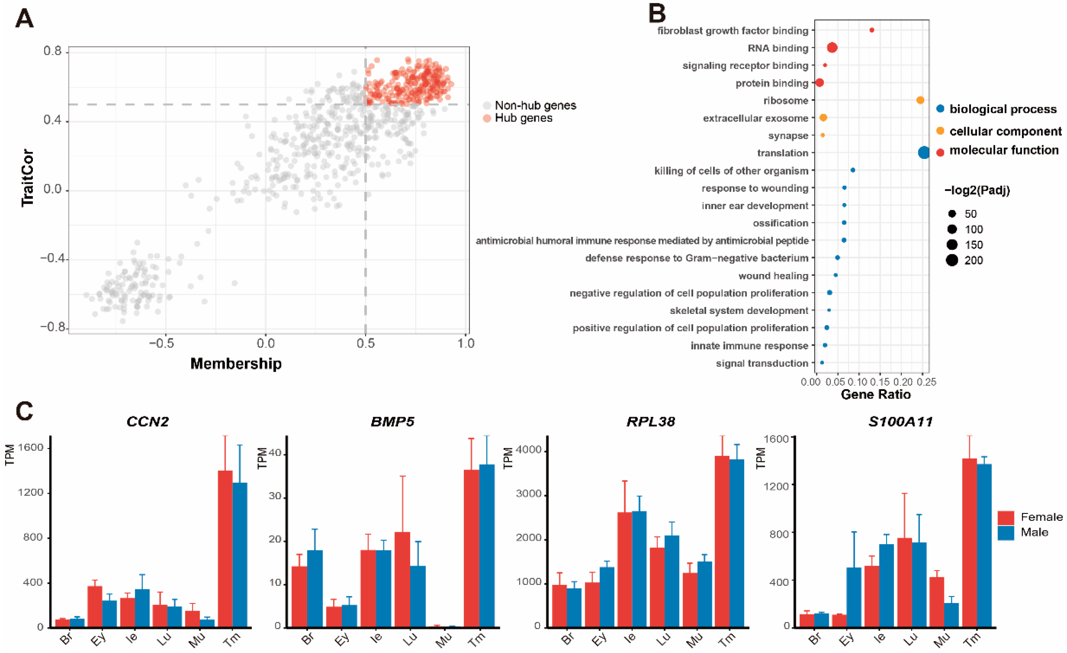
3.3. Functional Enrichment Analysis of Significant Modules

In the gene module of the inner ear, 200 hub genes (GS > 0.6 and MM > 0.6) were selected to reveal the biological functions of the inner ear (Figure 3A). The GO functional enrichment analysis assigned these genes to three categories: biological process, molecular function, and cellular component (Figure 3B and Table S3). For the GO category biological process, the hub genes of the inner ear were mainly enriched in intracellular protein transport (GO:0006886), signal transduction (GO:0007165), and regulation of sodium ion transport (GO:0002028). These pathways are closely related to cellular signal transmission. Other highlighted biological processes included inner ear morphogenesis (GO:0042472) and neural crest cell development (GO:0014032), which are related to structural formation in the auditory system. For the GO category molecular function, the hub genes of the inner ear were enriched in potassium ion binding (GO:0030955), magnesium ion binding (GO:0000287), and calcium ion binding (GO:0005509), which are related to mechanoelectrical transduction. For the GO category cellular component, extracellular exosome (GO:0070062) and plasma membrane (GO:0005886) suggested cell activity in the inner ear. From significant pathways enriched in the inner ear, we selected four featured genes in these GO categories as hearing-related genes, as follows: SOX9 (“signal transduction”, GO:0007165), FGFR1 (“inner ear morphogenesis”, GO:0042472), ATP1A1 (“potassium ion binding”, GO:0030955), and IDS (“calcium ion binding”, GO: 0005509). Notably, the expression of these four genes was higher in the inner ear than in other tissues (Figure 3C).
In the gene module of the tympanic membrane, 228 hub genes (GS > 0.5, MM > 0.5) were selected (Figure 4A). GO terms belonging to biological processes included signaling receptor binding (GO:0005102) and signal transduction (GO:0007165), indicating sound processing of the tympanic membrane. The others were associated with biological processes including inner ear development (GO:0048839) and skeletal system development (GO:0001501), which are related to development of the auditory system. Moreover, innate immune response (GO:0045087) and response to wounding (GO:0009611) suggested active immune responses in the tympanic membrane. The cellular components included ribosomes (GO:0005840), synapses (GO:0045202), and extracellular exosomes (GO:0070062), suggesting robust protein synthesis and secretion in this tissue (Figure 4B and Table S4). From significant pathways enriched in the tympanic membrane, we selected four featured genes, as follows: BMP5 (“negative/positive regulation of cell population proliferation”, GO:0007165/0,008,284 and “ossification”, GO:0001503), RPL38 (“ossification”, GO: 0001503), CCN2, and S100A11 (“protein binding”, GO:0005515 and “signal transduction”, GO:0007165). These four genes exhibited higher expression levels in the tympanic membrane than in other organs (Figure 4C).

3.4. Expression of DEGs and Screening of Genes in Pathways

Next, we compared the transcriptional differences in the inner ear and tympanic membrane between male and female T. scripta elegans. Twenty-three DEGs were identified in the tympanic membrane; however, this number is negligible and is not discussed (Table S6). There were 153 DEGs in the inner ear identified, among which the expression levels of 135 and 18 genes were upregulated in males and females, respectively (Figure 5A and Table S5). KEGG enrichment analysis revealed that these DEGs were mostly related to synaptic exaction and inhibition of neurons and neuronal signaling, including the GABAergic synapse, glutamatergic, and neurotrophin signaling pathways (Figure 5B). Notably, the GABAergic synapse pathway, which modulates nerve excitation, was significant among KEGG terms. In the GABAergic synapse pathway, six DEGs (GABRA1, GABRG2, GABBR2, GNAO1, SLC38A1, and SLC12A5) were identified, all of which exhibited higher expression levels in male T. scripta elegans than in females (Figure 5C). These genes play critical roles in synaptic activity, and their combined action leads to hyperpolarization and decreased excitability in postsynaptic cells (Figure 6).

4. Discussion

Currently, only limited studies exist on gene expression related to auditory functions in turtles. Here, we comprehensively probed the molecular functions of the inner ear and tympanic membrane of T. scripta elegans based on comparative transcriptomics and revealed that it is highly correlated with auditory organ development and signal transduction. We identified six candidate genes in the GABAergic synapse pathway, which were determined to be involved in differences in hearing sensitivity between sexes.

4.1. Molecular Functions in the Inner Ear

The inner ear is an indispensable component of the auditory system in vertebrates, and its primary function is to detect sound and to convey signals to the brain [36]. Our results indicated that SOX9, FGFR1, ATP1A1, and IDS were expressed at high levels in the inner ear. SOX9 is an evolutionarily conserved transcription factor, and studies on zebrafish, African clawed frogs, chickens, and mice have demonstrated its essential function in the inner ear and cranial neural crest development [37,38,39,40]. Fibroblast growth factor receptor 1 (FGFR1), a tyrosine kinase receptor, is expressed in the inner ear of mice and is necessary for the development of the auditory sensory epithelium [41]. Functional enrichment analysis showed that FGFR1 is involved in numerous biological functions, such as inner ear morphogenesis, protein binding, and calcium ion binding, which suggests that it plays a vital role in the function of the inner ear, specifically in receiving and processing sound, in T. scripta elegans. The ATP1A1 gene encodes Na/K-ATPase α1 isoforms [42], which are critical for maintaining hyperpolarized membrane potential by controlling cellular sodium and potassium concentrations [43]. This indicates that ATP1A1 is essential for mechanoelectrical transduction in the inner ear. Hair cells of the inner ear contribute to the initial steps in the neural processing of sound and balance in vertebrates [44]. IDS is an important gene in cochlea development in mice and chickens; further, it regulates hair cell differentiation and is expressed in the prosensory domains of the otic vesicle [45]. Overall, these four genes were confirmed to be associated with the development of auditory organs and sound transduction and are indispensable for the functional maturation of the inner ear in T. scripta elegans. These findings will allow us to further explore the molecular mechanisms underlying inner ear development and hearing in turtles.

4.2. Molecular Function of the Tympanic Membrane

The tympanic membrane is the fundamental “sound receiver”, as confirmed by audiograms of T. scripta elegans [23]. In addition to being a critical component of the auditory system that transmits sound to the inner ear, the tympanic membrane also participates in the defense system of the middle ear and can respond to immunological stimulation [46]. We identified significant pathways and genes implicated in the tympanic membrane. BMP5 belongs to the transforming growth factor (TGF)-β superfamily [47] and is essential for the formation of particular skeletal elements and the development of several soft tissues [48]. Our results revealed a higher expression level of BMP5 in the tympanic membrane than in the other five tissues, indicating that this gene is critical for the fundamental morphogenesis of auditory organs. Furthermore, Rpl38 is indispensable during the development of the middle ear, and its deletion results in ectopic ossification and cholesterol crystal deposition in the middle ear cavity, an enlarged Eustachian tube, and chronic inflammation with effusion, all of which cause conductive hearing loss in mice [49]. CCN2 encodes the connective tissue growth factor (CTGF) protein, a matricellular protein involved in many biological processes, including protein binding, cell proliferation and differentiation, and signal transduction [50]. The molecular and biological characteristics of CCN2 might be crucial for the development and function of the tympanic membrane. S100A11, a member of the calcium-binding protein S100 family, might mediate signal transduction with various stimuli [51]. Moreover, S100A11 also has biological functions, including the regulation of enzyme activity and inflammatory responses [52]. Our results collectively suggest that the tympanic membrane is vital for maintaining normal hearing functions in T. scripta elegans.

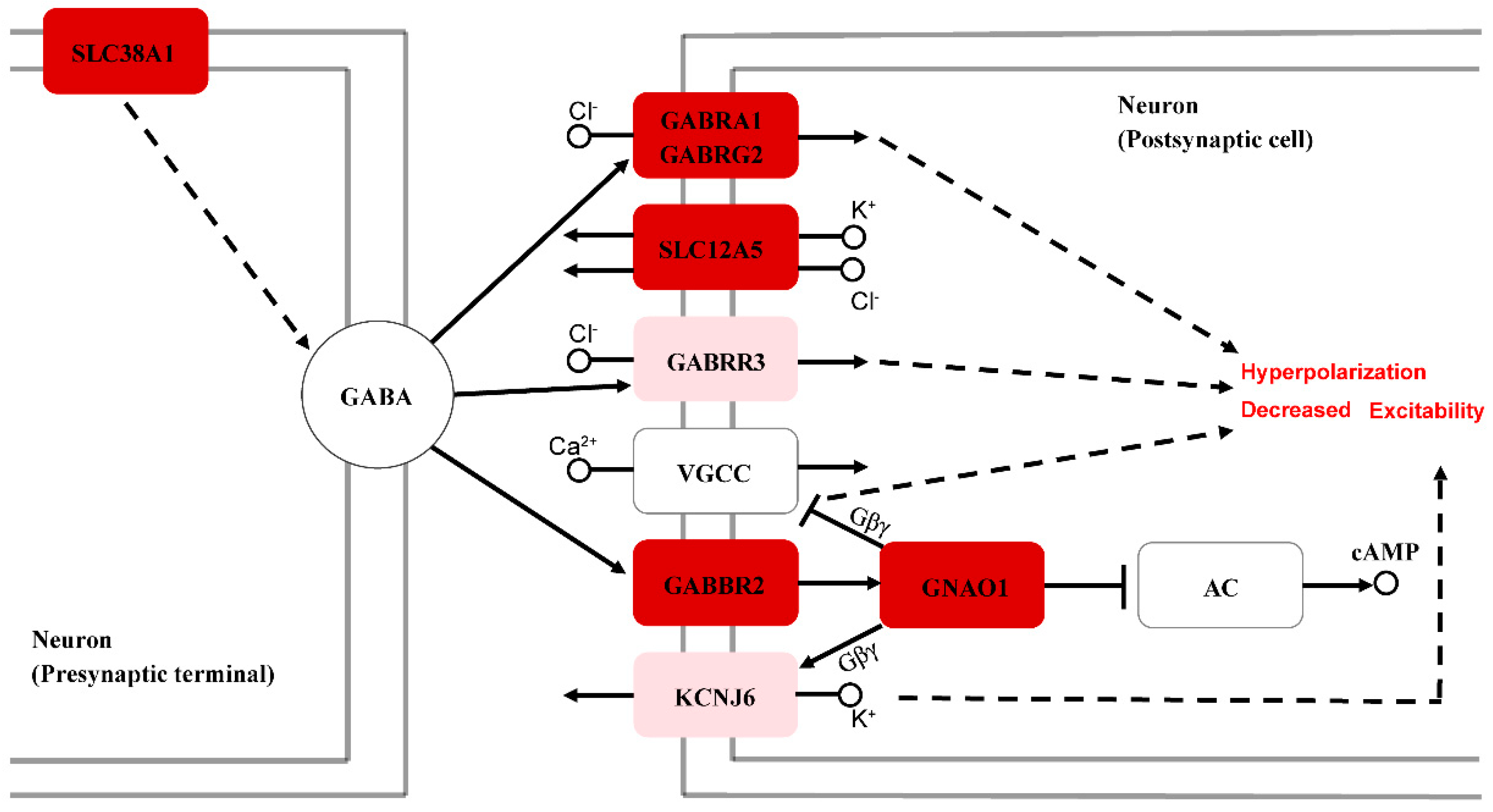
4.3. Molecular Mechanisms Underlying the Differences in Hearing Sensitivity between Sexes

Wang et al. [30] reported that female and male T. scripta elegans have a similar range of frequency sensitivities (0.2–0.9 kHz). However, the auditory brainstem response (ABR) threshold of males is significantly higher than that of females, suggesting that sexually dimorphic hearing sensitivity has emerged in turtles. In this study, numerous genes with different expression patterns between males and females in T. scripta elegans were identified, of which six auditory-related genes (i.e., GABRA1, GABRG2, GABBR2, GNAO1, SLC38A1, and SLC12A5) are involved in the GABAergic synapse pathway and might influence hearing sensitivity. In the mature central nervous system (CNS), γ-aminobutyric acid (GABA) is the chief inhibitory neurotransmitter, and it exerts its effects through ionotropic (GABAA and GABAC) and G protein-linked metabotropic (GABAB) receptors to achieve fast synaptic inhibition [53,54].
GABAA receptors are essential for the formation and development of GABAergic synapses [55]. GABRG2 and GABRA1 encode the GABAA receptor subunits α1 and γ2, respectively, and function in controlling neuronal excitability [56]. The functions of GABAA receptors depend on their expression levels, distribution patterns, and chloride (Cl) transmembrane gradients. SLC12A5 encodes potassium chloride cotransporter 2 (KCC2), which can mediate GABAergic ionic plasticity [57,58]. GABBR2 encodes GABAB receptor subunit 2, and the GABAB receptor is a G protein-coupled receptor (GPCR) that is specifically associated with a subset of G proteins (pertussis toxin-sensitive Gi/o family) that modulate voltage-gated calcium channels (VGCCs). Activation of this receptor triggers the GTP-dependent release of G protein heterotrimers (Gα-GTP and Gβγ), which mediate the direct inhibition of vesicle release, along with indirect inhibition by the suppression of VGCCs [59,60,61]. GNAO1 encodes a specific α-subunit of heterotrimeric guanine nucleotide-binding proteins (Gαo, a member of the Gi/o family), which are abundant in tissues of the mammalian CNS [62,63]. In addition, GIRK2, as a member of the G protein-gated inwardly rectifying potassium (GIRK) channel family, is also directly gated by G proteins, specifically the Gβγ dimer [64,65], and GIRK channel activation via Gi/o-coupled GPCR results in hyperpolarization of the neuron, thereby inhibiting neuronal activity [66,67,68]. Moreover, overexpression of the KCNJ6 gene, which encodes the GIRK2 channel, can lead to enhanced GABAB receptor-mediated GIRK currents in mice [69]. Therefore, the upregulation of GABAB receptor subunit 2 (GABBR2) expression can regulate GABAB–GIRK currents, which are considered inhibitory. GABAc receptors (encoded by GABRR3) also play an important role in inhibiting postsynaptic cells [70]. Experiments in mice have demonstrated that solute carrier 38 member a1 (SLC38A1) plays a key role in GABA synthesis [71,72]. For male T. scripta elegans, these six upregulated genes were determined to be involved in the GABAergic synapse pathway (Figure 5C and Figure 6), which eventually leads to hyperpolarization and decreased excitability of the signals for neurons. When exposed to sound stimulation, GABAergic neurotransmission can balance the glutamatergic excitatory drive to control the neuronal output by providing the majority of synaptic inhibition. Therefore, the hearing of male T. scripta elegans might be insensitive compared to that of females, which is consistent with the phenomenon that males require a higher ABR threshold to hear. Our results reflect the differences in the differentiation and regulation of hearing-related neurons between males and females.
Revealing the differences in auditory sensitivity between males and females of T. scripta elegans provides not only better insight into the causes of greater hearing sensitivity in females but also an opportunity to explore the probable molecular mechanisms of sexual differences in auditory characteristics. Further analysis and verification are required to explore additional factors that might contribute to this difference in hearing sensitivity.

5. Conclusions

Our research complements the molecular details underlying auditory ability or at least the types of candidate genes and pathways involved. This study offers a new perspective to focus on the hearing characteristics and auditory adaptations in turtles.

Supplementary Materials

The following supporting information can be downloaded at https://www.mdpi.com/article/10.3390/ani12182410/s1, Figure S1: Mapping rate of 36 samples to the reference genome; Figure S2: Expression levels of GABRR3 and KCNJ6 in the inner ear of male and female Trachemys scripta elegans; Table S1: Summary of samples and RNA sequencing; Table S2: Summary of transcriptome assembly mapping to the genome; Table S3: Significant GO terms enriched based on genes in the blue module; Table S4: Significant GO terms enriched based on genes in the red module; Table S5: Differential expression of genes in the inner ear; Table S6: Differential expression of genes in the tympanic membrane.

Author Contributions

Conceptualization, N.L., T.W., H.S. and J.W.; methodology, N.L. and J.W.; software, N.L.; formal analysis, N.L.; investigation, N.L., B.C., J.Q. and J.L.; resources, T.W., H.S. and J.W.; data curation, N.L. and J.W.; writing—original draft preparation, N.L.; writing—review and editing, N.L. and J.W.; visualization, N.L.; supervision, J.W.; project administration, T.W. and J.W. All authors have read and agreed to the published version of the manuscript.

Funding

This research was funded by the National Natural Science Foundation of China (31860608).

Institutional Review Board Statement

The research was reviewed and approved by the Animal Research Ethics Committee of the Hainan Provincial Education Centre for Ecology and Environment, Hainan Normal University (HNECEE-2018-001).

Informed Consent Statement

Not applicable.

Data Availability Statement

The sequencing data were submitted to the Genome Sequence Archive (GSA, https://bigd.big.ac.cn/gsa/, accessed on 14 June 2022) under accession number CRA007204.

Acknowledgments

We sincerely thank Xiaofei Zhai, Wei Wu, and Qiheng Chen for their assistance with this study.

Conflicts of Interest

All authors declare no conflict of interest.

References

  1. Lee, N.; Christensen-Dalsgaard, J.; White, L.A.; Schrode, K.M.; Bee, M.A. Lung mediated auditory contrast enhancement improves the Signal-to-noise ratio for communication in frogs. Curr. Biol. 2021, 31, 1488–1498. [Google Scholar] [CrossRef] [PubMed]
  2. Yu, K.; Wood, W.E.; Theunissen, F.E. High-capacity auditory memory for vocal communication in a social songbird. Sci. Adv. 2020, 6, eabe0440. [Google Scholar] [CrossRef] [PubMed]
  3. Egnor, S.R.; Seagraves, K.M. The contribution of ultrasonic vocalizations to mouse courtship. Curr. Opin. Neurobiol. 2016, 38, 1–5. [Google Scholar] [CrossRef] [PubMed]
  4. Manley, G.A. Some aspects of the evolution of hearing in vertebrates. Nature 1971, 230, 506–509. [Google Scholar] [CrossRef] [PubMed]
  5. Flaherty, M.; Libert, K.; Monson, B.B. Extended high-frequency hearing and head orientation cues benefit children during speech-in-speech recognition. Hear. Res. 2021, 406, 108230. [Google Scholar] [CrossRef]
  6. Baotic, A.; Sicks, F.; Stoeger, A.S. Nocturnal “humming” vocalizations: Adding a piece to the puzzle of giraffe vocal communication. BMC Res. Notes 2015, 8, 425. [Google Scholar] [CrossRef]
  7. Nachtigall, P.E.; Supin, A.Y.; Smith, A.B.; Pacini, A.F. Expectancy and conditioned hearing levels in the bottlenose dolphin (Tursiops truncatus). J. Exp. Biol. 2016, 219, 844–850. [Google Scholar] [CrossRef]
  8. Heffner, R.S.; Koay, G.; Heffner, H.E. Hearing in American leaf-nosed bats. IV: The Common vampire bat, Desmodus rotundus. Hear. Res. 2013, 296, 42–50. [Google Scholar] [CrossRef]
  9. Simmons, A.M.; Hom, K.N.; Simmons, J.A. Big brown bats (Eptesicus fuscus) maintain hearing sensitivity after exposure to intense band-limited noise. J. Acoust. Soc. Am. 2017, 141, 1481. [Google Scholar] [CrossRef]
  10. Duque, F.G.; Carruth, L.L. Vocal communication in hummingbirds. Brain Behav. Evol. 2022, 97, 241–252. [Google Scholar] [CrossRef]
  11. Feng, A.S.; Narins, P.M. Ultrasonic communication in concave-eared torrent frogs (Amolops tormotus). J. Comp. Physiol. A Neuroethol. Sens. Neural Behav. Physiol. 2008, 194, 159–167. [Google Scholar] [CrossRef] [PubMed]
  12. Chapuis, L.; Kerr, C.C.; Collin, S.P.; Hart, N.S.; Sanders, K.L. Underwater hearing in sea snakes (Hydrophiinae): First evidence of auditory evoked potential thresholds. J. Exp. Biol. 2019, 222, jeb198184. [Google Scholar] [CrossRef] [PubMed]
  13. Berman, D.S.; Regal, P.J. The loss of the ophidian middleear. Evolution 1967, 21, 641–643. [Google Scholar] [CrossRef] [PubMed]
  14. Wever, E.G.; Vernon, J.A. The sensitivity of the turtle’s ear as shown by its electrical potentials. Proc. Natl. Acad. Sci. USA 1956, 42, 213–220. [Google Scholar] [CrossRef] [PubMed]
  15. Martin, K.J.; Alessi, S.C.; Gaspard, J.C.; Tucker, A.D.; Bauer, G.B.; Mann, D.A. Underwater hearing in the loggerhead turtle (Caretta caretta): A comparison of behavioral and auditory evoked potential audiograms. J. Exp. Biol. 2012, 215, 3001–3009. [Google Scholar] [CrossRef]
  16. Piniak, W.E.; Mann, D.A.; Harms, C.A.; Jones, T.T.; Eckert, S.A. Hearing in the Juvenile Green Sea Turtle (Chelonia mydas): A Comparison of Underwater and Aerial Hearing Using Auditory Evoked Potentials. PLoS ONE 2016, 11, e0159711. [Google Scholar] [CrossRef]
  17. Ridgway, S.H.; Wever, E.G.; McCormick, J.G.; Palin, J.; Anderson, J.H. Hearing in the giant sea turtle, Chelonia mydas. Proc. Natl. Acad. Sci. USA 1969, 64, 884–890. [Google Scholar] [CrossRef]
  18. Fay, R.R.; Popper, A.N. Evolution of hearing in vertebrates: The inner ears and processing. Hear. Res. 2000, 149, 1–10. [Google Scholar] [CrossRef]
  19. Anthwal, N.; Thompson, H. The development of the mammalian outer and middle ear. J. Anat. 2016, 228, 217–232. [Google Scholar] [CrossRef]
  20. Van Dijk, P.; Mason, M.J.; Schoffelen, R.L.; Narins, P.M.; Meenderink, S.W. Mechanics of the frog ear. Hear. Res. 2011, 273, 46–58. [Google Scholar] [CrossRef]
  21. Montefeltro, F.C.; Andrade, D.V.; Larsson, H.C. The evolution of the meatal chamber in crocodyliforms. J. Anat. 2016, 228, 838–863. [Google Scholar] [CrossRef] [PubMed]
  22. Takechi, M.; Kitazawa, T.; Hirasawa, T.; Hirai, T.; Iseki, S.; Kurihara, H.; Kuratani, S. Developmental mechanisms of the tympanic membrane in mammals and non-mammalian amniotes. Congenit. Anom. 2016, 56, 12–17. [Google Scholar] [CrossRef] [PubMed]
  23. Christensen-Dalsgaard, J.; Brandt, C.; Willis, K.L.; Christensen, C.B.; Ketten, D.; Edds-Walton, P.; Fay, R.R.; Madsen, P.T.; Carr, C.E. Specialization for underwater hearing by the tympanic middle ear of the turtle, Trachemys scripta elegans. Proc. R. Soc. B 2012, 279, 2816–2824. [Google Scholar] [CrossRef] [PubMed]
  24. Chen, Z.; Corey, D.P. An inner ear gene expression database. J. Assoc. Res. Otolaryngol. 2002, 3, 140–148. [Google Scholar] [CrossRef]
  25. Cho, Y.; Gong, T.W.; Stöver, T.; Lomax, M.I.; Altschuler, R.A. Gene expression profiles of the rat cochlea, cochlear nucleus, and inferior colliculus. J. Assoc. Res. Otolaryngol. 2002, 3, 54–67. [Google Scholar] [CrossRef]
  26. Chen, Z.; Corey, D.P. Understanding inner ear development with gene expression profiling. J. Neurobiol. 2002, 53, 276–285. [Google Scholar] [CrossRef]
  27. Schimmang, T.; Maconochie, M. Gene expression profiling of the inner ear. J. Anat. 2016, 228, 255–269. [Google Scholar] [CrossRef]
  28. Dong, D.; Lei, M.; Liu, Y.; Zhang, S. Comparative inner ear transcriptome analysis between the Rickett’s big-footed bats (Myotis ricketti) and the greater short-nosed fruit bats (Cynopterus sphinx). BMC Genom. 2013, 14, 916. [Google Scholar] [CrossRef]
  29. Chen, Z.; Liu, Y.; Liang, R.; Cui, C.; Zhu, Y.; Zhang, F.; Zhang, J.; Chen, X. Comparative transcriptome analysis provides insights into the molecular mechanisms of high-frequency hearing differences between the sexes of Odorrana tormota. BMC Genom. 2022, 23, 296. [Google Scholar] [CrossRef]
  30. Wang, T.; Li, H.; Cui, J.; Zhai, X.; Shi, H.; Wang, J. Auditory brainstem responses in the red-eared slider Trachemys scripta elegans (Testudoformes: Emydidae) reveal sexually dimorphic hearing sensitivity. J. Comp. Physiol. A Neuroethol. Sens. Neural Behav. Physiol. 2019, 205, 847–854. [Google Scholar] [CrossRef] [Green Version]
  31. Zhu, W.; Liu, L.; Wang, X.; Gao, X.; Jiang, J.; Wang, B. Transcriptomics reveals the molecular processes of light-induced rapid darkening of the non-obligate cave dweller Oreolalax rhodostigmatus (Megophryidae, Anura) and their genetic basis of pigmentation strategy. BMC Genom. 2018, 19, 422. [Google Scholar] [CrossRef] [PubMed]
  32. Brian Simison, W.; Parham, J.F.; Papenfuss, T.J.; Lam, A.W.; Henderson, J.B. An Annotated Chromosome-Level Reference Genome of the Red-Eared Slider Turtle (Trachemys scripta elegans). Genome Biol. Evol. 2020, 12, 456–462. [Google Scholar] [CrossRef] [PubMed]
  33. Langfelder, P.; Horvath, S. WGCNA: An R package for weighted correlation network analysis. BMC Bioinform. 2008, 9, 559. [Google Scholar] [CrossRef] [PubMed]
  34. Love, M.I.; Huber, W.; Anders, S. Moderated estimation of fold change and dispersion for RNA-seq data with DESeq2. Genome Biol. 2014, 15, 550. [Google Scholar] [CrossRef] [PubMed]
  35. Bu, D.; Luo, H.; Huo, P.; Wang, Z.; Zhang, S.; He, Z.; Wu, Y.; Zhao, L.; Liu, J.; Guo, J.; et al. KOBAS-i: Intelligent prioritization and exploratory visualization of biological functions for gene enrichment analysis. Nucleic Acids Res. 2021, 49, W317–W325. [Google Scholar] [CrossRef] [PubMed]
  36. Brown, R.; Groves, A.K. Hear, Hear for Notch: Control of Cell Fates in the Inner Ear by Notch Signaling. Biomolecules 2020, 10, 370. [Google Scholar] [CrossRef]
  37. Spokony, R.F.; Aoki, Y.; Saint-Germain, N.; Magner-Fink, E.; Saint-Jeannet, J.P. The transcription factor Sox9 is required for cranial neural crest development in Xenopus. Development 2002, 129, 421–432. [Google Scholar] [CrossRef]
  38. Cheung, M.; Briscoe, J. Neural crest development is regulated by the transcription factor Sox9. Development 2003, 130, 5681–5693. [Google Scholar] [CrossRef]
  39. Saint-Germain, N.; Lee, Y.H.; Zhang, Y.; Sargent, T.D.; Saint-Jeannet, J.P. Specification of the otic placode depends on Sox9 function in Xenopus. Development 2004, 131, 1755–1763. [Google Scholar] [CrossRef]
  40. Bagheri-Fam, S.; Barrionuevo, F.; Dohrmann, U.; Günther, T.; Schüle, R.; Kemler, R.; Mallo, M.; Kanzler, B.; Scherer, G. Long-range upstream and downstream enhancers control distinct subsets of the complex spatiotemporal Sox9 expression pattern. Dev. Biol. 2006, 291, 382–397. [Google Scholar] [CrossRef] [Green Version]
  41. Pirvola, U.; Ylikoski, J.; Trokovic, R.; Hébert, J.M.; McConnell, S.K.; Partanen, J. FGFR1 is required for the development of the auditory sensory epithelium. Neuron 2002, 35, 671–680. [Google Scholar] [CrossRef]
  42. Liu, W.; Rask-Andersen, H. Na/K-ATPase Gene Expression in the Human Cochlea: A Study Using mRNA in situ Hybridization and Super-Resolution Structured Illumination Microscopy. Front. Mol. Neurosci. 2022, 15, 857216. [Google Scholar] [CrossRef] [PubMed]
  43. Pivovarov, A.S.; Calahorro, F.; Walker, R.J. Na(+)/K(+)-pump and neurotransmitter membrane receptors. Invertebr. Neurosci. 2018, 19, 1. [Google Scholar] [CrossRef] [PubMed]
  44. Kamaid, A.; Neves, J.; Giráldez, F. Id gene regulation and function in the prosensory domains of the chicken inner ear: A link between Bmp signaling and Atoh1. J. Neurosci. 2010, 30, 11426–11434. [Google Scholar] [CrossRef] [PubMed]
  45. Jones, J.M.; Montcouquiol, M.; Dabdoub, A.; Woods, C.; Kelley, M.W. Inhibitors of differentiation and DNA binding (Ids) regulate Math1 and hair cell formation during the development of the organ of Corti. J. Neurosci. 2006, 26, 550–558. [Google Scholar] [CrossRef]
  46. Ichimiya, I.; Kurono, Y.; Mogi, G. Immunological potential of the tympanic membrane. Observation under normal and inflammatory conditions. Am. J. Otolaryngol. 1997, 18, 165–172. [Google Scholar] [CrossRef]
  47. Kingsley, D.M. The TGF-beta superfamily: New members, new receptors, and new genetic tests of function in different organisms. Genes Dev. 1994, 8, 133–146. [Google Scholar] [CrossRef]
  48. King, J.A.; Marker, P.C.; Seung, K.J.; Kingsley, D.M. BMP5 and the molecular, skeletal, and soft-tissue alterations in short ear mice. Dev. Biol. 1994, 166, 112–122. [Google Scholar] [CrossRef]
  49. Noben-Trauth, K.; Latoche, J.R. Ectopic mineralization in the middle ear and chronic otitis media with effusion caused by RPL38 deficiency in the Tail-short (Ts) mouse. J. Biol. Chem. 2011, 286, 3079–3093. [Google Scholar] [CrossRef]
  50. Ramazani, Y.; Knops, N.; Elmonem, M.A.; Nguyen, T.Q.; Arcolino, F.O.; van den Heuvel, L.; Levtchenko, E.; Kuypers, D.; Goldschmeding, R. Connective tissue growth factor (CTGF) from basics to clinics. Matrix Biol. 2018, 68–69, 44–66. [Google Scholar] [CrossRef]
  51. Zhang, L.; Zhu, T.; Miao, H.; Liang, B. The Calcium Binding Protein S100A11 and Its Roles in Diseases. Front. Cell Dev. Biol. 2021, 9, 693262. [Google Scholar] [CrossRef] [PubMed]
  52. He, H.; Li, J.; Weng, S.; Li, M.; Yu, Y. S100A11: Diverse function and pathology corresponding to different target proteins. Cell Biochem. Biophys. 2009, 55, 117–126. [Google Scholar] [CrossRef] [PubMed]
  53. Ben-Ari, Y.; Gaiarsa, J.L.; Tyzio, R.; Khazipov, R. GABA: A pioneer transmitter that excites immature neurons and generates primitive oscillations. Physiol. Rev. 2007, 87, 1215–1284. [Google Scholar] [CrossRef] [PubMed]
  54. Zhou, Z.; Sun, H.; Li, X.; Li, Y.; Zhao, S.; Zhang, D.; Yao, Z.; Li, J. A local GABAergic system is functionally expressed in human fallopian tube. Biochem. Biophys. Res. Commun. 2010, 398, 237–241. [Google Scholar] [CrossRef]
  55. Lu, W.; Bromley-Coolidge, S.; Li, J. Regulation of GABAergic synapse development by postsynaptic membrane proteins. Brain Res. Bull. 2017, 129, 30–42. [Google Scholar] [CrossRef]
  56. Farrant, M.; Nusser, Z. Variations on an inhibitory theme: Phasic and tonic activation of GABA(A) receptors. Nat. Rev. Neurosci. 2005, 6, 215–229. [Google Scholar] [CrossRef]
  57. Rivera, C.; Voipio, J.; Kaila, K. Two developmental switches in GABAergic signalling: The K+-Cl− cotransporter KCC2 and carbonic anhydrase CAVII. J. Physiol. 2005, 562, 27–36. [Google Scholar] [CrossRef]
  58. Kaila, K.; Price, T.J.; Payne, J.A.; Puskarjov, M.; Voipio, J. Cation-chloride cotransporters in neuronal development, plasticity and disease. Nat. Rev. Neurosci. 2014, 15, 637–654. [Google Scholar] [CrossRef]
  59. Bormann, J. Electrophysiology of GABAA and GABAB receptor subtypes. Trends Neurosci. 1988, 11, 112–116. [Google Scholar] [CrossRef]
  60. Padgett, C.L.; Slesinger, P.A. GABAB Receptor Coupling to G-proteins and Ion Channels. Adv. Pharmacol. 2010, 58, 123–147. [Google Scholar]
  61. Zurawski, Z.; Page, B.; Chicka, M.C.; Brindley, R.L.; Wells, C.A.; Preininger, A.M.; Hyde, K.; Gilbert, J.A.; Cruz-Rodriguez, O.; Currie, K.P.M.; et al. Gbetagamma directly modulates vesicle fusion by competing with synaptotagmin for binding to neuronal SNARE proteins embedded in membranes. J. Biol. Chem. 2017, 292, 12165–12177. [Google Scholar] [CrossRef] [PubMed] [Green Version]
  62. Huff, R.M.; Axton, J.M.; Neer, E.J. Physical and immunological characterization of a guanine nucleotide-binding protein purified from bovine cerebral cortex. J. Biol. Chem. 1985, 260, 10864–10871. [Google Scholar] [CrossRef]
  63. Feng, H.; Khalil, S.; Neubig, R.R.; Sidiropoulos, C. A mechanistic review on GNAO1-associated movement disorder. Neurobiol. Dis. 2018, 116, 131–141. [Google Scholar] [CrossRef] [PubMed]
  64. Logothetis, D.E.; Kurachi, Y.; Galper, J.; Neer, E.J.; Clapham, D.E. The beta gamma subunits of GTP-binding proteins activate the muscarinic K+ channel in heart. Nature 1987, 325, 321–326. [Google Scholar] [CrossRef]
  65. Whorton, M.R.; MacKinnon, R. X-ray structure of the mammalian GIRK2-betagamma G-protein complex. Nature 2013, 498, 190–197. [Google Scholar] [CrossRef]
  66. Hibino, H.; Inanobe, A.; Furutani, K.; Murakami, S.; Findlay, I.; Kurachi, Y. Inwardly rectifying potassium channels: Their structure, function, and physiological roles. Physiol. Rev. 2010, 90, 291–366. [Google Scholar] [CrossRef]
  67. Lüscher, C.; Slesinger, P.A. Emerging roles for G protein-gated inwardly rectifying potassium (GIRK) channels in health and disease. Nat. Rev. Neurosci. 2010, 11, 301–315. [Google Scholar] [CrossRef]
  68. Dascal, N. Signalling via the G protein-activated K+ channels. Cell. Signal. 1997, 9, 551–573. [Google Scholar] [CrossRef]
  69. Cortez, M.A.; Shen, L.; Wu, Y.; Aleem, I.S.; Trepanier, C.H.; Sadeghnia, H.R.; Ashraf, A.; Kanawaty, A.; Liu, C.C.; Stewart, L.; et al. Infantile spasms and Down syndrome: A new animal model. Pediatric Res. 2009, 65, 499–503. [Google Scholar] [CrossRef]
  70. Bormann, J.; Feigenspan, A. GABAC receptors. Trends Neurosci. 1995, 18, 515–519. [Google Scholar] [CrossRef]
  71. Qureshi, T.; Sørensen, C.; Berghuis, P.; Jensen, V.; Dobszay, M.B.; Farkas, T.; Dalen, K.T.; Guo, C.; Hassel, B.; Utheim, T.P.; et al. The Glutamine Transporter Slc38a1 Regulates GABAergic Neurotransmission and Synaptic Plasticity. Cereb. Cortex 2019, 29, 5166–5179. [Google Scholar] [CrossRef] [PubMed]
  72. Qureshi, T.; Bjørkmo, M.; Nordengen, K.; Gundersen, V.; Utheim, T.P.; Watne, L.O.; Storm-Mathisen, J.; Hassel, B.; Chaudhry, F.A. Slc38a1 Conveys Astroglia-Derived Glutamine into GABAergic Interneurons for Neurotransmitter GABA Synthesis. Cells 2020, 9, 1686. [Google Scholar] [CrossRef] [PubMed]
Figure 1. Biological functions of organs in Trachemys scripta elegans.
Animals 12 02410 g001
Figure 2. Co-expression analysis of all genes in the six tested tissues. (A,B) Analysis of soft-thresholding powers based on (A) scale independence and (B) mean connectivity. (C) Cluster dendrogram of genes. Modules with high similarity were merged. (D) Heatmap of correlation between modules and traits. The color scale shows the strength of correlation (Br: brain; Ey: eye; Ie: inner ear; Lu: lung; Mu: muscle; Tm: tympanic membrane).
Animals 12 02410 g002
Figure 3. Screening and analysis of module genes associated with the inner ear. (A) Selection of hub genes in the dot plot. (B) Main Gene Ontology (GO) functional items enriched based on hub genes (Padj = corrected p-value). (C) Expression patterns of four featured genes in the six tissues.
Animals 12 02410 g003
Figure 4. Enrichment analysis and expression of hub genes related to the tympanic membrane. (A) Selection of hub genes in the dot plot. (B) Main Gene Ontology (GO) functional items enriched based on hub genes (Padj = corrected p-value). (C) Expression patterns of four featured genes in the six tissues.
Animals 12 02410 g004
Figure 5. Comparison of transcriptional expression in the inner ear between males and females. (A) differentially expressed gene (DEG) screening in the inner ear between sexes. (B) KEGG functional items enriched based on DEGs (Padj = q-value). (C) Expression of six genes in the GABAergic synapse pathway. ***: q < 0.001, *: q < 0.05.
Animals 12 02410 g005
Figure 6. Regulatory mechanisms of six genes in the GABAergic synapse pathway (AC: adenylyl cyclase). The genes with high expression in males are shown in different shades of red boxes (fold change increases from light to dark red). The expression levels of genes between males and females in the light red boxes are shown in Figure S2.
Animals 12 02410 g006
Publisher’s Note: MDPI stays neutral with regard to jurisdictional claims in published maps and institutional affiliations.

