Article

Comparison of Sources and Methods for the Isolation of Equine Adipose Tissue-Derived Stromal/Stem Cells and Preliminary Results on Their Reaction to Incubation with 5-Azacytidine

1
Equine Clinic: Surgery and Radiology, Department of Veterinary Medicine, Freie Universität Berlin, Oertzenweg 19b, 14163 Berlin, Germany
2
Clinical Unit of Equine Internal Medicine, Department for Companion Animals and Horses, University of Veterinary Medicine Vienna, Veterinärplatz 1, 1210 Vienna, Austria
3
Institute of Immunology, Department of Veterinary Medicine, Freie Universität Berlin, Robert-von-Ostertag-Str. 7, 14163 Berlin, Germany
4
Institute of Veterinary Physiology, Department of Veterinary Medicine, Freie Universität Berlin, Oertzenweg 19b, 14163 Berlin, Germany
5
Institute for Veterinary Epidemiology and Biostatistics, Department of Veterinary Medicine, Freie Universität Berlin, Königsweg 67, 14163 Berlin, Germany
*
Author to whom correspondence should be addressed.
Shared first authorship. These authors contributed equally to this work.
Animals 2022, 12(16), 2049; https://doi.org/10.3390/ani12162049
Submission received: 29 April 2022 / Revised: 4 August 2022 / Accepted: 8 August 2022 / Published: 11 August 2022
(This article belongs to the Special Issue Latest Advancements in Equine Cardiology)

Abstract

:

Simple Summary

The function of the equine heart is different from that in other species, and a species-specific in vitro model would simplify investigations in the field of equine cardiology. The recent advances in stem cell research and the availability of adipose tissue-derived stromal/stem cells (ASCs) could be a promising starting point for the development of such an in vitro model. In order to test the hypothesis that equine ASCs can be differentiated into cells resembling heart cells, we isolated ASCs from abdominal, retrobulbar, and subcutaneous adipose tissue after collagenase digestion or from direct cultivation of explants. Both techniques resulted in similar yields of cells displaying morphological, immunophenotypical, and molecular biological characteristics of mesenchymal stem cells. Abdominal adipose tissue was found to be most suitable for ASC isolation in equines. However, contrasting earlier studies performed with ASCs from other species, equine ASCs were refractory to 5-azacytidine-induced upregulation of markers characteristic for the differentiation into heart cells. Hence, further studies are required to establish equine cardiomyocyte induction.

Abstract

Physiological particularities of the equine heart justify the development of an in vitro model suitable for investigations of the species-specific equine cardiac electrophysiology. Adipose tissue-derived stromal/stem cells (ASCs) could be a promising starting point from which to develop such a cardiomyocyte (CM)-like cell model. Therefore, we compared abdominal, retrobulbar, and subcutaneous adipose tissue as sources for the isolation of ASCs applying two isolation methods: the collagenase digestion and direct explant culture. Abdominal adipose tissue was most suitable for the isolation of ASCs and both isolation methods resulted in comparable yields of CD45-/CD34-negative cells expressing the mesenchymal stem cell markers CD29, CD44, and CD90, as well as pluripotency markers, as determined by flow cytometry and real-time quantitative PCR. However, exposure of equine ASCs to 5-azacytidine (5-AZA), reportedly inducing CM differentiation from rats, rabbits, and human ASCs, was not successful in our study. More precisely, neither the early differentiation markers GATA4 and NKX2-5, nor the late CM differentiation markers TNNI3, MYH6, and MYH7 were upregulated in equine ASCs exposed to 10 µM 5-AZA for 48 h. Hence, further work focusing on the optimal conditions for CM differentiation of equine stem cells derived from adipose tissue, as well as possibly from other origins, are needed.

1. Introduction

Studies in equine cardiac electrophysiology have shown important electrophysiological differences for horses in comparison to other species, including humans. Thus, according to the pattern of propagation of electrical activity in the equine heart muscle, horses have been classified in the activation pathway group II, represented by pigs and including most animals [1]. This group has some important electrophysiological differences concerning the activation and propagation of electrical activity compared to humans (classified together with primates and carnivores in group I) [1]. Furthermore, the cardiac action potential lasts longer in horses than in other species and has an impressive ability to adapt to different heart rates [2]. These electrophysiological particularities distinguishing between horses and humans make it difficult to extrapolate results gained from one species directly to the other species. Therefore, species-specific studies are needed. Various in vitro models have been used to study cardiac electrophysiology in the past, including the two-electrode voltage clamp or patch clamp techniques with heterogeneously expressed channels in oocytes or mammalian cells [3,4,5,6]. Further myocardium wedge preparations have been used as in vitro models to study equine cardiac electrophysiology [3,4,5,6]. However, all these models have some limitations, including, for example, only one channel studied in the two-electrode voltage clamp or patch clamp technique. Technical difficulties with measurements hamper the wedge preparations. An equine cardiomyocyte (eCM)-like cell in vitro model would be extremely helpful to overcome some of these limitations.
Regenerative medicine and studies on the differentiation potential of pluri- or multipotent stem cells have made huge progress in the last few years [7,8,9]. However, most studies in equines have focused on orthopedic problems [10,11] and studies on other tissues are rare. Moreover, recent research identified adipose tissue as an abundant and easily accessible source of mesenchymal stem cells (MSCs) in comparison to bone marrow, umbilical cord or embryonic stem cells [12,13]. Furthermore, several protocols to isolate adipose tissue-derived stromal/stem cells (ASCs) have been described over the years in many species, including the horse [14,15,16,17,18,19,20,21,22,23,24]. Typically, ASCs, among other nucleated cells, were either isolated from the stromal vascular fraction (SVF) [15,18,19,20,21,22,23,25] or derived from a direct culture of explants of the adipose tissue [17,24,26,27].
Characteristics of MSCs have been demonstrated for ASCs [20,21,28,29], including multilineage differentiation potential (i.e., adipogenic, osteogenic, chondrogenic, myogenic, and neurogenic) in horses [18,19,20,21,22,23,30] and in other species [15,18,25,31]. Although earlier studies reported the potential of ASCs to differentiate between cardiomyocyte (CM)-like cells in rodents, rabbits, and humans [32,33,34,35], proof of CM differentiation of equine ASCs is lacking. A substantial number of these former studies used the DNA methylation inhibitor 5-azacytidine (5-AZA) to achieve the differentiation into CM-like cells [32,33,34,35]. The supposed mechanism behind the differentiation reported is the epigenetic activation or silencing of target genes [36]. Considerable variation in 5-AZA-mediated CM differentiation is reported, especially for human ASCs [32,35,37,38,39]. Nevertheless, results from these former studies [32,33,34,35] make ASCs a potential source for the development of an eCM-like cell model.
Adipose tissue is not uniform, and functional differences exist between white, visceral and brown adipose tissue [40,41]. It is well-known that the secretion of adipokines and pro-inflammatory mediators by the visceral adipose tissue contributes to the pathogenesis of human and equine metabolic syndrome [41,42]. Moreover, ASCs harvested from different sources of adipose tissue have recently been shown to present heterogeneous secretory profiles in culture and varying microenvironmental signals present at the original site might affect the phenotype of the ASCs [40,43]. Similarly, human and canine ASCs from different tissue sources displayed substantial differences in their differentiation and proliferation potential [32,44,45,46]. However, most studies in horses used subcutaneous (sc) adipose tissue as a starting material [18,19,20,21,22,23], and little is known about the differentiation potential of equine ASCs harvested from other sources of adipose tissue, such as abdominal (abd) adipose tissue [47,48].
Therefore, the current study aimed primarily to compare the yield and the morphological and molecular biological characteristics of equine ASCs derived from three different sources of adipose tissue applying two isolation methods. We hypothesized that the equine ASCs isolated by the two isolation methods of three sources of adipose tissue would show similar differences as those described for human and canine cells. The second aim was to investigate whether equine ASCs could be differentiated into eCM-like cells by using the DNA methylation inhibitor 5-AZA as a differentiation factor at a concentration used in other species.

2. Materials and Methods

2.1. Isolation and Cultivation of Equine ASCs

Regarding the harvesting of ASCs, abd, retrobulbar (rb), and sc adipose tissue was obtained from a total of 16 horses within 8 h post euthanasia at the Equine Hospital of the Freie Universität Berlin or after slaughter in commercial abattoirs. The reasons for euthanasia were unrelated to the present study. The horses were of different breeds, sexes, and ages (Supplementary Table S1). The abd adipose tissue was obtained from the properitoneal fat in the tela subserosa of the peritoneum near the linea alba; the rb adipose tissue was harvested after enucleation of the bulbus occuli; and the sc adipose tissue was obtained from the region next to the tail head over the superficial gluteal muscle. Sampling was performed in accordance with the German animal welfare legislation (reference StN 008/20–IV C 10).
After extraction, the adipose tissue samples (abd and sc: tissue specimen of approximately 9 g; rb: tissue specimen of approximately 6 g) were washed in sterile Dulbecco’s phosphate-buffered saline (DPBS) with calcium and magnesium (DPBS w Ca2+/Mg2+, Sigma Aldrich, Taufkirchen, Germany) and stored for 2 to 8 h at 4 °C in precooled DBPS w Ca2+/Mg2+, supplemented further with 1% penicillin-streptomycin (100 U/mL and 100 µg/mL, respectively, Sigma Aldrich, Taufkirchen, Germany) and 1% amphotericin B (2.5 µg/mL, BioWest, Riverside, CA, USA) in order to be transported to the lab of the Institute of Veterinary Physiology. In the lab, the adipose tissue was washed a second time, cut into smaller pieces (2 × 2 × 2 mm), and cleared of macroscopically visible blood vessels. Hereafter, ASCs were obtained either by collagenase digestion [18,19,20,21,22,23,25] or by the explant technique, described by others [17,24,26,27].
In brief, regarding the enzymatic digestion, the minced adipose tissue was incubated with 0.1% collagenase type I (1 mg/mL, Life Technologies GmbH, Karlsruhe, Germany) for 1 h at 37 °C under regular shaking and subsequently filtered through a 100-µm cell strainer (Geyer GmbH & Co. KG, Renningen, Germany). After a centrifugation step (260× g, 5 min, room temperature (RT), acceleration 2, brake 0), the cell pellet containing the SVF was seeded in 75 cm2 cell culture flasks (tissue culture flask 75, TPP Techno Plastic Products AG, Trasadingen, Switzerland) and incubated as indicated below in basal medium (B-M), composed of Dulbecco’s Modified Eagle Medium high glucose (DMEM high glc., Life Technologies GmbH, Karlsruhe, Germany) supplemented with 20% fetal bovine serum (FBS, Sigma Aldrich, Taufkirchen, Germany), 1% penicillin/streptomycin (100 U/mL and 100 µg/mL) and 1% amphotericin B (2.5 µg/mL). Typically, ASCs obtained from the SVF (ASCs-SVF) could be identified at approximately three to four days after seeding and were further cultivated. Utilizing the direct explant method, adipose tissue specimens were placed directly onto 12-well cell culture plates (tissue culture test plate 12, TPP Techno Plastic Products AG, Trasadingen, Switzerland) and incubated in 200 μl of B-M in order to allow the cell attachment to the bottom of the culture plates. A substantial number of explant-derived ASCs (ASCs-EXP) had attached and began to spread within one week, and any remaining adipose tissue was removed. Attached cells were propagated further under standard culture conditions at 37 °C, 5% CO2 and a humidified atmosphere with both methods. After the first passage, FBS content for both ASCs-SVF and ASCs-EXP was reduced from 20% to 10% (hereinafter the medium is referred to as expansion medium, E-M). The medium was changed every two to three days, and cells were passaged at 80–90% confluence. During passaging, the cells were detached by using trypsin-EDTA (Trypsin-EDTA solution, 0.25% Trypsin/0.02% EDTA, Sigma Aldrich, Taufkirchen, Germany), according to the procedure described in Jurek et al. [49].
At this point, the success of harvesting precursor cells from the different adipose tissues by both isolation methods was analyzed by using a binary isolation success scoring system (0 points: nongrowth of cells, isolation not possible; 1 point: growth of cells, successful isolation; contamination was not taken into account). Each isolation was scored, and points were awarded for the isolation technique and adipose tissue localization in cells of passage 0 (P0). The 12 wells used for isolation were summed up in an overall score for the explant technique, whereas each isolation in the enzymatic digestion was done in a single-cell culture flask and scored.
In order to have sufficient cells available for all further experiments, the cells were expanded up to passage 2 or 3 (P2–P3), trypsinized, counted, and cryopreserved in E-M supplemented with 5% DMSO (2 × 10⁶ cells/mL, dimethyl sulfoxide, sterile filtered, Sigma Aldrich, Taufkirchen, Germany). The expanded ASCs were further characterized morphologically by transmitted light microscopy, immunophenotypically by flow cytometry, and molecular biologically by real-time quantitative PCR.

2.2. Antibody Staining and Flow Cytometry

In accordance with the criteria defined by the International Society of Cellular Therapy (ISCT) for work with human MSCs [28], the purity of the equine ASCs-EXP and ASCs-SVF populations was surveyed based on a stem cell-specific profile of the surface marker expression adapted for horses [21,29,50,51]. The ASCs-EXP or ASCs-SVF derived from the most suitable adipose tissue localization were detached from cell culture flasks by Accutase Cell Detachment Solution (PAN Biotech GmbH, Aidenbach, Germany), washed in FACS buffer (DPBS, Pan Biotech GmbH, Aidenbach, Germany, supplemented with 0.2% bovine serum albumin (BSA) and 2 mM EDTA) and counted with a Neubauer chamber. Cells were transferred at a concentration of 100,000 cells per well to V-shape 96-well plates (Greiner Bio-one, Kremsmünster, Austria) and stained in phosphate-buffered saline (PBS) with 0.2% BSA for 10 min on ice with the following monoclonal antibodies: CD45 (clone F10-89-4, biotin; BIO-Rad Laboratories GmbH, Feldkirchen, Germany), CD34 (clone 581, PE phycoerythrin; BioLegend, San Diego, CA, USA), CD29 (clone TS2/16, APC allophycocyanin; BioLegend, San Diego, CA, USA), CD44 (clone IM7, PerCP peridinin-chlorophyll protein; BioLegend, San Diego, CA, USA), CD105 (clone SN6, PE-Cy7 phycoerythrin-cyanine7; ThermoFisher Scientific Inc., Waltham, MA, USA), and CD90.1 (clone OX-7, Pacific Blue; BioLegend, San Diego, CA, USA). Streptavidin-Alexa Fluor 488 (BioLegend, San Diego, CA, USA) was used as a secondary conjugate and dead cells were excluded by using the fixable viability dye eFluor780 (ThermoFisher Scientific Inc., Waltham, MA, USA). The following isotype controls were used to verify the specificity of antibody binding: clone OKT3 (biotin; ThermoFisher Scientific Inc., Waltham, MA, USA), MOPC-173 (PE; BioLegend, San Diego, CA, USA), and P3.6.2.8.1 (APC and PE-Cy7; ThermoFisher Scientific Inc., Waltham, MA, USA). Similar to other studies (e.g., Paebst et al. [29]), equine peripheral blood mononuclear cells (PBMC) were included in the flow cytometry approaches as additional controls in order to prove the binding of the antibody clones applied. To that end, EDTA blood was harvested from five donor horses through venipuncture at the jugular vein and kept on ice until PBMC isolation
The samples were diluted 1:2 in 0.9% NaCl and transferred into tubes filled with 12.5 mL human Pancoll (PAN-Biotech GmbH, Aidenbach, Germany). Following centrifugation (800× g, 20 min, RT, acceleration 1, brake 0), the PBMC were collected from the interphase. After a further washing step, the pellets were suspended in FACS buffer, counted in a CASY Cell Counter (Roche-Innovatis, Reutlingen, Germany) and subjected to single and combined stains with the anti-target antibodies or respective isotype controls, as specified above.
Cells were acquired on a BD FACSCanto II (BD Biosciences, NJ, USA) and analyzed by using FlowJo software (version 9.9.6, Tree star Inc., Ashland, OR, USA).

2.3. Cardiomyogenic Induction

Based on the results of the flow cytometry and the analysis of isolation success, the abd ASCs-SVF were used for this part of the study. When abd ASCs-SVF reached 80–90% confluence (time point T0, before induction), the cells were incubated with E-M containing 10 µM 5-AZA for 48 h (Figure 1). Thereafter, the cells were further cultivated in E-M with medium changes every two to three days and harvested on day 21 (time point T3, three weeks after induction).
At this time point (T3), the expression of pluripotency and cardiac markers was investigated by SYBR Green real-time quantitative PCR and compared to the expression in non-induced abd ASCs-SVF at day 0 (T0). Untreated control cells cultivated in E-M until T3 served as negative controls (nc). Cells were cultivated in uncoated 75 cm2 cell culture flasks (tissue culture flask 75, TPP Techno Plastic Products AG, Trasadingen, Switzerland). Cell morphology (CM appearance, spontaneous beating of cells) was subjectively assessed every two to three days by light microscopy within the induction period of three weeks. Each experiment was conducted with abd ASCs-SVF of five horses in two experimental runs to ensure reproducibility.

2.4. Real-Time Quantitative PCR

Real-time quantitative PCR was performed at T0 and T3 in order to analyze and compare the expression of pluripotency markers OCT4/POU5F1 and MYC, cardiac markers GATA4, NKX2-5, TNNI3, MYH6, and MYH7, and the muscle marker MYF6 (for excluding myogenic differentiation) [52,53,54]. The successful cardiomyogenic induction would be expected to be accompanied initially by the downregulation of the pluripotency markers typical for MSCs (OCT4/POU5F1, MYC) [38], and the upregulation of the early cardiogenic transcription factors GATA4 and NKX2-5 among others [54,55]. Subsequently, structure proteins, such as the alpha isoform (gene MYH6) or the beta isoform of the myosin heavy chain (gene MYH7), and cardiac troponin (gene TNNI3), would be expressed [54,56]. Regarding the real-time quantitative PCR, the RNA extraction of T0 and T3 cells was performed by using the extraction kit Nucleospin RNA II (Macherey Nagel GmbH & Co. KG, Düren, Germany), according to the manufacturer’s instructions. The quantity of RNA extracted and the degree of impurities caused by salts and proteins were analyzed spectrophotometrically with a Nanophotometer™ (Implen, GmbH, Munich, Germany). The quality of the RNA was assessed by the quotient of ribosomal RNA 18S/28S microelectrophoretically via Agilent-Bioanalyzer-Nano (Agilent RNA 6000 Nano Kit, Agilent Technologies, Böblingen, Germany) as a “lab-on-a-chip” method. The cDNA synthesis was performed with the iScript™ cDNA Synthesis Kit (BIO-Rad Laboratories GmbH, Feldkirchen, Germany), following the manufacturer’s instructions. Primers, listed in Table 1, were designed based on the gene database NCBI (Primer-BLAST) and software Primer3Plus.
After reverse transcription, specific cDNA could be amplified by means of the forward and reverse primers by using conventional PCR, which was subsequently sequenced (Microsynth Seqlab GmbH, Göttingen, Germany). The SYBR Green real-time quantitative PCR was carried out by using an iCycler iQ™ (BIO-Rad Laboratories GmbH, Feldkirchen, Germany), according to the following conditions: 1 cycle at 95 °C for 2 min (denaturation) and 40 cycles in three steps at 95 °C for 5 s, at 55 °C for 10 s and at 72 °C for 20 s (amplification). Because SYBR-Green can lead to unspecific products unless probe-based real-time quantitative PCR is used, a melting curve including 80 cycles from 55 °C in 0.5 °C in increments of 10 s each per temperature step was appended after each 40-cycle PCR run. The DNA contaminations were excluded based on nc containing water (H2O) and samples without reverse transcriptase (NRT). A composite sample taken from all samples (inter-run-control) with a reference gene (GAPDH) on each plate was detected and, after passing all plates, was normalized thereon to avoid plate differences of various PCR runs. A positive control sample was entrained on each PCR plate, depending on the gene.
Investigations included mRNA quantification in atrial and ventricular tissue for cardiac-specific genes, in muscle tissue for muscle markers, and in ASCs for pluripotency markers as reference populations. Respective samples were recovered from heart muscle tissue obtained directly post-mortem from six euthanized horses provided by the Department of Veterinary Clinical Sciences Section of Large Animal Medicine and Surgery of the University of Copenhagen and muscle tissue harvested directly post-mortem from two donor horses provided by the Equine Clinic of Freie Universität Berlin. Relative quantification using the 2−ΔΔCt method according to Livak and Schmittgen [57,58] revealed that cardiac markers and the muscle marker were amplified with high Ct values and were, therefore, in the negative area of gene regulation. Hence, the Livak method was not used.

2.5. Statistical Analyses

Data analysis was performed visually and with commercially available statistical software (Graph Pad Prism, Version 9.1.2 for Mac, GraphPad Software, San Diego, CA, USA and IBM SPSS, Version 27, Armonk, NY, USA). The isolation success of ASCs was assessed with linear mixed models and defined as the target. The source of adipose tissue, isolation methods and the interaction between the source of adipose tissue and the isolation method were fixed factors and horse was the random factor. Statistical tests were performed for the analysis of the results of the real-time quantitative PCR only when Ct mean values of individual markers and treatments were more than four cycles away from the mean values of the nc, NRT and H2O. In these cases, a one-way ANOVA with Tukey’s HSD or Hochberg post-hoc test for multiple comparisons was applied. Results are shown as means ± standard deviation. Statistical significance was set at p < 0.05.

3. Results

3.1. Isolation and Cultivation of Equine ASCs

Self-renewable ASCs could be obtained from abd, rb, and sc adipose tissue with both the explant culture and collagenase digestion methods. Cells were plastic-adherent with typical fibroblast-like morphology. Although ASCs-SVF attached mostly three to four days after seeding to the bottom of cell culture flasks, ASCs-EXP grew out of the explants within one week. The linear mixed model used to compare the isolation success of ASCs showed a significant influence of the source of adipose tissue (p = 0.003) but not of the isolation method (p = 0.12). Furthermore, there was no significant interaction between the source of adipose tissue and the isolation method (p = 0.19). The isolation of abd ASCs-EXP and rb ASCs-EXP was comparably successful, but the abd adipose tissue appeared to be most suitable for the isolation of ASCs-SVF, as shown in Figure 2. Furthermore, ASCs harvested from sc adipose tissue by both isolation procedures displayed the lowest isolation success, i.e., fewer cells adhered (Figure 2). In the following, the characterization of the ASCs and the induction experiments were conducted only with ASCs obtained from abd adipose tissue, as this adipose tissue localization showed the best isolation success overall. Furthermore, in order to facilitate comparison with other studies, cells obtained from the SVF were used for the induction experiments because the isolation method did not seem to influence the isolation success.

3.2. Flow Cytometry

Flow cytometry indicated similar viability for abd ASCs-EXP (mean ± SD, 70.4 ± 10.8%) and abd ASCs-SVF (69.3 ± 7.75%) and confirmed the absence of the negative markers CD34 (abd ASCs-EXP: 0.44 ± 0.46%; abd ASCs-SVF: 0.38 ± 0.30%) and CD45 (abd ASCs-EXP: 1.42 ± 0.41%; abd ASCs-SVF: 1.40 ± 0.64%) on the surface of abd ASCs-EXP and abd ASCs-SVF. Furthermore, both cell preparations uniformly expressed the typical stem cell surface marker molecules CD29 (abd ASCs-EXP: 99.7 ± 0.42%; abd ASCs-SVF: 100 ± 0.00%), CD44 (abd ASCs-EXP: 99.9 ± 0.26%; abd ASCs-SVF: 99.9 ± 0.05%) and CD90 (abd ASCs-EXP: 96.7 ± 4.20%; abd ASCs-SVF: 96.4 ± 3.23%) (Figure 3A,B). The surface marker CD105 reported to be expressed at variable levels by equine stem cells in earlier studies [20,21,23,29,47,50,51,59] was not detectable in the present study (abd ASCs-EXP: 1.68 ± 2.01%; abd ASCs-SVF: 1.18 ± 0.77%) (Figure 3A,B). Equine PBMC stained in parallel to ASCs confirmed the binding patterns expected for CD90 (expressed by peripheral T cells; PBMC: 29.1 ± 17.0), CD45 (expressed in several splice variants by all hematopoietic cells; PBMC: 22.3 ± 12.9%), CD29 (b1 integrin expressed to varying degrees on hematopoietic cells; PBMC: 36.8 ± 16.1%), and CD44 (upregulated on activated B and T cells; PBMC: 94.3 ± 7.4%). By contrast, CD34 (primarily expressed by hematopoietic stem/progenitor cells) and CD105 (expressed by endothelial cells and activated macrophages, reported earlier to be present on equine stem cells) were largely absent from equine PBMC (Figure 3C,D). Specific background signals of primary antibodies could be excluded by using isotype controls.

3.3. Cardiomyogenic Differentiation

3.3.1. Cell Morphology

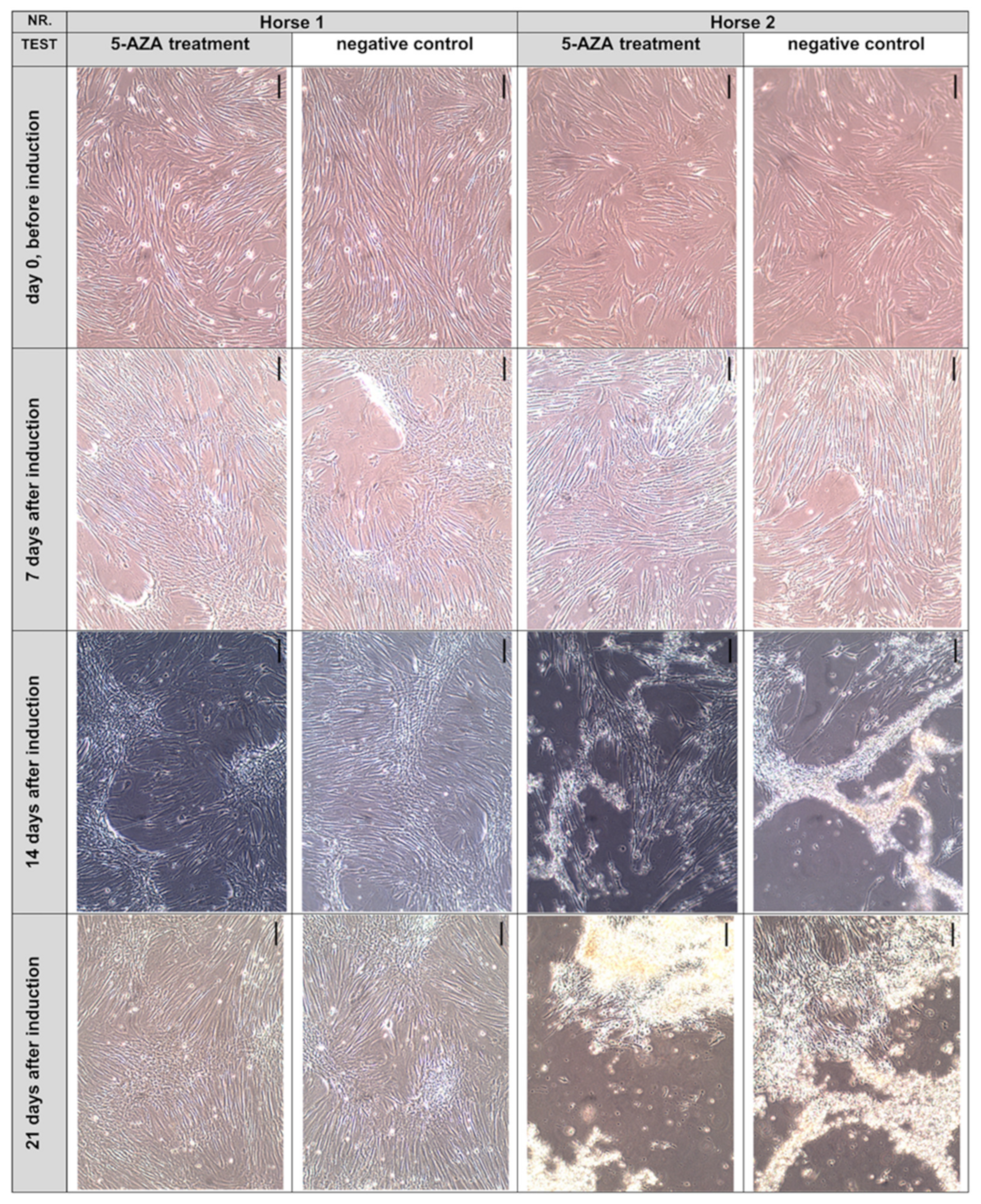
Previous studies [33,34,35,38] reported morphological changes of ASCs after exposure to 5-AZA, including the appearance of a stick-like morphology or ball-like cells, the formation of cell-to-cell connections, and myotube-like structures. However, such morphological modifications were absent in our cultures of equine abd ASCs-SVF derived from passage 3 or 4 and exposed to 5-AZA for 48 h (Figure 4). Furthermore, no spontaneous beating activity was seen.
The abd ASCs-SVF of two out of five donors maintained their spindle-shaped, fibroblast-like cell morphology, regardless of whether they were exposed to 5-AZA or not. A moderate, incipient “dome” formation, indicative of overconfluent growth, was detected in cultures derived from two out of five donors, and a dome formation in cells of the other three out of five donors was partly more severe. These cells cultivated in uncoated culture flasks showed a partially strong cell detachment and cell death independent of the treatment. We identified atypical small cells with a bright shining nucleus and atypical mitotic-like cells by light microscopy as indicators of cell detachment. Subjectively, no differences in the morphological appearance between the exposed and nonexposed abd ASCs-SVF could be detected at any time. The following Figure 4 illustrates a survey of transmitted light microcopy images.

3.3.2. Reverse-Transcription Real-Time Quantitative PCR

The RNA extracted from all samples showed a high quality and quantity, as proven via spectralphotometric measurements in a Nanophotometer™ (Ratio: close to 2) and investigations in a Bioanalyzer-Nano (RIN value: close to 10; desired value: 6–10).
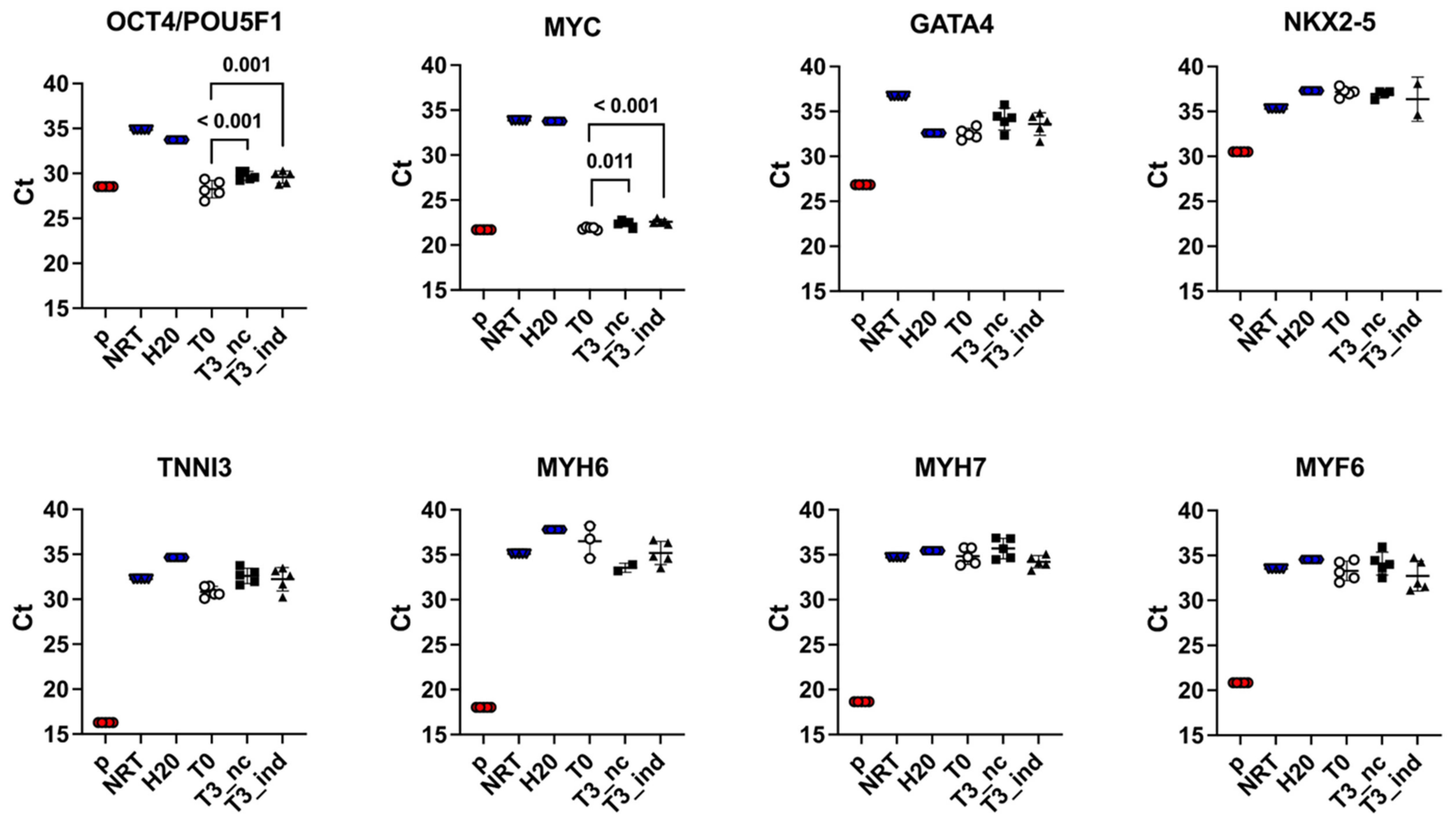
Gene expression of the two pluripotency markers OCT4/POU5F1 and MYC was observed at T0 and, together with the morphological and immunophenotypical results, confirmed that the cells harvested were ASCs. At T3, both markers were significantly downregulated, both in 5-AZA-induced cells (OCT4/POU5F1: T0 vs. T3_nc: p < 0.001; MYC: T0 vs. T3_nc: p = 0.011) and non-induced controls (OCT4/POU5F1: T0 vs. T3_ind: p = 0.001; MYC: T0 vs. T3_ind: p < 0.001).
Statistical tests were applied when the mean Ct values in the real-time quantitative PCR of a given marker or treatment differed from the mean values of negative controls (nc), NRT and H2O by at least four cycles, as described above.
Contrary to expectations, the 5-AZA treatment did not result in an upregulation of cardiac marker genes GATA4, NKX2-5, TNNI3, MYH6, and MYH7 compared to positive controls (atrial and ventricular heart muscle tissue, respectively) and, therefore, the cardiomyogenic induction was unsuccessful in our setting. Statistical tests were not performed in this part of the experiments because the Ct mean values of individual markers and treatments were consistently less than four cycles away from the mean value of the nc, NRT and H2O. A differentiation towards a myogenic cell line could be excluded as no upregulation of the muscle marker gene MYF6 was observed.
The Ct mean values of the real-time quantitative PCR with standard deviation are shown in Figure 5 as scatter dot plots and described in detail in the Supplements (Supplementary Table S2).

4. Discussion

The main finding of our study was that ASCs could be isolated from all sources of adipose tissue sampled in horses, as expected [18,19,20,21,22,23,24,47]. In line with results from human and canine studies [32,44,45,46], we could show that the source of adipose tissue influences the isolation success. The ASCs-SVF from abd adipose tissue showed a better isolation success than the ASCs-SVF harvested from the two other sources in our study (i.e., retrobulbar and subcutaneous adipose tissue). On the other hand, the isolation success for both isolation method was equivalent in our experiments. Furthermore, the cell viability determined by flow cytometry for ASCs-SVF and ASCs-EXP was similar, confirming that both methods can be used for the isolation of ASCs in horses [24], as described for other species [17,24,26,27]. However, in contrast to studies with rodent or rabbit cells [33,34,60], we were not successful in differentiating ASCs-SVF into eCM-like cells after exposure to 5-AZA for 48 h.
The first aim of the present study was to compare different sources of adipose tissue and different isolation methods for the isolation of equine ASCs. Adipose tissue is abundant and easily accessible compared to bone marrow and umbilical cord cells [12,13]. This accessibility confers a great potential on to ASCs to establish in vitro cell models, whereas further criteria, such as the immunogenicity of the cells used, have to be considered for an in vivo allogeneic application to avoid immunological rejection [43].
Some former studies in humans and horses [18,19,20,21,22,23,31] have assessed the similarity of ASCs to other MSCs, their differentiation potential and their phenotypical characteristics [12,13]. Concerning the source of adipose tissue, the sc location for harvesting adipose tissue to isolate ASCs has been widely used in horses [18,19,20,21,22,23,30], whereas the abd source has been used more often in cats [25,61], dogs [45,46,62], and humans [32]. Regarding horses, two studies reported the use of adipose tissue harvested abdominally around the mesentery of the small colon [48], in the linea alba and in lipomas [47]. Nevertheless, direct comparisons are sparse [41,42]. The structural and functional heterogeneity of adipose tissue has been shown in vivo and in vitro [13,40,41,44]. Considering the higher isolation success from the abd location, our results are in line with studies showing that the number of cells isolated from abd adipose tissue was higher than in those harvested at the tail head and that those cells had better viability [44,45,47,48]. However, these results should be confirmed by including a larger number of samples and a more homogenous population of horses with regard to age and breed. Special care should be taken regarding the breed and body conditions of the donor horses. It is well-known that easy keeper type horses have a particular tendency to adiposity and insulin dysregulation [63,64]. Such factors might influence the differentiation potential of MSCs, as has been suggested recently [65]. The difference between the rb and sc adipose tissue is more difficult to explain, but could be related to the fact that mainly structural white adipose tissue can be found at these locations. White adipose tissue has less proliferation potential compared to adipose tissue harvested from the abdomen or from a peri- or epicardial origin based on exposure to a completely different microenvironment [32,44].
Regarding the isolation methods, the collagenase digestion is an established method of isolating ASCs and has been used in different species including horses [15,18,19,20,21,22,23,25]. The explant method has been described more recently as a simple method by which to isolate human [26,27,66], equine [24], and bovine MSCs [17]. In a direct comparison, the fibroblast-like aspect, the adhesion to plastic and the isolation success were similar in cells obtained with both methods in the present study, which is in accordance with results of a previous study in horses [24]. Furthermore, the expression of CD markers was similar in both populations of cells in our experiments. When comparing the CD profile of equine cells, some differences have to be considered. According to the criteria defined by the International Society of Cellular Therapy (ISCT), ≥95% of human MSCs express the surface markers CD105, CD73, and CD90, and MSCs largely lack the expression of CD45, CD34, CD14, CD11b, CD79α, CD19, or HLA-DR (present in ≤2% of the cell population) [28]. Regarding equine MSCs, previous work defined the expression of CD29, CD44, and CD90 and the absence of CD34 and CD45 as a typical marker profile [20,21,23,29,47,59]. In compliance with these earlier works on equine MSCs, both ASCs-EXP and ASCs-SVF generated in the present study displayed MSCs characteristics evident in the homogenous expression of CD29, CD44, and CD90 and the lack of CD34 and CD45. On the other hand, a difference to human MSC concerns CD 105. Although several earlier studies reported the varying expression of CD105 by equine MSCs [20,21,23,29,47,50,51,59], our study confirms the work by Ranera et al. [21] reporting the lack of CD105 surface expression on bone marrow MSCs and ASCs. These data, as well as the cell viability of about 70% determined for both abd ASCs-EXP and abd ASCs-SVF, confirm the explant method as equivalent to the digestion method for isolating ASCs from horses [24]. Moreover, our results are in line with results from a study comparing the two methods in respect to ASCs generation from human synovial tissue [27]. In summary, ASCs-EXP and ASCs-SVF were highly comparable concerning expression profiles of MSC markers and, different from previous studies [12,13,24], cells obtained by the SVF technique did not comprise elevated numbers of hematopoietic lineage cells, endothelial cells, pericytes, or other stromal cells in our hands [12,13,24].
These results were obtained with a time frame that extended up to 8 h for a few samples; however, even these cells still seemed to have good viability. Good viability and proliferation was shown previously for bone marrow MSCs harvested 30 min after euthanasia in horses [51]. Taken together, this could indicate that viable MSCs might be harvested much longer after death than expected. However, this topic should be investigated in studies specifically designed to address this question.
In addition to the flow cytometry approach, we also monitored the expression of a group of well-known transcription factors: OCT4/POU5F1, NANOG, SOX2, and MYC, which were shown to be associated with pluripotency and self-renewal of especially embryonic stem cells [59,67,68,69,70]. Confirming earlier reports of OCT4/POU5F1 expression in equine ASCs [69,71] and MYC expression in human ASCs [72], we detected the expression of both transcription factors in ASCs-SVF at T0. These two pluripotency markers showed downregulation over three weeks in induced and non-induced cells. Similar downregulation of pluripotency markers have been reported by Safwani et al. [38]. Although they reported no change in the expression of OCT4/POU5F1, they showed a downregulation over time of SOX2 and NANOG when cells were exposed to 5-AZA [38]. The reduction of the stem cell markers in both the stimulated and non-stimulated cells, as seen in our experiment, could be a sign of senescence of cells and, therefore, indicate a loss of viability, although good viability has been reported until passage 4 or 10 for canine ASCs [45,46,62] or passage 7 for human ASCs [73].
In the second part of the study, we investigated the reaction of equine ASCs to the incubation with 5-AZA. Several previous studies reported the 5-AZA-driven differentiation of ASCs into CM-like cells in cells isolated from rats, rabbits, and, inconsistently, from human ASCs. The differentiation was documented by showing morphological changes [33,34,35,37] and the upregulation of heart-specific genes, such as myosin heavy chains, sarcomeric alpha-actinin, connexin 43, or heart-specific ion channels [33,34,35,37,60]. In one report, even the spontaneous beating of cells was described [34]. To see whether abd ASCs-SVF generated in our study responded in a similar manner, we exposed the cells to 10 µM 5-AZA for 48 h. Under these conditions, we did not observe any upregulation of cardiac differentiation markers, morphological changes or beating behavior. It is notable that conflicting reports exist on the effects of 5-AZA on ASCs isolated from human tissue. One study showed that ASCs exposed to 6 μM 5-AZA for 24 h responded with an upregulation of heart-specific genes [37], and exposure for 48 h was required according to another study [35]. By clear contrast, other studies applied 5 to 10 μM 5-AZA for 24 h (or even repeatedly) and found no evidence of any upregulated expression of cardiac genes [32,38,39]. Although the reason for these conflicting results is not entirely clear, it has to be borne in mind that 5-AZA is an unspecific differentiation agent [36]. As an unspecific gene activator, 5-AZA has been used in combination with resveratrol to avoid the aging of ASCs [74], and this result might show a promising new application for 5-AZA in equine regenerative medicine [75]. However, the unspecific activation of gene expression could be an explanation for the variable success that has been reported with 5-AZA in human cells and in our equine cells; more cardiac-specific transformation protocols should probably be investigated in the future [12,56,76,77]. As several reports indicated impaired cell proliferation and elevated cell death for prolonged or repeated exposure of ASCs to 5-AZA as well as in response to a higher concentration [34,39], we did not expose our cells to a higher concentration or beyond 48 h. In line with the unaltered morphology and the lack of cardiac differentiation markers, we found no evidence of the elevated expression of GATA4 and NKX2-5, two transcription factors known to be essential in early stages of cardiac development [54,55,68]. This result is also in line with other studies reporting little or no impact of 5-AZA on the differentiation potential of human ASCs into CM-like cells. Furthermore, constitutively higher expression of GATA4 was shown in epicardial ASCs, possibly indicating a slightly better differentiation potential in these cells compared to ASCs isolated from omental adipose tissue [32]. Differences in the differentiation potential were also reported in cells isolated from dogs, in which the number of MSCs isolated was higher in omental adipose tissue [45], but the pluripotent differentiation potential was maintained longer in subcutaneously harvested ASCs [46]. These different results highlight that differentiation depends partially on the origin of the adipose tissue and probably also on the microenvironment of the cells harvested. In the absence of an upregulation of early cardiac genes, we could not see an upregulation of the more specific cardiac genes (TNNI3, MYH6, and MYH7) in contrast to former studies [33,34,35,37,60].
Apart from the origin of adipose tissue, species, individual characteristics, or culture technics might have hampered our results and should be discussed as limitations. Concerning species, studies in rodents, where it is much easier to harvest tissue from a homogenous laboratory-bred population, have shown much better results, as there are several studies where both the early and specific cardiac markers were upregulated in experiments using the same or other induction protocols, independently of the isolation technique [33,34,56,60]. Secondly, it has recently been highlighted that the characteristics of MSCs depend on the physiological status of the donors [65]. The MSCs have shown signs of early senescence in the case of cells harvested from subjects with diabetes or metabolic syndrome [65]. Therefore, the inhomogeneous population from which our ASCs have been harvested must be considered as a limitation that should be optimized in further studies. Third, the culture conditions seem to be a non-negligible factor. The different culture mediums used in the mentioned studies could explain parts of the discrepancies in the reported results [32,35,37,38,39,78,79] and further an improvement of the cardiomyogenic differentiation potential could be obtained by using laminin-coated culture dishes, as shown for human ASCs [80]. In addition, an increasing passage number might reduce the proliferation and differentiation potential. The passage number in our studies was similar to that applied in other successful studies [32,33,35,37,60]. However, it is still possible that the high passage number (>passage 6) with possibly reduced proliferation or differentiation potential, as reported in men and dogs [45,46,62,73], might have contributed to the reduced transdifferentiation success in the present and previous studies [38]. Lastly, the cardiomyogenic differentiation potential might vary depending also on the origin of MSCs. Although in some species bone marrow-derived MSCs and ASCs could be successfully differentiated into CM-like cells after exposure to 5-AZA, their umbilical cord counterparts did not always show such potential [33,34,35,37,78,79,81].

5. Conclusions

Adipose tissue-derived stem cells could be isolated from different sources of adipose tissue in horses, as expected based on previous studies [18,19,20,21,22,23,24,32,44,45,46,47]. In line with these studies from human and dogs [32,44,45], the ASCs isolated from abd adipose tissue showed a better isolation success than ASCs harvested from the two other sources. The cells isolated showed immunological and molecular biological characteristics of MSCs; however, they could not be transformed into eCM-like cells with our induction protocol that was based on protocols for successful CM differentiation in other species [33,34,35,37,60]. Therefore, further studies will be needed to optimize sources of ASCs, protocols and laboratory methods to achieve the successful induction of eCM differentiation.

Supplementary Materials

The following supporting information can be downloaded at: https://www.mdpi.com/article/10.3390/ani12162049/s1, Table S1: Study collective; Table S2: Numeric results of the real time quantitative PCR before and after exposition to 5-azacytidine; Figure S1: Gating strategies applied for the characterization of equine adipose tissue-derived mesenchymal stem cells (ASCs) by flow cytometry.

Author Contributions

Conceptualization, D.S.T., H.J.S., S.R., J.R.A. and H.G.; methodology, D.S.T., H.J.S., S.R., S.T., K.S., G.S., J.R.A. and H.G.; formal analysis, D.S.T., H.J.S., S.R. and R.M.; investigation, D.S.T., H.J.S., S.R., S.T., K.S. and G.S.; resources, D.S.T., H.J.S., S.R., J.R.A. and H.G.; writing—original draft preparation, D.S.T. and H.J.S.; writing—review and editing, D.S.T., H.J.S., S.R., S.T., K.S., G.S., J.R.A. and H.G.; supervision, D.S.T., J.R.A. and H.G.; project administration, D.S.T., J.R.A. and H.G.; funding acquisition, D.S.T., H.J.S., J.R.A. and H.G. All authors have read and agreed to the published version of the manuscript. The results presented in this publication will contribute to the Dr. med. vet. thesis of H.J.S. Parts of this study have been presented as a poster at the 30th annual online-meeting of the Fachgruppe Innere Medizin und klinische Labordiagnostik (InnLab) of the German Veterinary Medical Society (DVG) held on 28–29 January 2022.

Funding

This research was funded by an Internal Research Funding via the Research Committee of the Freie Universität Berlin, attributed to D.S.T., and a scholarship of the Elsa-Neumann foundation, grant Number 071045, attributed to H.J.S.

Institutional Review Board Statement

The study and sampling protocol was reviewed by the State Office of Health and Social Affairs Berlin (LaGeSo) and was, according to the German animal welfare legislation, not classified as an animal experiment (reference StN 008/20–IV C 10).

Data Availability Statement

All data is contained within the manuscript and its supplementary material.

Acknowledgments

We would like to thank the Department of Veterinary Clinical Sciences Section of Large Animal Medicine and Surgery at the University of Copenhagen for the generous gift of the heart and muscle tissue for the validation of the real-time quantitative PCR. We would also like to thank the employees of the abattoirs and the equine hospital of the Freie Universität Berlin for support while collecting adipose tissue, and Yvonne Weber for the technical support with the FACS apparatus.

Conflicts of Interest

The authors declare no conflict of interest. The funders had no role in the design of the study; in the collection, analyses, or interpretation of data; in the writing of the manuscript, or in the decision to publish the results.

References

  1. Hamlin, R.L. QRS in pigs versus in dogs. J. Pharmacol. Toxicol. Methods 2010, 62, 4–5. [Google Scholar] [CrossRef] [PubMed]
  2. Rabkin, S.W. Measurement of the QT interval: Lessons from thirty-two animal species for the correction of the QT interval by heart rate. Int. J. Clin. Cardiol. 2018, 5, 127. [Google Scholar] [CrossRef]
  3. Trachsel, D.S.; Tejada, M.A.; Groesfjeld Christensen, V.; Pedersen, P.J.; Kanters, J.K.; Buhl, R.; Calloe, K.; Klaerke, D.A. Effects of trimethoprim-sulfadiazine and detomidine on the function of equine Kv 11.1 channels in a two-electrode voltage-clamp (TEVC) oocyte model. J. Vet. Pharmacol. Ther. 2018, 41, 536–545. [Google Scholar] [CrossRef] [PubMed]
  4. Calloe, K.; Rognant, S.; Friis, S.; Shaughnessy, C.; Klaerke, D.A.; Trachsel, D.S. Compounds commonly used in equine medicine inhibits the voltage-gated potassium channel Kv11.1. Res. Vet. Sci. 2019, 123, 239–246. [Google Scholar] [CrossRef]
  5. Pedersen, P.J.; Thomsen, K.B.; Flak, J.B.; Tejada, M.A.; Hauser, F.; Trachsel, D.; Buhl, R.; Kalbfleisch, T.; DePriest, M.S.; MacLeod, J.N.; et al. Molecular cloning and functional expression of the K+ channel KV7.1 and the regulatory subunit KCNE1 from equine myocardium. Res. Vet. Sci. 2017, 113, 79–86. [Google Scholar] [CrossRef]
  6. Pedersen, P.J.; Thomsen, K.B.; Olander, E.R.; Hauser, F.; Tejada Mde, L.; Poulsen, K.L.; Grubb, S.; Buhl, R.; Calloe, K.; Klaerke, D.A. Molecular cloning and functional expression of the equine K+ channel KV11.1 (Ether à Go-Go-Related/KCNH2 gene) and the regulatory subunit KCNE2 from equine myocardium. PLoS ONE 2015, 10, e0138320. [Google Scholar] [CrossRef]
  7. Liu, G.; David, B.T.; Trawczynski, M.; Fessler, R.G. Advances in pluripotent stem cells: History, mechanisms, technologies, and applications. Stem Cell Rev. Rep. 2020, 16, 3–32. [Google Scholar] [CrossRef]
  8. Kornicka, K.; Geburek, F.; Rocken, M.; Marycz, K. Stem cells in equine veterinary practice-current trends, risks, and perspectives. J. Clin. Med. 2019, 8, 675. [Google Scholar] [CrossRef]
  9. Gattegno-Ho, D.; Argyle, S.A.; Argyle, D.J. Stem cells and veterinary medicine: Tools to understand diseases and enable tissue regeneration and drug discovery. Vet. J. 2012, 191, 19–27. [Google Scholar] [CrossRef]
  10. Przadka, P.; Buczak, K.; Frejlich, E.; Gasior, L.; Suliga, K.; Kielbowicz, Z. The Role of mesenchymal stem cells (MSCs) in veterinary medicine and their use in musculoskeletal disorders. Biomolecules 2021, 11, 1141. [Google Scholar] [CrossRef]
  11. Arnhold, S.; Wenisch, S. Adipose tissue derived mesenchymal stem cells for musculoskeletal repair in veterinary medicine. Am. J. Stem. Cells 2015, 4, 1–12. [Google Scholar]
  12. Khazaei, S.; Keshavarz, G.; Bozorgi, A.; Nazari, H.; Khazaei, M. Adipose tissue-derived stem cells: A comparative review on isolation, culture, and differentiation methods. Cell Tissue Bank. 2021, 23, 1–16. [Google Scholar] [CrossRef]
  13. Bourin, P.; Bunnell, B.A.; Casteilla, L.; Dominici, M.; Katz, A.J.; March, K.L.; Redl, H.; Rubin, J.P.; Yoshimura, K.; Gimble, J.M. Stromal cells from the adipose tissue-derived stromal vascular fraction and culture expanded adipose tissue-derived stromal/stem cells: A joint statement of the International Federation for Adipose Therapeutics and Science (IFATS) and the International Society for Cellular Therapy (ISCT). Cytotherapy 2013, 15, 641–648. [Google Scholar] [CrossRef]
  14. Shen, J.F.; Sugawara, A.; Yamashita, J.; Ogura, H.; Sato, S. Dedifferentiated fat cells: An alternative source of adult multipotent cells from the adipose tissues. Int. J. Oral Sci. 2011, 3, 117–124. [Google Scholar] [CrossRef]
  15. Matsumoto, T.; Kano, K.; Kondo, D.; Fukuda, N.; Iribe, Y.; Tanaka, N.; Matsubara, Y.; Sakuma, T.; Satomi, A.; Otaki, M.; et al. Mature adipocyte-derived dedifferentiated fat cells exhibit multilineage potential. J. Cell. Physiol. 2008, 215, 210–222. [Google Scholar] [CrossRef]
  16. Jumabay, M.; Bostrom, K.I. Dedifferentiated fat cells: A cell source for regenerative medicine. World J. Stem. Cells 2015, 7, 1202–1214. [Google Scholar] [CrossRef]
  17. Sandhu, M.A.; Jurek, S.; Trappe, S.; Kolisek, M.; Sponder, G.; Aschenbach, J.R. Influence of bovine serum lipids and fetal bovine serum on the expression of cell surface markers in cultured bovine preadipocytes. Cells Tissues Organs 2017, 204, 13–24. [Google Scholar] [CrossRef]
  18. Schwarz, C.; Leicht, U.; Rothe, C.; Drosse, I.; Luibl, V.; Rocken, M.; Schieker, M. Effects of different media on proliferation and differentiation capacity of canine, equine and porcine adipose derived stem cells. Res. Vet. Sci. 2012, 93, 457–462. [Google Scholar] [CrossRef]
  19. Vidal, M.A.; Kilroy, G.E.; Lopez, M.J.; Johnson, J.R.; Moore, R.M.; Gimble, J.M. Characterization of equine adipose tissue-derived stromal cells: Adipogenic and osteogenic capacity and comparison with bone marrow-derived mesenchymal stromal cells. Vet. Surg. 2007, 36, 613–622. [Google Scholar] [CrossRef]
  20. Hillmann, A.; Ahrberg, A.B.; Brehm, W.; Heller, S.; Josten, C.; Paebst, F.; Burk, J. Comparative characterization of human and equine mesenchymal stromal cells: A basis for translational studies in the equine model. Cell Transplant. 2016, 25, 109–124. [Google Scholar] [CrossRef]
  21. Ranera, B.; Lyahyai, J.; Romero, A.; Vázquez, F.J.; Remacha, A.R.; Bernal, M.L.; Zaragoza, P.; Rodellar, C.; Martín-Burriel, I. Immunophenotype and gene expression profiles of cell surface markers of mesenchymal stem cells derived from equine bone marrow and adipose tissue. Vet. Immunol. Immunopathol. 2011, 144, 147–154. [Google Scholar] [CrossRef]
  22. Burk, J.; Ribitsch, I.; Gittel, C.; Juelke, H.; Kasper, C.; Staszyk, C.; Brehm, W. Growth and differentiation characteristics of equine mesenchymal stromal cells derived from different sources. Vet. J. 2013, 195, 98–106. [Google Scholar] [CrossRef]
  23. Barberini, D.J.; Freitas, N.P.; Magnoni, M.S.; Maia, L.; Listoni, A.J.; Heckler, M.C.; Sudano, M.J.; Golim, M.A.; da Cruz Landim-Alvarenga, F.; Amorim, R.M. Equine mesenchymal stem cells from bone marrow, adipose tissue and umbilical cord: Immunophenotypic characterization and differentiation potential. Stem Cell Res. Ther. 2014, 5, 25. [Google Scholar] [CrossRef]
  24. Gittel, C.; Brehm, W.; Burk, J.; Juelke, H.; Staszyk, C.; Ribitsch, I. Isolation of equine multipotent mesenchymal stromal cells by enzymatic tissue digestion or explant technique: Comparison of cellular properties. BMC Vet. Res. 2013, 9, 221. [Google Scholar] [CrossRef]
  25. Kono, S.; Kazama, T.; Kano, K.; Harada, K.; Uechi, M.; Matsumoto, T. Phenotypic and functional properties of feline dedifferentiated fat cells and adipose-derived stem cells. Vet. J. 2014, 199, 88–96. [Google Scholar] [CrossRef]
  26. Priya, N.; Sarcar, S.; Majumdar, A.S.; SundarRaj, S. Explant culture: A simple, reproducible, efficient and economic technique for isolation of mesenchymal stromal cells from human adipose tissue and lipoaspirate. J. Tissue Eng. Regen. Med. 2014, 8, 706–716. [Google Scholar] [CrossRef]
  27. Lee, D.H.; Joo, S.D.; Han, S.B.; Im, J.; Lee, S.H.; Sonn, C.H.; Lee, K.M. Isolation and expansion of synovial CD34CD44+CD90+ mesenchymal stem cells: Comparison of an enzymatic method and a direct explant technique. Connect. Tissue Res. 2011, 52, 226–234. [Google Scholar] [CrossRef]
  28. Dominici, M.L.B.K.; Le Blanc, K.; Mueller, I.; Slaper-Cortenbach, I.; Marini, F.C.; Krause, D.S.; Deans, R.J.; Keating, A.; Prockop, D.J.; Horwitz, E.M. Minimal criteria for defining multipotent mesenchymal stromal cells. The International Society for Cellular Therapy position statement. Cytotherapy 2006, 8, 315–317. [Google Scholar] [CrossRef]
  29. Paebst, F.; Piehler, D.; Brehm, W.; Heller, S.; Schroeck, C.; Tarnok, A.; Burk, J. Comparative immunophenotyping of equine multipotent mesenchymal stromal cells: An approach toward a standardized definition. Cytom. Part A 2014, 85, 678–687. [Google Scholar] [CrossRef]
  30. Braun, J.; Hack, A.; Weis-Klemm, M.; Conrad, S.; Treml, S.; Kohler, K.; Walliser, U.; Skutella, T.; Aicher, W.K. Evaluation of the osteogenic and chondrogenic differentiation capacities of equine adipose tissue-derived mesenchymal stem cells. Am. J. Vet. Res. 2010, 71, 1228–1236. [Google Scholar] [CrossRef]
  31. Zuk, P.A.; Zhu, M.; Ashjian, P.; De Ugarte, D.A.; Huang, J.I.; Mizuno, H.; Alfonso, Z.C.; Fraser, J.K.; Benhaim, P.; Hedrick, M.H. Human adipose tissue is a source of multipotent stem cells. Mol. Biol. Cell 2002, 13, 4279–4295. [Google Scholar] [CrossRef] [PubMed]
  32. Wystrychowski, W.; Patlolla, B.; Zhuge, Y.; Neofytou, E.; Robbins, R.C.; Beygui, R.E. Multipotency and cardiomyogenic potential of human adipose-derived stem cells from epicardium, pericardium, and omentum. Stem. Cell Res. Ther. 2016, 7, 84. [Google Scholar] [CrossRef] [PubMed]
  33. Carvalho, P.H.; Daibert, A.P.; Monteiro, B.S.; Okano, B.S.; Carvalho, J.L.; Cunha, D.N.; Favarato, L.S.; Pereira, V.G.; Augusto, L.E.; Del Carlo, R.J. Differentiation of adipose tissue-derived mesenchymal stem cells into cardiomyocytes. Arq. Bras. Cardiol. 2013, 100, 82–89. [Google Scholar] [CrossRef] [PubMed]
  34. Rangappa, S.; Fen, C.; Lee, E.H.; Bongso, A.; Sim, E.K. Transformation of adult mesenchymal stem cells isolated from the fatty tissue into cardiomyocytes. Ann. Thorac. Surg. 2003, 75, 775–779. [Google Scholar] [CrossRef]
  35. Yang, J.; Song, T.; Wu, P.; Chen, Y.; Fan, X.; Chen, H.; Zhang, J.; Huang, C. Differentiation potential of human mesenchymal stem cells derived from adipose tissue and bone marrow to sinus node-like cells. Mol. Med. Rep. 2012, 5, 108–113. [Google Scholar] [CrossRef]
  36. Sulewska, A.; Niklinska, W.; Kozlowski, M.; Minarowski, L.; Naumnik, W.; Niklinski, J.; Dabrowska, K.; Chyczewski, L. DNA methylation in states of cell physiology and pathology. Folia Histochem. CytoBiol. 2007, 45, 149–158. [Google Scholar]
  37. Kakkar, A.; Nandy, S.B.; Gupta, S.; Bharagava, B.; Airan, B.; Mohanty, S. Adipose tissue derived mesenchymal stem cells are better respondents to TGFβ1 for in vitro generation of cardiomyocyte-like cells. Mol. Cell Biochem. 2019, 460, 53–66. [Google Scholar] [CrossRef]
  38. Safwani, W.K.; Makpol, S.; Sathapan, S.; Chua, K.H. 5-Azacytidine is insufficient for cardiogenesis in human adipose-derived stem cells. J. Negat. Results Biomed. 2012, 11, 3. [Google Scholar] [CrossRef]
  39. Lee, W.C.; Sepulveda, J.L.; Rubin, J.P.; Marra, K.G. Cardiomyogenic differentiation potential of human adipose precursor cells. Int. J. Cardiol. 2009, 133, 399–401. [Google Scholar] [CrossRef]
  40. Ong, W.K.; Chakraborty, S.; Sugii, S. Adipose Tissue: Understanding the Heterogeneity of Stem Cells for Regenerative Medicine. Biomolecules 2021, 11, 918. [Google Scholar] [CrossRef]
  41. Ibrahim, M.M. Subcutaneous and visceral adipose tissue: Structural and functional differences. Obes. Rev. 2010, 11, 11–18. [Google Scholar] [CrossRef]
  42. Reynolds, A.; Keen, J.A.; Fordham, T.; Morgan, R.A. Adipose tissue dysfunction in obese horses with equine metabolic syndrome. Equine. Vet. J. 2019, 51, 760–766. [Google Scholar] [CrossRef]
  43. Tan, K.; Zheng, K.; Li, D.; Lu, H.; Wang, S.; Sun, X. Impact of adipose tissue or umbilical cord derived mesenchymal stem cells on the immunogenicity of human cord blood derived endothelial progenitor cells. PLoS ONE 2017, 12, e0178624. [Google Scholar] [CrossRef]
  44. Russo, V.; Yu, C.; Belliveau, P.; Hamilton, A.; Flynn, L.E. Comparison of human adipose-derived stem cells isolated from subcutaneous, omental, and intrathoracic adipose tissue depots for regenerative applications. Stem Cells Transl. Med. 2014, 3, 206–217. [Google Scholar] [CrossRef]
  45. Bahamondes, F.; Flores, E.; Cattaneo, G.; Bruna, F.; Conget, P. Omental adipose tissue is a more suitable source of canine Mesenchymal stem cells. BMC Vet. Res. 2017, 13, 166. [Google Scholar] [CrossRef]
  46. Yaneselli, K.M.; Kuhl, C.P.; Terraciano, P.B.; de Oliveira, F.S.; Pizzato, S.B.; Pazza, K.; Magrisso, A.B.; Torman, V.; Rial, A.; Moreno, M.; et al. Comparison of the characteristics of canine adipose tissue-derived mesenchymal stem cells extracted from different sites and at different passage numbers. J. Vet. Sci. 2018, 19, 13–20. [Google Scholar] [CrossRef]
  47. Arnhold, S.; Elashry, M.I.; Klymiuk, M.C.; Geburek, F. Investigation of stemness and multipotency of equine adipose-derived mesenchymal stem cells (ASCs) from different fat sources in comparison with lipoma. Stem Cell Res. Ther. 2019, 10, 309. [Google Scholar] [CrossRef]
  48. Metcalf, G.L.; McClure, S.R.; Hostetter, J.M.; Martinez, R.F.; Wang, C. Evaluation of adipose-derived stromal vascular fraction from the lateral tailhead, inguinal region, and mesentery of horses. Can. J. Vet. 2016, 80, 294–301. [Google Scholar]
  49. Jurek, S.; Sandhu, M.A.; Trappe, S.; Bermudez-Pena, M.C.; Kolisek, M.; Sponder, G.; Aschenbach, J.R. Optimizing adipogenic transdifferentiation of bovine mesenchymal stem cells: A prominent role of ascorbic acid in FABP4 induction. Adipocyte 2020, 9, 35–50. [Google Scholar] [CrossRef]
  50. Spaas, J.H.; De Schauwer, C.; Cornillie, P.; Meyer, E.; Van Soom, A.; Van de Walle, G.R. Culture and characterisation of equine peripheral blood mesenchymal stromal cells. Vet. J. 2013, 195, 107–113. [Google Scholar] [CrossRef]
  51. Schröck, C.; Eydt, C.; Geburek, F.; Kaiser, L.; Päbst, F.; Burk, J.; Pfarrer, C.; Staszyk, C. Bone marrow-derived multipotent mesenchymal stromal cells from horses after euthanasia. Vet. Med. Sci. 2017, 3, 239–251. [Google Scholar] [CrossRef]
  52. Bentzinger, C.F.; Wang, Y.X.; Rudnicki, M.A. Building muscle: Molecular regulation of myogenesis. Cold Spring Harb. Perspect. Biol. 2012, 4, a008342. [Google Scholar] [CrossRef]
  53. Gayraud-Morel, B.; Chretien, F.; Flamant, P.; Gomes, D.; Zammit, P.S.; Tajbakhsh, S. A role for the myogenic determination gene Myf5 in adult regenerative myogenesis. Dev. Biol. 2007, 312, 13–28. [Google Scholar] [CrossRef]
  54. Paoletti, C.; Divieto, C.; Chiono, V. Impact of biomaterials on differentiation and reprogramming approaches for the generation of functional cardiomyocytes. Cells 2018, 7, 114. [Google Scholar] [CrossRef]
  55. McCulley, D.J.; Black, B.L. Transcription factor pathways and congenital heart disease. Curr. Top. Dev. Biol. 2012, 100, 253–277. [Google Scholar] [CrossRef]
  56. Jumabay, M.; Zhang, R.; Yao, Y.; Goldhaber, J.I.; Bostrom, K.I. Spontaneously beating cardiomyocytes derived from white mature adipocytes. Cardiovasc. Res. 2010, 85, 17–27. [Google Scholar] [CrossRef]
  57. Livak, K.J.; Schmittgen, T.D. Analysis of relative gene expression data using real-time quantitative PCR and the 2−ΔΔCT Method. Methods 2001, 25, 402–408. [Google Scholar] [CrossRef]
  58. Schmittgen, T.D.; Livak, K.J. Analyzing real-time PCR data by the comparative CT method. Nat. Protoc. 2008, 3, 1101–1108. [Google Scholar] [CrossRef]
  59. De Schauwer, C.; Meyer, E.; Van de Walle, G.R.; Van Soom, A. Markers of stemness in equine mesenchymal stem cells: A plea for uniformity. Theriogenology 2011, 75, 1431–1443. [Google Scholar] [CrossRef]
  60. Jiang, A.; Chen, Y.; Shi, L.; Li, F. Differentiation of brown adipose-derived stem cells into cardiomyocyte-like cells is regulated by a combination of low 5-azacytidine concentration and bone morphogenetic protein 4. Int. J. Clin. Exp. Pathol. 2018, 11, 5514–5524. [Google Scholar]
  61. Kim, H.R.; Lee, J.; Byeon, J.S.; Gu, N.Y.; Lee, J.; Cho, I.S.; Cha, S.H. Extensive characterization of feline intra-abdominal adipose-derived mesenchymal stem cells. J. Vet. Sci. 2017, 18, 299–306. [Google Scholar] [CrossRef] [PubMed]
  62. Guercio, A.; Di Bella, S.; Casella, S.; Di Marco, P.; Russo, C.; Piccione, G. Canine mesenchymal stem cells (MSCs): Characterization in relation to donor age and adipose tissue-harvesting site. Cell Biol. Int. 2013, 37, 789–798. [Google Scholar] [CrossRef] [PubMed]
  63. Carslake, H.B.; Pinchbeck, G.L.; McGowan, C.M. Equine metabolic syndrome in UK native ponies and cobs is highly prevalent with modifiable risk factors. Equine. Vet. J. 2021, 53, 923–934. [Google Scholar] [CrossRef] [PubMed]
  64. Durham, A.E.; Frank, N.; McGowan, C.M.; Menzies-Gow, N.J.; Roelfsema, E.; Vervuert, I.; Feige, K.; Fey, K. ECEIM consensus statement on equine metabolic syndrome. J. Vet. Intern. Med. 2019, 33, 335–349. [Google Scholar] [CrossRef]
  65. Kornicka, K.; Houston, J.; Marycz, K. Dysfunction of mesenchymal stem cells Isolated from metabolic syndrome and type 2 diabetic patients as result of oxidative stress and autophagy may limit their potential therapeutic use. Stem. Cell Rev. Rep. 2018, 14, 337–345. [Google Scholar] [CrossRef]
  66. Salehinejad, P.; Alitheen, N.B.; Ali, A.M.; Omar, A.R.; Mohit, M.; Janzamin, E.; Samani, F.S.; Torshizi, Z.; Nematollahi-Mahani, S.N. Comparison of different methods for the isolation of mesenchymal stem cells from human umbilical cord Wharton’s jelly. In Vitro Cell Dev. Biol. Anim. 2012, 48, 75–83. [Google Scholar] [CrossRef]
  67. Calloni, R.; Cordero, E.A.; Henriques, J.A.; Bonatto, D. Reviewing and updating the major molecular markers for stem cells. Stem. Cells Dev. 2013, 22, 1455–1476. [Google Scholar] [CrossRef]
  68. Später, D.; Hansson, E.M.; Zangi, L.; Chien, K.R. How to make a cardiomyocyte. Development 2014, 141, 4418–4431. [Google Scholar] [CrossRef]
  69. Esteves, C.L.; Sharma, R.; Dawson, L.; Taylor, S.E.; Pearson, G.; Keen, J.A.; McDonald, K.; Aurich, C.; Donadeu, F.X. Expression of putative markers of pluripotency in equine embryonic and adult tissues. Vet. J. 2014, 202, 533–535. [Google Scholar] [CrossRef]
  70. Kidder, B.L.; Yang, J.; Palmer, S. Stat3 and c-Myc genome-wide promoter occupancy in embryonic stem cells. PLoS ONE 2008, 3, e3932. [Google Scholar] [CrossRef]
  71. Ranera, B.; Remacha, A.R.; Álvarez-Arguedas, S.; Romero, A.; Vázquez, F.J.; Zaragoza, P.; Martín-Burriel, I.; Rodellar, C. Effect of hypoxia on equine mesenchymal stem cells derived from bone marrow and adipose tissue. BMC Vet. Res. 2012, 8, 142. [Google Scholar] [CrossRef]
  72. Kozlowska, U.; Krawczenko, A.; Futoma, K.; Jurek, T.; Rorat, M.; Patrzalek, D.; Klimczak, A. Similarities and differences between mesenchymal stem/progenitor cells derived from various human tissues. World J. Stem. Cells 2019, 11, 347–374. [Google Scholar] [CrossRef]
  73. Kern, S.; Eichler, H.; Stoeve, J.; Kluter, H.; Bieback, K. Comparative analysis of mesenchymal stem cells from bone marrow, umbilical cord blood, or adipose tissue. Stem. Cells 2006, 24, 1294–1301. [Google Scholar] [CrossRef]
  74. Kornicka, K.; Marycz, K.; Maredziak, M.; Tomaszewski, K.A.; Nicpon, J. The effects of the DNA methyltranfserases inhibitor 5-Azacitidine on ageing, oxidative stress and DNA methylation of adipose derived stem cells. J. Cell Mol. Med. 2017, 21, 387–401. [Google Scholar] [CrossRef]
  75. Kornicka-Garbowska, K.; Pedziwiatr, R.; Wozniak, P.; Kucharczyk, K.; Marycz, K. Microvesicles isolated from 5-azacytidine-and-resveratrol-treated mesenchymal stem cells for the treatment of suspensory ligament injury in horse-a case report. Stem. Cell Res. Ther. 2019, 10, 394. [Google Scholar] [CrossRef]
  76. Sliwa, A.; Balwierz, A.; Kiec-Wilk, B.; Polus, A.; Knapp, A.; Dembinska-Kiec, A. Differentiation of human adipose tissue SVF cells into cardiomyocytes. Genes Nutr. 2009, 4, 195–198. [Google Scholar] [CrossRef]
  77. Song, K.; Wang, Z.; Li, W.; Zhang, C.; Lim, M.; Liu, T. In vitro culture, determination, and directed differentiation of adult adipose-derived stem cells towards cardiomyocyte-like cells induced by angiotensin II. Appl. Biochem. Biotechnol. 2013, 170, 459–470. [Google Scholar] [CrossRef]
  78. Antonitsis, P.; Ioannidou-Papagiannaki, E.; Kaidoglou, A.; Papakonstantinou, C. In vitro cardiomyogenic differentiation of adult human bone marrow mesenchymal stem cells. The role of 5-azacytidine. Interact. Cardiovasc. Thorac. Surg. 2007, 6, 593–597. [Google Scholar] [CrossRef]
  79. Martin-Rendon, E.; Sweeney, D.; Lu, F.; Girdlestone, J.; Navarrete, C.; Watt, S.M. 5-Azacytidine-treated human mesenchymal stem/progenitor cells derived from umbilical cord, cord blood and bone marrow do not generate cardiomyocytes in vitro at high frequencies. Vox Sang. 2008, 95, 137–148. [Google Scholar] [CrossRef]
  80. Van Dijk, A.; Niessen, H.W.; Zandieh Doulabi, B.; Visser, F.C.; van Milligen, F.J. Differentiation of human adipose-derived stem cells towards cardiomyocytes is facilitated by laminin. Cell Tissue Res. 2008, 334, 457–467. [Google Scholar] [CrossRef]
  81. Soltani, L.; Rahmani, H.R.; Daliri Joupari, M.; Ghaneialvar, H.; Mahdavi, A.H.; Shamsara, M. Ovine fetal mesenchymal stem cell differentiation to cardiomyocytes, effects of co-culture, role of small molecules; reversine and 5-azacytidine. Cell Biochem. Funct. 2016, 34, 250–261. [Google Scholar] [CrossRef]
Figure 1. Timeline showing the protocol for the induction experiment with 5-azacytidine (5-AZA). E-M, expansion medium; FBS, fetal bovine serum; DMEM, Dulbecco’s Modified Eagle Medium.
Animals 12 02049 g001
Figure 2. Isolation success of adipose tissue-derived stem/stromal cells (ASCs) harvested by explant culture (ASCs-EXP, red) and collagenase digestion (ASCs-SVF, black) from abdominal (abd), retrobulbar (rb), and subcutaneous (sc) adipose tissue based on the isolation success scoring.
Animals 12 02049 g002
Figure 3. Characterization of equine abdominal adipose tissue-derived stem/stromal cells (ASCs) by flow cytometry. (A) Surface marker expression by ASCs-EXP and ASCs-SVF. Representative overlay plots including fully stained cells (target, blue) and isotype controls (black) are shown. (B) Marker expression by ASCs generated with the two protocols. Data pooled from two (ASCs-SVF) and four (ASCs-EXP) independent experiments, each performed with cells from one to three individual donors. (C) Equine peripheral blood mononuclear cells (PBMC, n = 5) were used as positive controls to survey the binding of the antibodies applied for the characterization of equine stem cells. Representative overlay plots report fluorescence signals detected with fully stained cells (target, red) and isotype controls (black). The CD90 was stained in all samples. Numbers indicate the percentage of cells expressing the target molecules. (D) Marker expression by PBMC isolated from five donors. Gating strategies are reported in Supplementary Figure S1. FSC, forward scatter; SSC, side scatter.
Animals 12 02049 g003
Figure 4. Morphology of abdominal adipose tissue-derived stem/stromal cells (ASCs) obtained by collagenase digestion before (day 0) and after (day 7, 14 and 21) exposure to 5-azacytidine (5-AZA) (exemplary for n = 2 donors). The cells of horse 1 exhibited the typical fibroblast-like morphology and moderate “dome” formation. Cell detachment was seen in horse 2 after two to three weeks as well as atypical small cells indicative of this. No differences between the induced and negative control cells were visible in either horse. Scale bar: 0.1 mm.
Animals 12 02049 g004
Figure 5. Results of the SYBR Green real-time quantitative PCR for the pluripotency, cardiac, and myogenic markers expressed in equine adipose-derived stem/stromal cells (ASCs) after exposure to 5-azacytidine (5-AZA) for 48 h. Gene expression at T0 (nonexposed) and T3 (exposed T3_ind, induced and T3_nc, negative control) as well as positive (p, red color symbols) and negative control (nc as NRT and H2O controls, blue color symbols) are shown. Dot plots represent Ct mean values with standard deviation from two experimental runs (n = 5 donors). One-way ANOVA (post hoc tests: Hochberg, Tukey’s HSD) was used for the statistical analysis. Statistical significance was set at p < 0.05.
Animals 12 02049 g005
Table 1. List of primers used for SYBR Green quantitative reverse-transcription real-time PCR.
GenemRNAGene IDSequence 5′-3′Exon NoIntron LengthProduct Length
ACTB1NM_001081838.1100033878For: GCCAACCGCGAGAAGATGAC2448124
Rev: AGTCCATCACGATGCCAGTG3
GAPDH1NM_001163856.1100033897For: AAGAAGGTGGTGAAGCAGG986116
Rev: GCATCGAAGGTGGAAGAGTGGG10
RN18S1NW_019643269.1100861557For: ACTCACACGGGAAACCTCAC10122
Rev: AACCAGACAAATCGCTCCAC1
MYC2XM_001497991.1100068097For: CAGCGACTCTGAAGAAGAAC11069241
Rev: ACTGTCCAACTTAGCCCTC2
OCT4/
POU5F12
XM_001490108100050785For: AGCAATTTGCCAAGCTCC2633235
Rev: GTCTCTGCTTTGCATATCTCC3–4
GATA43XM_023636259.1100065126For: CAGAAAACGGAAGCCAAAGAAC42747218
Rev: ACATCGCACTGACCGAGAAC6
NKX2-53XM_005614765.3100069632For: AAGGACCCTCGAGGCGATAA11508247
Rev: ACCAGATCTTGACCTGCGTG2
TNNI33NM_001081904.1100034065For: TGGATGAGGAGAGATACGATG6547101
Rev: CTTAAACTTGCCCCGAAGG7
MYH63XM_023622391.1111767446For: GCGCATCGAGTTCAAGAAG181254188
Rev: TGATACGCCCAAACTCCTCC19
MYH73NM_001081758791234For: TGAGAAGGGCAAAGGCAAG15385129
Rev: ATGATGCAACGCACGAAG16
MYF64NM_001317257.1100050603For: CAGCTACAGACCCAAGCAAGA1539202
Rev: AGGAGAGTTTGCGTTCCTCC3
1 Reference gene, 2 stem cell gene, 3 heart muscle gene, 4 muscle gene.
Publisher’s Note: MDPI stays neutral with regard to jurisdictional claims in published maps and institutional affiliations.