Article

Functional Identification of Porcine DLK1 during Muscle Development

1
National Engineering Laboratory for Animal Breeding, College of Animal Science and Technology, China Agricultural University, Beijing 100193, China
2
College of Animal Science, Tibet Agriculture and Animal Husbandry University, Linzhi 860000, China
*
Authors to whom correspondence should be addressed.
These authors contributed equally to this work.
Animals 2022, 12(12), 1523; https://doi.org/10.3390/ani12121523
Submission received: 5 May 2022 / Revised: 5 June 2022 / Accepted: 8 June 2022 / Published: 11 June 2022
(This article belongs to the Section Pigs)

Abstract

:

Simple Summary

Skeletal muscle is the largest tissue and serves as a protein reservoir and energy reservoir in the human and animal body. It also serves as the main metabolic activity site. The formation of skeletal muscle mainly depends on the differentiation and fusion of myocytes and other complex ordered processes; each step is regulated by various factors. In this study, we investigated the expression profiles, functional identification, and regulatory pathways of Delta-like 1 homolog (DLK1) in pigs and myocytes. We found that DLK1 was highly expressed in the muscle tissues of pigs. DLK1 promoted myocyte proliferation, migration, differentiation, fusion, and muscular hypertrophy, but suppressed muscle degradation. DLK1 also inhibited the Notch signaling pathway by regulating the expression of key factors in the pathway, thereby producing a phenotype in which DLK1 promotes muscle development. These findings provide valuable information to improve our understanding of the functional mechanisms of DLK1 that underly myogenesis to accelerate the process of animal genetic improvement.

Abstract

DLK1 is paternally expressed and is involved in metabolism switching, stem cell maintenance, cell proliferation, and differentiation. Porcine DLK1 was identified in our previous study as a candidate gene that regulates muscle development. In the present study, we characterized DLK1 expression in pigs, and the results showed that DLK1 was highly expressed in the muscles of pigs. In-vitro cellular tests showed that DLK1 promoted myoblast proliferation, migration, and muscular hypertrophy, and at the same time inhibited muscle degradation. The expression of myogenic and fusion markers and the formation of multinucleated myotubes were both upregulated in myoblasts with DLK1 overexpression. DLK1 levels in cultured myocytes were negatively correlated with the expression of key factors in the Notch pathway, suggesting that the suppression of Notch signaling pathways may mediate these processes. Collectively, our results suggest a biological function of DLK1 as an enhancer of muscle development by the inhibition of Notch pathways.

1. Introduction

Skeletal muscle development and growth are complex processes regulated by various factor networks [1,2,3], such as fibroblast growth factor [4], ferulic acid [5], signaling pathways [6], amino acids, and insulin-like growth factors [7]. Myogenesis includes a series of morphological changes from the embryonic stage and involves the proliferation, migration, differentiation, and fusion of muscle cells [8,9]. Embryonic myogenesis is essential for muscle fiber formation, whereas postnatal muscle growth mostly results from fiber hypertrophy [10]. Myofiber types and the number of satellite cell progenitors are also different throughout the developmental stages [11]. The meat production performance of agricultural animals is mainly dependent on muscle growth and development, and insight into the factors that regulate muscle development is crucial for treating muscle diseases. Therefore, it is important to study the development of skeletal muscle.
DLK1, also known as preadipocyte factor 1 (Pref-1), was first discovered in neuroblastoma because of its inhibitory effect on preadipocyte differentiation [12]. DLK1 is a transmembrane glycoprotein with six tandem repeat epidermal growth factor (EGF)-like extracellular motifs [13]. DLK1 is expressed in developing myofibers, associated satellite cells, and various tumors [14,15]. It is also widely expressed in various tissues during embryogenesis [16], whereas its expression is ceased in adult muscles and becomes restricted to neuroendocrine tissues and preadipocytes in both humans and mice [17,18]. The expression level of porcine DLK1 in the embryo and during a short period after birth is significantly higher than that in other periods [19,20]. DLK1 mRNA is markedly enhanced in muscles from callipyge sheep at 120 days of gestation through to 12 weeks of age [21]. DLK1 expression was significantly increased in hypertrophied muscles [22]. The DLK1 level rises in the presence of different myopathies, such as muscular dystrophies, following intense exercise and injuries [14,23,24,25]. These changes of DLK1 expression affect the fate of cell differentiation [26].
In the metabolism, DLK1 regulates fat formation and cell differentiation [27,28]. DLK1 suppresses adipocyte differentiation, and DLK1 interference enhances adipogenesis, showing that DLK1 may maintain the preadipose state [29]. Pregnancy serum DLK1 concentrations are related to indices of insulin resistance and secretion [30]. Glucocorticoids reduce DLK1 expression, resulting in increasing adipose differentiation [31]. Lee found that DLK1-transgenic mice with a substantial loss of adipose tissue exhibited decreased insulin sensitivity, glucose intolerance, and hypertriglyceridemia [32]. Studies have also indicated a significant increase in muscle mass and a decrease in fat deposition at the DLK1 locus in pigs [33,34]. Furthermore, DLK1-knockout mice display skeletal deformity, growth retardation, and obesity [35]. Muscle-specific DLK1-deletion also resulted in reduced skeletal muscle mass due to a reduction in the number of myofibers and the expression of the MyoD and Myh4 genes [36]. Additionally, DLK1 overexpression enhanced the differentiation of cultured myoblasts [36]. Sheep DLK1 gene-coding mice showed muscle hypertrophy and pathobolism [37]. DLK1 might also be involved in muscle regeneration [25,38]. DLK1 is a member of the family of EGF-like repeat-containing proteins that include Notch/Delta/Serrate, which regulate cell fate determination, differentiation, and adipose tissue homeostasis [39]. Notch signaling has been found to be a key regulator of stem cell self-renewal and myogenesis in normal skeletal muscle [40]; it inhibits myogenic differentiation by the suppression of MyoD expression [41,42].
Existing information on the DLK1 gene focuses on adipose differentiation; however, the specific regulatory role and mechanism of DLK1 (especially the porcine DLK1 gene) in muscle development is still poorly defined. In this study, we compared DLK1 expression in pigs with different growth rates, explored its effects on various cytological processes in myogenesis, and elucidated the potential role and regulation pathway of DLK1 in muscle development. Our data provide a basis for further research on the molecular regulation of muscle development in agricultural animals, including pigs, and will accelerate the process of animal genetic improvement.

2. Materials and Methods

2.1. Animal Samples

All animal procedures were approved by the China Agricultural University Animal Care and Use Committee (permit number SKLAB-2012-04-07). Embryonic tissue samples were taken from sacrificed Tibetan (TP), Wujin (WJ), and Yorkshire (YY) pregnant sows 60 days after insemination. TP and WJ pigs show slow growth characteristics, and YY are fast-growing pigs. The longissimus dorsi (LD) muscle tissues were sampled from the 12th rib. All animals were raised at the Tibet Agriculture and Animal Husbandry College.

2.2. Cell Cultures and Reagents

The C2C12 myoblast cell line (Type Culture Collection of the Chinese Academy of Sciences, Shanghai, China) was cultured in Dulbecco’s modified Eagle’s medium (DMEM, Gibco, Grand Island, NY, USA) containing 10% fetal bovine serum (FBS, Gibco, Grand Island, NY, USA) and 1% penicillin-streptomycin; the cell line was differentiated in DMEM supplemented with 2% horse serum (Gibco, Grand Island, NY, USA). The medium was changed every alternate day. The incubation environment was set to 37 °C and 5% CO2.

2.3. Vector Construction and Transfection

Porcine DLK1 complementary DNA (cDNA) (GenBank accession number: NM_001048187.1) was amplified by polymerase chain reaction (PCR) (forward: AAGCTTATGACCGCGACCGCA. Reverse: CTCGAGGCTTAGATCTCCTCGTCCCC) and then cloned into the pCDH vector from our laboratory. C2C12 cells were transiently transfected with DLK1 overexpression plasmid to investigate the effects of porcine DLK1 on myoblasts. Transfection was performed with Lipofectamine 2000 (Invitrogen, Carlsbad, CA, USA) according to the manufacturer’s instructions.

2.4. RNA Extraction, cDNA Synthesis, and Expression Analysis

Total RNA from cells or tissues was extracted using TRIzol Reagent (Invitrogen, Carlsbad, CA, USA) according to the manufacturer’s instructions, and then reverse transcribed into cDNA using the Transcriptase Kit (TIANGEN, Beijing, China). For expression analysis, semi-quantitative real-time PCR (SqRT-PCR) was performed as previously described [43], and the PCR products were analyzed using 1% agarose gel electrophoresis. Quantitative real-time PCR (qRT-PCR) was carried out on a Bio-Rad PCR System using SYBR Green Master Mix (TIANGEN, Beijing, China) and gene-specific primers. GAPDH was used as an internal control. Fold changes in the indicated genes were analyzed using the 2−ΔΔCT method [44]. Proliferation-positive and -negative marker genes were chosen as in previous studies [45,46,47]. Primer sequences are listed in Table S1.

2.5. Proliferation Assay

The proliferation of the control and overexpression groups at 0 h and 12 h was observed under a microscope (Leica, Heidelberg, Germany). Cell Counting Kit-8 (CCK8) and 5-ethynyl-2-deoxyuridine (EdU) assays were used to analyze cell proliferation, which were performed as previously described [43]. Briefly, control and transfected cells were incubated with 10% CCK8 (Beyotime Biotechnology, Shanghai, China) at 37 °C for 1 h in the dark, and the absorbance was measured at 450 nm to determine the proliferation ability. For EdU staining, cells were incubated with 50 mM EdU (Ribobio, Guangzhou, China) at 37 °C for 2 h. EdU-positive cells were analyzed in the different treatment groups with Image J software.

2.6. Migration Assay

Cell migration was tested using Transwell and wound healing assays. Transfected cells were seeded into the upper Transwell chamber (6.5 mm diameter, 8.0 µm pore size; Corning Inc., Corning, NY, USA) with serum-free medium, and a complete medium was added to the lower chamber. After 12 h of incubation, the migrated cells were stained with crystal violet and observed under a microscope (ZEISS, Jena, Germany). Cells were seeded in 6-well plates and transfected. A wound line was created across the surface of the plates using a sterile plastic tip. The wounded cells were removed using PBS (Gibco, Grand Island, NY, USA) and cultured in 2% serum DMEM for 24 h. Migrated cells were photographed using a microscope (ZEISS, Jena, Germany).

2.7. Immunofluorescent Staining

Cells were fixed in 4% paraformaldehyde after washing with PBS. Fixed cells were permeabilized with 0.3% TritonX-100 and blocked for 1 h. The cells were then incubated with mouse anti-myosin heavy chain antibody (cat. no. M4276; Sigma-Aldrich, St. Louis, Missouri, USA, 1:500) at 4 °C overnight. Finally, the cells were incubated with fluorescently labeled secondary antibodies (cat. no. A11032; Thermo Fisher Scientific, Wilmington, DE, USA, 1:400) for 1 h at room temperature (about 25 °C) and DAPI (4′,6-diamidino-2-phenylindole) for 5 min. Digital images were captured using a fluorescence microscope (Leica image analysis system, model Q500MC). The fusion index was measured by dividing the number of nuclei found within the myotubes by the total number of nuclei in each image [48].

2.8. Statistical Analysis

Results are expressed as the mean ± standard deviation (SD) of three independent technical replicates and biological experiments, respectively. Student’s t-test was used to determine statistical significance, with * p < 0.05. considered significant [49]. Values of p and n are listed in the figure legends.

3. Results

3.1. Expression of DLK1 in Tissues of Pig Embryos

DLK1 was widely expressed in various tissues of pig embryos at 60 days post-insemination, a time that is in the middle of a crucial period of myofiber ontogenesis. The electrophoretogram displayed that its expression was much higher in the tissues of the longissimus dorsi muscle (LD), back fat (BF), hypothalamus, and leg muscle (Figure 1A). The expression of DLK1 in the LD was lower in TP than in YY and WJ (Figure 1B), which was consistent with our previous transcriptomic results [50]. These results indicate that DLK1 can regulate pig growth and development.

3.2. DLK1 Promotes Myoblast Proliferation

The DLK1 overexpression vector was constructed to investigate its biological functions in myoblasts (Figure 2A). Microscopic examination showed that the number of proliferating cells in the overexpression group was greater than that in the control group during the same growth period (Figure 2B). The EdU and CCK8 results showed that DLK1 overexpression substantially improved EdU positivity compared with that of the control (Figure 2C), and it markedly increased the absorbance of cells after CCK8 treatment (Figure 2D). In addition, the expression of proliferation marker genes (Ki67, CDK4, and Cyclin B) was elevated, whereas proliferative inhibitors were decreased in C2C12 cells with DLK1 overexpression (Figure 2E); this further increased the possibility that DLK1 accelerates myoblast proliferation.

3.3. DLK1 Accelerates Myoblast Migration

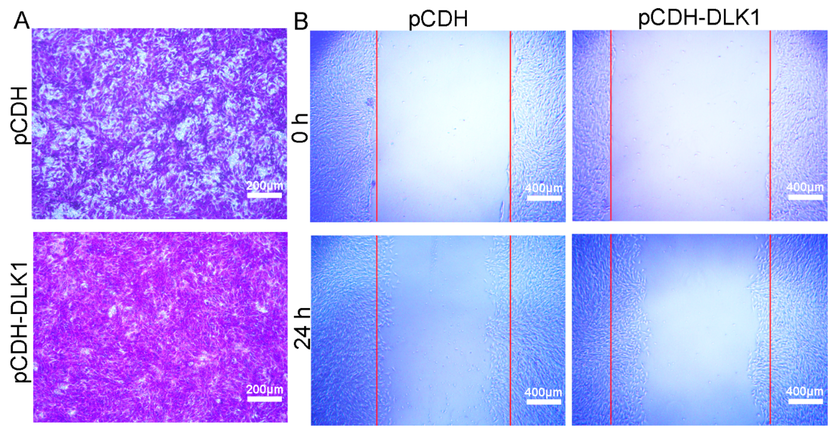
Transwell migration assays indicated that the number of migrated cells with DLK1 overexpression was higher than that with the vector alone, suggesting that DLK1 promoted the migration of C2C12 cells (Figure 3A). To confirm these results, wound healing assays were performed; C2C12/DLK1 cells displayed a higher migration ability than C2C12/vector cells after 24 h (Figure 3B).

3.4. DLK1 Positively Regulates Myogenic Differentiation and Myogenin Expression

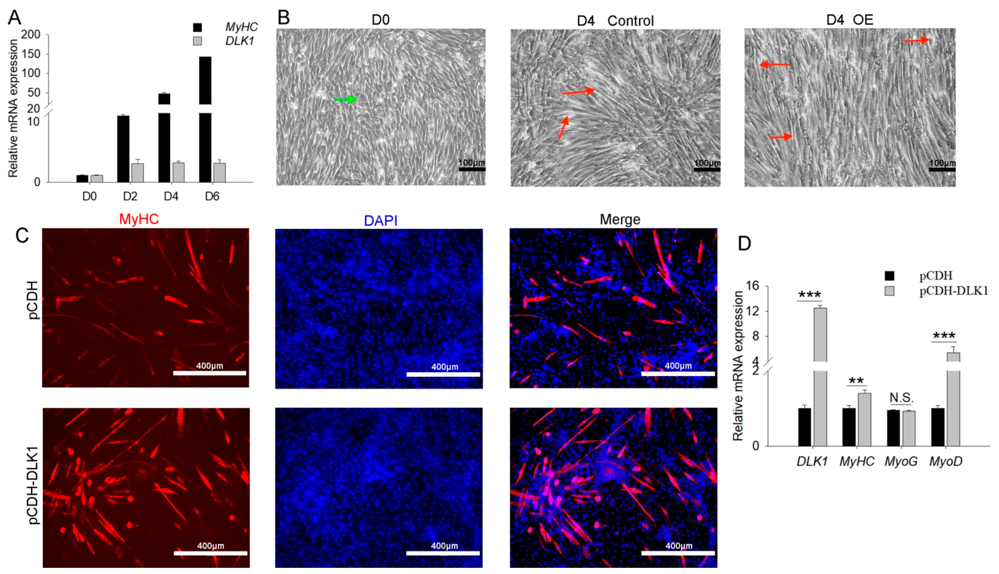
The time course of the changes in the myogenic and DLK1 gene expression was detected in C2C12 myoblasts during differentiation. DLK1 was elevated during the myogenic differentiation of C2C12 myoblasts, which was consistent with the changes in the expression levels of the myogenic marker, MyHC (Figure 4A). Microscopic views showed that DLK1-overexpressed myoblasts induced more orderly bundles of muscle tubes than the control during differentiation, and undifferentiated myoblasts displayed a disorderly and irregular morphology (Figure 4B). Immunocytochemical staining further confirmed that more myotubes were formed by myocytes following DLK1 overexpression compared to control vector-transfected cells (Figure 4C). As expected, enforced DLK1 expression in C2C12 cells dramatically enhanced the levels of myogenic markers (MyHC and MyoD), but MyoG expression was not significantly different (Figure 4D). Taken together, these results indicate that DLK1 positively regulates myogenin transcription and myogenesis.

3.5. DLK1 Promotes Myogenic Fusion and Muscular Hypertrophy but Inhibits Muscle Degradation

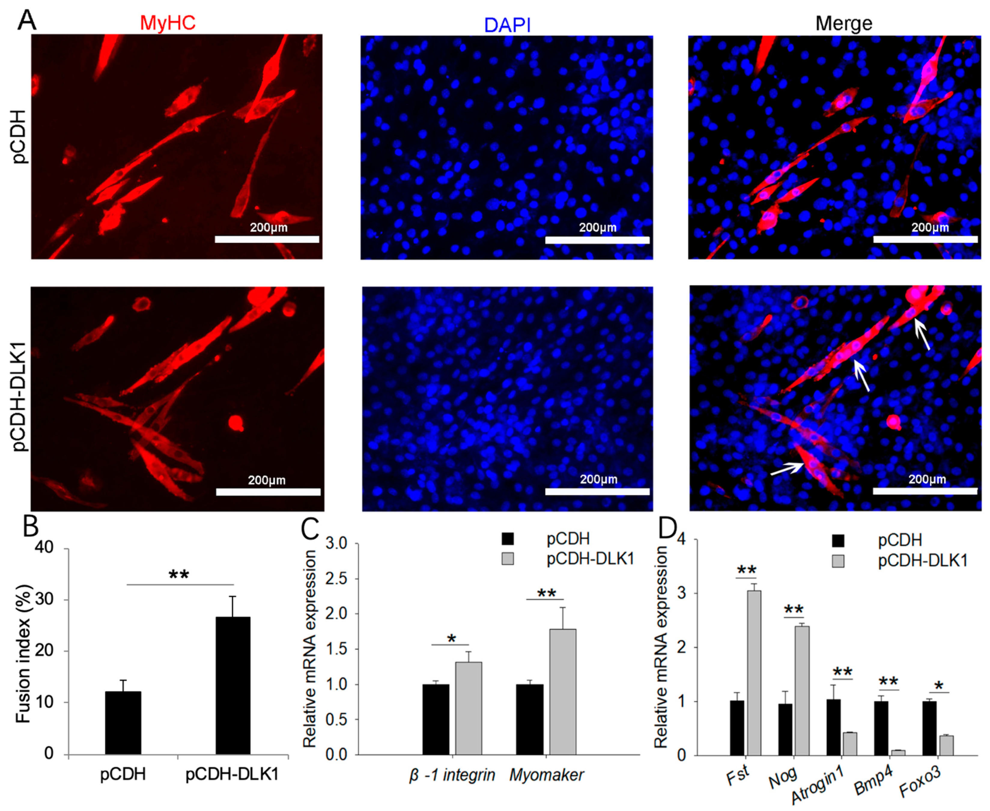
Myoblasts overexpressing DLK1 displayed accelerated fusion kinetics, as demonstrated by the appearance of thick, large myotubes containing many myonuclei (Figure 5A,B). Consistent with the staining results, DLK1 overexpression markedly increased the expression of fusion markers (Myomaker and β-1integrin) (Figure 5C). The DLK1-overexpressed treatment resulted in the significantly elevated expression of muscle hypertrophy genes, including Fst and Nog, whereas it downregulated the expression of muscle degradation markers (Atrogin1, Bmp4, and Foxo3) (Figure 5D).

3.6. Regulatory Pathway of DLK1 on Myogenesis

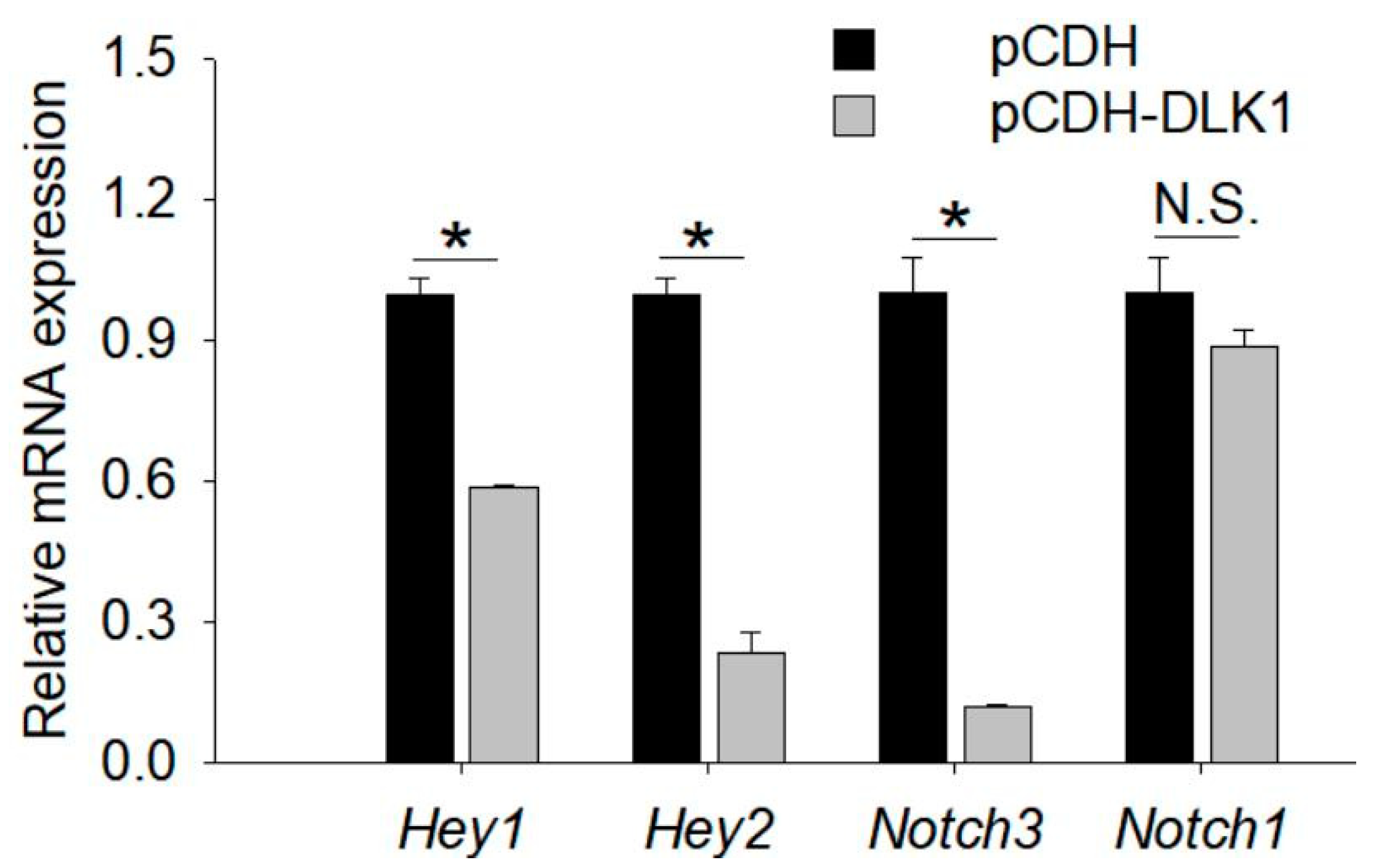
The Notch signaling pathway has been reported to be involved in muscle development [40]. Several genes related to the Notch signaling pathway were selected for validation by qRT-PCR (Figure 6). Notch-pathway-related genes (Hey1/2 and Notch3) were prominently reduced at the mRNA level by DLK1 overexpression in C2C12 cells. Among these Notch-related genes, only the expression of Notch1 showed an insignificant downward trend with enhanced DLK1 expression. These data indicate that DLK1 may regulate muscle growth and development by inhibiting the Notch pathway.

4. Discussion

DLK1 is crucial for the proper development of several mammalian tissues [38]. However, the specific functions and signaling mechanisms of DLK1 in skeletal muscle cells remain unclear. We revealed that DLK1 is expressed in a variety of tissues and highly expressed in the muscle tissue, reflecting the close association between DLK1 and muscle development. This study was designed to elucidate the role of porcine DLK1 in skeletal muscle development by overexpression of DLK1. Moore et al. [51] and Ohno et al. [52] showed that DLK1 is preferentially expressed in fetal stromal cell lines and supports hematopoietic stem cell growth, directly demonstrating that exogenous DLK1 is a positive regulator of murine stem cell growth. Stromal DLK1 promotes proliferation of the intestinal epithelium during development [53]. Similarly, our data showed that mouse myoblast cells overexpressing porcine DLK1 grew faster than the controls, and DLK1 overexpression upregulated the expression of proliferation markers, thereby confirming a positive role of DLK1 in myocyte proliferation. A study that upregulated DLK1 in transfected K562 cells also enhanced myocyte proliferation, providing evidence for such a role [54]. However, the results of the present study are somewhat inconsistent with some previous results in terms of proliferation. Jolena et al. indicated that DLK1 overexpression inhibited cell proliferation [36]. The lack of DLK1 showed an enhanced number of cells in another study [55]. DLK1 in hematopoietic cells inhibited proliferation and differentiation [56]. These functional differences may exist because different biological functions of DLK1 are realized by alternative splicing of exon 5, and different subtypes seem to have different functions [57]. The function of DLK1 may also be tissue specific.
Muscle differentiation is an important process in skeletal muscle development. Skeletal muscle satellite cells differentiate into myoblasts, and MyoD and MyHC are important genetic indicators of this process. Several previous studies have indicated that DLK1 may be involved in regulating cell differentiation [55,56,58]. Our data that the expression levels of DLK1 gradually improved with the extension of differentiation time of C2C12 cells is consistent with the results observed by other investigators [36]. Compared with the control cells, both the number of differentiated myoducts and the expression of differentiated factors were increased in the DLK1 gene overexpression group, which further proves the positive regulation of DLK1 on cell differentiation. A new observation was reported in the current study. DLK1 is associated not only with myocyte proliferation and differentiation, but also with cell migration.
Myoblast fusion is a complex and highly regulated process and is one of the key steps in myogenesis [9]. Many genes and their products may regulate myoblast fusion, and their precise multilevel interactions are essential for myoblast fusion [59]. Myomaker and β-1integrin genes are mainly expressed on the surface of myoblasts and play an important role in regulating myoblast fusion [60]. In the current study, DLK1 positively regulated myocyte fusion. Overexpression of DLK1 led to more multinuclear fusions in differentiated cells, resulting in thicker and longer myotube formation and upregulated expression of the fusion markers, Myomaker and β-1integrin. Davis et al. [37] reported increased immunostaining of DLK1 in callipyge LD skeletal muscle at 8 weeks of age, suggesting the potential role of DLK1 in muscle hypertrophy. Our study showed that over-expressed DLK1 significantly enhanced the expression level of muscle hypertrophy genes but reduced the expression of muscle degradation genes. These results are consistent with that in a previous report [21].
The signaling pathways that result in myogenesis are complex [61], and little is known about DLK1 signaling in skeletal muscles. Notch has been widely studied as a key signaling pathway in skeletal muscle development in mice [62]. The expression of myogenic factors and the differentiation of multinucleated myotubes are suppressed by activating the Notch pathway [52,63,64]. The Notch signaling pathway has also been shown to regulate satellite cell activation, proliferation, differentiation, and muscle regeneration in mice [65]. Studies have suggested that DLK1 is involved in the regulation of the Notch signaling pathway [66,67]. However, specific regulatory sites remain unclear. The findings of the current study support these reports, since DLK1 inhibited Notch signaling by modulating Hey1/2 and Notch3 expression. Notably, DLK1 did not affect Notch1. These results further demonstrate that DLK1 acts as an inhibitor of the Notch pathway and regulates muscle development.
Taken together, these observations suggest that DLK1 has a positive regulatory effect on muscle growth and development, which may be mediated by the inhibition of the Notch signaling pathway. This finding provides a foundation for research on the mechanisms of muscle growth and development in pigs. DLK1 may have many other functional roles that need to be explored; it remains to be determined whether there is a balance between the regulatory effects of different subtypes of DLK1 on muscle development. In addition, the regulatory relationship between DLK1 and the Notch pathway can be further verified by means of the specific activators of the Notch pathway, and it is necessary to clarify the form through which DLK1 inhibits key loci of the Notch pathway.

5. Conclusions

We report the expression, functional identification, and regulatory pathways of porcine DLK1. DLK1 is highly expressed in the muscle tissue of pigs. Cooperating cell phenotype and expression profile analyses showed that DLK1 promoted myocyte proliferation, migration, differentiation, polynuclear fusion, and muscle hypertrophy, but inhibited muscle atrophy. In addition, DLK1 also suppressed the Notch signaling pathway by regulating the expression of key factors in the pathway. These results lead to the conclusion that DLK1 might promote muscle growth and development by inhibiting the Notch signaling pathway, providing new insights and a foundation for further research on the molecular mechanisms underlying porcine myogenesis.

Supplementary Materials

The following supporting information can be downloaded from https://www.mdpi.com/article/10.3390/ani12121523/s1: Table S1 Primer sequences for SqRT-PCR and qRT-PCR.

Author Contributions

Investigation, writing—original draft preparation, visualization, Y.F.; conceptualization, methodology, formal analysis, X.H.; validation, resources, P.S.; supervision, Y.C.; software, data curation, conceptualization, B.Z.; writing—review and editing, project administration, funding acquisition, H.Z. All authors have read and agreed to the published version of the manuscript.

Funding

This research was funded by the Tibet Major Science and Technology Project (grant number XZ202101ZD005N) and the National Natural Science Foundation of China (grant number 32060736).

Institutional Review Board Statement

All experiments were approved by the Animal Welfare Committee of the State Key Laboratory for Agro-Biotechnology of China Agricultural University (Permit Number: SKLAB-2012-04-07).

Informed Consent Statement

Not applicable.

Data Availability Statement

Not applicable.

Acknowledgments

We are grateful to Qingyong Meng for providing the antibodies. We would like to thank Xiaoxiang Hu and Zhengxing Lian for providing the fluorescence microscope and microplate reader, respectively.

Conflicts of Interest

The authors declare no conflict of interest.

References

  1. Abe, T.; Nahar, V.K.; Young, K.C.; Patterson, K.M.; Stover, C.D.; Lajza, D.G.; Tribby, A.C.; Geddam, D.A.; Ford, M.A.; Bass, M.A.; et al. Skeletal muscle mass, bone mineral density, and walking performance in masters cyclists. Rejuvenation Res. 2014, 17, 291–296. [Google Scholar] [CrossRef] [PubMed]
  2. Buckingham, M. Skeletal muscle development and the role of the myogenic regulatory factors. Biochem. Soc. Trans. 1996, 24, 506–509. [Google Scholar] [CrossRef]
  3. Mohammadabadi, M.; Bordbar, F.; Jensen, J.; Du, M.; Guo, W. Key Genes Regulating Skeletal Muscle Development and Growth in Farm Animals. Animals 2021, 11, 835. [Google Scholar] [CrossRef] [PubMed]
  4. Cortes-Araya, Y.; Stenhouse, C.; Salavati, M.; Dan-Jumbo, S.O.; Ho, W.; Ashworth, C.J.; Clark, E.; Esteves, C.L.; Donadeu, F.X. KLB dysregulation mediates disrupted muscle development in intrauterine growth restriction. J. Physiol. 2022, 600, 1771–1790. [Google Scholar] [CrossRef] [PubMed]
  5. Yin, X.; Liu, W.; Chen, H.; Qi, C.; Chen, H.; Niu, H.; Yang, J.; Kwok, K.W.H.; Dong, W. Effects of ferulic acid on muscle development and intestinal microbiota of zebrafish. J. Anim. Physiol. Anim. Nutr. 2022, 106, 429–440. [Google Scholar] [CrossRef]
  6. Roy, A.; Kumar, A. Supraphysiological activation of TAK1 promotes skeletal muscle growth and mitigates neurogenic atrophy. Nat. Commun. 2022, 13, 2201. [Google Scholar] [CrossRef]
  7. Duran, B.O.S.; Zanella, B.T.T.; Perez, E.S.; Mareco, E.A.; Blasco, J.; Dal-Pai-Silva, M.; Garcia de la Serrana, D. Amino Acids and IGF1 Regulation of Fish Muscle Growth Revealed by Transcriptome and microRNAome Integrative Analyses of Pacu (Piaractus mesopotamicus) Myotubes. Int. J. Mol. Sci. 2022, 23, 1180. [Google Scholar] [CrossRef]
  8. Horsley, V.; Pavlath, G.K. Forming a multinucleated cell: Molecules that regulate myoblast fusion. Cells Tissues Organs 2004, 176, 67–78. [Google Scholar] [CrossRef]
  9. Abmayr, S.M.; Pavlath, G.K. Myoblast fusion: Lessons from flies and mice. Development 2012, 139, 641–656. [Google Scholar] [CrossRef]
  10. Manneken, J.D.; Dauer, M.V.P.; Currie, P.D. Dynamics of muscle growth and regeneration: Lessons from the teleost. Exp. Cell Res. 2022, 411, 112991. [Google Scholar] [CrossRef]
  11. Chal, J.; Pourquie, O. Making muscle: Skeletal myogenesis in vivo and in vitro. Development 2017, 144, 2104–2122. [Google Scholar] [CrossRef] [PubMed]
  12. Jensen, C.H.; Teisner, B.; Højrup, P.; Rasmussen, H.B.; Madsen, O.D.; Nielsen, B.; Skjødt, K. Studies on the isolation, structural analysis and tissue localization of fetal antigen 1 and its relation to a human adrenal-specific cDNA, pG2. Hum. Reprod. 1993, 8, 635–641. [Google Scholar] [CrossRef] [PubMed]
  13. Macedo, D.B.; Kaiser, U.B. DLK1, Notch Signaling and the Timing of Puberty. Semin. Reprod. Med. 2019, 37, 174–181. [Google Scholar] [CrossRef] [PubMed]
  14. Andersen, D.C.; Petersson, S.J.; Jørgensen, L.H.; Bollen, P.; Jensen, P.B.; Teisner, B.; Schroeder, H.D.; Jensen, C.H. Characterization of DLK1+ cells emerging during skeletal muscle remodeling in response to myositis, myopathies, and acute injury. Stem Cells 2009, 27, 898–908. [Google Scholar] [CrossRef]
  15. Kawakami, T.; Chano, T.; Minami, K.; Okabe, H.; Okada, Y.; Okamoto, K. Imprinted DLK1 is a putative tumor suppressor gene and inactivated by epimutation at the region upstream of GTL2 in human renal cell carcinoma. Hum. Mol. Genet. 2006, 15, 821–830. [Google Scholar] [CrossRef]
  16. Miller, A.J.; Cole, S.E. Multiple Dlk1 splice variants are expressed during early mouse embryogenesis. Int. J. Dev. Biol. 2014, 58, 65–70. [Google Scholar] [CrossRef] [PubMed]
  17. Andersen, D.C.; Laborda, J.; Baladron, V.; Kassem, M.; Sheikh, S.P.; Jensen, C.H. Dual role of delta-like 1 homolog (DLK1) in skeletal muscle development and adult muscle regeneration. Development 2013, 140, 3743–3753. [Google Scholar] [CrossRef] [PubMed]
  18. Jensen, C.H.; Meyer, M.; Schroder, H.D.; Kliem, A.; Zimmer, J.; Teisner, B. Neurons in the monoaminergic nuclei of the rat and human central nervous system express FA1/dlk. Neuroreport 2001, 12, 3959–3963. [Google Scholar] [CrossRef] [PubMed]
  19. Deiuliis, J.A.; Li, B.; Lyvers-Peffer, P.A.; Moeller, S.J.; Lee, K. Alternative splicing of delta-like 1 homolog (DLK1) in the pig and human. Comp. Biochem. Physiol. Part B Biochem. Mol. Biol. 2006, 145, 50–59. [Google Scholar] [CrossRef]
  20. Oczkowicz, M.; Piestrzyska-Kajtoch, A.; Piórkowska, K.; Rejduch, B.; Rózycki, M. Expression of DLK1 and MEG3 genes in porcine tissues during postnatal development. Genet. Mol. Biol. 2010, 33, 790–794. [Google Scholar] [CrossRef]
  21. White, J.D.; Vuocolo, T.; McDonagh, M.; Grounds, M.D.; Harper, G.S.; Cockett, N.E.; Tellam, R. Analysis of the callipyge phenotype through skeletal muscle development; association of Dlk1 with muscle precursor cells. Differ. Res. Biol. Divers. 2008, 76, 283–298. [Google Scholar] [CrossRef] [PubMed]
  22. Yu, H.; Waddell, J.N.; Kuang, S.; Tellam, R.L.; Cockett, N.E.; Bidwell, C.A. Identification of genes directly responding to DLK1 signaling in Callipyge sheep. BMC Genom. 2018, 19, 283. [Google Scholar] [CrossRef] [PubMed]
  23. Crameri, R.M.; Langberg, H.; Magnusson, P.; Jensen, C.H.; Schroder, H.D.; Olesen, J.L.; Suetta, C.; Teisner, B.; Kjaer, M. Changes in satellite cells in human skeletal muscle after a single bout of high intensity exercise. J. Physiol. 2004, 558, 333–340. [Google Scholar] [CrossRef] [PubMed]
  24. Shin, J.; Velleman, S.G.; Latshaw, J.D.; Wick, M.P.; Suh, Y.; Lee, K. The ontogeny of delta-like protein 1 messenger ribonucleic acid expression during muscle development and regeneration: Comparison of broiler and Leghorn chickens. Poult. Sci. 2009, 88, 1427–1437. [Google Scholar] [CrossRef] [PubMed]
  25. Jørgensen, L.H.; Sellathurai, J.; Davis, E.E.; Thedchanamoorthy, T.; Al-Bader, R.W.; Jensen, C.H.; Schrøder, H.D. Delta-like 1 homolog (dlk1): A marker for rhabdomyosarcomas implicated in skeletal muscle regeneration. PLoS ONE 2013, 8, e60692. [Google Scholar] [CrossRef] [PubMed]
  26. Grassi, E.S.; Pietras, A. Emerging Roles of DLK1 in the Stem Cell Niche and Cancer Stemness. J. Histochem. Cytochem. 2022, 70, 17–28. [Google Scholar] [CrossRef] [PubMed]
  27. Sul, H.S. Minireview: Pref-1: Role in adipogenesis and mesenchymal cell fate. Mol. Endocrinol. 2009, 23, 1717–1725. [Google Scholar] [CrossRef]
  28. Albrecht, E.; Kuzinski, J.; Komolka, K.; Gotoh, T.; Maak, S. Localization and abundance of early markers of fat cell differentiation in the skeletal muscle of cattle during growth—Are DLK1-positive cells the origin of marbling flecks? Meat Sci. 2015, 100, 237–245. [Google Scholar] [CrossRef]
  29. Smas, C.M.; Sul, H.S. Pref-1, a protein containing EGF-like repeats, inhibits adipocyte differentiation. Cell 1993, 73, 725–734. [Google Scholar] [CrossRef]
  30. Petry, C.J.; Burling, K.A.; Barker, P.; Hughes, I.A.; Ong, K.K.; Dunger, D.B. Pregnancy Serum DLK1 Concentrations Are Associated with Indices of Insulin Resistance and Secretion. J. Clin. Endocrinol. Metab. 2021, 106, e2413–e2422. [Google Scholar] [CrossRef]
  31. Smas, C.M.; Chen, L.; Zhao, L.; Latasa, M.J.; Sul, H.S. Transcriptional repression of pref-1 by glucocorticoids promotes 3T3-L1 adipocyte differentiation. J. Biol. Chem. 1999, 274, 12632–12641. [Google Scholar] [CrossRef] [PubMed]
  32. Lee, K.; Villena, J.A.; Moon, Y.S.; Kim, K.H.; Lee, S.; Kang, C.; Sul, H.S. Inhibition of adipogenesis and development of glucose intolerance by soluble preadipocyte factor-1 (Pref-1). J. Clin. Investig. 2003, 111, 453–461. [Google Scholar] [CrossRef] [PubMed]
  33. Kim, K.S.; Kim, J.J.; Dekkers, J.C.; Rothschild, M.F. Polar overdominant inheritance of a DLK1 polymorphism is associated with growth and fatness in pigs. Mamm. Genome Off. J. Int. Mamm. Genome Soc. 2004, 15, 552–559. [Google Scholar] [CrossRef] [PubMed]
  34. Li, X.P.; Do, K.T.; Kim, J.J.; Huang, J.; Zhao, S.H.; Lee, Y.; Rothschild, M.F.; Lee, C.K.; Kim, K.S. Molecular characteristics of the porcine DLK1 and MEG3 genes. Anim. Genet. 2008, 39, 189–192. [Google Scholar] [CrossRef]
  35. Moon, Y.S.; Smas, C.M.; Lee, K.; Villena, J.A.; Kim, K.H.; Yun, E.J.; Sul, H.S. Mice lacking paternally expressed Pref-1/Dlk1 display growth retardation and accelerated adiposity. Mol. Cell. Biol. 2002, 22, 5585–5592. [Google Scholar] [CrossRef] [PubMed]
  36. Waddell, J.N.; Zhang, P.; Wen, Y.; Gupta, S.K.; Yevtodiyenko, A.; Schmidt, J.V.; Bidwell, C.A.; Kumar, A.; Kuang, S. Dlk1 is necessary for proper skeletal muscle development and regeneration. PLoS ONE 2010, 5, e15055. [Google Scholar] [CrossRef]
  37. Davis, E.; Jensen, C.H.; Schroder, H.D.; Farnir, F.; Shay-Hadfield, T.; Kliem, A.; Cockett, N.; Georges, M.; Charlier, C. Ectopic expression of DLK1 protein in skeletal muscle of padumnal heterozygotes causes the callipyge phenotype. Curr. Biol. CB 2004, 14, 1858–1862. [Google Scholar] [CrossRef]
  38. Zhang, L.; Uezumi, A.; Kaji, T.; Tsujikawa, K.; Andersen, D.C.; Jensen, C.H.; Fukada, S.I. Expression and Functional Analyses of Dlk1 in Muscle Stem Cells and Mesenchymal Progenitors during Muscle Regeneration. Int. J. Mol. Sci. 2019, 20, 3269. [Google Scholar] [CrossRef]
  39. Sánchez-Solana, B.; Nueda, M.L.; Ruvira, M.D.; Ruiz-Hidalgo, M.J.; Monsalve, E.M.; Rivero, S.; García-Ramírez, J.J.; Díaz-Guerra, M.J.; Baladrón, V.; Laborda, J. The EGF-like proteins DLK1 and DLK2 function as inhibitory non-canonical ligands of NOTCH1 receptor that modulate each other’s activities. Biochim. Biophys. Acta 2011, 1813, 1153–1164. [Google Scholar] [CrossRef]
  40. Mu, X.D.; Tang, Y.; Lu, A.P.; Takayama, K.; Usas, A.; Wang, B.; Weiss, K.; Huard, J. The role of Notch signaling in muscle progenitor cell depletion and the rapid onset of histopathology in muscular dystrophy. Hum. Mol. Genet. 2015, 24, 2923–2937. [Google Scholar] [CrossRef]
  41. Buas, M.F.; Kadesch, T. Regulation of skeletal myogenesis by Notch. Exp. Cell Res. 2010, 316, 3028–3033. [Google Scholar] [CrossRef] [PubMed]
  42. Bröhl, D.; Vasyutina, E.; Czajkowski, M.T.; Griger, J.; Rassek, C.; Rahn, H.-P.; Purfürst, B.; Wende, H.; Birchmeier, C. Colonization of the satellite cell niche by skeletal muscle progenitor cells depends on Notch signals. Dev. Cell 2012, 23, 469–481. [Google Scholar] [CrossRef] [PubMed]
  43. Fu, Y.; Shang, P.; Zhang, B.; Tian, X.; Nie, R.; Zhang, R.; Zhang, H. Function of the Porcine TRPC1 Gene in Myogenesis and Muscle Growth. Cells 2021, 10, 147. [Google Scholar] [CrossRef] [PubMed]
  44. Rao, X.; Huang, X.; Zhou, Z.; Lin, X. An improvement of the 2ˆ(-delta delta CT) method for quantitative real-time polymerase chain reaction data analysis. Biostat. Bioinform. Biomath. 2013, 3, 71–85. [Google Scholar]
  45. Weinlander, E.; Somnay, Y.; Harrison, A.D.; Wang, C.; Cheng, Y.Q.; Jaskula-Sztul, R.; Yu, X.M.; Chen, H. The novel histone deacetylase inhibitor thailandepsin A inhibits anaplastic thyroid cancer growth. J. Surg. Res. 2014, 190, 191–197. [Google Scholar] [CrossRef]
  46. Singhal, J.; Nagaprashantha, L.D.; Vatsyayan, R.; Awasthi, S.; Singhal, S.S. Didymin induces apoptosis by inhibiting N-Myc and upregulating RKIP in neuroblastoma. Cancer Prev. Res. 2012, 5, 473–483. [Google Scholar] [CrossRef]
  47. Weng, X.; Zhu, S.Q.; Cui, H.J. Artesunate inhibits proliferation of glioblastoma cells by arresting cell cycle. Zhongguo Zhong Yao Za Zhi = Zhongguo Zhongyao Zazhi = China J. Chin. Mater. Med. 2018, 43, 772–778. [Google Scholar] [CrossRef]
  48. Reza, M.M.; Subramaniyam, N.; Sim, C.M.; Ge, X.; Sathiakumar, D.; McFarlane, C.; Sharma, M.; Kambadur, R. Irisin is a pro-myogenic factor that induces skeletal muscle hypertrophy and rescues denervation-induced atrophy. Nat. Commun. 2017, 8, 1–17. [Google Scholar] [CrossRef]
  49. Seo, S.; Jeon, S.; Ha, J.K. Guidelines for experimental design and statistical analyses in animal studies submitted for publication in the Asian-Australasian Journal of Animal Sciences. Asian-Australas. J. Anim. Sci. 2018, 31, 1381–1386. [Google Scholar] [CrossRef]
  50. Shang, P.; Wang, Z.; Chamba, Y.; Zhang, B.; Zhang, H.; Wu, C. A comparison of prenatal muscle transcriptome and proteome profiles between pigs with divergent growth phenotypes. J. Cell. Biochem. 2019, 120, 5277–5286. [Google Scholar] [CrossRef]
  51. Moore, K.A.; Pytowski, B.; Witte, L.; Hicklin, D.; Lemischka, I.R. Hematopoietic activity of a stromal cell transmembrane protein containing epidermal growth factor-like repeat motifs. Proc. Natl. Acad. Sci. USA 1997, 94, 4011–4016. [Google Scholar] [CrossRef]
  52. Ohno, N.; Izawa, A.; Hattori, M.; Kageyama, R.; Sudo, T. dlk inhibits stem cell factor-induced colony formation of murine hematopoietic progenitors: Hes-1-independent effect. Stem Cells 2001, 19, 71–79. [Google Scholar] [CrossRef] [PubMed]
  53. Ichinose, M.; Suzuki, N.; Wang, T.; Wright, J.A.; Lannagan, T.R.M.; Vrbanac, L.; Kobayashi, H.; Gieniec, K.A.; Ng, J.Q.; Hayakawa, Y. Stromal DLK1 promotes proliferation and inhibits differentiation of the intestinal epithelium during development. Am. J. Physiol.-Gastrointest. Liver Physiol. 2021, 320, G506–G520. [Google Scholar] [CrossRef]
  54. Qi, X.; Chen, Z.; Liu, D.; Cen, J.; Gu, M. Expression of Dlk1 gene in myelodysplastic syndrome determined by microarray, and its effects on leukemia cells. Int. J. Mol. Med. 2008, 22, 61–68. [Google Scholar] [CrossRef]
  55. Raghunandan, R.; Ruiz-Hidalgo, M.; Jia, Y.; Ettinger, R.; Rudikoff, E.; Riggins, P.; Farnsworth, R.; Tesfaye, A.; Laborda, J.; Bauer, S.R. Dlk1 influences differentiation and function of B lymphocytes. Stem Cells Dev. 2008, 17, 495–507. [Google Scholar] [CrossRef] [PubMed]
  56. Li, L.; Forman, S.J.; Bhatia, R. Expression of DLK1 in hematopoietic cells results in inhibition of differentiation and proliferation. Oncogene 2005, 24, 4472–4476. [Google Scholar] [CrossRef] [PubMed]
  57. Mei, B.; Zhao, L.; Chen, L.; Sul, H.S. Only the large soluble form of preadipocyte factor-1 (Pref-1), but not the small soluble and membrane forms, inhibits adipocyte differentiation: Role of alternative splicing. Biochem. J. 2002, 364, 137–144. [Google Scholar] [CrossRef]
  58. Wust, S.; Drose, S.; Heidler, J.; Wittig, I.; Klockner, I.; Franko, A.; Bonke, E.; Gunther, S.; Gartner, U.; Boettger, T.; et al. Metabolic Maturation during Muscle Stem Cell Differentiation Is Achieved by miR-1/133a-Mediated Inhibition of the Dlk1-Dio3 Mega Gene Cluster. Cell Metab. 2018, 27, 1026–1039.e6. [Google Scholar] [CrossRef]
  59. Millay, D.P.; Sutherland, L.B.; Bassel-Duby, R.; Olson, E.N. Myomaker is essential for muscle regeneration. Genes Dev. 2014, 28, 1641–1646. [Google Scholar] [CrossRef]
  60. Millay, D.P.; O’Rourke, J.R.; Sutherland, L.B.; Bezprozvannaya, S.; Shelton, J.M.; Bassel-Duby, R.; Olson, E.N. Myomaker is a membrane activator of myoblast fusion and muscle formation. Nature 2013, 499, 301–305. [Google Scholar] [CrossRef]
  61. Shavlakadze, T.; Grounds, M. Of bears, frogs, meat, mice and men: Complexity of factors affecting skeletal muscle mass and fat. BioEssays News Rev. Mol. Cell. Dev. Biol. 2006, 28, 994–1009. [Google Scholar] [CrossRef]
  62. Zhao, H.B.; Qi, S.N.; Dong, J.Z.; Ha, X.Q.; Li, X.Y.; Zhang, Q.W.; Yang, Y.S.; Bai, J.; Zhao, L. Salidroside induces neuronal differentiation of mouse mesenchymal stem cells through Notch and BMP signaling pathways. Food Chem. Toxicol. Int. J. Publ. Br. Ind. Biol. Res. Assoc. 2014, 71, 60–67. [Google Scholar] [CrossRef] [PubMed]
  63. Buas, M.F.; Kabak, S.; Kadesch, T. Inhibition of myogenesis by Notch: Evidence for multiple pathways. J. Cell. Physiol. 2009, 218, 84–93. [Google Scholar] [CrossRef]
  64. Kitamoto, T.; Hanaoka, K. Notch3 null mutation in mice causes muscle hyperplasia by repetitive muscle regeneration. Stem Cells 2010, 28, 2205–2216. [Google Scholar] [CrossRef] [PubMed]
  65. Conboy, I.M.; Rando, T.A. The regulation of Notch signaling controls satellite cell activation and cell fate determination in postnatal myogenesis. Dev. Cell 2002, 3, 397–409. [Google Scholar] [CrossRef]
  66. Nueda, M.L.; Baladrón, V.; Sánchez-Solana, B.; Ballesteros, M.A.; Laborda, J. The EGF-like protein dlk1 inhibits notch signaling and potentiates adipogenesis of mesenchymal cells. J. Mol. Biol. 2007, 367, 1281–1293. [Google Scholar] [CrossRef]
  67. Traustadottir, G.A.; Lagoni, L.V.; Ankerstjerne, L.B.S.; Bisgaard, H.C.; Jensen, C.H.; Andersen, D.C. The imprinted gene Delta like non-canonical Notch ligand 1 (Dlk1) is conserved in mammals, and serves a growth modulatory role during tissue development and regeneration through Notch dependent and independent mechanisms. Cytokine Growth Factor Rev. 2019, 46, 17–27. [Google Scholar] [CrossRef]
Figure 1. Expressions of DLK1 in the embryonic tissues of pigs. (A) DLK1 expression in the different tissues of TP pigs at the embryonic stage by SqRT-PCR. LD, longissimus dorsi; BF, back fat; (B) the mRNA expression levels of DLK1 in the LD of three pig breeds. YY, Yorkshire (n = 6); WJ, Wujin pig (n = 6); TP, Tibetan pig (n = 6). Each bar represents the mean ± SD. ** p < 0.01.
Animals 12 01523 g001
Figure 2. DLK1 promotes myoblast proliferation. (A) Efficiency of the detection of plasmid overexpression; (B) microscopic view of cell proliferation; scale bar = 400 µm; (C) EdU staining for proliferated cells following pCDH-DLK1 transfection. Nuclei are stained with DAPI; red indicates EdU-positive proliferating cells. Representative images are shown in the left panel, and the statistical graphs in the right panel indicate the proliferating cells 48 h after transfection; n = 3 in each group; scale bar = 400 µm; (D) CCK8 assay of proliferated myoblasts transfected with overexpression fragments; (E) the mRNA expression levels of proliferation marker genes. The data represent the mean ± SD of three independent experiments. GAPDH was used as a reference gene. * p < 0.05, ** p < 0.01, *** p < 0.001, N.S. represents not significant.
Animals 12 01523 g002
Figure 3. DLK1 facilitates myoblast migration. (A) The effect of DLK1 on cell migration was investigated using a Transwell migration assay. Purple represents migrated cells stained with 0.1% crystal violet; scale bar = 200 µm; (B) the wound-healing migration assay of C2C12 myoblasts. The red line represents the wound healing area; scale bar = 400 µm.
Animals 12 01523 g003
Figure 4. DLK1 improved cell differentiation. (A) Quantitative real-time polymerase chain reaction (qRT-PCR) results showed the expression profiles of the DLK1 gene during differentiation. MyHC is the myogenic-differentiation-indicator gene; (B) microscopic view of differentiated cells; D0 and D4 refer to differentiation after zero and four days. Control and OE represent cells transfected with pCDH and pCDH-DLK1, respectively; scale bar = 100 µm; green arrows represent undifferentiated myoblasts that displayed a disorderly and irregular morphology; red arrows represent orderly bundles of muscle tubes. (C) Immunofluorescence staining for MyHC protein in pCDH- or pCDH-DLK1-treated myoblasts that were cultured for four days in differentiation medium. MyHC and the nucleus are stained in red and blue (DAPI), respectively; scale bar = 400 µm; (D) the mRNA expression of DLK1 and the differentiation marker genes, MyoD, MyoG, and MyHC, was quantified using qRT-PCR. ** p < 0.01, *** p < 0.001, N.S., not significant.
Animals 12 01523 g004
Figure 5. DLK1 stimulated myoblast fusion and muscle hypertrophy but suppressed muscle degradation. (A) Myoblast fusion analysis by immunofluorescence staining for MyHC after four days of differentiation, and the white arrows represent the multinucleated myotubes; white scale bar = 200 µm; (B) the fusion index was measured by dividing the number of nuclei found within the myotubes by the total number of nuclei; (C) the mRNA expression of fusion marker genes was quantified by a quantitative real-time polymerase chain reaction (qRT-PCR); (D) the mRNA expression of muscle hypertrophy genes (Fst and Nog) and muscle degradation markers (Atrogin1, Bmp4, and Foxo3) was quantified by qRT-PCR. The data represent the mean ± SD of three independent experiments. * p < 0.05, ** p < 0.01.
Animals 12 01523 g005
Figure 6. DLK1 negatively regulated Notch signaling pathways. The mRNA expression of Notch-related genes in the control and DLK1-overexpressed myoblasts. The data represent the mean ± SD of three independent experiments. * p < 0.05, N.S., not significant.
Animals 12 01523 g006
Publisher’s Note: MDPI stays neutral with regard to jurisdictional claims in published maps and institutional affiliations.