
Canine Blood Group Prevalence and Geographical Distribution around the World: An Updated Systematic Review


 , , ,
, , ,  and
and
Abstract
Simple Summary
Abstract
1. Introduction
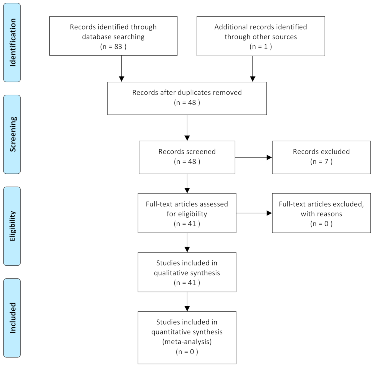
2. Materials and Methods
2.1. Search Strategy
2.2. Study Selection
2.3. Data and Quality Assessment
3. Results
3.1. Blood Groups
3.2. Blood-Typing Methods
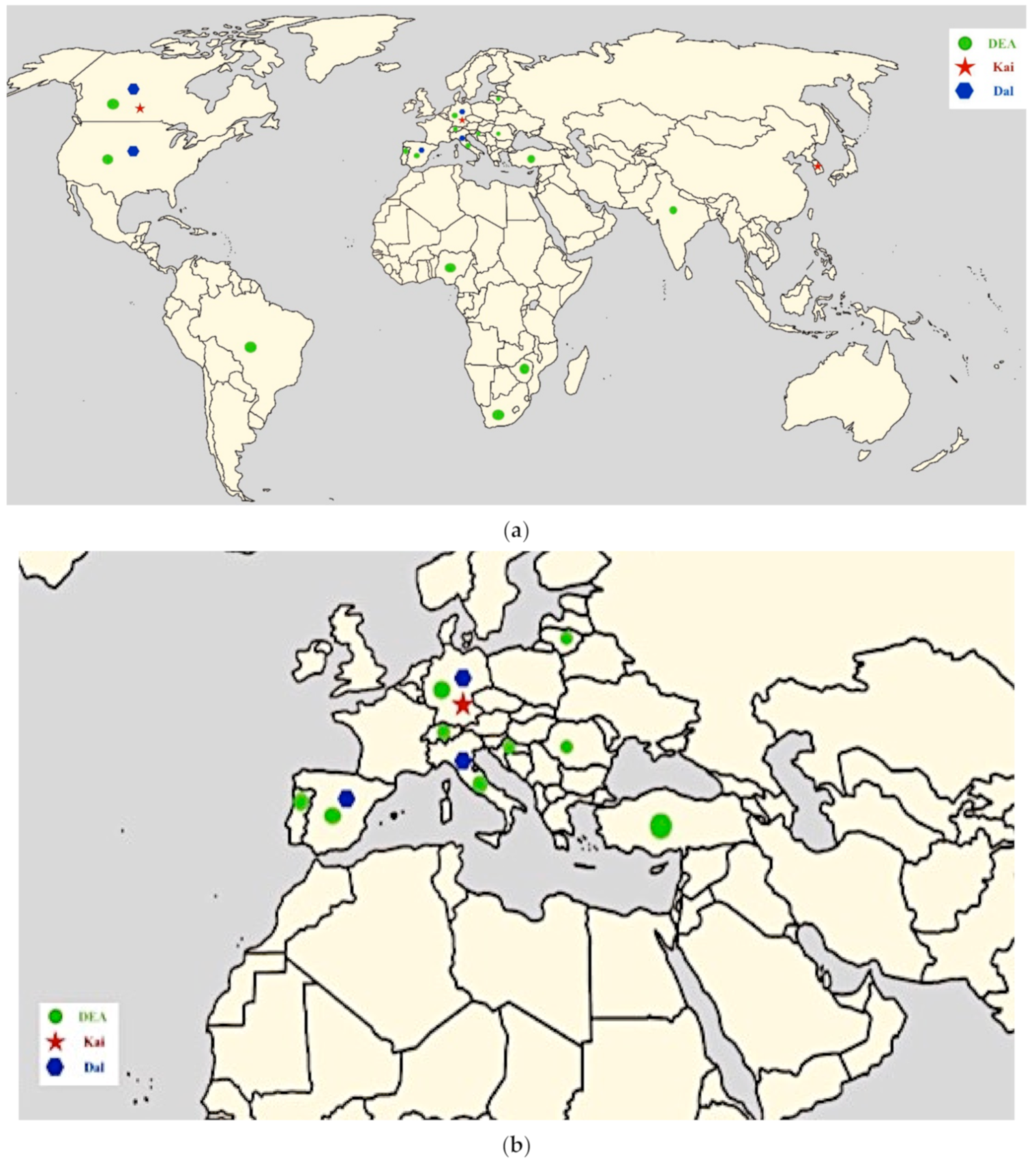
3.3. Prevalence of Blood Groups in Different Dog Breeds and Countries
4. Discussion
5. Conclusions
Author Contributions
Funding
Institutional Review Board Statement
Data Availability Statement
Acknowledgments
Conflicts of Interest
References
- Bowdler, A.J.; Bull, R.W.; Dries, C.; Slating, R.; Swisher, S.N. Representation of the ABH blood group system in the dog. Vox Sang. 1973, 24, 228–235. [Google Scholar] [CrossRef] [PubMed]
- Colling, D.T.; Saison, R. Canine blood groups. 2. Description of a new allele in the Tr blood group system. Anim. Blood Groups Biochem. Genet. 1980, 11, 13–20. [Google Scholar] [CrossRef] [PubMed]
- Hall, D.E. A naturally occurring red cell antigen antibody system in beagle dogs. J. Small Anim. Pract. 1970, 11, 543–551. [Google Scholar] [CrossRef] [PubMed]
- Zaremba, R.; Brooks, A.; Thomovsky, E. Transfusion Medicine: An Update on Antigens, Antibodies and Serologic Testing in Dogs and Cats. Top. Companion Anim. Med. 2019, 34, 36–46. [Google Scholar] [CrossRef]
- Bowling, A. Red blood cell antigens and blood groups in the horse. In Schalm’s Veterinary Hematology; Feldman, B., Zinkl, J., Jain, N., Eds.; Lippincott, Williams, & Wilkins: Baltimore, MD, USA, 2000; pp. 774–777. [Google Scholar]
- Lanevschi, A.; Wardrop, K.J. Principles of transfusion medicine in small animals. Can. Vet. J. 2001, 42, 447–454. [Google Scholar]
- Hale, A.S. Canine blood groups and their importance in veterinary transfusion medicine. Vet. Clin. N. Am. Small Anim. Pract. 1995, 25, 1323–1332. [Google Scholar] [CrossRef]
- Symons, M.; Bell, K. Canine blood groups: Description of 20 specificities. Anim. Genet. 1992, 23, 509–515. [Google Scholar] [CrossRef]
- Davidow, B. Transfusion medicine in small animals. Vet. Clin. N. Am. Small Anim. Pract. 2013, 43, 735–756. [Google Scholar] [CrossRef]
- Kessler, R.J.; Reese, J.; Chang, D.; Seth, M.; Hale, A.S.; Giger, U. Dog erythrocyte antigens 1.1, 1.2, 3, 4, 7, and Dal blood typing and cross-matching by gel column technique. Vet. Clin. Pathol. 2010, 39, 306–316. [Google Scholar] [CrossRef]
- Hale, A.S.; Werfelmann, J.; Lemmons, M.; Smiler, B.; Gerlach, J. An evaluation of 9570 dogs by breed and dog erythrocyte antigen typing. J. Vet. Intern. Med. 2008, 22, 740. [Google Scholar]
- Acierno, M.M.; Raj, K.; Giger, U. DEA 1 expression on dog erythrocytes analyzed by immunochromatographic and flow cytometric techniques. J. Vet. Intern. Med. 2014, 28, 592–598. [Google Scholar] [CrossRef] [PubMed]
- Blais, M.C.; Berman, L.; Oakley, D.A. Canine Dal blood type: A red cell antigen lacking in some Dalmatians. J. Vet. Intern. Med. 2007, 21, 281–286. [Google Scholar] [CrossRef] [PubMed]
- Goulet, S.; Blais, M.C. Characterization of Anti-Dal Alloantibodies Following Sensitization of Two Dal-Negative Dogs. Vet. Pathol. 2018, 55, 108–115. [Google Scholar] [CrossRef] [PubMed]
- Proverbio, D.; Lubas, G.; Spada, E.; Medina Valentin, A.A.; Viñals Florez, L.M.; Del Rosario Perlado Chamizo, M.; Perego, R.; Pennisi, M.G.; Ferro, E.; Baggiani, L.; et al. Prevalence of Dal blood type and dog erythrocyte antigens (DEA) 1, 4, and 7 in canine blood donors in Italy and Spain. BMC Vet. Res. 2020, 16, 126. [Google Scholar] [CrossRef]
- Ebelt, A.K.; Fuchs, S.; Weber, C.; Müller, E.; Giger, U. Survey of Blood Groups DEA 1, DEA 4, DEA 5, Dal, and Kai 1/Kai 2 in Different Canine Breeds from a Diagnostic Laboratory in Germany. Front. Vet. Sci. 2020, 7, 85. [Google Scholar] [CrossRef]
- Lee, J.H.; Giger, U.; Kim, H.Y. Kai 1 and Kai 2: Characterization of these dog erythrocyte antigens by monoclonal antibodies. PLoS ONE 2017, 12, e0179932. [Google Scholar] [CrossRef]
- Euler, C.C.; Lee, J.H.; Kim, H.Y.; Raj, K.; Mizukami, K.; Giger, U. Survey of Two New (Kai 1 and Kai 2) and Other Blood Groups in Dogs of North America. J. Vet. Intern. Med. 2016, 30, 1642–1647. [Google Scholar] [CrossRef]
- Polak, K.; Acierno, M.M.; Raj, K.; Mizukami, K.; Siegel, D.L.; Giger, U. Dog erythrocyte antigen 1: Mode of inheritance and initial characterization. Vet. Clin. Pathol. 2015, 44, 369–379. [Google Scholar] [CrossRef]
- Giger, U.; Stieger, K.; Palos, H. Comparison of various canine blood-typing methods. Am. J. Vet. Res. 2005, 66, 1386–1392. [Google Scholar] [CrossRef]
- Novais, A.A.; Santana, A.E.; Vicentin, L.A. Prevalence of DEA 1 canine blood group system in dogs (Canis familiaris, Linnaeus, 1758) reared in Brazil. Braz. J. Vet. Res. Anim. Sci. 1999, 36. [Google Scholar] [CrossRef]
- Van der Merwe, L.L.; Jacobson, L.S.; Pretorius, G.J. The breed prevalence of dog erythrocyte antigen 1.1 in the Onderstepoort area of South Africa and its significance in selection of canine blood donors. J. S. Afr. Vet. Assoc. 2002, 73, 53–56. [Google Scholar] [CrossRef] [PubMed]
- Lucidi, C.A.; Takahira, R.K.; Gerlach, J.A.; Davis, J.M.; Schwartz, K.A.; Scott, M.A. Flow cytometric assessment of canine erythrocytes and platelets for dog erythrocyte antigen 1.1. Vet. Clin. Pathol. 2011, 40, 435–443. [Google Scholar] [CrossRef] [PubMed]
- Nottidge, H.O.; Omobowale, T.O.; Washio, M.; Ajadir, A.; Toisumi, S.H.; Takahshi, K. The prevalence of the dog erythrocytre antigen (Dea 1.1 and Dea 1.2) in Nigerian indigenous dogs. Folia Vet. 2006, 50, 66–68. [Google Scholar]
- Santos, S.C.S.; dos Moroz, L.R.; Santos, M.M.; dos Santos, A.S.; dos Trindade, S.C.; Meyer, R.; Costa, M.D.F.D. Detection of canine anti-DEA 1 antibodies using flow cytometry in dogs following DEA 1-positive blood transfusion. Braz. J. Vet. Res. Anim. Sci. 2018, 55, 1–7. [Google Scholar] [CrossRef][Green Version]
- Santos, S.C.S.; Santos, M.M.; Rodrigues, W.F.; Meyer, R.; Costa, M.F.D. Blood typing in positive DEA 1 dogs: Comparative analysis between immunochromatography, hemagglutination and flow cytometry. Braz. J. Vet. Res. Anim. Sci. 2020, 57, e151444. [Google Scholar] [CrossRef]
- Kohn, B.; Classe, G.; Weingart, C. Clinical evaluation of the QuickVet/RapidVet canine dog erythrocyte antigen 1.1 blood-typing test. J. Vet. Diagn. Investig. 2012, 24, 539–545. [Google Scholar] [CrossRef]
- Blois, S.L.; Richardson, D.M.; Abrams-Ogg, A.C. Comparison of a gel column blood typing method and a point-of-care cartridge for dog erythrocyte antigen 1.1. J. Vet. Emerg. Crit. Care 2013, 23, 340–343. [Google Scholar] [CrossRef]
- Sargeant, J.M.; O’Connor, A.M. Introduction to systematic reviews in animal agriculture and veterinary medicine. Zoonoses Public Health 2014, 61 (Suppl. 1), 3–9. [Google Scholar] [CrossRef] [PubMed]
- Moher, D.; Shamseer, L.; Clarke, M.; Ghersi, D.; Liberati, A.; Petticrew, M.; Shekelle, P.; Stewart, L.A.; PRISMA-P Group. Preferred reporting items for systematic review and meta-analysis protocols (PRISMA-P) 2015 statement. Syst. Rev. 2005, 4, 1–9. [Google Scholar] [CrossRef]
- Delgado-Rodríguez, M.; Sillero-Arenas, M. Systematic review and meta-analysis. Med. Intensiva 2018, 42, 444–453. [Google Scholar] [CrossRef]
- Uman, L.S. Systematic reviews and meta-analyses. J. Can. Acad. Child Adolesc. Psychiatry 2011, 20, 57–59. [Google Scholar] [PubMed]
- Esteves, V.S.; Lacerda, L.A.; Lasta, C.S.; Pedralli, V.; González, F.H.D. Frequencies of DEA blood types in a purebred canine blood donor population in Porto Alegre, RS, Brazil. Pesqui. Vet. Bras. 2011, 31, 178–181. [Google Scholar] [CrossRef]
- Živčić, V.; Bedrica, L.; Šperanda, M.; Gračner, D.; Bošković, I.; Florijančić, T.; Đidara, M. Prevalence of DEA 1.1. blood group in Croatian indigenous breeds of dog: Posavaz Hound and Tornjak Hound. Vet. Arh. 2013, 83, 633–638. [Google Scholar]
- Medina Valentin, A.A.; Gavazza, A.; Lubas, G. Prevalence of Dog Erythrocyte Antigen 1 in 7414 Dogs in Italy. Vet. Med. Int. 2017, 2017, 5914629. [Google Scholar] [CrossRef]
- Ognean, L. Testing of Some Canine Blood Types in Transfusion Compatibility Assessment. Pak. Vet. J. 2014, 34, 96–99. [Google Scholar]
- Spada, E.; Proverbio, D.; Viñals Flórez, L.M.; Del Rosario Perlado Chamizo, M.; Serra, Y.; Gómez de la Serna, B.; Perego, R.; Baggiani, L. Prevalence of naturally occurring antibodies against dog erythrocyte antigen 7 in a population of dog erythrocyte antigen 7-negative dogs from Spain and Italy. Am. J. Vet. Res. 2016, 77, 877–881. [Google Scholar] [CrossRef]
- Villarnovo, D.; Burton, S.A.; Horney, B.S.; MacKenzie, A.L.; Vanderstichel, R. Preliminary evaluation of a gel tube agglutination major cross-match method in dogs. Vet. Clin. Pathol. 2016, 45, 411–416, Correction: Vet. Clin. Pathol. 2016, 45, 733. [Google Scholar] [CrossRef]
- Proverbio, D.; Perego, R.; Baggiani, L.; Spada, E. A card agglutination test for dog erythrocyte antigen 1 (DEA 1) blood typing in donor dogs: Determining an appropriate cutoff to detect positivity using a receiver operating characteristic curve. Vet. Clin. Pathol. 2019, 48, 630–635. [Google Scholar] [CrossRef]
- Ergul Ekiz, E.; Arslan, M.; Ozcan, M.; Gultekin, G.I.; Gulay, O.Y.; Kirmizibayrak, T.; Giger, U. Frequency of dog erythrocyte antigen 1.1 in 4 breeds native to different areas in Turkey. Vet. Clin. Pathol. 2011, 40, 518–523. [Google Scholar] [CrossRef]
- Ferreira, R.R.; Gopegui, R.R.; Matos, A.J. Frequency of dog erythrocyte antigen 1.1 expression in dogs from Portugal. Vet. Clin. Pathol. 2011, 40, 198–201. [Google Scholar] [CrossRef]
- Goulet, S.; Giger, U.; Arsenault, J.; Abrams-Ogg, A.; Euler, C.C.; Blais, M.C. Prevalence and Mode of Inheritance of the Dal Blood Group in Dogs in North America. J. Vet. Intern. Med. 2017, 31, 751–758. [Google Scholar] [CrossRef] [PubMed]
- Riond, B.; Schuler, E.; Rogg, E.; Hofmann-Lehmann, R.; Lutz, H. Prevalence of dog erythrocyte antigen 1.1 in dogs in Switzerland evaluated with the gel column technique. Schweiz. Arch. Tierheilkd. 2011, 153, 369–374. [Google Scholar] [CrossRef] [PubMed]
- Arikan, S.; Guzel, M.; Mamak, N.; Oğrak, Y. Frequency of blood types DEA 1.1, 3, 4, 5, and 7 in Kangal dog. Rev. Med. Vet. 2009, 160, 180–183. [Google Scholar]
- Iazbik, M.C.; O’Donnell, M.; Marin, L.; Zaldivar, S.; Hudson, D.; Couto, C.G. Prevalence of dog erythrocyte antigens in retired racing Greyhounds. Vet. Clin. Pathol. 2010, 39, 433–435. [Google Scholar] [CrossRef]
- Spada, E.; Perego, R.; Viñals Flórez, L.M.; Del Rosario Perlado Chamizo, M.; Baggiani, L.; Dall’Ara, P.; Proverbio, D. Comparison of cross-matching method for detection of DEA 7 blood incompatibility. J. Vet. Diagn. Investig. 2018, 30, 911–916. [Google Scholar] [CrossRef]
- Carli, E.; Carminato, A.; Ravagnan, S.; Capello, K.; Antognoni, M.T.; Miglio, A.; Furlanello, T.; Proverbio, D.; Spada, E.; Stefani, A.; et al. Frequency of DEA 1 antigen in 1037 mongrel and PUREBREED dogs in ITALY. Vet. Res. 2017, 13, 364. [Google Scholar] [CrossRef]
- Baranidharan, G.R.; Dhanan, J.R.; Prathaban, S.; Nambi, A.P.; Lubas, G.; Medina Valentin, A. Prevalence of dog erythrocyte antigen (DEA) 1 amongst the dog blood donors at Tamil Nadu veterinary and animal sciences university animal blood bank (TABB), India. Hematol. Transfus. Int. J. 2018, 6. [Google Scholar] [CrossRef]
- Dhliwayo, S.; Makonese, T.A.; Whittall, B.; Chikerema, S.M.; Pfukenyi, D.M.; Tivapasi, M.T. A study on the prevalence of dog erythrocyte antigen 1.1 and detection of canine Babesia by polymerase chain reaction from apparently healthy dogs in a selected rural community in Zimbabwe. J. S. Afr. Vet. Assoc. 2016, 87, e1–e5. [Google Scholar] [CrossRef][Green Version]
- Mesa-Sanchez, I.; Ruiz de Gopegui-Fernández, R.; Granados-Machuca, M.M.; Galan-Rodriguez, A. Prevalence of dog erythrocyte antigen 1.1 in galgos (Spanish greyhounds). Vet. Rec. 2014, 174, 351. [Google Scholar] [CrossRef]
- Paleckaitis, M.; Rakickaitė, G.; Tolpežnikaitė, E.; Buckiuniene, V.; Racevičiūtė-Stupelienė, A.; Alijosius, S.; Trepėnaitienė, R. Breed and gender dependency of blood type in dogs. Veterinarija ir Zootechnika 2018, 76, 98. [Google Scholar]
- Yagi, K.; Holowaychuck, M.K. Manual of Veterinary Transfusion Medicine and Blood Banking, 1; John Wiley & Sons: Hoboken, NJ, USA, 2016. [Google Scholar]
- Lower, R. The method observed in transfusing the bloodout of one live animal into another. Philos. Trans. R. Soc. Lond. 1965, 1, 353–358. [Google Scholar]
- Colling, D.; Saison, R. Canine blood groups: I, description of new erythrocyte specificities. Anim. Genet. 1980, 11, 1–12. [Google Scholar] [CrossRef] [PubMed]
- Giger, U. Blood typing and crossmatching to ensure blood compatibility. In Kirk’s Current Veterinary Therapy, 15th ed.; Bonagura, J.D., Twedt, D.C., Eds.; WB Saunders: St. Louis, MO, USA, 2014. [Google Scholar]
- Seth, M.; Jackson, K.V.; Winzelberg, S.; Giger, U. Comparison of gel column, card, and cartridge techniques for dog erythrocyte antigen 1.1 blood typing. Am. J. Vet. Res. 2012, 73, 213–219. [Google Scholar] [CrossRef] [PubMed]


| Table 2005 | Country | Reference |
|---|---|---|
| MSU Michigan State University test | USA Brazil | Giger et al. (2005) [20] Novais et al. (1999) [21] Esteves et al. (2011) [33] |
| Serological agglutination reaction, RapidVet-H (Canine DEA 1.1., dms/Agrolaboproducts ag Neuhausen am Rheinfall, Switzerland) | Croatia | Živčić et al. (2013) [34] |
| South Korea | Lee et al. (2017) [17] | |
| Italy | Medina Valentin et al. (2017) [35] | |
| Romania | Ognean (2014) [36] | |
| Spain | Spada et al. (2016) [37] | |
| South Africa | van der Merwe et al. (2002) [22] | |
| Canada | Villarnovo et al. (2016) [38] | |
| Italy | Proverbio et al. (2019) [39] | |
| USA | Blais et al. (2007) [13] | |
| Typing card test for DEA 1 | USA | Giger et al. (2005) [20] |
| Gel column method | USA | Blais et al. (2007) [13] |
| Blois et al. (2013) [28] | ||
| Germany | Kohn et al. (2012) [27] | |
| Ebelt et al. (2020) [16] | ||
| Turkey | Ergul Ekiz et al. (2011) [40] | |
| Portugal | Ferreira et al. (2011) [41] | |
| North America | Giger et al. (2005) [20] | |
| Kessler et al. (2010) [10] | ||
| Blois et al. (2013) [28] | ||
| Euler et al. (2016) [18] | ||
| Goulet et al. (2017) [42] | ||
| Goulet et al. (2018) [14] | ||
| Italy | Proverbio et al. (2020) [15] | |
| Spain | Spada et al. (2016) [37] | |
| Switzerland | Riond et al. (2011) [43] | |
| Tube test | Turkey | Arikan et al. (2009) [44] |
| USA | Iazbik et al. (2010) [45] | |
| Kessler et al. (2010) [10] | ||
| Lucidi et al. (2011) [23] | ||
| Nigeria | Nottidge et al. (2006) [24] | |
| Romania | Ognean (2014) [36] | |
| Spain | Spada et al. (2018) [46] | |
| Canada | Villarnovo et al. (2016) [38] | |
| Brazil | Santos et al. 2020 [47] | |
| Flow cytometry | Lucidi et al. (2011) [23] | |
| USA | Acierno et al. (2014) [12] | |
| Polak et al. (2015) [19] | ||
| Brazil | Santos et al. (2018) [25] | |
| Santos et al. (2020) [26] | ||
| Automated canine cartridge dog erythrocyte antigen (DEA) 1.1 blood-typing method (QuickVet/RapidVet) | Germany | Kohn et al. (2012) [27] |
| Immunochromatographic test | Italy | Carli et al. (2017) [47] |
| Medina Valentin et al. (2017) [35] | ||
| Proverbio et al. (2019) [39] | ||
| Proverbio et al. (2020) [15] | ||
| India | Baranidharan et al. (2018) [48] | |
| USA | Acierno et al. (2014) [12] | |
| Polak et al. (2015) [19] | ||
| Zimbabwe | Dhliwayo et al. (2016) [49] | |
| Germany | Ebelt et al. (2020) [16] | |
| North America | Blois et al. (2013) [28] | |
| Euler et al. (2016) [18] | ||
| Spain | Mesa-Sanchez et al. (2014) [50] | |
| Spada et al. (2018) [46] | ||
| Proverbio et al. (2020) [15] | ||
| Lithuania | Paleckaitis et al. (2018) [51] | |
| Brazil | Santos et al. 2020 [26] | |
| New card agglutination tests for DEA 4 and DEA 5 | Germany | Ebelt et al. (2020) [16] |
| Blood Groups. Positive | Breed | Number of Dogs n (%) | Reference |
|---|---|---|---|
| DEA 1 | Mixed Breed | 172 (59%) | Lucidi et al. (2011) [23] |
| Mixed Breed | 66 (87%) | Acierno et al. (2014) [12] | |
| Mixed Breed | 96 (55%) | Kohn et al. (2012) [27] | |
| Mixed Breed | 206 (59.2%) | Ebelt et al. (2020) [16] | |
| Mixed Breed | 178 (65.2%) | Ergul Ekiz et al. (2011) [40] | |
| Mixed Breed | 198 (61.1%) | Arikan et al. (2009) [44] | |
| Mixed Breed | 274 (56.9%) | Ferreira et al. (2011) [41] | |
| Mixed Breed | 43 (46.5–58%) | Kessler et al. (2010) [10] | |
| Mixed Breed | 503 (59.6%) | Euler et al. (2016) [18] | |
| Mixed Breed | 320 (42.8%) | Proverbio et al. (2020) [15] | |
| Ibizan Hounds | 92 (75%) | Spada et al. (2016) [37] | |
| Mixed Breed | 304 (50%) | Riond et al. (2011) [43] | |
| Greyhound | 206 (13.1%) | Iazbik et al. (2010) [45] | |
| Mixed Breed | 66 (60.6%) | Iazbik et al. (2010) [45] | |
| Nigerian Indigenous | 178 (39.89%) | Nottidge et al. (2006) [24] | |
| Mixed Breed | 1037 (62%) | Carli et al. (2017) [47] | |
| Mixed Breed | 7414 (61.2%) | Medina Valentin et al. (2017) [35] | |
| Mixed Breed | 125 (61.6%) | Baranidharan et al. (2018) [48] | |
| Mixed-Breed | 100 (78%) | Dhliwayo et al. (2016) [49] | |
| Galgos | 88 (55.7%) | Mesa-Sanchez et al. (2014) [50] | |
| Mixed Breed | 118 (51.7%) | Mesa-Sanchez et al. (2014) [50] | |
| Mixed Breed | 233 (47%) | van der Merwe et al. (2002) [22] | |
| Mixed Breed | 150 (91.3%) | Novais et al. (1999) [21] | |
| Posavaz Hound | 30 (60%) | Živčić et al. (2013) [34] | |
| Tornjak Hounds | 30 (53.3%) | Živčić et al. (2013) [34] | |
| Mixed Breed | 100 (83%) | Esteves et al. (2011) [33] | |
| Mixed Breed | 69 (56%) | Santos et al. 2020 [26] | |
| DEA 3 | Mixed Breed | 198 (23.2%) | Arikan et al. (2009) [44] |
| Mixed Breed | 75 (10.6–13.3%) | Kessler et al. (2010) [10] | |
| Mixed Breed | 100(7%) | Esteves et al. (2011) [33] | |
| DEA 4 | Mixed Breed | 206 (100%) | Ebelt et al. (2020) [16] |
| Mixed Breed | 198 (100%) | Arikan et al. (2009) [44] | |
| Mixed Breed | 75 (100%) | Kessler et al. (2010) [10] | |
| Mixed Breed | 320 (100%) | Proverbio et al. (2020) [15] | |
| Ibizan Hounds | 92 (98.8%) | Spada et al. (2016) [37] | |
| Mixed Breed | 100 (100%) | Esteves et al. (2011) [33] | |
| DEA 5 | Mixed Breed | 206 (9–11%) | Ebelt et al. (2020) [16] |
| Mixed Breed | 198 (55.5%) | Arikan et al. (2009) [44] | |
| Mixed Breed | 100 (9%) | Esteves et al. (2011) [33] | |
| DEA 7 | Mixed Breed | 198 (71.7%) | Arikan et al. (2009) [44] |
| Mixed Breed | 75 (12–22.6%) | Kessler et al. (2010) [10] | |
| Mixed Breed | 320 (13.4%) | Proverbio et al. (2020) [15] | |
| Ibizan Hounds | 92 (25%) | Spada et al. (2016) [37] | |
| Spanish Greyhounds | 42 (4.7%) | Spada et al. (2018) [46] | |
| Mixed Breed | 100 (16%) | Esteves et al. (2011) [33] | |
| Dal | Mixed Breed | 206 (89.3%) | Ebelt et al. (2020) [16] |
| Mixed Breed | 63 (100%) | Kessler et al. (2010) [10] | |
| Dalmatians | 128 (85.5–100%) | Goulet et al. (2017) [42] | |
| D. Pinschers | 432 (43.3–78.6%) | Goulet et al. (2017) [42] | |
| Shih Tzus | 21 (21.4–100%) | Goulet et al. (2017) [42] | |
| Mixed Breed | 549 (98.6–100%) | Goulet et al. (2017) [42] | |
| Mixed Breed | 320 (2.2%) | Proverbio et al. (2020) [15] | |
| Kai 1 | Mixed Breed | 206 (96.6%) | Ebelt et al. (2020) [16] |
| Mixed Breed | 503 (2.94%) | Euler et al. (2016) [18] | |
| Kai 2 | Mixed Breed | 206 (2.9%) | Ebelt et al. (2020) [16] |
| Mixed Breed | 503 (1%) | Euler et al. (2016) [18] |
Publisher’s Note: MDPI stays neutral with regard to jurisdictional claims in published maps and institutional affiliations. |
© 2021 by the authors. Licensee MDPI, Basel, Switzerland. This article is an open access article distributed under the terms and conditions of the Creative Commons Attribution (CC BY) license (http://creativecommons.org/licenses/by/4.0/).
Share and Cite
Mangiaterra, S.; Rossi, G.; Antognoni, M.T.; Cerquetella, M.; Marchegiani, A.; Miglio, A.; Gavazza, A. Canine Blood Group Prevalence and Geographical Distribution around the World: An Updated Systematic Review. Animals 2021, 11, 342. https://doi.org/10.3390/ani11020342
Mangiaterra S, Rossi G, Antognoni MT, Cerquetella M, Marchegiani A, Miglio A, Gavazza A. Canine Blood Group Prevalence and Geographical Distribution around the World: An Updated Systematic Review. Animals. 2021; 11(2):342. https://doi.org/10.3390/ani11020342
Chicago/Turabian StyleMangiaterra, Sara, Giacomo Rossi, Maria Teresa Antognoni, Matteo Cerquetella, Andrea Marchegiani, Arianna Miglio, and Alessandra Gavazza. 2021. "Canine Blood Group Prevalence and Geographical Distribution around the World: An Updated Systematic Review" Animals 11, no. 2: 342. https://doi.org/10.3390/ani11020342
APA StyleMangiaterra, S., Rossi, G., Antognoni, M. T., Cerquetella, M., Marchegiani, A., Miglio, A., & Gavazza, A. (2021). Canine Blood Group Prevalence and Geographical Distribution around the World: An Updated Systematic Review. Animals, 11(2), 342. https://doi.org/10.3390/ani11020342