Article

GDF-9 and BMP-15 mRNA Levels in Canine Cumulus Cells Related to Cumulus Expansion and the Maturation Process

Laboratory of Animal Reproduction, Department of Animal Production, Faculty of Veterinary Sciences, University of Chile, Santiago 8320000, Chile
*
Author to whom correspondence should be addressed.
Animals 2020, 10(3), 462; https://doi.org/10.3390/ani10030462
Submission received: 20 February 2020 / Revised: 4 March 2020 / Accepted: 6 March 2020 / Published: 10 March 2020
(This article belongs to the Section Animal Reproduction)

Abstract

:

Simple Summary

The knowledge of physiological events associated with canine reproduction involving oocyte developmental potential is essential to increase the success of reproductive biotechnologies in this species. In mammals, the oocytes are closely surrounded by a group of cells known as the cumulus cells. Although it is not well-known how these cells interact with the oocyte to promote maturation, they may provide important answers concerning oocyte development. The competence to undergo expansion is a unique characteristic of cumulus cells which is critical for normal oocyte maturation, however, the complete expansion of these cells takes longer in canines, which has been associated with the lengthy maturation process of the oocyte. Growth Differentiation Factor 9 (GDF-9) and Bone Morphogenetic Protein 15 (BMP-15) are described as relevant players in the oocyte–cumulus cells’ regulatory mechanisms. Cumulus cells express many important genes from a very early stage, therefore, we proposed to study the gene expression of GDF-9 and BMP-15 in canine cumulus cells in relation to cumulus expansion and the maturation process. We demonstrate, for the first time, that these genes are differentially expressed in canine cumulus cells throughout the estrous cycle and that this expression is related to cumulus expansion and maturity status, suggesting specific regulation.

Abstract

The competence to undergo expansion is a characteristic of cumulus cells (CCs). The aim was to investigate the expression of GDF-9 and BMP-15 mRNA in canine cumulus cells in relation to cumulus expansion and meiotic development over the estrous cycle. CCs were recovered from nonmatured and in vitro-matured (IVM) dog cumulus oocyte complexes (COCs), which were obtained from antral follicles at different phases of the estrous cycle. Quantitative real-time polymerase chain reaction (q-PCR) was used to evaluate the relative abundance of GDF-9 and BMP-15 transcripts from the CCs with or without signs of expansion. The results were evaluated by ANOVA and logistic regression. The maturity of the oocyte and the expansion process affected the mRNA levels in CCs. There were differences (p < 0.05) in GDF-9 and BMP-15 gene expression in CCs isolated from nonmatured COCs when comparing the reproductive phases. Lower mRNA levels (p < 0.05) were observed in anestrus and proestrus in comparison to those in estrus and diestrus. In contrast, when comparing GDF-9 mRNA levels in IVM COCs, no differences were found among the phases of the estrous cycle in expanded and nonexpanded CCs (p < 0.05). However, the highest (p < 0.05) BMP-15 gene expression in CCs that did not undergo expansion was exhibited in anestrus and the lowest (p < 0.05) expression was observed in estrus in expanded CCs. Although the stage of the estrous cycle did not affect the second metaphase (MII )rates, the expanded CCs obtained at estrus coexisted with higher percentages of MII (p < 0.05). In conclusion, the differential expression patterns of GDF-9 and BMP-15 mRNA transcripts might be related to cumulus expansion and maturation processes, suggesting specific regulation and temporal changes in their expression.

Graphical Abstract

1. Introduction

Cumulus cells (CCs), which are closely adjacent to the oocyte, are differentiated cells derived from preantral granulosa cells, the primary cell type in the ovary that provides physical support and the microenvironment required for the developing oocyte. This intimate association allows CCs to play vital roles, such as supporting the maturation of the oocyte and relaying endocrine and paracrine signals. This relationship between the oocyte and the CCs involves a complex and varied set of interactions [1]. The growth of the oocyte and its CC compartment in the follicle takes place in a highly coordinated and mutually dependent manner [2].
During the late stages of preantral follicles, the formation of the follicular antrum begins and separates the granulosa cells into the cumulus and the mural granulosa cells [3]. The mechanisms responsible for this differentiation have not yet been fully described, however, the paracrine factors, such as Growth Differentiation Factor 9 (GDF-9) and Bone Morphogenetic Protein15 (BMP-15), were shown to be involved in promoting the differentiation of preantral granulosa cells into cumulus cells [4,5]. These proteins are members of the Transforming Growth Factor beta (TGF-β) superfamily and are described as relevant players in the oocyte–cumulus cells’ regulatory mechanisms [6], and essential for follicular cell processes and fundamental to oocyte maturation [7,8]. Both GDF-9 and BMP-15 were described to be involved in the communication between oocytes and the adjacent cumulus cells by regulating amino acid uptake, glycolysis, and cholesterol biosynthesis from cumulus cells via gap junctions to the oocyte [5,9]. In addition to the regulation of cumulus cell metabolism, GDF-9 and BMP-15 regulate diverse processes and gene expression during the preovulatory stage [10].
GDF-9 and BMP-15 were identified in canine granulosa/theca cells [11] and in canine oocytes during in vitro maturation (IVM) [12], and their changes during follicular development throughout the dog estrous cycle were also reported [13]. Recent studies using recombinant GDF-9 and BMP-15 in vitro demonstrated that both growth factors in combination also play a significant role in meiotic development in dogs [14].
Cumulus cells communicate with each other and with the oocyte through specialized gap junctions that allow metabolic exchange and the transport of signaling molecules [15]. In mice, during meiotic arrest, cyclic guanosine monophosphate (cGMP) from CCs passes into the oocyte through gap junctions and inhibits the hydrolysis of cyclic adenosine monophosphate (cAMP) by phosphodiesterase (PDE) [16]. This inhibition maintains a high concentration of cAMP in the oocyte and blocks meiotic progression [17]. In most mammals, after the preovulatory peak of luteinizing hormone (LH), cumulus expansion occurs as a critical aspect in the final stages of follicular development [15]. The loss of gap junctions between cumulus cells and the oocyte during cumulus expansion has been closely related to oocyte meiotic progression [1,18], and this process is modulated in part by BMP-15 and GDF-9 [16,19]. Both factors induce a cascade of reactions involving downstream genes [20]. This process initiates the secretion of extracellular matrix materials, such as hyaluronic acid (HA), which is synthesized by the enzyme hyaluronan synthase 2 (HAS2), HA-binding protein pentraxin-3 (PTX3), and tumor necrosis factor-alpha-induced protein 6 (TNFAIP6) [21] (Hussein et al., 2006), thereby enabling the cumulus cells to undergo mucification, which is a critical process in the final stages of follicular development [15].
In canine oocytes, the LH rise is not enough to stimulate meiotic resumption, and although the CCs expand, the innermost layers adjacent to the oocyte remain closely linked to the oocyte for two or three days after ovulation and continue to support the oocyte [22,23]. The role of these paracrine factors in mucification in dogs and the dynamic expression in canine CCs is intriguing. Thus, the aim of this study was to investigate the expression of GDF-9 and BMP-15 mRNA in CCs from canine COCs in order to test the hypothesis that the expression of these factors is related to the expansion of CCs and to meiotic development after maturation.

2. Material and Methods

2.1. Animals and Sample Preparation

Healthy adult (1–6 year) non-pregnant bitches (n = 88) undergoing routine ovariohysterectomy at the local Veterinary Hospital were used in this study. All animals were classified considering the stages of estrous cycle and treated according to the guidelines of the Chilean Ethics Committee of the National Commission for Scientific and Technological Research (CONICYT). The owners of the animals gave informed consent for the use of samples.
Serum progesterone (P4) analyses of blood samples obtained on the day of surgery of each donor was used to confirm the physiological status of each bitch. This was assessed by an enzyme-linked fluorescence assay (ELFA) on the Mini-Vidas automated analyzer (Biomerieux, Marcy l’Etoile, France) [24] using P4 canine kits (VIDAS® Progesterone #30409, Biomerieux, Marcy l’Etoile, France).
The reproductive phase was also assessed according to the ovarian structures by examination of the type of growing follicles and corpus luteum (CL) [25].

2.2. COCs Processing

Ovaries were kept in saline solution (0.9%, w/v NaCl) containing 100 IU/mL of penicillin G and 0.1 mg/mL of streptomycin sulfate (Merck KGaA, Darmstadt, Germany), then transported to the laboratory within 20 min. During transport, the temperature of the ovaries was maintained at 37 °C. After washing in phosphate-buffered saline (PBS) (137 mM NaCl, 2.7 mM KCL, 10 mM Na2HPO4, 2 mM KH2PO4) at 38 °C and pH 7.4, the ovaries were mechanically dissected.
Only antral follicles from 0.5 mm to 4.9 mm (anestrus, proestrus and diestrus) or 5 to 10 mm (estrus) were punctured with a narrow bore Pasteur pipette, and cumulus–oocyte complexes (COCs) with more than three tightly wrapped layers of CCs and uniformly distributed oocyte cytoplasm were selected under a dissecting microscope (Meiji Techno SKT, Tokyo, Japan). The selected COCs retrieved from pooled antral follicles of individual bitches according to each reproductive phase were collected separately in Petri dishes containing PBS.

2.3. Experimental Design

Two experimental groups of COCs were set up according to the experimental design in each replicate (Figure 1).

2.3.1. Noncultured COCs

Groups of COCs at each phase of the estrous cycle (~800–1000 total) were subjected to CC denudation by gentle pipetting under a stereomicroscope, washed twice in 5% PBS (Merck KGaA Darmstadt, Germany), and evaluated on the basis of satisfactory cumulus morphology without signs of mucification. Cumulus cells were stripped off from COCs with a small-bore pipette and transferred into a 1.5 mL conical tube and washed three times in PBS [5]. The denuded oocytes were removed from the dish and were examined for presence of a visible germinal vesicle (GV), as previously described [26].

2.3.2. COCs Subjected to In Vitro Maturation (IVM)

Cumulus–oocyte complexes (~400) at each phase of the estrous cycle were in vitro-maturated for 72 h in accordance with our previous studies [14]. In brief, COCs were incubated in 100 μL drops of Tissue Culture Medium 199 (TCM199-25 mM Hepes; Earle’s salt, Invitrogen, Carlsbad, CA, USA), with 10% fetal calf serum (FCS) (Merck KGaA), 0.25 mM pyruvate, 10 IU/mL of hCG, 300 IU/ mL of penicillin, 20 μg/mL of streptomycin and, 2 μg/mL of ß-estradiol (E2) (E8875-1G. Merck KGaA) for 72 h at 38.5 °C in a humidified atmosphere with 5% CO2.
After the IVM period, each COC was examined under an inverted microscope (Nikon TMS 301953 Tokyo, Japan). The cumulus was evaluated and classified on the basis of cumulus expansion as either nonexpanded or expanded. Cumulus cells were mechanically removed from COCs by passage through a narrow glass pipette. The collected CCs that underwent expansion and those that did not expand were washed separately by gentle centrifugation in an Eppendorf 5415D centrifuge (Eppendorf AG, Hamburg; Germany).
The denuded IVM oocytes were washed in PBS solution and fixed in 4% paraformaldehyde in PBS. For meiotic evaluation, the oocytes were incubated with 4,5-diamidino-2phenylindole (DAPI) solution (Thermo Fisher Scientific Inc., Rockford, IL, USA), 1 µg/mL in PBS for 5 min at room temperature (22 °C) and visualized on an inverted epifluorescence microscope (Olympus IX71, Tokyo, Japan). Each oocyte was classified according to the chromatin configuration as a germinal vesicle (GV), meiosis resumption or germinal vesicle breakdown (GVBD), first metaphase (MI), or second metaphase (MII), as previously described [26], considering the phase of the estrous cycle and cumulus expansion.

2.4. Total RNA Extraction and Reverse Transcription Analysis by Real-Time Polymerase Chain Reaction (RT-qPCR)

Cumulus cells from either group of COCs (non-cultured and IVM) were kept in RNAlater (Ambion®, Thermo Fisher Scientific Inc.) and subsequently stored at −80 °C until total RNA isolation for further q-PCR analysis.
Total RNA was extracted from the CCs using the column affinity Purification Kit GeneJETTM RNA (Thermo Fisher Scientific Inc.), following the manufacturer’s instructions. The RNA concentration was determined using a Qubit® Fluorometer (Invitrogen, Eugene, OR, USA) with the quantification kit Qubit® RNA Assay (Molecular Probes, Invitrogen). The RNA samples were stored at −80 °C until use. DNA contamination was removed by DNAse I treatment and reverse transcription (RT) was performed using the enzyme conjugate SuperScriptTM First-Strand Synthesis System (Invitrogen). The complementary DNA concentration was determined using the Quantification Kit ssDNA Qubit® Assay (Invitrogen, Molecular Probes). All samples were run in duplicates using 10 ng of complementary DNA (cDNA).
Quantitative real-time polymerase chain reaction (RT-qPCR) was used to evaluate the relative abundance of GDF-9 and BMP-15 mRNA transcripts in CCs in nonmatured and IVM COCs throughout the estrous cycle. Considering our previous studies, we previously used the Norm Finder algorithm to generate a stability measure, for which a lower value indicates increased stability in gene expression, and used samples taken from different groups to allow a direct estimation of the variation in expression of different genes (ACTB, H2A, EEF1B2, GAPDH, RPS18, and PGK1). Therefore, it was possible to rank genes using the similarity of their expression profiles. This method estimated intra- and intergroup variation, which were then included when calculating the value of stability of each reference gene. With this information, the gene that was more suitable for normalization was ACTB [25]. Canine-specific primers for both genes and β-actin (ACTB) as the reference gene were used (Table 1). The results were obtained by the comparative Ct method using ΔΔCt.

2.5. Statistical Analysis

A comparison of the relative expression levels of GDF-9 and BMP-15 mRNA in expanded and nonexpanded cumulus cells and between the phases of the cycle was performed using ANOVA. The model included the main effect of cumulus expansion, the phase of the estrous cycle, and the interaction between these two factors. Differences among means were identified with Duncan’s tests. The percentage of oocytes in each stage of meiotic maturation in each condition was evaluated by logistic regression. Differences with p < 0.05 were considered significantly different.
All analyses were performed using InfoStat Professional Program (Version 2018; National University of Córdoba, Argentina).

3. Results

3.1. Estrous Cycle Determination

The different phases of the estrous cycle were determined according to P4 levels obtained from blood samples of each bitch. The P4 values were accompanied by the corresponding ovarian examinations in all samples. Thirty bitches showed serum P4 values less than 0.19 ng/mL and an absence of follicles or corpore lutea (CLs) in the ovarian surface, and were therefore considered in anestrus; 12 bitches were in proestrus with P4 values greater than 0.2 ng/mL up to 2 ng/mL and growing small–medium follicles on the surface of the ovaries; 10 were in estrus, showing 2–18 mg/mL P4 and large follicles on the surface of the ovaries; and 26 were at diestrus, with more than 20 ng/mL P4 and with mainly predominant CLs on the ovaries. The intra-assay coefficient of variation (CV) was 15%–23% and the inter-assay CV was 17%–25%.

3.2. Expansion of Cumulus Cells

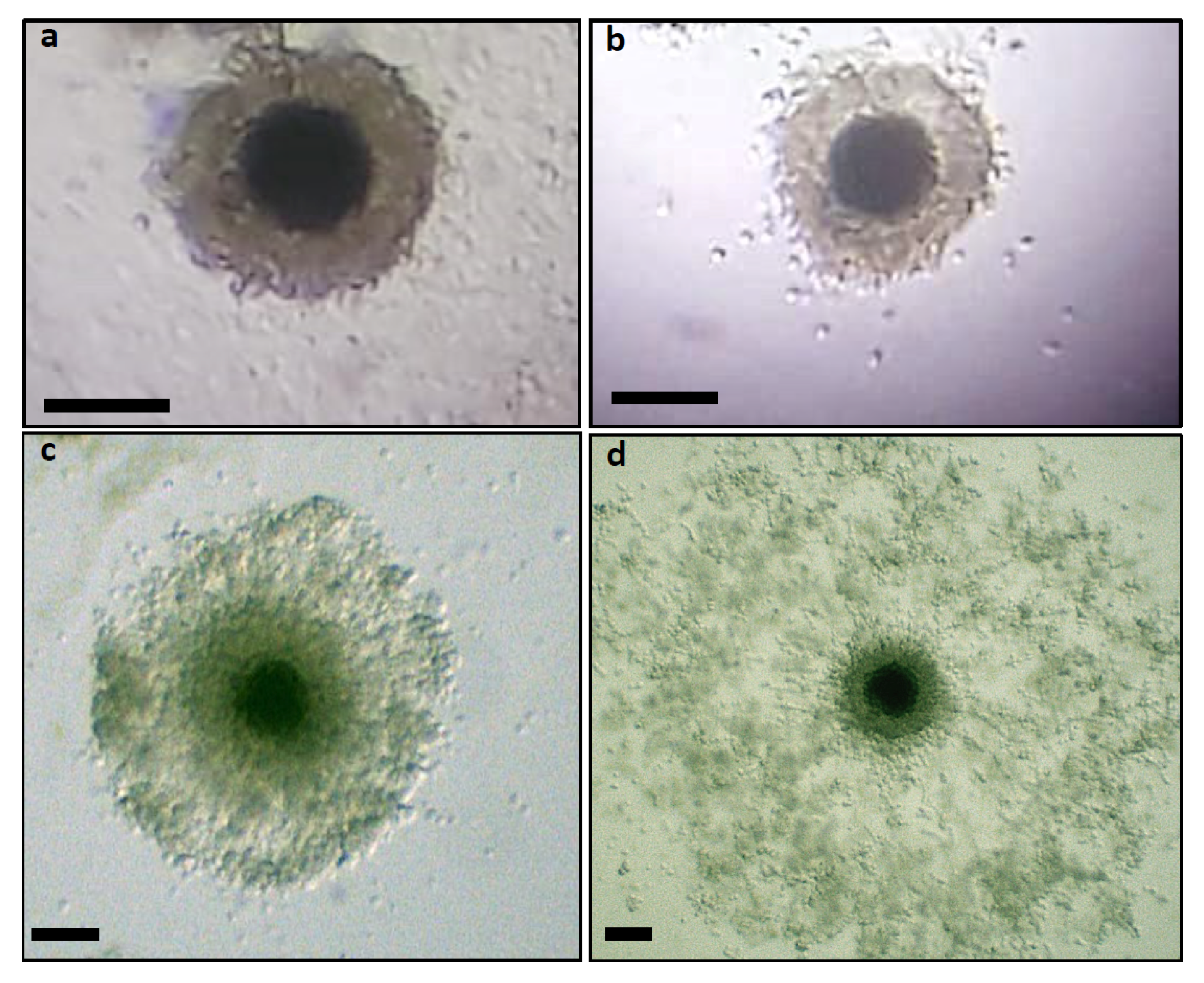
All noncultured COCs had compact CC layers. COCs incubated for IVM showed different patterns of cumulus mucification (Figure 2). Nonexpanded are shown in Figure 2a,b and expanded in Figure 2c,d.

3.3. Expression of Cumulus Expansion-Related Transcripts by Cumulus Cells

Real-time polymerase chain reaction was performed in CCs from COCs that were noncultured and from those submitted to IVM in order to evaluate the mRNA transcripts of GDF-9 and BMP-15 in dog CCs over the estrous cycle. This evaluation was made considering the maturation process and cumulus expansion.
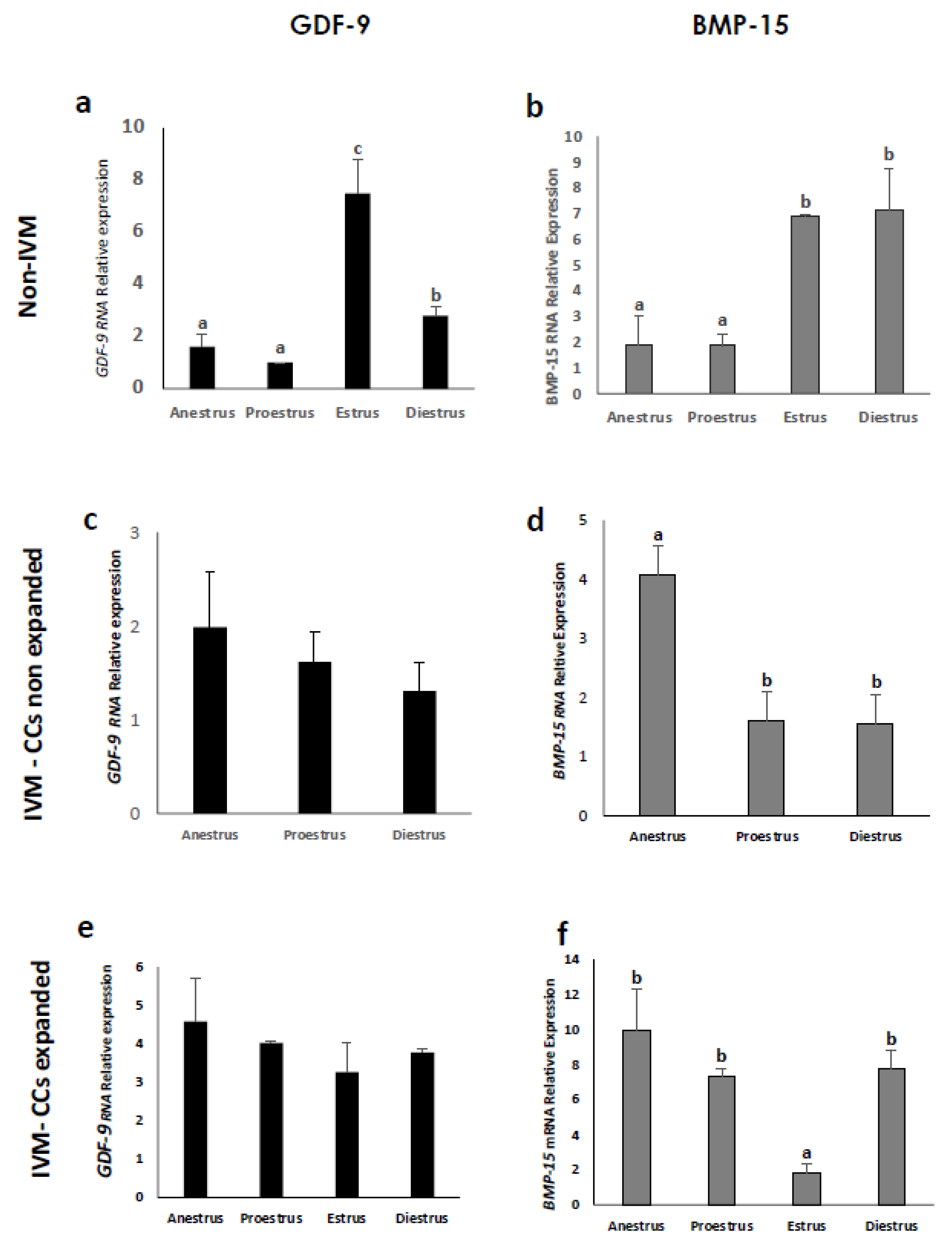
Both genes were expressed in CCs of COCs originating from all phases of the estrous cycle, whether they were submitted to IVM or not. However, the relative abundance of these genes was different (p < 0.05) according to CC expansion and the maturation process (Figure 3). Cumulus cells from noncultured COCs obtained in anestrus and proestrus showed the lowest GDF-9 mRNA levels (Figure 3a; p < 0.05) and the highest was observed in those at estrus (p < 0.05). On the contrary, no differences were found between the estrous phases in GDF-9 mRNA levels in nonexpanded (Figure 3c) or expanded (Figure 3e) CCs from IVM COCs. Regarding BMP-15 expression, CCs from nonmatured COCs obtained at anestrus and proestrus also showed lower (p < 0.05) mRNA levels than those at estrus and diestrus (Figure 3b), whereas CCs from IVM COCs that did not show signs of mucification expressed the highest (p < 0.05) BMP-15 mRNA levels at anestrus (Figure 3d). The BMP-15 gene expression in expanded CCs (Figure 3f) registered the lowest (p < 0.05) value in the estrus phase.
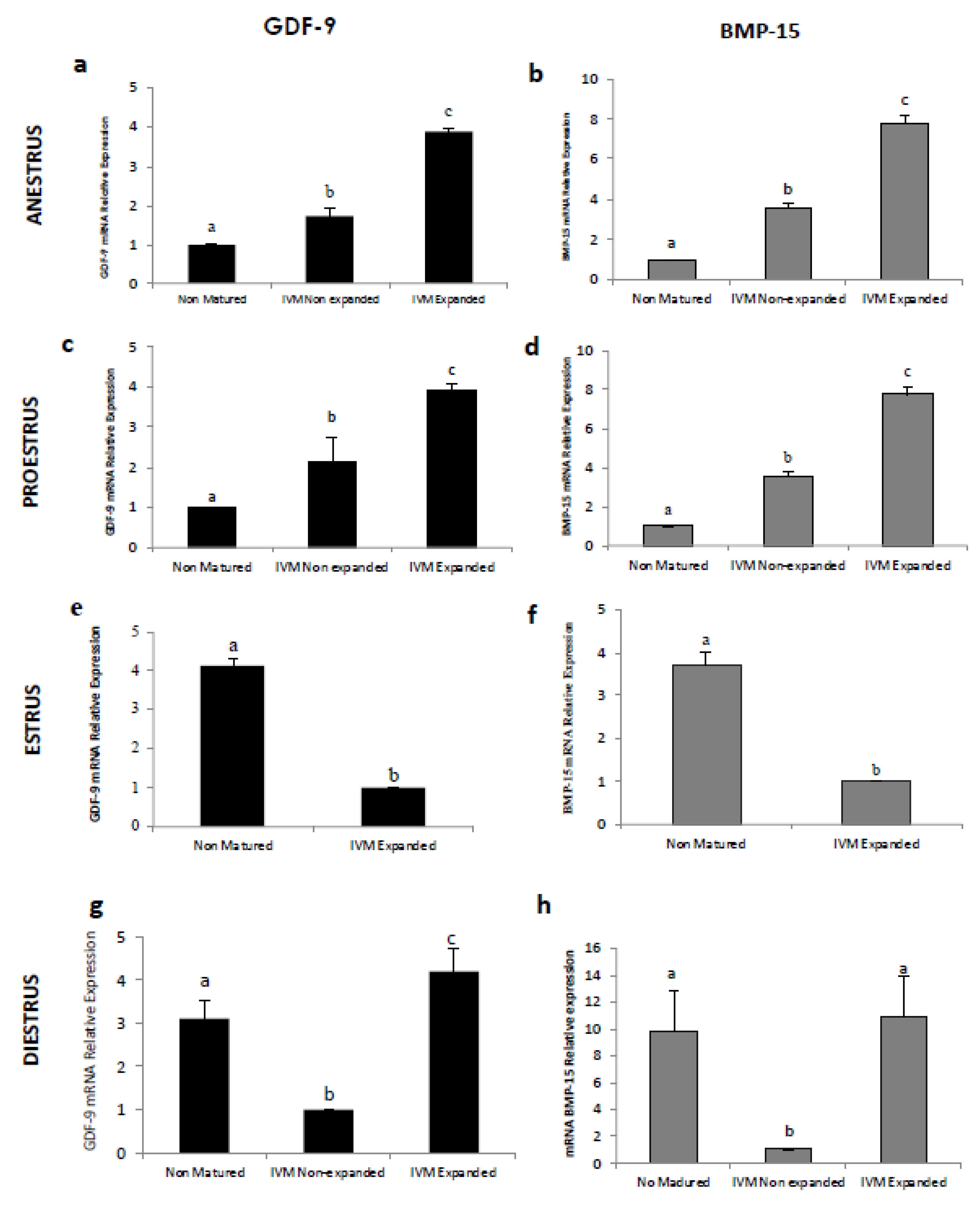
Comparing gene expression between CCs retrieved from non-cultured COCs and those from COCs after IVM, both expanded and nonexpanded (Figure 4), CCs surrounding IVM oocytes obtained in anestrus (Figure 4a,b) and proestrus (Figure 4c,d) expressed significantly higher mRNA levels of GDF-9 and BMP-15 than those from nonmatured oocytes. In contrast, gene expression of both genes in CCs collected from IVM COCs during the estrus phase (Figure 4c,f) was lower in IVM COCs (p < 0.05) than in those from nonmatured COCs. The majority of COCs after IVM obtained in the estrus phase exhibited almost only expanded CCs, so the number of CCs that did not mucify was too small to extract RNA from. At diestrus (Figure 4g,h), CCs from nonmatured COCs exhibited higher GDF-9 and BMP-15 mRNA levels (p < 0.05) than nonexpanded CCs after IVM. However, COCs with cumulus expansion after IVM expressed higher (p < 0.05) mRNA levels compared to nonmatured COCs.

3.4. Meiotic Growth and Cumulus Expansion

All oocytes from nonmature COCs were at the GV stage without signs of mucification. The oocyte meiotic maturity status after IVM according to CC expansion was recorded for each phase of the estrous cycle (Table 2). The percentage of oocytes that remained in GV class was similar to expanded and nonexpanded CCs after IVM in estrus and diestrus, but no oocytes at the GV stage were observed from COCs with expanded CCs in anestrus and proestrus. The resumption of meiosis (GVBD) at proestrus and diestrus was higher (p < 0.05) in oocytes with expanded CCs in comparison to those that did not expand. In the estrus phase, only oocytes with expanded CCs reached the MII stage.

4. Discussion

The expression of the cumulus expansion-related transcripts GDF-9 and BMP-15 was studied in canine CCs using nonmatured COCs and IVM COCs. The gene expression of these paracrine factors found in CCs in this work was previously reported in canine ovaries [25,27], as in other animal species [28,29]. The expression levels of these transcripts in CCs were stage dependent over the estrous cycle, which was in agreement with our previous studies performed in canine oocytes and granulosa/theca cells ex vivo [25].
The Figure 3 shows the major mRNA levels of both genes observed in noncultured COCs at estrus and diestrus compared to those at anestrus and proestrus, which may have been influenced by the rise in LH. Cumulus–oocyte complexes obtained at estrus and diestrus were retrieved from follicles that were already exposed to the LH surge in vivo, in contrast to those obtained in the other phases. In dogs, anestrus and proestrus precede the LH peak [30], with the LH surge being a critical event in the CCs development in many species [31], including canines [23]. It was reported that human chorionic gonadotropin (hCG, with LH-like activity) supplementation more effectively promoted CC development at 24 and 36 h of IVM of silver fox oocytes [32]. Similarly, the addition of hCG to culture medium had a highly significant effect on cumulus and oocyte maturation in dogs [33]. In mice, the transcriptome and function of CCs is highly affected by the LH surge [34]. Gonadotropin signal transduction by the somatic compartment requires de novo mRNA synthesis within CCs [35]. Indeed, the LH peak is believed to induce multiple intracellular signaling and second messengers in the follicular cells of several species [36], thereby influencing gene expression.
Interestingly, the dynamic expression of GDF-9 and BMP-15 genes in CCs changed according to the maturity process (IVM vs. non-cultured,) and mucification patterns in the different phases of the estrous cycle, as indicated in Figure 4. Cumulus cells obtained from COCs during anestrus and proestrus submitted to IVM increased the relative abundance of GDF-9 and BMP-15 mRNA during culture, especially in CCs that could expand. The culture medium was supplemented with hCG and estrogen, which enhanced CC development, as previously described in dogs [33,37]. This increase in gene expression in IVM COCs was coincident with other reports in humans [38] that describe GDF-9 and BMP-15 mRNA to be significantly correlated with the maturation process. However, it was found that CCs from canine COCs retrieved at estrus showed lower levels of GDF-9 and BMP-15 mRNA in expanded CCs post-IVM compared with nonmatured COCs. A decrease in the relative abundance of transcripts was suggested as the major change occurring during the transition to the large antral stage of development, which coincides with the acquisition of oocyte developmental competence [39,40]. This finding is supported by other studies that showed reduced GDF-9 gene expression in human CCs of matured oocytes [41]. Possibly, the reduced transcriptional activity observed in other species in COCs from preovulatory follicles is achieved only after in vitro maturation in canines. In the present study, all COCs obtained in the estrus phase came from large or preovulatory follicles, therefore, the COCs from these matured follicles may have been able to accumulate a large number of transcripts during their growth, resulting in diminished transcriptional activity during the final stages of development in vitro, which possibly occurs in vivo in canines post-ovulation. Accordingly, the protein expression level of GDF-9 in dog oocytes and CCs declined during in vitro maturation and was not elevated at the time of cumulus expansion [12].
Cumulus cells obtained from COCs during diestrus and submitted to IVM increased (GDF-9) or maintained (BMP-15) the transcript levels in expanded CCs only compared to those from noncultured COCs. This suggested that the COCs with low levels of GDF-9 and BMP-15 gene expression in their CCs during diestrus were not able to undergo expansion. The presence of a corpus luteum (CL) and/or progesterone in mice affects estradiol concentration, which is required for the acquisition and maintenance of CCs to undergo expansion [42]. In bovines, the presence of a CL in the ovary exerts a negative effect on the expansion of CCs, suggesting that the CL receives the highest rate of blood flow out of all the other ovarian tissues [43].
Clearly, there was a relationship between CC expansion and the relative gene expression of GDF-9 and BMP-15, which changed according to the reproductive status. Therefore, the ability to express GDF-9 and BMP-15 genes in the CCs at different stages seemed to be directly influenced by the previous follicular environment, which changes in every reproductive phase.
The effect of the estrous cycle on in vitro meiotic development of canine oocytes has been described in several reports, however, few studies have evaluated the possible effects of the estrous cycle on CC expansion and gene expression related to meiotic growth. The competence to undergo expansion is a unique characteristic of CC differentiation [44], which is critical for normal oocyte maturation in many mammals [45]. Transcriptome analysis in CCs in other species indicated that CC gene expression correlates with oocyte developmental potential [46]. However, in canines, it was suggested that oocytes with large expanded CCs after IVM do not necessarily achieve meiosis [47]. In the present study, we found a clear difference in the expression of the GDF-9 and BMP-15 genes in CCs according to the phases of the cycle, however, we did not find such differences in meiotic maturation rates (see Table 2). Meiotic development in oocytes from follicles at different estrous phases was low and within the range reported for in vitro-matured canine oocytes [12,14,27,47]. Although the percentages of MII oocytes were not different among the reproductive phases, the most remarkable result of the meiotic development analyses was the relationship of the cumulus expansion with the percentage of oocytes at the MII stage, but only during the estrus phase. At the same time, there was a significantly lower expression of both genes in expanded CCs from IVM COCs retrieved at estrus in comparison to non-cultured COCs, suggesting that other factors possibly play a role in the meiotic stimulus at the final stage of development. However, these proteins seem to be important in acquiring meiotic competence, because the addition of recombinant GDF-9 and BMP-15 in oocyte maturation media was shown to enhance the competence of canine oocytes matured in vitro [14]. This effect is possible achieved in the early development stages. The process of competence, which includes coordinated sequences of molecular events required for the oocyte to fulfill maturation, is poorly understood in canines. Thus, the dynamics of these signals in regulating meiotic maturation in growing oocytes implies that the oocytes pass through different endocrine and paracrine environments that influence the subsequent expression of different genes. The reproductive cycle in canines is quite long, therefore, the growing oocytes need to pass slowly under these different environments inside the follicle in order to acquire the necessary competence to resume meiosis. Consequently, the fate of individual transcripts stored in the CCs can differ during development.

5. Conclusions

Although we did not evaluate the gene expression in CCs from COCs at each meiotic stage separately, the observed differential expression patterns of GDF-9 and BMP-15 mRNA transcripts in CCs during the different estrous phases suggest a possible relationship with cumulus expansion and maturity condition, which could indicate specific regulation and temporal changes in their expression, indicative of different stimulation at precise stages of development.

Author Contributions

G.R. carried out the PCR, in vitro cultures and gene analysis. J.P. designed the primers and performed the transcriptional analysis. K.A. collaborated to the cell culture protocols. M.D.l.R. designed the experiments, analyzed the data, and drafted the manuscript. All authors have read and agree to the published version of the manuscript.

Funding

This research was supported by the grant FONDECYT 1171670 from The National Commission for Scientific and Technological Research (CONICYT) and grant; YV029/01-19, VID, University of Chile to M.D.l.R.

Acknowledgments

We thank the veterinarians from Pablo de Rokha Veterinary Center for providing the canine ovaries.

Conflicts of Interest

The authors declare that they have no conflict of interests. The funders had no role in the design of the study; in the collection, analyses, or interpretation of data; in the writing of the manuscript, or in the decision to publish the results.

References

  1. Huang, Z.; Wells, D. The human oocyte and cumulus cells relationship: New insights from the cumulus cell transcriptome. Mol. Hum. Reprod. 2010, 16, 715–725. [Google Scholar] [CrossRef] [Green Version]
  2. Kimura, N.; Totsukawa, K.; Sato, E. Significance of mammalian cumulus-oocyte complex Matrix in Oocyte meiotic maturation: Review of the synthetic control and possible roles of hyaluronan (HA) and HA-binding protein. J. Mamm. Ova Res. 2006, 23, 36–51. [Google Scholar] [CrossRef]
  3. Kidde, G.M.; Vanderhyden, B.C. Bidirectional communication between oocytes and follicle cells: Ensuring oocyte developmental competence. Can. J. Physiol. Pharmacol. 2010, 88, 399–413. [Google Scholar] [CrossRef] [Green Version]
  4. Li, R.; Norman, R.J.; Armstrong, D.T.; Gilchrist, R.B. Oocyte-secreted factors determine functional differences between bovine mural granulosa cells and cumulus cells. Biol. Reprod. 2000, 63, 839–845. [Google Scholar] [CrossRef] [Green Version]
  5. Su, Y.Q.; Sugiura, K.; Wigglesworth, K.; O’Brien, M.J.; Affourtit, J.P.; Pangas, S.A.; Eppig, J.J. Oocyte regulation of metabolic co- operativity between mouse cumulus cells and oocytes: BMP15 and GDF9 control cholesterol biosynthesis in cumulus cells. Development 2008, 135, 111–121. [Google Scholar] [CrossRef] [Green Version]
  6. Gilchrist, R.B.; Ritter, L.J.; Armstrong, D.T. Oocyte-somatic cell interactions during follicle development in mammals. Anim. Reprod. Sci. 2004, 82–83, 431–446. [Google Scholar] [CrossRef]
  7. Eppig, J.J. Oocyte control of ovarian follicular development and function in mammals. Reproduction 2001, 122, 829–838. [Google Scholar] [CrossRef]
  8. Belli, M.; Shimasaki, S. Molecular aspects and clinical Relevance of GDF9 and BMP15 in ovarian function. Vitam Horm. 2018, 107, 317–348. [Google Scholar]
  9. Gilchrist, R.B.; Thompson, J.G. Oocyte maturation: Emerging concepts and technologies to improve developmental potential in vitro. Theriogenology 2007, 67, 2–5. [Google Scholar] [CrossRef]
  10. Haas, C.S.; Rovani, M.T.; Oliveira, F.C.; Vieira, A.D.; Bordignon, V.; Gonçalves, P.B.; Ferreira, R.; Gasperin, B.G. Expression of growth and differentiation factor 9 and cognate receptor s during final follicular growth in cattle. Anim. Reprod. 2015, 13, 756–761. [Google Scholar] [CrossRef]
  11. Maupeu, D.; Palomino, J.; De los Reyes, M. Immunohistochemistry localization of growth differentiation factor 9 (GDF-9) and bone morphogenetic protein 15 (BMP-15) in canine follicles throughout estrus cycle. Reprod. Fertil. Dev. 2014, 27, 182. [Google Scholar] [CrossRef]
  12. De los Reyes, M.; Parraguez, V.H.; Rojas, C.; Palomino, J. Expression of growth differentiation factor 9 (GDF-9) during in vitro maturation in canine oocytes. Theriogenology 2013, 80, 587–596. [Google Scholar] [CrossRef]
  13. Fernandez, T.; Palomino, J.; Parraguez, V.H.; Peralta, O.A.; De los Reyes, M. Differential expression of GDF-9 and BMP- 15 during follicular development in canine ovaries evaluated by flow cytometry. Anim. Reprod. Sci. 2016, 167, 59–67. [Google Scholar] [CrossRef]
  14. Garcia, P.; Aspee, K.; Ramirez, G.; Dettleff, P.; Palomino, J.; Peralta, O.A.; Parraguez, V.H.; De los Reyes, M. Influence of growth differentiation factor 9 and bone morphogenetic protein 15 on in vitro maturation of canine oocytes. Reprod. Dom. Anim. 2019, 54, 373–380. [Google Scholar] [CrossRef]
  15. Uyar, A.; Torrealday, S.; Seli, E. Cumulus and granulosa cell markers of oocyte and embryo quality. Fertil. Steril. 2013, 15, 29–33. [Google Scholar] [CrossRef] [Green Version]
  16. Russel, D.L.; Gilchrist, R.B.; Brown, H.M.; Thomsom, J.G. Bidirectional communication between cumulus cells and the oocyte: Old hands and new players? Theriogenology 2016, 86, 62–68. [Google Scholar] [CrossRef] [Green Version]
  17. Zeng, H.T.; Richani, D.; Sutton-McDowall, M.L.; Ren, Z.; Smitz, J.E.; Stokes, Y.; Gilchrist, R.B.; Thompson, J.G. Prematuration with cyclic adenosine monophosphate modulators alters cumulus cell and oocyte metabolism and enhances developmental competence of in vitro- matured mouse oocytes. Biol. Reprod. 2014, 91, 1–11. [Google Scholar] [CrossRef]
  18. Elvin, J.A.; Clarck, A.T.; Wang, P.; Wolfman, N.M.; Matzuk, M.M. Paracrine Actions of Growth Differentiation Factor-9 in the mammalian ovary. Mol. Endocrinol. 1999, 13, 1035–1048. [Google Scholar] [CrossRef]
  19. Gilchrist, R.B.; Lane, M.; Thompson, J.G. Oocyte-secreted factors: Regulators of cumulus cell function and oocyte quality. Hum. Reprod. Up 2008, 14, 159–177. [Google Scholar] [CrossRef] [Green Version]
  20. Sanfins, A.; Rodrigues, P.; Albertini, D.F. GDF-9 and BMP-15 direct the follicle symphony. J. Assist. Reprod. Gen. 2018, 35, 1741–1750. [Google Scholar] [CrossRef]
  21. Hussein, T.S.; Thompson, J.G.; Gilchrist, R.B. Oocyte-secreted factors enhance oocyte developmental competence. Dev. Biol. 2006, 296, 514–521. [Google Scholar] [CrossRef]
  22. Reynaud, K.; Fontbonne, A.; Marseloo, N.; Thoumire, S.; Chebrout, M.; de Lesegno, C.V.; Chastant-Maillard, S. In vivo meiotic resumption, fertilization and early embryonic development in the bitch. Reproduction 2005, 130, 193–201. [Google Scholar] [CrossRef] [Green Version]
  23. Viaris de Lesegno, C.; Reynaud, K.; Pechoux, C.; Thoumire, S.; Chastant-Maillard, S. Ultrastructure of canine oocytes during in vivo maturation. Mol. Reprod. Dev. 2008, 75, 115–125. [Google Scholar] [CrossRef]
  24. Brugger, N.; Otzdorff, C.; Walter, B.; Hoffmann, B.; Braun, J. Quantitative Determination of Progesterone (P4) in Canine Blood Serum Using an Enzyme-linked Fluorescence Assay. Repro Dom Anim. 2011, 46, 870–873. [Google Scholar] [CrossRef]
  25. Palomino, J.; De los Reyes, M. Temporal expression of GDF-9 and BMP-15 in canine ovarian follicles. Theriogenology 2016, 86, 1541–1549. [Google Scholar] [CrossRef]
  26. De los Reyes, M.; Palomino, J.; Parraguez, V.H.; Hidalgo, M.; Saffie, P. Mitochondrial distribution and meiotic progression in canine oocytes during in vivo and in vitro maturation. Theriogenology 2011, 75, 346–353. [Google Scholar] [CrossRef]
  27. Cho, S.J.; Lee, K.L.; Kim, Y.G.; Kim, D.H.; Yoo, J.G.; Yang, B.C.; Kong, I.K. Differential gene-expression profiles from canine cumu- lus cells of ovulated versus in vitro-matured oocytes. Reprod. Fertil. Dev. 2016, 28, 278–285. [Google Scholar] [CrossRef]
  28. Prochazka, R.; Nemcova, L.; Nagyova, E.; Kanka, J. Expression of Growth Differentiation Factor 9 Messenger RNA in Porcine Growing and Preovulatory Ovarian Follicles. Biol. Reprod. 2004, 71, 1290–1295. [Google Scholar] [CrossRef] [Green Version]
  29. Hosoe, M.; Kaneyam, K.; Ushizawa, K.; Hayashi, K.; Takahashi, T. Quantitative analysis of bone morphogenetic protein 15 (BMP15) and growth differentiation factor 9 (GDF9) gene expression in calf and adult bovine ovaries. Reprod. Biol. Endocrin. 2011, 9, 33. [Google Scholar] [CrossRef] [Green Version]
  30. Concannon, P.W. Reproductive cycles of the domestic bitch. Anim. Reprod. Sci. 2011, 124, 200–210. [Google Scholar] [CrossRef]
  31. Peng, X.R.; Hsueh, A.J.; LaPolt, P.S.; Bjersing, L.; Ny, T. Localization of luteinizing hormone receptor messenger ribonucleic acid expression in ovarian cell types during follicle development and ovulation. Endocrinology 1991, 129, 3200–3207. [Google Scholar] [CrossRef]
  32. Wen, X.H.; Feng, H.L.; Sun, Q.Y. In vitro maturation of follicular oocytes of the silver fox (Vulpes fulva). Theriogenology 1994, 41, 333. [Google Scholar] [CrossRef]
  33. De los Reyes, M.; de Lange, J.; Miranda, P.; Palomino, J.; Barros, C. Effect of human chorionic gonadotrophin supplementation during different culture periods on in vitro maturation of canine oocytes. Theriogenology 2005, 64, 1–11. [Google Scholar] [CrossRef]
  34. Juengel, J.L.; Hudson, N.L.; Heath, D.A.; Smith, P.; Reader, K.L.; Lawrence, S.B.; Groome, N.P. Growth differentiation factor 9 and bone morphogenetic protein 15 are essential for ovarian follicular development in sheep. Biol. Reprod. 2002, 67, 1777–1789. [Google Scholar] [CrossRef] [Green Version]
  35. Assidi, M.; Dieleman, S.J.; Sirard, M.A. Cumulus cell gene expression following the LH surge in bovine preovulatory follicles: Potential early markers of oocyte competence. Reproduction 2010, 140, 835–852. [Google Scholar] [CrossRef] [Green Version]
  36. Mattioli, M.; Barboni, B. Signal transduction mechanism for LH in the cumulus–oocyte complex. Mol. Cell Endocrinol. 2000, 161, 19–23. [Google Scholar] [CrossRef]
  37. Kim, M.K.; Fibrianto, Y.H.; Oh, H.J.; Jang, G.; Kim, H.J.; Lee, K.S.; Kang, S.K.; Lee, B.C.; Hwang, W.S. Effects of estradiol-17β and progesterone supplementation on in vitro nuclear maturation of canine oocytes. Theriogenology 2005, 63, 1342–1353. [Google Scholar] [CrossRef]
  38. Li, Y.; Li, R.Q.; Ou, S.B.; Zhang, N.F.; Ren, L.; Wei, L.; Zhang, Q.X.; Yang, D.Z. Increased GDF9 and BMP15 mRNA levels in cumulus granulosa cells correlate with oocyte maturation, fertilization, and embryo quality in humans. Reprod. Biol. Endocrinol. 2014, 12, 1–9. [Google Scholar] [CrossRef] [Green Version]
  39. Pant, D.; Reynolds, L.P.; Luther, J.S.; Borowicz, P.P.; Stenbak, T.M.; Bilski, J.J.; Weigl, R.M.; Lopes, F.; Petry, K.; Johnson, M.L.; et al. Expression of connexin 43 and gap junctional intercellular communication in the cumulus–oocyte complex in sheep. Reproduction 2005, 129, 191–200. [Google Scholar] [CrossRef] [Green Version]
  40. Sanchez, F.; Adriaenssens, T.; Romero, S.; Smitz, J. Quantification of oocyte-specific transcripts in follicle-enclosed oocytes during antral development and maturation in vitro. Mol. Hum. Reprod. 2009, 15, 539–550. [Google Scholar] [CrossRef] [Green Version]
  41. Zhao, S.Y.; Qiao, J.; Chen, Y.J.; Liu, P.; Li, J.; Yan, J. Expression of growth differentiation factor-9 and bone morphogenetic protein-15 in oocytes and cumulus granulosa cells of patients with polycystic ovary syndrome. Fertil. Steril. 2010, 94, 261–267. [Google Scholar] [CrossRef]
  42. Sugiura, K.; Su, Y.Q.; Li, Q.; Wigglesworth, K.; Matzuk, M.M.; Eppig, J.J. Estrogen promotes the development of mouse cumulus cells in coordination with oocyte-derived GDF9 and BMP15. Mol. Endocrinol. 2010, 24, 2303–2314. [Google Scholar] [CrossRef] [PubMed]
  43. Quezada-Casasola, A.; Martínez-Armendáriz, K.E.; Itzá-Ortiz, M.F.; Escárcega-Ávila, A.M.; Pérez-Eguía, E.; Filipiak, Y.; Larocca, C.; Carrera-Chávez, J.M. Effect of presence of corpora lutea on cumulus expansion of in vitro matured bovine oocytes selected by trypan blue and brilliant cresyl blue tests. J. Appl. Anim. Res. 2018, 46, 967–972. [Google Scholar] [CrossRef] [Green Version]
  44. Diaz, F.J.; Wigglesworth, K.; Eppig, J.J. Oocytes are required for the preantral granulosa cell to cumulus cell transition in mice. Dev. Biol. 2007, 305, 300–311. [Google Scholar] [CrossRef] [PubMed] [Green Version]
  45. Nevoral, J.; Orsák, M.; Petr, J.; Dvořakova, M.; Weingartová, I.; Vyskočilova, A.; Aámostna, K.; Krejčová, T.; Jílek, F. Cumulus cell expansion, its role in oocyte biology and perspectives of measurement: A review. Sci. Agric. Bohem. 2014, 45, 12–225. [Google Scholar] [CrossRef] [Green Version]
  46. Adriaenssens, T.; Wathlet, S.; Segers, I.; Verheyen, G.; De Vos, A.; Van der Elst, J.; Coucke, W.; Devroey, P.; Smitz, J. Cumulus cell gene expression is associated with oocyte developmental quality and influenced by patient and treatment characteristics. Hum. Reprod. 2010, 25, 1259–1270. [Google Scholar] [CrossRef] [Green Version]
  47. Rodrigues, B.A.; Rodrigues, J.L. Cumulus cell features and nuclear chromatin configuration of in vitro matured canine COCs and the influence of in vivo serum progesterone concentrations of ovary donors. Zygote 2009, 17, 79–91. [Google Scholar] [CrossRef]
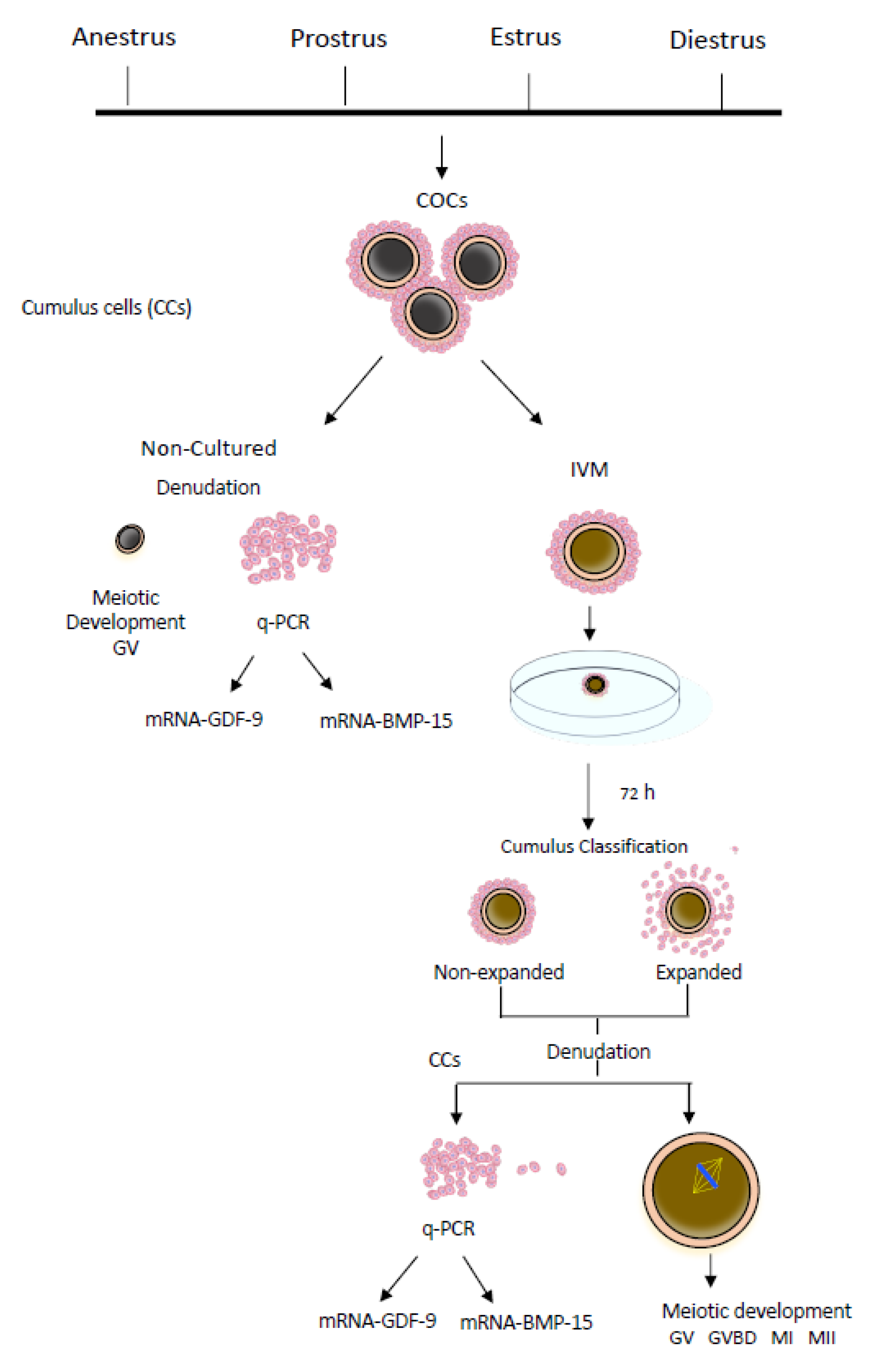
Figure 1. Experimental design. CCs: cumulus cells; COCs: cumulus–oocyte complexes; IVM: in vitro maturation; q-PCR: quantitative real-time polymerase chain reaction; GV: germinal vesicle; GVBD: resumption of meiosis or germinal vesicle breakdown; MI: first metaphase; MII: second metaphase.
Animals 10 00462 g001
Figure 2. Canine cumulus–oocyte complexes (COCs) after 72 h of in vitro maturation. (a,b) COCs without cumulus cell expansion; (c,d) COCs with different grades of cumulus cell expansion (bar: 100 μm).
Animals 10 00462 g002
Figure 3. Relative gene expression of Growth Differentiation Factor 9 (GDF-9) and Bone Morphogenetic Protein 15 (BMP-15) from cumulus cells of nonmatured COCs (a,b) (~350 COCs at each phase of the estrous cycle), in vitro-matured non-expanded COCs (c,d) (~300 COCs at anestrus, proestrus and diestrus), and in vitro-matured expanded cumulus cells (e,f) (~300 at each phase). The mRNA levels were expressed in relation to β-Actin mRNA as the control or housekeeping gene. Different letters above the bars indicate differences at p < 0.05.
Animals 10 00462 g003
Figure 4. Relative gene expression of Growth Differentiation Factor 9 (GDF-9) and Bone Morphogenetic Protein 15 (BMP-15) in canine cumulus cells according to the maturation and expansion of the cumulus cells at anestrus (a,b), proestrus (c,d), estrus (e,f), and diestrus (g,h). The GDF-9 and BMP-15 mRNA levels were expressed in relation to β-Actin mRNA as the control or housekeeping gene. For each gene (GDF-9 or BMP-15), bars with different superscripts show a significant difference at p < 0.05.
Animals 10 00462 g004
Table 1. Sequences of GDF-9 and BMP-15-specific primers and reference gene used in this study for qRT-PCR analysis.
GeneOligos (5’–3’)Amplicon (bp)Accession NumberReferences
ACTBF: ATTGTCATGGACTCTGGGGATG191AF021873.2[25]
R: TCCTTGATGTCACGCACGAT
GDF-9F: TACCCCCATCCCTGCTTTTA155NM001168013.1[27]
R: TCCACCTTCAGTCGATTCCT
BMP-15F: CCCTGCCCCTGATTCGGGAG82XM003640274.3[25]
R: CCGCAAAGGATGCCCAAGGAC
qPCR: quantitative real-time polymerase chain reaction.
Table 2. In vitro canine meiotic development according to the expansion of cumulus cells and the estrous cycle.
Oocytes Meiotic Stage (%)
CycleExpGVGVBDMIMIIN
Anestrus+0 a37 a,b53 a,b10 a,b51
7 b37 a,b48 a,b7 a54
Proestrus+0 a60 c36 a4 a53
9 b21 a57 a,b13 a,b56
Estrus+6 b21 a56 a,b18 b34
5 b26 a68 b0 a19
Diestrus+4 b50 b,c39 a7 a46
5 b22 a56 a,b17 b59
GV: germinal vesicle; GVBD: germinal vesicle breakdown; MI: first metaphase; MII: second metaphase; N: number of evaluable oocytes. Expansion: (+) cumulus cells in expansion; (−) nonexpanded cumulus cells. COCs were cultured for 72 h; a,b,c: Within a column, values with different superscript letters differ significantly (p < 0.05).