Production Significance of Bovine Respiratory Disease Lesions in Slaughtered Beef Cattle

 ,
,  ,
,
Abstract
:Simple Summary
Abstract
1. Introduction
2. Material and Methods
2.1. Animals
2.2. Sampling
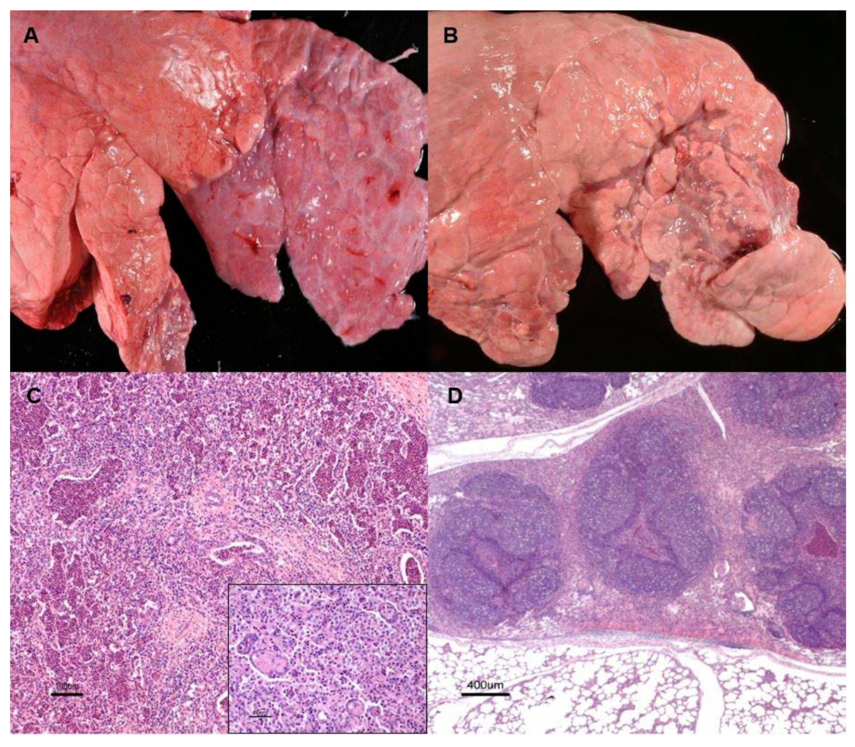
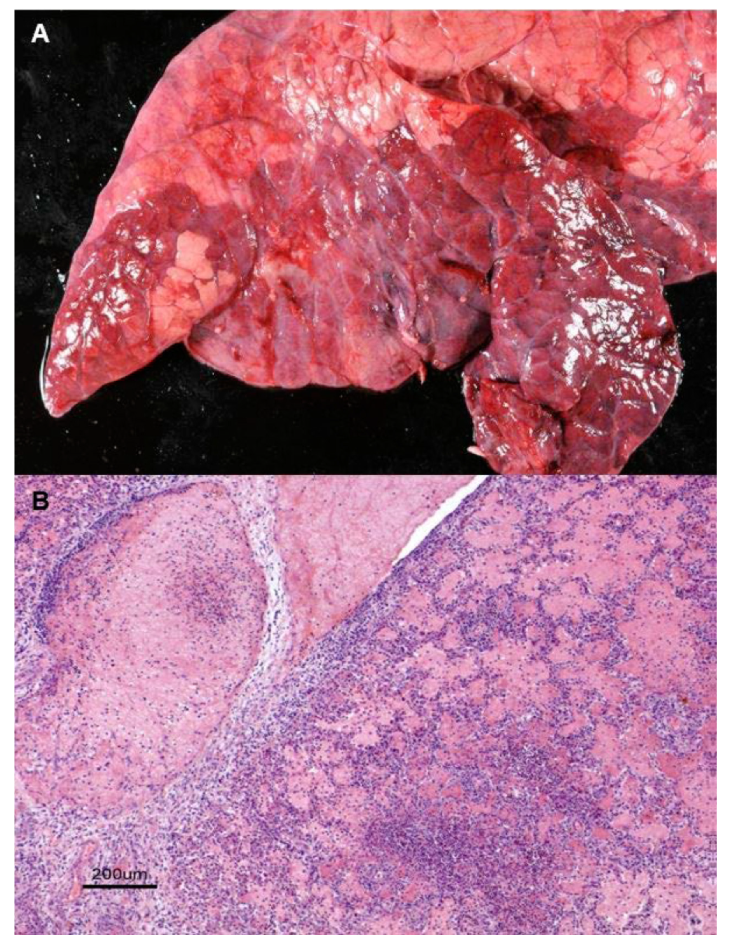
2.3. Macroscopic and Microscopic Inspection of the Lungs
2.4. Etiological Identification by PCR
2.5. Statistical Analysis
3. Results
3.1. Prevalence of Pneumonia
3.2. Type of Pneumonia
3.3. Affected Area
3.4. Carcass Weight
3.5. Age at Slaughter
3.6. Prevalence of Etiological Agents
4. Discussion
5. Conclusions
Supplementary Materials
Author Contributions
Funding
Acknowledgments
Conflicts of Interest
References
- Griffin, D. Economic impact associated with respiratory disease in beef cattle. Vet. Clin. N. Am. Food Anim. Pract. 1997, 13, 367–377. [Google Scholar] [CrossRef]
- Griffin, D. The monster we don’t see: Subclinical BRD in beef cattle. Anim. Health Res. Rev. 2014, 15, 138–141. [Google Scholar] [CrossRef] [PubMed]
- Pardon, B.; Hostens, M.; Duchateau, L.; Dewulf, J.; De Bleecker, K.; Deprez, P. Impact of respiratory disease, diarrhea, otitis and arthritis on mortality and carcass traits in white veal calves. BMC Vet. Res. 2013, 9, 79. [Google Scholar] [CrossRef] [PubMed] [Green Version]
- Fulton, R.W.; Blood, K.S.; Panciera, R.J.; Payton, M.E.; Ridpath, J.F.; Confer, A.W.; Saliki, J.T.; Burge, L.T.; Welsh, R.D.; Johnson, B.J.; et al. Lung pathology and infectious agents in fatal feedlot pneumonias and relationship with mortality, disease onset, and treatments. J. Vet. Diagn Investig. 2009, 21, 464–477. [Google Scholar] [CrossRef] [Green Version]
- Miles, D.G. Overview of the North American beef cattle industry and the incidence of bovine respiratory disease (BRD). Anim. Health Res. Rev. 2009, 10, 101–103. [Google Scholar] [CrossRef]
- Gagea, M.I.; Bateman, K.G.; van Dreumel, T.; McEwen, B.J.; Carman, S.; Archambault, M.; Shanahan, R.A.; Caswell, J.L. Diseases and pathogens associated with mortality in Ontario beef feedlots. J. Vet. Diagn. Investig. 2006, 18, 18–28. [Google Scholar] [CrossRef] [Green Version]
- Francoz, D.; Buczinski, S.; Bélanger, A.; Forte, G.; Labrecque, O.; Tremblay, D.; Wellemans, V.; Dubuc, J. Respiratory pathogens in québec dairy calves and their relationship with clinical status, lung consolidation, and average daily gain. J. Vet. Intern. Med. 2015, 29, 381–387. [Google Scholar] [CrossRef]
- Fulton, R.W.; Cook, B.J.; Step, D.L.; Confer, A.W.; Saliki, J.T.; Payton, M.E.; Burge, L.J.; Welsh, R.D.; Blood, K.S. Evaluation of health status of calves and the impact on feedlot performance: Assessment of a retained ownership program for postweaning calves. Can. J. Vet. Res. Rev. Can. Rech. Vet. 2002, 66, 173–180. [Google Scholar]
- Delabouglise, A.; James, A.; Valarcher, J.-F.; Hägglund, S.; Raboisson, D.; Rushton, J. Linking disease epidemiology and livestock productivity: The case of bovine respiratory disease in France. PLoS ONE 2017, 12, e0189090. [Google Scholar] [CrossRef] [Green Version]
- Garcia, M.D.; Thallman, R.M.; Wheeler, T.L.; Shackelford, S.D.; Casas, E. Effect of bovine respiratory disease and overall pathogenic disease incidence on carcass traits1,2. J. Anim. Sci. 2010, 88, 491–496. [Google Scholar] [CrossRef]
- Noyes, N.R.; Benedict, K.; Gow, S.; Booker, C.; Hannon, S.; McAllister, T.A.; Morley, P.S. Mannheimia haemolyticain Feedlot Cattle: Prevalence of recovery and associations with antimicrobial use, resistance, and health outcomes. J. Vet. Intern. Med. 2015, 29, 705–713. [Google Scholar] [CrossRef] [PubMed]
- Wolfger, B.; Timsit, E.; White, B.J.; Orsel, K. A systematic review of bovine respiratory disease diagnosis focused on diagnostic confirmation, early detection, and prediction of unfavorable outcomes in feedlot cattle. Vet. Clin. N. Am. Food Anim. Pr. 2015, 31, 351–365. [Google Scholar] [CrossRef] [PubMed]
- Mosier, D. Review of BRD pathogenesis: The old and the new. Anim. Heal Res. Rev. 2014, 15, 166–168. [Google Scholar] [CrossRef] [PubMed]
- Taylor, J.D.; Fulton, R.W.; Lehenbauer, T.W.; Step, D.L.; Confer, A.W. The epidemiology of bovine respiratory disease: What is the evidence for preventive measures? Can. Vet. J. Rev. Vet. Can. 2010, 51, 1351–1359. [Google Scholar]
- Caswell, J.L. Failure of respiratory defenses in the pathogenesis of bacterial pneumonia of cattle. Vet. Pathol. 2013, 51, 393–409. [Google Scholar] [CrossRef] [Green Version]
- Guterbock, W.M. The impact of BRD: The current dairy experience. Anim. Heal Res. Rev. 2014, 15, 130–134. [Google Scholar] [CrossRef]
- Haines, D.M.; Martin, K.M.; Clark, E.G.; Jim, G.K.; Janzen, E.D. The immunohistochemical detection of Mycoplasma bovis and bovine viral diarrhea virus in tissues of feedlot cattle with chronic, unresponsive respiratory disease and/or arthritis. Can. Vet. J. Rev. Vet. Can. 2001, 42, 857–860. [Google Scholar]
- Griffin, D.; Chengappa, M.; Kuszak, J.; McVey, D.S. Bacterial pathogens of the bovine respiratory disease complex. Vet. Clin. N. Am. Food Anim. Pr. 2010, 26, 381–394. [Google Scholar] [CrossRef]
- Radaelli, E.; Luini, M.; Loria, G.R.; Nicholas, R.; Scanziani, E. Bacteriological, serological, pathological and immunohistochemical studies of Mycoplasma bovis respiratory infection in veal calves and adult cattle at slaughter. Res. Vet. Sci. 2008, 85, 282–290. [Google Scholar] [CrossRef]
- Rezac, D.J.; Thomson, D.U.; Bartle, S.J.; Osterstock, J.B.; Prouty, F.L.; Reinhardt, C.D. Prevalence, severity, and relationships of lung lesions, liver abnormalities, and rumen health scores measured at slaughter in beef cattle1. J. Anim. Sci. 2014, 92, 2595–2602. [Google Scholar] [CrossRef] [Green Version]
- Brscic, M.; Leruste, H.; Heutinck, L.; Bokkers, E.; Wolthuis-Fillerup, M.; Stockhofe, N.; Gottardo, F.; Lensink, B.; Cozzi, G.; Van Reenen, C. Prevalence of respiratory disorders in veal calves and potential risk factors. J. Dairy Sci. 2012, 95, 2753–2764. [Google Scholar] [CrossRef] [PubMed]
- Ministerio de Agricultura, Pesca y Alimentación (MAPA). 2018. Available online: https://www.mapa.gob.es/es/estadistica/temas/estadísticas-agrarias/resultados_provisionales_nov2018_bovino_webmapa_tcm30–502922.pdf (accessed on 20 December 2019).
- Fábregas, X.; Simón, J.A.; Canada, L. Departament de Salut. Generalitat de Catalunya. 2015. (Official Data). Available online: https://ddd.uab.cat/record/130598 (accessed on 20 December 2019).
- Maxie, G.; Jubb, K. Palmer’s Pathology of Domestic Animals, 6th ed.; Elsevier: St Louis, MO, USA, 2015. [Google Scholar]
- Zachary, J.F. Pathologic Basis of Veterinary Disease, 6th ed.; Elsevier: St Louis, MO, USA, 2016. [Google Scholar]
- Lundborg, G.K.; Svensson, E.C.; Oltenacu, P.A. Herd-level risk factors for infectious diseases in Swedish dairy calves aged 0–90 days. Prev. Vet. Med. 2005, 68, 123–143. [Google Scholar] [CrossRef] [PubMed]
- Dargatz, D.A.; Lombard, J.E. Summary of BRD data from the 2011 NAHMS feedlot and dairy heifer studies. Anim. Health Res. Rev. 2014, 15, 123–125. [Google Scholar] [CrossRef] [PubMed]
- Murray, G.M.; More, S.J.; Sammin, D.; Casey, M.J.; McElroy, M.C.; O’Neill, R.G.; Byrne, W.J.; Earley, B.; Clegg, T.A.; Ball, H.; et al. Pathogens, patterns of pneumonia, and epidemiologic risk factors associated with respiratory disease in recently weaned cattle in Ireland. J. Vet. Diagn. Investig. 2017, 29, 20–34. [Google Scholar] [CrossRef] [PubMed]
- Shane, D.D.; McLellan, J.G.; White, B.J.; Larson, R.L.; Amrine, D.; Sanderson, M.W.; Apley, M.D. Evaluation of animal-to-animal and community contact structures determined by a real-time location system for correlation with and prediction of new bovine respiratory disease diagnoses in beef cattle during the first 28 days after feedlot entry. Am. J. Vet. Res. 2018, 79, 1277–1286. [Google Scholar] [CrossRef] [PubMed]
- Dubrovsky, S.; Van Eenennaam, A.L.; Karle, B.; Rossitto, P.; Lehenbauer, T.; Aly, S.S. Bovine respiratory disease (BRD) cause-specific and overall mortality in preweaned calves on California dairies: The BRD 10K study. J. Dairy Sci. 2019, 102, 7320–7328. [Google Scholar] [CrossRef]
- Murray, G.M.; More, S.J.; Clegg, T.A.; Earley, B.; O’Neill, R.G.; Johnston, D.; Gilmore, J.; Nosov, M.; McElroy, M.C.; Inzana, T.J.; et al. Risk factors associated with exposure to bovine respiratory disease pathogens during the peri-weaning period in dairy bull calves. BMC Vet. Res. 2018, 14, 1–11. [Google Scholar] [CrossRef]
- Loneragan, G.H.; Dargatz, D.A.; Morley, P.S.; Smith, M.A. Trends in mortality ratios among cattle in US feedlots. J. Am. Vet. Med. Assoc. 2001, 219, 1122–1127. [Google Scholar] [CrossRef]
- Weeks, C.A.; McNally, P.W.; Warriss, P.D. Influence of the design of facilities at auction markets and animal handling procedures on bruising in cattle. Vet. Rec. 2002, 150, 743–748. [Google Scholar] [CrossRef]
- Schneider, M.; Tait, J.R.; Busby, W.D.; Reecy, J.M. An evaluation of bovine respiratory disease complex in feedlot cattle: Impact on performance and carcass traits using treatment records and lung lesion scores1,2. J. Anim. Sci. 2009, 87, 1821–1827. [Google Scholar] [CrossRef]
- Mosier, D. Progression of BRD lung lesions. In Proceedings of the Academy of Veterinary Consultants, Springs, CO, USA, 4–6 August 2006; pp. 55–69. [Google Scholar]
- Thompson, P.N.; Stone, A.; Schultheiss, W.A. Use of treatment records and lung lesion scoring to estimate the effect of respiratory disease on growth during early and late finishing periods in South African feedlot cattle. J. Anim. Sci. 2006, 84, 488–498. [Google Scholar] [CrossRef] [PubMed]
- Toaff-Rosenstein, R.L.; Gershwin, L.J.; Tucker, C.B. Fever, feeding, and grooming behavior around peak clinical signs in bovine respiratory disease. J. Anim. Sci. 2016, 94, 3918–3932. [Google Scholar] [CrossRef] [PubMed]
- Kiser, J.N.; Lawrence, T.E.; Neupane, M.; Seabury, C.M.; Taylor, J.F.; Womack, J.E.; Neibergs, H.L. Rapid Communication: Subclinical bovine respiratory disease—Loci and pathogens associated with lung lesions in feedlot cattle. J. Anim. Sci. 2017, 95, 2726–2731. [Google Scholar] [CrossRef]
- Fulton, R.W.; Confer, A.W. Laboratory test descriptions for bovine respiratory disease diagnosis and their strengths and weaknesses: Gold standards for diagnosis, do they exist? Can. Vet. J. 2012, 53, 754–761. [Google Scholar] [PubMed]
- Bell, C.J.; Blackburn, P.; Elliott, M.; Patterson, T.I.A.P.; Ellison, S.; Lahuerta-Marin, A.; Ball, H.J. Investigation of polymerase chain reaction assays to improve detection of bacterial involvement in bovine respiratory disease. J. Vet. Diagn. Investig. 2014, 26, 631–634. [Google Scholar] [CrossRef] [Green Version]
- Singh, K.; Ritchey, J.W.; Confer, A.W. Mannheimia haemolytica: Bacterial-host interactions in bovine pneumonia. Vet. Pathol. 2011, 48, 338–348. [Google Scholar] [CrossRef] [Green Version]


| Management System | Veals | Yearlings | Total | ||||
|---|---|---|---|---|---|---|---|
| ♀ | ♂ | Total | ♀ | ♂ | Total | ||
| Intensive | 509 | 127 | 636 | 135 | 136 | 271 | 907 |
| Mixed | 37 | 52 | 89 | 22 | 30 | 52 | 141 |
| Extensive | 7 | 21 | 28 | 9 | 16 | 25 | 53 |
| 553 | 200 | 753 | 166 | 182 | 348 | 1101 | |
| LS Mean (SE) 1 | Odds Ratio (95% CI) 2 | p-Value | Crude Prevalence 3 | Adjusted Estimated Prevalence 4 | |
|---|---|---|---|---|---|
| Management system | |||||
| Intensive | −1.93 (0.732) | Reference | 0.170 | 0.127 | |
| Mixed | −2.40 (0.949) | 0.59 (0.03–10.79) | 0.703 | 0.225 | 0.083 |
| Extensive | −1.41 (0.986) | 1.61 (0.09–27.28) | 0.683 | 0.245 | 0.197 |
| Type of animal | |||||
| Veal | −1.24 (0.530) | Reference | 0.194 | 0.223 | |
| Yearling | −2.58 (0.583) | 0.26 (0.13–0.54) | 0.0003 | 0.153 | 0.070 |
| Sex | |||||
| Male | −2.48 (0.559) | Reference | 0.186 | 0.078 | |
| Female | −1.39 (0.552) | 3.09 (1.54–6.19) | 0.001 | 0.178 | 0.206 |
| System × Animal type | |||||
| Intensive Veal | −1.79 (0.735) | Reference | 0.175 | 0.143 | |
| Intensive Yearling | −2.07 (0.746) | 0.76 (0.25–2.26) | 0.204 | 0.159 | 0.111 |
| Mixed Veal | −1.49 (0.933) | Reference | 0.278 | 0.184 | |
| Mixed Yearling | −3.32 (1.042) | 0.17 (0.05–0.48) | 0.001 | 0.135 | 0.035 |
| Extensive Veal | −0.46 (1.034) | Reference | 0.357 | 0.388 | |
| Extensive Yearling | −2.35 (1.134) | 0.15 (0.02–0.88) | 0.036 | 0.120 | 0.087 |
| System × Sex | |||||
| Intensive male | −1.81 (0.743) | Reference | 0.202 | 0.141 | |
| Intensive female | −2.05 (0.736) | 0.78 (2.63–20.06) | 0.242 | 0.157 | 0.114 |
| Mixed male | −3.40 (1.019) | Reference | 0.134 | 0.032 | |
| Mixed female | −1.41 (0.947) | 7.23 (0.89–29.82) | 0.0001 | 0.350 | 0.196 |
| Extensive male | −2.23 (1.044) | Reference | 0.189 | 0.097 | |
| Extensive female | −0.59 (1.121) | 5.16 (0.52–1.17) | 0.067 | 0.375 | 0.358 |
| LS mean (SE) 1 | Odds Ratio (95% CI) 2 | p-Value | Crude Prevalence 3 | Estimated Prevalence 4 | |
|---|---|---|---|---|---|
| Management system | |||||
| Intensive | −2.22 (0.292) | Reference | 0.091 | 0.098 | |
| Mixed | −1.17 (0.448) | 2.86 (1.03–7.915) | 0.043 | 0.219 | 0.236 |
| Extensive | −0.47 (0.594) | 5.77 (1.61–20.642) | 0.007 | 0.385 | 0.384 |
| Type of animal | |||||
| Veal | −1.23 (0.448) | Reference | 0.123 | 0.206 | |
| Yearling | −1.35 (0.298) | 1.12 (0.41–3.07) | 0.820 | 0.151 | 0.226 |
| Sex | |||||
| Male | −1.00 (0.344) | Reference | 0.268 | 0.268 | |
| Female | −1.58 (0.386) | 0.56 (0.22–1.42) | 0.224 | 0.171 | 0.171 |
| Production System | Type of Animal | Sex | p-Values 1,2 | ||||||||
|---|---|---|---|---|---|---|---|---|---|---|---|
| IM | MM | EM | Veal | Yearling | ♀ | ♂ | p | p × Sys | p × Type | p × Sex | |
| No lesions | 273 b | 266 | 258 | 234 c | 297 | 234 | 297 b | 0.772 | 0.082 | 0.214 | 0.0006 |
| Pneumonia | 256 a | 268 | 262 | 225 d | 299 | 244 | 280 a | ||||
| SED 3 | 4.4 | 9.9 | 15.9 | 6.8 | 8.6 | 7.9 | 7.4 | ||||
| Production System | Type of Animal | Sex | p-Values 1,2 | ||||||||
|---|---|---|---|---|---|---|---|---|---|---|---|
| IM | MM | EM | Veal | Yearling | ♀ | ♂ | p | p × Sys | p × Type | p × Sex | |
| No lesions | 12.7 b | 12.7 c | 12.5 | 10.0 | 15.2 | 12.3 | 13.0 d | 0.878 | 0.042 | 0.964 | 0.005 |
| Pneumonia | 12.3 a | 13.4 d | 12.0 | 10.0 | 15.2 | 12.7 | 12.4 c | ||||
| SED 3 | 0.19 | 0.38 | 0.63 | 0.28 | 0.36 | 0.32 | 0.31 | ||||
| Etiological Diagnosis Real-Time PCR (RT-PCR) | Chronic % (n = 40) | Acute % (n = 10) | Total % (n = 50) |
|---|---|---|---|
| Mannheimia haemolytica | 37.5% (15/40) | 50% (5/10) | 40% (20/50) |
| Mycoplasma bovis | 65% (26/40) | 70% (7/10) | 60% (33/50) |
| Histophilus somni | 15% (6/40) | 20% (2/10) | 16% (8/50) |
| Pasteurella multocida | 27.5% (11/40) | 10% (1/10) | 24% (12/50) |
| Bovine Herpesvirus type I | 5% (2/40) | 0 | 4% (2/50) |
| Bovine Respiratory sincitial virus | 10% (4/40) | 0 | 8% (4/50) |
| Parainfluenza virus type 3 | 10% (4/40) | 0 | 8% (4/50) |
| Bovine Viral Diarrhea virus | 2.5% (1/40) | 0 | 2% (1/50) |
© 2020 by the authors. Licensee MDPI, Basel, Switzerland. This article is an open access article distributed under the terms and conditions of the Creative Commons Attribution (CC BY) license (http://creativecommons.org/licenses/by/4.0/).
Share and Cite
Fernández, M.; Ferreras, M.d.C.; Giráldez, F.J.; Benavides, J.; Pérez, V. Production Significance of Bovine Respiratory Disease Lesions in Slaughtered Beef Cattle. Animals 2020, 10, 1770. https://doi.org/10.3390/ani10101770
Fernández M, Ferreras MdC, Giráldez FJ, Benavides J, Pérez V. Production Significance of Bovine Respiratory Disease Lesions in Slaughtered Beef Cattle. Animals. 2020; 10(10):1770. https://doi.org/10.3390/ani10101770
Chicago/Turabian StyleFernández, Miguel, María del Carmen Ferreras, Francisco Javier Giráldez, Julio Benavides, and Valentín Pérez. 2020. "Production Significance of Bovine Respiratory Disease Lesions in Slaughtered Beef Cattle" Animals 10, no. 10: 1770. https://doi.org/10.3390/ani10101770
APA StyleFernández, M., Ferreras, M. d. C., Giráldez, F. J., Benavides, J., & Pérez, V. (2020). Production Significance of Bovine Respiratory Disease Lesions in Slaughtered Beef Cattle. Animals, 10(10), 1770. https://doi.org/10.3390/ani10101770