Article

Ammonia Production by Streptomyces Symbionts of Acromyrmex Leaf-Cutting Ants Strongly Inhibits the Fungal Pathogen Escovopsis

Chemical Ecology/Biological Chemistry, University of Konstanz, Universitätsstrasse 10, 78457 Konstanz, Germany
*
Author to whom correspondence should be addressed.
Microorganisms 2021, 9(8), 1622; https://doi.org/10.3390/microorganisms9081622
Submission received: 5 July 2021 / Revised: 25 July 2021 / Accepted: 26 July 2021 / Published: 29 July 2021

Abstract

:
Leaf-cutting ants live in mutualistic symbiosis with their garden fungus Leucoagaricus gongylophorus that can be attacked by the specialized pathogenic fungus Escovopsis. Actinomyces symbionts from Acromyrmex leaf-cutting ants contribute to protect L. gongylophorus against pathogens. The symbiont Streptomyces sp. Av25_4 exhibited strong activity against Escovopsis weberi in co-cultivation assays. Experiments physically separating E. weberi and Streptomyces sp. Av25_4 allowing only exchange of volatiles revealed that Streptomyces sp. Av25_4 produces a volatile antifungal. Volatile compounds from Streptomyces sp. Av25_4 were collected by closed loop stripping. Analysis by NMR revealed that Streptomyces sp. Av25_4 overproduces ammonia (up to 8 mM) which completely inhibited the growth of E. weberi due to its strong basic pH. Additionally, other symbionts from different Acromyrmex ants inhibited E. weberi by production of ammonia. The waste of ca. one third of Acomyrmex and Atta leaf-cutting ant colonies was strongly basic due to ammonia (up to ca. 8 mM) suggesting its role in nest hygiene. Not only complex and metabolically costly secondary metabolites, such as polyketides, but simple ammonia released by symbionts of leaf-cutting ants can contribute to control the growth of Escovopsis that is sensitive to ammonia in contrast to the garden fungus L. gongylophorus.

1. Introduction

Leaf-cutting ants belonging to the genus Acromyrmex or Atta (Hymenoptera: Formicidae: Myrmicinae), are particular among ants because they cultivate the fungus Leucoagaricus gongylophorus (Möller, 1893, Agaricales: Agaricaceae) in designated chambers of their nests. Leaf-cutting ants live in mutualistic symbiosis with their garden fungus. The ants cut plant material and bring it to their nests where ant workers process the plant material into small pieces and inoculate their garden fungus with them. The ants perform this fungiculture because they depend on L. gongylophorus as food source [1,2]. L. gongylophorus can efficiently degrade the plant material and thus makes the nutrients from the plant tissue available for the leaf-cutting ants [3,4]. In addition to the supply with plant material, leaf-cutting ants have to take great care of their garden fungus removing any old and suspicious material, such as introduced spores and bacteria, to waste chambers in order to avoid infection of the garden fungus. The survival of the whole ant colony depends on a healthy garden fungus [5,6,7]. The garden fungus can be devastated by pathogens such as Escovopsis (Hypocreales: Hypocreaceae) [6,8,9], Fusarium (Hypocreales: Nectriaceae) [10], Syncephalastrum (Mucorales: Syncephalastraceae) [10] or Trichoderma (Hypocreales: Hypocreaceae) [10]. In particular, the specialized fungal pathogen Escovopsis weberi (Muchovej and Della Lucia 1990, Hypocreales: Hypocreaceae) can destroy the ants’ colonies [6,9] using toxins targeting L. gongylophorus and the leaf-cutting ants [11,12,13]. Therefore, leaf-cutting ants make use of both mechanical cleaning and chemical treatment with metapleural gland secretions, to protect L. gongylophorus as well as to stimulate its growth [14,15,16,17,18].
In 1999, Currie et al. discovered that antibiotic-producing Actinobacteria, Pseudonocardia (Pseudonocardiales: Pseudonocardiaceae), growing on the integument of leaf-cutting ants in biofilms, support the ants as mutualistic symbiotic partners to fight infections [19]. In the following years it turned out that the microcosmos of leaf-cutting ants is much more complex than the interactions between a leaf-cutting ant species, its garden fungus, its Pseudonocardia symbiont and the pathogen E. weberi. Many microorganisms are found in the leaf-cutting ants’ microcosomos fulfilling different ecological functions: there are pathogens [5,6,9,10], as well as microbial symbionts protecting and supporting the growth of the garden fungus [19,20,21,22,23,24,25].
With the identification of the antifungal compounds from leaf-cutting ant symbionts, we started to gain an in-depth picture on the chemistry of the interspecies interactions in the microcosmos of leaf-cutting ants. In the last decade, a growing variety of polyketide and non-ribosomal peptide antifungals, e.g., candicidin [22], dentigerumycin [23,26], nystatin P1 [25], antimycins [27,28] or attinimycins [29], have been characterized from microbial symbionts of different leaf-cutting ant species indicating the diversity of defense mechanisms. Some of the antifungals appear to be common and widespread among symbionts of leaf-cutting ants and even occur at different geographical locations [22,25,29]. Some secondary metabolites from microbial symbionts of leaf-cutting ants were directly identified in their biological context (e.g., on the ants’ integument or in waste material) thus strongly suggesting their ecological function [27,29]. So far, all antimicrobial compounds from microbial symbionts were—as expected—complex secondary metabolites of polyketide or non-ribosomal peptide origin.
Among the isolated Actinomycetes from the leaf-cutting ant Acromyrmex volcanus (Wheeler 1937, Formicidae: Myrmicinae) we had obtained Streptomyces sp. Av25_4 [22]. This strain exhibited pronounced activity against E. weberi. Here, we describe the simple but efficient strategy Streptomyces sp. Av25_4 and other Streptomyces symbionts use in addition to complex secondary metabolites to control the fungal pathogen Escovopsis.

2. Materials and Methods

2.1. Chemicals

Activated charcoal, agar agar Kobe I, and mannitol were purchased from Carl Roth (Karlsruhe, Germany). Soy flour (Hensel Bio-Voll-Soja) was from W. Schoenenberger GmBH Co. KG (Magstadt, Germany). HPLC grade methanol was purchased from VWR International GmbH (Bruchsal, Germany). Ethyl acetate was distilled prior to use. For preparation of media, double distilled water was used. All other chemicals were from Sigma-Aldrich (Taufkirchen, Germany). Petri dishes were purchased from Greiner Bio-One GmbH (Frickenhausen, Germany).

2.2. Streptomyces Symbionts, Fungal Strains and Cultivation Conditions

Streptomyces strains were isolated previously from A. volcanus and Acromyrmex echinatior (Forel 1899, Formicidae: Myrmicinae) [22]. Streptomyces strains were cultivated on soy flour mannitol (SFM) agar plates (20 g soy flour, 20 g mannitol, 20 g agar 1 l ddH2O) [30] at 28 °C. E. weberi CBS 110660 and Escovopsis aspergilloides (Seifert, Samson and Chapela 1995, Hypocreales: Hypocreaceae) CBS423.93 were obtained from the Westerdijk Fungal Biodiversity Institute (Utrecht, the Netherlands). Fusarium equiseti FSU5459 (Corda Sacc. 1886, Hypocreales: Nectriaceae) was from the Jena Microbial Resource Collection (Jena, Germany).
E. weberi, E. aspergilloides, and F. equiseti were cultivated at 28 °C on either SFM agar or potato dextrose agar (PDA) plates. L. gongylophorus was isolated from an Acromyrmex octospinosus (Reich 1793, Formicidae: Myrmicinae) nest maintained at University of Kaiserslautern by Dr. Rainer Wirth. L. gongylophorus was cultivated on potato dextrose agar medium (PDA) or SFM agar plates at 30 °C, at ca. 40–50% humidity in the dark.

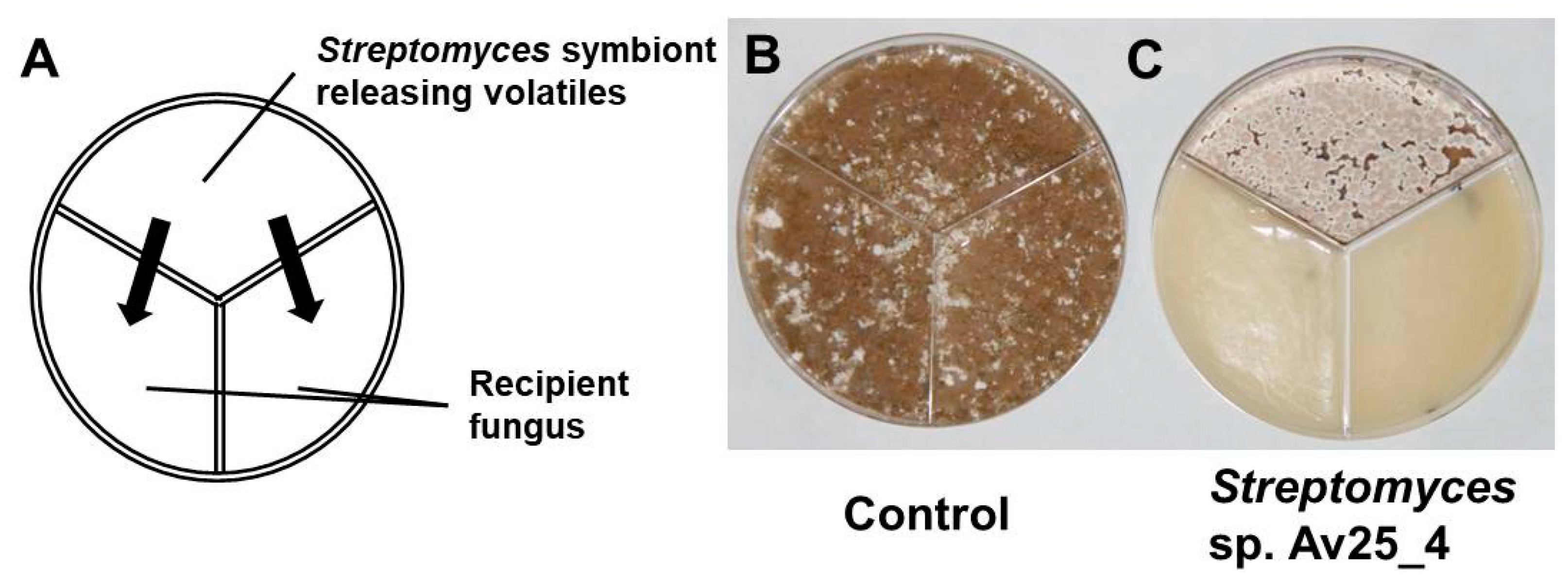
2.3. Screening Streptomyces Symbionts for Antifungal Volatile Compounds

Eight Streptomyces isolates [22] (Table S1) from leaf-cutting ants were screened for their ability to produce antifungal volatiles using three compartment Petri dishes divided by a plastic barrier.
A Streptomyces symbiont was spread onto SFM medium in one compartment. The plates were wrapped with parafilm in order to avoid potential contamination with volatiles from the other cultures in the incubator and incubated at 28 °C. After 3 d, 80 µL of an E. weberi spore suspension was spread uniformly on the two remaining compartments of the divided plate containing SFM medium, and plates were again sealed with parafilm. As control, E. weberi was grown alone in the same way. The growth of E. weberi was monitored 5 d after inoculation of E. weberi. All bioassays were performed in triplicate. Streptomyces sp. Av25_4 inhibited the growth of E. weberi mycelium completely and was therefore selected for detailed investigations. In addition, it was tested whether Streptomyces sp. Av25_4 inhibits the growth of E. aspergilloides in the same way.

2.4. Trapping Volatiles Released by Streptomyces sp. Av25_4 and Evaluation of the Consequences on E. weberi Growth

Three compartment Petri dishes were used for the experiment (Figure 1, Figure S1). As a positive control, Streptomyces sp. Av25_4 was spread onto SFM agar in one compartment, and E. weberi was introduced as an agar plug (6 mm diameter) to PDA agar in a second compartment. The agar plug originated from a PDA plate of actively growing E. weberi. The third compartment was left empty.
For the volatile trapping experiment, the third compartment was filled with 2 g of activated charcoal in order to absorb the volatiles released by Streptomyces sp. Av25_4 and investigate their effect on the growth of E. weberi. The Petri dishes were sealed with parafilm after inoculation with E. weberi and further incubated for 5 d at 28 °C (Figure S2). As negative control, E. weberi was grown alone in the compartment containing PDA medium. The area of fungal growth was calculated by using image J 1.52a. Each bioassay was at least performed in triplicate.

2.5. Closed Loop Stripping (CLS) of Volatiles: GC-MS Analysis and Bioassays with the Collected Organic Volatiles

Streptomyces sp. Av25_4 was cultivated on SFM agar plates at 28 °C for 7d. The emitted volatiles were then sampled from Streptomyces sp. Av25_4. Through two holes in the side of the agar plate a closed-loop stripping pump (3V power supply, Fürgut, Tannheim, Germany,) fitted with an activated charcoal filter (1.5 mg, CLSA, le Ruisseau de Montbrun, Daumazan sur Arize, France) was inserted into the head space and the volatiles were collected for 1 h (Figure S3) [31]. The collected volatiles were eluted from the charcoal filter with 3 × 25 μL ethyl acetate. The extract was analyzed by GC-MS using a Trace GC Ultra hyphenated with an ISQ quadrupole mass spectrometer (Thermofisher, Dreieich, Germany). Separation was performed with an Optima 5 MS column (30 m × 0.25 mm, film 0.25 um, Macherey-Nagel, Düren, Germany). GC-MS conditions: inlet temperature 280 °C, splitless injection, hydrogen served as a carrier gas (0.9 mL/min). The volatile metabolite profiles were recorded using the following temperature programme: 50 °C isotherm for 3 min, 6 °C/min to 200 °C, 20 °C/min to 260 °C, 260 °C isotherm for 1 min. The mass spectrometer was operated in electron impact positive ionization mode at 70 eV. Compounds were identified by analysis of their mass spectra and comparison of their retention times as well as mass spectra to authentic reference compounds.
Volatiles collected (from days 4 to 9 for 24 h each) by closed loop stripping from 10 agar plates of Streptomyces sp. Av25_4 in a 2.5 L desiccator were tested for their inhibitory potential against E. weberi. The volatiles were eluted from the charcoal filters with 120 μL ethylacetate. In three compartment Petri dishes E. weberi was cultivated for 5 d. In the second compartment, a filter paper (1.5 cm diameter) was added. In total, 20 μL of the extract or ethylacetate as control was applied daily to the filter paper in the three compartment Petri dish bioassay. After 10 d the growth of E. weberi was analyzed by determining the covered area using image J 1.52a.

2.6. Change of the pH upon Growth of Streptomyces sp. Av25_4

The change of the pH in the growth medium was monitored using phenol red as pH indicator [32]. Streptomyces sp. Av25_4 was spread on the SFM medium of the first compartment. The other two compartments contained SFM medium with 0.002% of phenol red as pH indicator. The plates were sealed with parafilm and the color change in the medium was recorded by taking pictures at 4, 7, and 10 d after incubation.

2.7. Detection of Ammonia

Whether Streptomyces sp. Av25_4 produces ammonia or not was investigated by using ortho-phathaldialdehyde (OPA) derivatization and fluorescence detection of the resulting derivative [33] as well as by 1H NMR analysis [34]. The OPA derivatization reagent was prepared by mixing 10.5 mg OPA dissolved in 210 μL methanol and 10.5 mg of sodium sulfite dissolved in 630 μL of sodium phosphate buffer (100 mM, pH adjusted to 12.0 with 2M NaOH). Streptomyces sp. Av25_4 was grown for 7 d at 28 °C on SFM agar plates. Ten plates with fully-grown Streptomyces sp. Av25_4 were transferred into a desiccator (2.5 L) that was closed with a septum. A CLS pump was used to directly transfer the volatiles released within 1 h by Streptomyces sp. Av25_4 from the desiccator into the OPA derivatization solution (20 μL of OPA reagent in 500 μL of phosphate buffer) in a 4 mL glass vial.
Similarly, a CLS pump was used to directly transfer the volatiles released within 3 d by 6 d grown Streptomyces sp. Av25_4 from the desiccator (size 2.5 L, 10 SFM agar plates) into the 250 mM HCl solution (500 μL prepared in ddH2O) in a 4 mL glass vial. DMSO-d6 (50 μL) was added for calibration of the NMR spectrometer. 1H-NMR spectra were recorded for the obtained solution of the collected volatiles, a solvent control and an equally prepared ammonium standard using a Bruker Avance III 600 spectrometer (Bruker BioSpin GmbH, Rheinstetten, Germany) with water suppression. Data was recorded using the Bruker Topspin software. Data was analyzed using MestReNova 14 software (Mestrelab Research, Santiago de Compostela, Spain).

2.8. Quantification of Ammonia

From the E. weberi/Streptomyces sp. Av25_4 co-culture bioassays, where the strains were separated by a plastic barrier using the three compartment plates (see above), an agar plug (1 × 1 cm area) was taken from the PDA agar compartment in which E. weberi was inoculated to quantify ammonia. The agar plugs were collected into 2 mL Eppendorf tubes at different time points (2, 4, 6, 8, 10, and 12d) and stored at −20 °C. After freezing and thawing of the samples, the released liquid was collected by centrifugation at 16,873 g for 3 min. Then, 20 μL of the obtained liquid was diluted with 500 μL of 0.1 M phosphate buffer (pH 12.0) and derivatized with 20 µL of OPA reagent. The samples were kept in the dark for 30 min at room temperature and analyzed using a Dionex Ultimate 3000 UHPLC system connected to a Dionex RF 2000 fluorescence detector. UHPLC method: HPLC column: Thermo Scientific Accucore RP-MS column: 150 × 2.1, 1.5 μm); solvent A: H2O 0.1% acetic acid, solvent B: MeOH 0.1% acetic acid; gradient elution: 35–100% B in 20 min, 5 min 100% B, flow rate: 0.25 mL/min.
The fluorescence intensity of thus formed fluorescent product was measured at emission wavelength of 420 nm after excitation at 360 nm [33].
Ammonia was quantified by comparison of the peak areas to an external calibration curve (2.5, 5, 7.5, 10 mM ammonia). All values where at least determined in triplicate.
Similarly, the amount of ammonia that accumulated in the PDA agar compartment when 5 mL ammonia (5, 7, 9, and 11 mM) was supplied into one compartment without medium of the three compartment agar plate was determined. Number of biological replicates per data point was four.

2.9. Influence of Ammonia on the Growth of E. weberi, E. aspergilloides, F. equiseti, and L. gongylophorus

In order to study how ammonia affects fungal growth, three or two compartment Petri dishes were used. The first compartment containing PDA medium was inoculated with an E. weberi, E. aspergilloides, F. equiseti fungal hyphae/spore solution or an L. gongylophorus agar plug (6 mm or 1.5 cm diameter). The second compartment was supplemented with 5 mL of 5, 7, 9, 11, 15 mM or 10 mM (in case of L. gongylophorus) ammonia solution and the third compartment was left empty. The Petri dishes with E. weberi, E. aspergilloides, or F. equiseti were sealed with parafilm and further incubated for 5–10 d at 28 °C. In case of L. gongylophorus the plates were incubated for 3 weeks at 28 °C and 40–50% humidity.
For control plates, sterile water was added instead of the ammonia solution. Each concentration was at least tested three times. The area of fungal growth was calculated by using image J 1.52a.

2.10. Influence of Medium Alkalinization on the Growth of E. weberi

The growth of E. weberi on SFM agar plates at pH 7.5, 8.0, and 8.5 was monitored. The SFM agar plates were inoculated with an E. weberi agar plug (6 mm diameter). The pH of SFM medium was adjusted by using 3M sodium hydroxide solution. The growth of E. weberi was analyzed after 5 d of incubation. Each pH was at least reproduced three times.

2.11. Determination of the pH and Quantification of Ammonia from Waste of Leaf-Cutting Ants

In order to determine the pH of waste from different Atta and Acromyrmex leaf-cutting ant nests (Table S2) 2 g of waste was suspended in 20 mL double distilled water (pH7).
The pH was determined with a pH electrode after 3 min.
The presence and amount of ammonia in waste of leaf-cutting ants was determined by collecting ammonia with molecular sieves (0.5 nm pore size, Merck, Darmstadt, Germany) and comparing the values to an ammonia reference curve. Molecular sieves (0.5 g) were added into a 4 mL glass vial that was posed into a 50 mL flask with 2 g of waste or 2 g of an (2.5, 5, 10 mM) ammonia solution. Then the flask was closed. After 16 h, the molecular sieve was collected and ammonia was eluted with 1 mL of 250 mM HCl and ultrasonication for 10 min. The solution was centrifuged at 16,873 g for 1 min and the supernatant was collected. Ammonia was quantified using the indophenol-blue method adapted from the DIN 38406–5norm (DIN-Normenausschuss Wasserwesen 1983). Then, 50 μL of solution A (0.65 g sodium salicylate, 0.65 g trisodium citrate dihydrate and 10 mg sodium nitroprusside dihydrate in 5 mL ddH2O) and 50μL of solution B (10 mg sodium dichloroisocyanurate in 5 mL NaOH (32 g L−1)) were added to a 1/10 dilution of 500 μL molecular sieve extracts of the ammonium standards or the molecular-sieve extracts from waste. Reaction mixtures were incubated for 3 h in the dark. The absorption was measured at 655 nm and the amount of ammonia was calculated based on the ammonia reference values. The measurements were performed in triplicate [34].

3. Results

3.1. Streptomyces sp. Av25_4 Produces Antifungal Volatiles

As Streptomyces strains are well known to produce a large variety of organic volatile compounds [35,36,37] we screened Streptomyces symbionts from leaf-cutting ants for whether the production of volatile compounds inhibits the growth of the pathogenic fungus. E. weberi and the Streptomyces isolates were cultivated in three compartment Petri dishes so that they grow physically separated from each other but can exchange volatile compounds (Figure 1). The volatile compounds of 3 out of 8 strains (Streptomyces sp. Av25_4, Streptomyces sp. Av26_5, Streptomyces sp. Av28_3) clearly inhibited E. weberi growth (Figure S1, Table S1). In particular, Streptomyces sp. Av25_4, nearly completely inhibited the growth of E. weberi (Figure 1). Therefore, Streptomyces sp. Av25_4 was selected in order to reveal which compound(s) is/are responsible for the observed antifungal activity. By adding activated charcoal to the assay to adsorb any volatiles produced, we observed a clear decrease of the antifungal effect against E. weberi, leading to 42 ± 11% increase in the area covered by E. weberi compared to the control (Figure 2, Figure S2). Notably, exposure to less ammonia due to the presence of activated charcoal (Figure 2D) still led to a change in pigmentation compared to the control (Figure 2B).

3.2. Analysis of Volatiles from Streptomyces sp. Av25_4

In order to study the volatile compounds released by Streptomyces sp. Av25_4, the volatile compounds were collected using closed loop stripping (Figure S3). GC-MS analysis revealed a range of organic compounds, such as alcohols, ketones, aldehydes, terpenes, and lactones. Five compounds, namely geosmin, 2-methyl-isoborneol, 2-phenylethanol, nonanal, and benzaldehyde were selected because they were both abundant and some of them we suspected to exert antifungal activity against E. weberi (Figure S4). Among the five compounds tested, only nonanal and benzaldehyde inhibited E. weberi growth at high concentrations and the aldehydes were also present in the SFM medium control. Thus, these compounds cannot account for the observed complete growth inhibition of E. weberi by volatiles from Streptomyces sp. Av25_4.
Moreover, organic volatiles collected for 24 h by closed loop stripping from 10 Streptomyces sp. Av25_4 plates and applied daily to the E. weberi growth assay in a two compartment Petri dish did not show any effect on the growth of E. weberi (Figure S4B,C).

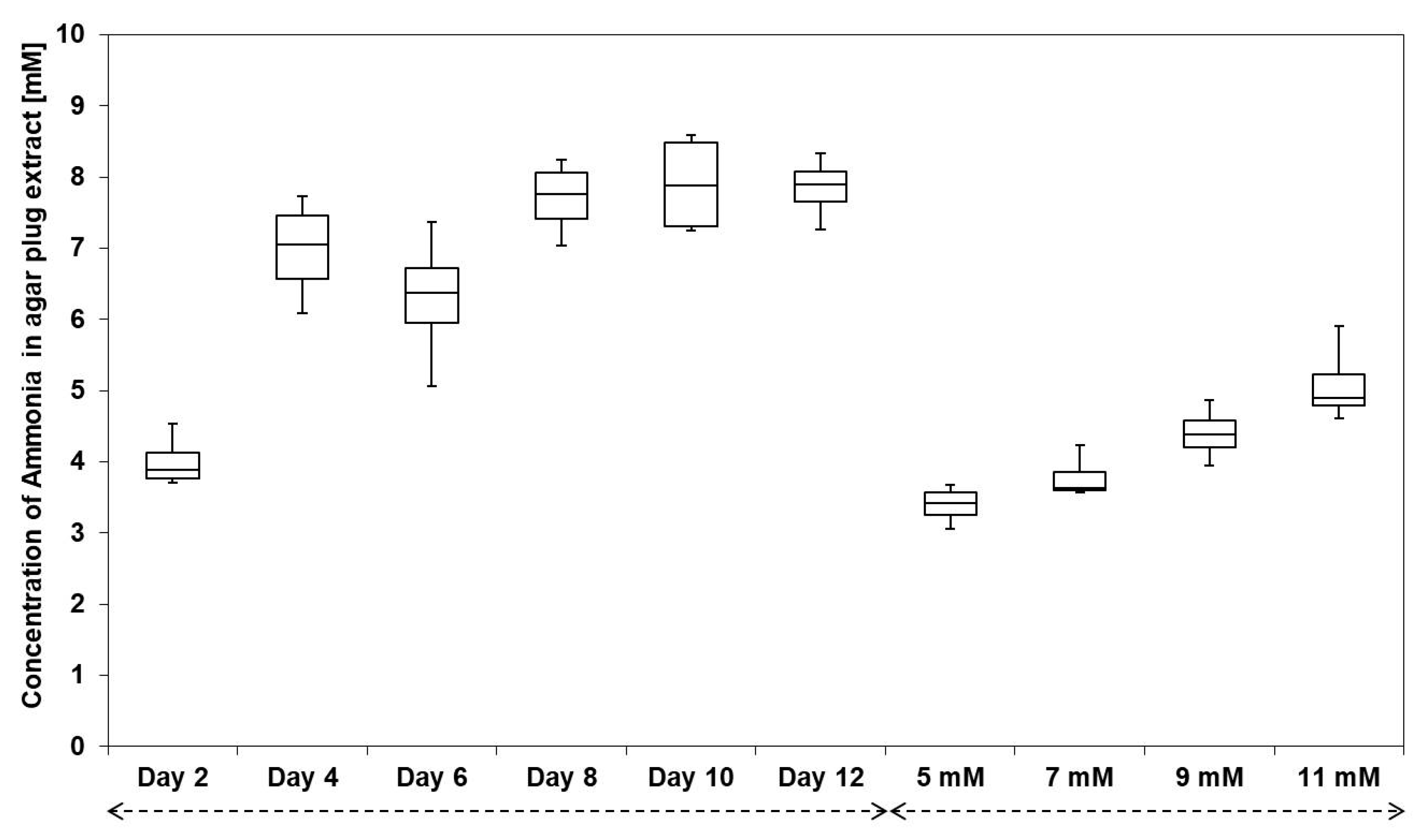
3.3. Streptomyces sp. Av25_4 Overproduces Ammonia

As the organic volatile extract from the closed loop stripping of Streptomyces sp. Av25_4 as well as the selected identified volatile organic compounds did not show activity against E. weberi, we suspected that Streptomyces sp. Av25_4 releases another potent antifungal volatile. Since our GC-MS approach was only suitable for nonpolar organic compounds, we suspected that polar volatiles may confer the observed antifungal activity.
Using the three compartment Petri dish we monitored whether volatiles from Streptomyces sp. Av25_4 alter the pH of the E. weberi PDA growth medium that was supplemented with the pH indicator phenol red [32]. Upon alkalinization phenol red changes its color form pale yellow (pH 1–7.3) to purple (pH > 7.3) [32]. At the beginning of the experiment, the pH increase was observed near Streptomyces sp. Av25_4. However, after 7 d the whole E. weberi growth medium appeared entirely pink, indicating the alkalinization is caused by a volatile compound from Streptomyces sp. Av25_4 (Figure S5).
Some Streptomyces strains can produce volatile amines such as ammonia [32,34] and trimethylamine [38]. Such volatile amines can alter the pH in the physically separated compartment with E. weberi. Therefore, we investigated if Streptomyces sp. Av25_4 produces volatile amines. The volatile amines were trapped in 250 mM HCl solution using the closed loop stripping pump (Figure S6). The collected sample was analyzed by 1H-NMR spectroscopy using water suppression. The 1H-NMR spectrum revealed an intense triplet signal at 7.01 ppm with a coupling constant of 51.6 Hz, proving that Streptomyces sp. Av25_4 produces a high amount of ammonia (Figure S7) [34]. Signals accounting for other volatile compounds were only detected in traces in the 1H-NMR spectrum.
Furthermore, the production of ammonia by Streptomyces sp. Av25_4 was quantified by derivatization with ortho-phthalaldehyde (OPA), HPLC separation and fluorescence detection (Figure S8) [19]. Streptomyces sp. Av25_4 emitted ammonia in large quantities (up to ca. 8 mM) in comparison to other symbionts (Figure 3). Trimethylamine was not detected in the volatile blend of Streptomyces sp. Av25_4.

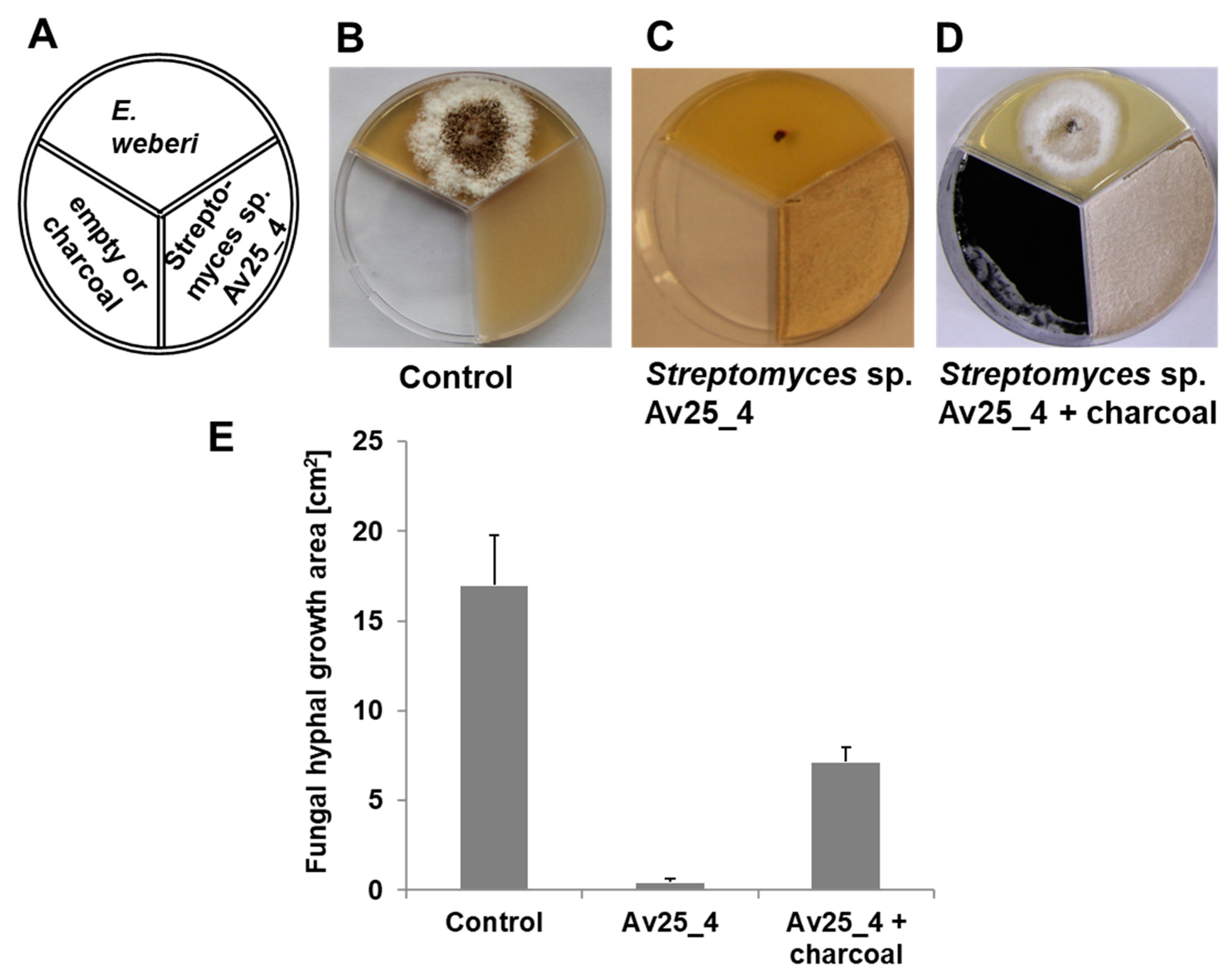
3.4. Ammonia from Streptomyces sp. Av25_4 Strongly Inhibits the Growth of Escovopsis

The growth inhibitory effect of ammonia against E. weberi was assessed by adding 5 mL of ammonia solutions (5, 7, 9, and 11 mM) in one compartment of a three compartment plate (Figure S9). The volatile ammonia received in the E. weberi compartment of an ammonia concentration of 5, 7, and 9 mM, respectively, inhibited almost 37 ± 9%, 56 ± 4%, and 77 ± 2% of the E. weberi growth (Figure S10). The 11 mM ammonia completely inhibited the growth of E. weberi. Additionally, E. aspergilloides was strongly inhibited by ammonia (15 mM) (Figure S11). However, ammonia (10 mM) neither affected the growth of F. equiseti nor of L. gongylophorus (Figures S12–S14).
In order to assess whether or not Streptomyces sp. Av25_4 releases sufficient volatile ammonia to inhibit E. weberi, the concentration of received volatile ammonia in the PDA agar compartment with E. weberi was quantified after 2, 4, 6, 8, 10, and 12 d of Streptomyces sp. Av25_4 growth.
The ammonia concentration in PDA agar extracts of the E. weberi growth compartment was quantified by OPA derivatization and fluorescence detection in comparison to a standard ammonia reference curve (y = 3136.8x + 3578.6, R2 = 0.999). After 2 d of growth, Streptomyces sp. Av25_4 caused an accumulation of ca. 4 mM ammonia in the PDA growth medium (Figure 3). After 10 d, the concentration of ammonia further increased to almost 8 mM. The ammonia concentrations of the PDA agar extract of the Streptomyces sp. Av25_4/E. weberi co-culture match to the concentration necessary to almost completely inhibit the growth of E. weberi.

3.5. Alkalinization of the Medium and Growth Inhibition

As the base ammonia was found to be sufficient to explain the observed growth inhibition of E. weberi, we studied if the alkalinization of the medium is the reason for the growth inhibition. Therefore, the growth of E. weberi was monitored at pH 7.5, 8.0, and 8.5 adjusted with sodium hydroxide instead of ammonia. E. weberi was 90 ± 5% inhibited at pH 7.5 (Figures S15 and S16). At pH 8.0, the growth of E. weberi was completely inhibited.

3.6. Waste of Leaf-cutting Ants Can Be Strongly Basic

Since the nests of A. volcanus and A. echinatior ants we previously isolated our ammonia producing Streptomyces strains from, in particular Streptomyces sp. Av25_4 (see above, Figure S1), were unfortunately not available to us any more, we investigated whether other nests of leaf-cutting ants exhibit a basic pH in their waste and produce ammonia that strongly inhibits the growth of Escovopsis. The pH of waste samples from 19 Atta and Acromyrmex colonies was determined (Table S2). Six out of 19 waste samples exhibited a basic pH of > 8. Interestingly, the pH of waste of colonies of the same leaf-cutting ant species varied and could be either acidic or basic. However, the pH of the individual nests remained throughout our experiments. All basic waste samples contained ammonia, as determined by collecting ammonia with molecular sieves followed by quantification using the Berthelot reaction [34]. The waste of Acromyrmex ambiguus (Emery 1888, Formicidae: Myrmicinae) colony L contained ammonia in high amounts (8 mM) that strongly inhibited Escovopsis growth.

4. Discussion

Microorganisms, in particular Actinomycetes, often produce a large variety of volatile compounds [35,36,37]. However, in contrast to volatiles from plants and animals, the biological function of microbial volatiles, in particular in the ecological context, remained in most cases unknown. As volatiles can diffuse over long distances, they are ideally suited for communication as well as deterrence of competitors [35]. For some microbial volatile compounds, e.g., the terpene albaflavenone [39] antibiotic activity was established. Studying the biological role of volatile compounds from Actinomycetes recently gained attention among researchers interested in the chemical ecology of Actinomycetes [35,36,37]. Huang et al. revealed that Solenopsis invicta ants favor soils rich in Actinomyces bacteria by recognizing their characteristic volatiles, 2-methylisoborneol and geosmin [40]. In the context of leaf-cutting ants, Silva-Junior et al. recently discovered that Actinomyces symbionts produce the same pyrazines [41] that the ants use as infochemicals [42,43,44]. These findings nicely demonstrate that volatiles should be investigated for their ecological role in interspecies interactions between ants and their microbial symbionts.
Now we demonstrate that the microbial symbiont of A. volcanus ants, Streptomyces sp. Av25_4, overproduces ammonia up to 8 mM. Moreover, also other Streptomyces symbionts from leaf-cutting ants we investigated produce ammonia, however, less pronounced than Streptomyces sp. Av25_4 (Figure S1, Table S1). Microbial symbionts from A. volcanus and A. echinatior produce ammonia, indicating that ammonia production is common among Streptomyces symbionts of leaf-cutting ants.
The 8 mM of ammonia (Figure 3), as produced by Streptomyces sp. Av25_4, strongly inhibited the growth of the pathogen E. weberi, explaining the observed inhibition of E. weberi in co-culture with Streptomyces sp. Av25_4 separated by a physical barrier. Our results are perfectly in line with the recent, detailed mechanistic study by Avalos et al. [32], which nicely demonstrated that many Streptomyces strains produce ammonia that can serve them to inhibit potential bacterial competitors. Moreover, volatile amines play important physiological roles in some Streptomyces strains as trimethylamine was found to induce exploratory growth [38], while ammonia induces droplet production [34], and exerts antimicrobial [32] and anti-oomycetal effect [45]. In addition, ammonia can potentiate the activity of antibiotic secondary metabolites [32]. Both tested Escovopsis strains, E. weberi and E. aspergilloides, were sensitive to ammonia whereas the garden fungus L. gongylophorus and F. equiseti were not affected by ammonia rendering the defense by ammonia highly suitable to fight the specialized pathogen of leaf-cutting ants Escovopsis.
However, the fungus garden of leaf-cutting ants appears to be maintained at an acidic pH of 5.2 [46,47]. In order to avoid infections by Escovopsis and other pathogens, leaf-cutting ants carefully weed their garden fungus, remove any old and suspicious material containing pathogens from their garden fungus and deposit the waste in special waste chambers. Thus, the outbreak of infections in the nest of leaf-cutting ants can mostly be kept under control [15,17,18]. In order to avoid reintroduction of pathogens from the waste sites by leaf-cutting ants performing the waste management back into the nest efficient disinfection of the waste is required. Thus, we reasoned whether the ammonia from microbial symbionts may contribute to inactivate the waste material of leaf-cutting ants.
Indeed, the waste of 6 out of 19 (Table S2) ant nests exhibited a pH of ca. 8, that we showed to strongly inhibit Escovopsis growth (Figures S15 and S16). With the example of the A. ambiguus nest colony L we proved that the basic pH is caused by ammonia. Alternatively, we observed a pH around 5 from 6 out of 19 leaf-cutting ant nests tested, indicating that both acidification and alkalization are used as an efficient and simple strategy to disinfect the waste. Indeed, the use of acids or bases is an efficient and common way of microorganisms to outcompete competitors, that humans make use of e.g., for preservation of food [48,49].
Interestingly the use of acids or bases is not species specific but rather specific for the individual nest potentially suggesting that it may depend on the acquired symbionts.
Future experiments are needed to study in-depth which antiinfective strategies are used to inactivate dangerous pathogens in the waste dumps of different leaf-cutting ant species and nests. In order to reveal the functioning of the microcosomos of leaf-cutting ants it is crucial to identify and consider the whole variety of low and high molecular weight compounds present. Our experiments demonstrate that one strategy to inhibit Escovopsis is to maintain a basic pH (Figure 3) using the volatile base ammonia. Compared to the complex secondary metabolites of Actinomyces symbionts of leaf-cutting ants, e.g., the polyketide candicidin [22], ammonia is a very simple but remarkably efficient compound. As established by Avalos et al., ammonia production can be a highly efficient way to outcompete competitors because of its biosynthetic simplicity and thus metabolically low cost of production. Streptomycetes often produce ammonia by degradation of amino acids [32]. Ammonia production only requires few enzymes in contrast to the mega-synthases needed for polyketide or non-ribosomal peptide production [32].
For the first time we demonstrated that in the microcosmos of leaf-cutting ants, microbial symbionts, such as Streptomyces sp. Av25_4, overproduce ammonia that strongly inhibits the growth of the fungal pathogen Escovopsis. With simple cost-effective ammonia, Streptomyces strains contribute to render the waste of leaf-cutting ants basic to discourage the growth and spreading of Escovopsis from their waste chambers.

Supplementary Materials

The following are available online at https://www.mdpi.com/article/10.3390/microorganisms9081622/s1. Figure S1. Screening Streptomyces symbionts from leaf-cutting ants for the production of volatile antifungal compounds that inhibit the growth of E. weberi using the three compartment Petri dishes to only allow growth inhibition by volatile compounds. Figure S2. Three compartment bioassay to identify volatile compounds inhibiting the growth of E. weberi. Figure S3. Closed loop stripping. Figure S4. Organic volatile compounds from Streptomyces sp. Av25_4. Figure S5. Monitoring pH change in Petri dishes. Figure S6. Detection of ammonia produced by Streptomyces sp. 25_4 using a closed loop stripping pump transferring the volatiles into the vial with the OPA derivatization reagent. Figure S7. Identification of ammonia as the pH changing principle using 1H-NMR spectroscopy. Figure S8. Identification of ammonia as pH changing principle using ortho-phthalaldehyde derivatization. Figure S9. Three compartment Petri dish bioassay evaluating the effect of ammonia on E. weberi growth. Figure S10. Effect of ammonia on E. weberi growth. Figure S11. Growth of E. aspergilloides in presence of ammonia on SFM agar plates. Figure S12. Growth of F. equiseti in presence of ammonia on SFM agar plates. Figure S13. Influence of ammonia on the growth of L. gongylophorus. Figure S14. Effect of ammonia on L. gongylophorus growth. Figure S15. Growth of E. weberi on SFM agar plates at different pH adjusted with sodium hydroxide. Figure S16. Effect of basic pH on E. weberi growth. Table S1. Streptomyces symbionts used to screen for volatiles with antifungal activity against E. weberi. Table S2. pH of the waste of different Acromyrmex and Atta leaf-cutting ant nests.

Author Contributions

Conceptualization, B.D. and D.S.; methodology, B.D. and D.S.; data analysis, B.D. and D.S.; writing, B.D. and D.S.; supervision, D.S.; funding acquisition, D.S. All authors have read and agreed to the published version of the manuscript.

Funding

This research was funded by the Deutsche Forschungsgemeinschaft: GSC 218: Konstanz Research School Chemical Biology (KoRS-CB), Project number: 39072414.

Data Availability Statement

Data is presented in the paper and the supporting information. Original data files will be available upon request from the authors.

Acknowledgments

We are very grateful to Rainer Wirth, Flavio Roces, Daniela Römer, and Christoph Kleineidam for leaf-cutting ants and waste samples. We thank Anke Friemel for the NMR measurement, Sören Radke for performing some of the bioassays, and Anthony Farlow for language editing. We are greatly indebted to the Konstanz Research School Chemical Biology funded by the Deutsche Forschungsgemeinschaft for financial support and a doctoral fellowship to BD.

Conflicts of Interest

The authors declare no conflict of interest.

References

  1. Hölldobler, B.; Wilson, E.O. The Ants; Harvard University Press: Cambridge, MA, USA, 1990. [Google Scholar]
  2. Wirth, R.; Beyschlag, W.; Ryel, R.; Herz, H.; Hölldobler, B. The Herbivory of Leaf-Cutting Ants. In A Case Study on Atta Colombica in the Tropical Rainforest of Panama; Springer: Berlin/Heidelberg, Germany; New York, NY, USA, 2003. [Google Scholar]
  3. Silva, A.; Bacci, M., Jr.; Gomes de Siqueira, C.; Correa Bueno, O.; Pagnocca, F.C.; Aparecida Hebling, M.J. Survival of Atta sexdens workers on different food sources. J. Insect. Physiol. 2003, 49, 307–313. [Google Scholar] [CrossRef]
  4. Aylward, F.O.; Burnum-Johnson, K.E.; Tringe, S.G.; Teiling, C.; Tremmel, D.M.; Moeller, J.A.; Scott, J.J.; Barry, K.W.; Piehowski, P.D.; Nicora, C.D.; et al. Leucoagaricus gongylophorus produces diverse enzymes for the degradation of recalcitrant plant polymers in leaf-cutter ant fungus gardens. Appl. Environ. Microbiol. 2013, 79, 3770–3778. [Google Scholar] [CrossRef] [PubMed] [Green Version]
  5. Fisher, P.J.; Stradling, D.J.; Sutton, B.C.; Petrini, L.E. Microfungi in the fungus gardens of the leaf-cutting ant Atta cephalotes: A preliminary study. Mycol. Res. 1996, 100, 541–546. [Google Scholar] [CrossRef]
  6. Currie, C.R.; Mueller, U.G.; Malloch, D. The agricultural pathology of ant fungus gardens. Proc. Natl. Acad. Sci. USA 1999, 96, 7998–8002. [Google Scholar] [CrossRef] [Green Version]
  7. Bot, A.N.M.; Currie, C.R.; Hart, A.G.; Boomsma, J. Waste management in leaf-cutting ants. Ethol. Ecol. Evol. 2001, 13, 225–237. [Google Scholar] [CrossRef]
  8. Muchovej, J.J.; Della Lucia, T.M. Escovopsis, a new genus from leaf-cutting ant nests to replace Phialocladus nomem invalidum. Mycotaxon 1990, 37, 191–195. [Google Scholar]
  9. Seifert, K.A.; Samson, R.A.; Chapela, I.H. Escovopsis aspergilloides, a rediscovered hyphomycete from leaf-cutting ant nests. Mycologia 1995, 87, 407–413. [Google Scholar] [CrossRef]
  10. Rodrigues, A.; Pagnocca, F.; Bacci, M.; Hebling, M.; Bueno, O.; Pfenning, L. Variability of non-mutualistic filamentous fungi associated with Atta sexdens rubropilosa nests. Folia Microbiol. 2005, 50, 421. [Google Scholar] [CrossRef]
  11. Boya, P.C.; Fernandez-Marin, H.; Mejia, L.C.; Spadafora, C.; Dorrestein, P.C.; Gutierrez, M. Imaging mass spectrometry and MS/MS molecular networking reveals chemical interactions among cuticular bacteria and pathogenic fungi associated with fungus-growing ants. Sci. Rep. 2017, 7, 5604. [Google Scholar] [CrossRef] [PubMed] [Green Version]
  12. Dhodary, B.; Schilg, M.; Wirth, R.; Spiteller, D. Secondary Metabolites from Escovopsis weberi and Their Role in Attacking the Garden Fungus of Leaf-Cutting Ants. Chemistry 2018, 24, 4445–4452. [Google Scholar] [CrossRef] [PubMed] [Green Version]
  13. Heine, D.; Holmes, N.A.; Worsley, S.F.; Santos, A.C.A.; Innocent, T.M.; Scherlach, K.; Patrick, E.H.; Yu, D.W.; Murrell, J.C.; Vieria, P.C.; et al. Chemical warfare between leafcutter ant symbionts and a co-evolved pathogen. Nat. Commun. 2018, 9, 2208. [Google Scholar] [CrossRef]
  14. Schildknecht, H.; Koob, K. Myrmicacin, the first insect herbicide. Angew. Chem. Int. Ed. 1971, 10, 124–125. [Google Scholar] [CrossRef]
  15. Ortius-Lechner, D.; Maile, R.; Morgan, E.D.; Boomsma, J.J. Metapleural gland secretion of the leaf-cutter ant Acromyrmex octospinosus: New compounds and their functional significance. J. Chem. Ecol. 2000, 26, 1667–1683. [Google Scholar] [CrossRef]
  16. Bot, A.N.M.; Ortius-Lechner, D.; Finster, K.; Maile, R.; Boomsma, J.J. Variable sensitivity of fungi and bacteria to compounds produced by the metapleural glands of leaf-cutting ants. Insect. Soc. 2002, 49, 363–370. [Google Scholar] [CrossRef]
  17. Fernández-Marín, H.; Zimmerman, J.K.; Rehner, S.A.; Wcislo, W.T. Active use of the metapleural glands by ants in controlling fungal infection. Proc. Roy. Soc. B Biol. Sci. 2006, 273, 1689–1695. [Google Scholar] [CrossRef] [PubMed] [Green Version]
  18. Fernández-Marín, H.; Nash, D.R.; Higginbotham, S.; Estrada, C.; van Zweden, J.S.; d’Ettorre, P.; Wcislo, W.T.; Boomsma, J.J. Functional role of phenylacetic acid from metapleural gland secretions in controlling fungal pathogens in evolutionarily derived leaf-cutting ants. Proc. Roy. Soc. B Biol. Sci. 2015, 282, 20150212. [Google Scholar] [CrossRef] [PubMed]
  19. Currie, C.R.; Scott, J.A.; Summerbell, R.C.; Malloch, D. Fungus-growing ants use antibiotic-producing bacteria to control garden parasites. Nature 1999, 398, 701–704. [Google Scholar] [CrossRef]
  20. Santos, A.V.; Dillon, R.J.; Dillon, V.M.; Reynolds, S.E.; Samuels, R.I. Ocurrence of the antibiotic producing bacterium Burkholderia sp. in colonies of the leaf-cutting ant Atta sexdens rubropilosa. FEMS Microbiol. Lett. 2004, 239, 319–323. [Google Scholar] [CrossRef] [PubMed] [Green Version]
  21. Kost, C.; Lakatos, T.; Böttcher, I.; Arendholz, W.R.; Redenbach, M.; Wirth, R. Non-specific association between filamentous bacteria and fungus-growing ants. Naturwissenschaften 2007, 94, 821–828. [Google Scholar] [CrossRef] [PubMed]
  22. Haeder, S.; Wirth, R.; Herz, H.; Spiteller, D. Candicidin-producing Streptomyces support leaf-cutting ants to protect their fungus garden against the pathogenic fungus Escovopsis. Proc. Natl. Acad. Sci. USA 2009, 106, 4742–4746. [Google Scholar] [CrossRef] [Green Version]
  23. Oh, D.-C.; Poulsen, M.; Currie, C.R.; Clardy, J. Dentigerumycin: A bacterial mediator of an ant-fungus symbiosis. Nature Chem. Biol. 2009, 5, 391–393. [Google Scholar] [CrossRef] [PubMed]
  24. Pinto-Tomas, A.A.; Anderson, M.A.; Suen, G.; Stevenson, D.M.; Chu, F.S.T.; Cleland, W.W.; Weimer, P.J.; Currie, C.R. Symbiotic nitrogen fixation in the fungus gardens of leaf-cutter ants. Science 2009, 326, 1120–1123. [Google Scholar] [CrossRef] [PubMed]
  25. Barke, J.; Seipke, R.F.; Grüschow, S.; Heavens, D.; Drou, N.; Bibb, M.J.; Goss, R.J.M.; Yu, D.W.; Hutchings, M.I. A mixed community of actinomycetes produce multiple antibiotics for the fungus farming ant Acromyrmex octospinosus. BMC Biol. 2010, 8, 109. [Google Scholar] [CrossRef] [Green Version]
  26. Holmes, N.A.; Innocent, T.M.; Heine, D.; Bassam, M.A.; Worsley, S.F.; Trottmann, F.; Patrick, E.H.; Yu, D.W.; Murrell, J.C.; Schiøtt, M. Genome analysis of two Pseudonocardia phylotypes associated with Acromyrmex leafcutter ants reveals their biosynthetic potential. Front. Microbiol. 2016, 7, 2073. [Google Scholar] [CrossRef] [PubMed] [Green Version]
  27. Schoenian, I.; Spiteller, M.; Ghaste, M.; Wirth, R.; Herz, H.; Spiteller, D. Chemical basis of the synergism and antagonism in microbial communities in the nests of leaf-cutting ants. Proc. Natl. Acad. Sci. USA 2011, 108, 1955–1960. [Google Scholar] [CrossRef] [Green Version]
  28. Seipke, R.F.; Barke, J.; Brearley, C.; Hill, L.; Yu, D.W.; Goss, R.J.M.; Hutchings, M.I. A single Streptomyces symbiont makes multiple antifungals to support the fungus farming ant Acromyrmex octospinosus. PLoS ONE 2011, 6, e22028. [Google Scholar] [CrossRef] [Green Version]
  29. Fukuda, T.T.H.; Helfrich, E.J.N.; Mevers, E.; Melo, W.G.P.; Van Arnam, E.B.; Andes, D.R.; Currie, C.R.; Pupo, M.T.; Clardy, J. Specialized Metabolites Reveal Evolutionary History and Geographic Dispersion of a Multilateral Symbiosis. ACS Cent. Sci. 2021, 7, 292–299. [Google Scholar] [CrossRef] [PubMed]
  30. Kieser, T.E.; Bibb, M.J.; Buttner, M.J.; Chater, K.F.; Hopwood, D.A. Practical Streptomyces Genetics; John Innes Foundation: Colney, UK, 2000. [Google Scholar]
  31. Grob, K. Organic substances in potable water and in its precursor: Part I. Methods for their determination by gas-liquid chromatography. J. Chrom. A 1973, 84, 255–273. [Google Scholar] [CrossRef]
  32. Avalos, M.; Garbeva, P.; Raaijmakers, J.M.; van Wezel, G.P. Production of ammonia as a low-cost and long-distance antibiotic strategy by Streptomyces species. ISME J. 2019, 1–15. [Google Scholar] [CrossRef]
  33. Genfa, Z.; Dasgupta, P.K. Fluorometric measurement of aqueous ammonium ion in a flow injection system. Anal. Chem. 1989, 61, 408–412. [Google Scholar] [CrossRef]
  34. Schmidt, K.; Spiteller, D. Ammonia released by Streptomyces aburaviensis induces droplet formation in Streptomyces violaceoruber. J. Chem. Ecol. 2017, 43, 806–816. [Google Scholar] [CrossRef] [Green Version]
  35. Schulz, S.; Dickschat, J.S. Bacterial volatiles: The smell of small organisms. Nat. Prod. Rep. 2007, 24, 814–842. [Google Scholar] [CrossRef]
  36. Schmidt, R.; Cordovez, V.; De Boer, W.; Raaijmakers, J.; Garbeva, P. Volatile affairs in microbial interactions. ISME J. 2015, 9, 2329–2335. [Google Scholar] [CrossRef] [Green Version]
  37. Avalos, M.; van Wezel, G.P.; Raaijmakers, J.M.; Garbeva, P. Healthy scents: Microbial volatiles as new frontier in antibiotic research? Curr. Opin. Microbiol. 2018, 45, 84–91. [Google Scholar] [CrossRef] [Green Version]
  38. Jones, S.E.; Ho, L.; Rees, C.A.; Hill, J.E.; Nodwell, J.R.; Elliot, M.A. Streptomyces exploration is triggered by fungal interactions and volatile signals. Elife 2017, 6, e21738. [Google Scholar] [CrossRef]
  39. Gürtler, H.; Pedersen, R.; Anthoni, U.; Christophersen, C.; Nielsen, P.H.; Wellington, E.M.H.; Pedersen, C.; Bock, K. Albaflavenone, a sesquiterpene ketone with a zizaene skeleton produced by a streptomycete with a new rope morphology. J. Antibiot. 1994, 47, 434–439. [Google Scholar] [CrossRef] [PubMed] [Green Version]
  40. Huang, H.; Ren, L.; Li, H.; Schmidt, A.; Gershenzon, J.; Lu, Y.; Cheng, D. The nesting preference of an invasive ant is associated with the cues produced by actinobacteria in soil. PLoS Pathog. 2020, 16, e1008800. [Google Scholar] [CrossRef] [PubMed]
  41. Silva-Junior, E.A.; Ruzzini, A.C.; Paludo, C.R.; Nascimento, F.S.; Currie, C.R.; Clardy, J.; Pupo, M.T. Pyrazines from bacteria and ants: Convergent chemistry within an ecological niche. Sci. Rep. 2018, 8, 2595. [Google Scholar] [CrossRef] [Green Version]
  42. Evershed, R.; Morgan, E. The amounts of trail pheromone substances in the venom of workers of four species of attine ants. Insect Biochem. 1983, 13, 469–474. [Google Scholar] [CrossRef]
  43. Cross, J.H.; West, J.R.; Silverstein, R.M.; Jutsum, A.R.; Cherrett, J.M. Trail pheromone of the leaf-cutting ant, Acromyrmex octospinosus (Reich),(Formicidae: Myrmicinae). J. Chem. Ecol. 1982, 8, 1119–1124. [Google Scholar] [CrossRef]
  44. de Oliveira, J.; Martinez, O.; Carnieri, N.; Vilela, E.; Reis, H. Components of the trail pheromone of the leaf-cutting ants Atta laevigata F. Smith and Atta bisphaerica Forel (Formicidae: Attini). An. Soc. Entomológica Bras. 1990, 19, 145–154. [Google Scholar]
  45. Howell, C.R.; Beier, R.C.; Stipanovic, R.D. Production of Ammonia by Enterobacter cloacae and Its Possible Role in the Biological Control of Pythium Preemergence Damping-off by the Bacterium. Phytopathology 1988, 78, 1075–1078. [Google Scholar] [CrossRef]
  46. Somera, A.F.; Lima, A.M.; Dos Santos-Neto, A.J.; Lancas, F.M.; Bacci, M., Jr. Leaf-cutter ant fungus gardens are biphasic mixed microbial bioreactors that convert plant biomass to polyols with biotechnological applications. Appl. Environ. Microbiol. 2015, 81, 4525–4535. [Google Scholar] [CrossRef] [PubMed] [Green Version]
  47. Semenova, T.A.; Hughes, D.P.; Boomsma, J.J.; Schiott, M. Evolutionary patterns of proteinase activity in attine ant fungus gardens. BMC Microbiol. 2011, 11, 15. [Google Scholar] [CrossRef] [Green Version]
  48. Parkouda, C.; Nielsen, D.S.; Azokpota, P.; Ouoba, L.I.; Amoa-Awua, W.K.; Thorsen, L.; Hounhouigan, J.D.; Jensen, J.S.; Tano-Debrah, K.; Diawara, B.; et al. The microbiology of alkaline-fermentation of indigenous seeds used as food condiments in Africa and Asia. Crit. Rev. Microbiol. 2009, 35, 139–156. [Google Scholar] [CrossRef] [PubMed]
  49. De Roos, J.; De Vuyst, L. Acetic acid bacteria in fermented foods and beverages. Curr. Opin. Biotechnol. 2018, 49, 115–119. [Google Scholar] [CrossRef] [PubMed]
Figure 1. Screening Streptomyces symbionts of leaf-cutting ants for volatile antifungal compounds that inhibit E. weberi. (A) Scheme of the experimental set-up in a three compartment Petri dish. (B) Growth of E. weberi (control), (C) volatiles emitted by Streptomyces sp. Av25_4 completely inhibited the growth of E. weberi.
Microorganisms 09 01622 g001
Figure 2. Antifungal activity of volatiles produced by Streptomyces sp. Av25_4 against E. weberi with and without activated charcoal in the third compartment. (A) Scheme of the experimental setup. (B) E. weberi grown for 4d in the first compartment. (C) E. weberi grown for 4d in first compartment and Streptomyces sp. Av25_4 grown for 7d in the second compartment. (D) E. weberi grown for 4 d in the first compartment, Streptomyces sp. Av25_4 grown for 7d in the second compartment and 2 g activated charcoal in the third compartment. (E) Quantification of the E. weberi growth area analyzed by image J.
Microorganisms 09 01622 g002
Figure 3. Box plot diagram showing the amount of ammonia released by Streptomyces sp. Av25_4 after 2, 4, 6, 8, 10, and 12 d in the agar plug of the PDA medium of the E. weberi compartment. Ammonia in the PDA agar plug of the E. weberi compartment after supplementation of ammonia (5, 7, 9, and 11 mM). The whiskers represent the minimum and maximum values and the segment inside the rectangle shows the median values (four biological replicates).
Microorganisms 09 01622 g003
Publisher’s Note: MDPI stays neutral with regard to jurisdictional claims in published maps and institutional affiliations.

