Article

Diversity and Oil Degradation Potential of Culturable Microbes Isolated from Chronically Contaminated Soils in Trinidad

by
Amanda C. Ramdass
and
Sephra N. Rampersad
*
Biochemistry Research Laboratory (Rm216), Department of Life Sciences, Faculty of Science and Technology, The University of the West Indies, Trinidad and Tobago, West Indies
*
Author to whom correspondence should be addressed.
Microorganisms 2021, 9(6), 1167; https://doi.org/10.3390/microorganisms9061167
Submission received: 15 May 2021 / Revised: 24 May 2021 / Accepted: 26 May 2021 / Published: 28 May 2021
(This article belongs to the Section Environmental Microbiology)

Abstract

:
Trinidad and Tobago is the largest producer of oil and natural gas in Central America and the Caribbean. Natural crude oil seeps, in addition to leaking petroleum pipelines, have resulted in chronic contamination of the surrounding terrestrial environments since the time of petroleum discovery, production, and refinement in Trinidad. In this study, we isolated microbes from soils chronically contaminated with crude oil using a culture-dependent approach with enrichment. The sampling of eight such sites located in the southern peninsula of Trinidad revealed a diverse microbial composition and novel oil-degrading filamentous fungi and yeast as single-isolate degraders and naturally occurring consortia, with specific bacterial species not previously reported in the literature. Multiple sequence comparisons and phylogenetic analyses confirmed the identity of the top degraders. The filamentous fungal community based on culturable species was dominated by Ascomycota, and the recovered yeast isolates were affiliated with Basidiomycota (65.23%) and Ascomycota (34.78%) phyla. Enhanced biodegradation of petroleum hydrocarbons is maintained by biocatalysts such as lipases. Five out of seven species demonstrated extracellular lipase activity in vitro. Our findings could provide new insights into microbial resources from chronically contaminated terrestrial environments, and this information will be beneficial to the bioremediation of petroleum contamination and other industrial applications.

1. Introduction

Trinidad and Tobago is the largest producer of oil and natural gas in the Caribbean and Central America [1,2]. As a result of its large reserves of oil and natural gas, exploitation of the latter now drives its economy. As such, Trinidad has one of the largest natural gas processing facilities in the Western Hemisphere. Trinidad was subject to more than a century of petroleum exploration, which began in 1857 and was based on the detection of oil discharge in terrestrial environments identified as natural crude oil seeps in the southwestern peninsula of the island [3]. The largest naturally-occurring petroleum seep in the world is the Pitch Lake in Trinidad, which was described in 1595 by Sir Walter Raleigh in his search for El Dorado. Current seepage detection by SAR (synthetic aperture radar) also indicates multiple seepage sites offshore [4]. Natural crude oil seeps, in addition to leaking petroleum pipelines, have resulted in the contamination of the surrounding terrestrial environments since the time of petroleum discovery, production, and refinement. It is hypothesized that the chronically contaminated sites in Trinidad are inhabited by novel, indigenous microorganisms with generalist, as well as specialist, metabolic functions for utilizing crude oil as a carbon source, which are required for sustaining the microbial community in this type of perturbed terrestrial ecosystem. Sutton et al. [5] indicated that conclusions based on the short-term simulated spiking of soils may not reflect long-term natural remediation conditions in situ; the authors support the need to investigate long-term contaminated sites.
Crude oil is a heterogeneous mixture composed of saturates, aromatic hydrocarbons (including polycyclic aromatic hydrocarbons, PAHs), asphaltenes, and resins [6,7] that are hazardous to animal and human health [8,9,10]. Of particular concern is the percolation of surface spills on land (leachates) and the subsequent partitioning into gaseous, aqueous, and pure phases in the subsurface strata and migration of BTEX (benzene, ethylbenzene, toluene, and xylene) components of crude oil into waterways, aquifers, and water tables, which result in subsurface persistence and pose long-term effects on groundwater quality [11,12,13,14,15,16]. PAHs are chemically stable, demonstrate extreme persistence in contaminated environments, and are proven to be carcinogenic, genotoxic, teratogenic, and mutagenic [17,18,19]. Remediation of oil-contaminated terrestrial environments is focused on physio-chemical and/or biological methods for restoration [20]. Bioremediation methods exploit the use of microorganisms and their diverse metabolic capabilities for the attenuation of the toxic effects of hydrocarbon chemicals by various routes, including transforming them into less toxic products than the parent compound, completely mineralizing and/or immobilizing them [10,21,22,23]. Bioremediation is considered to be ‘environmentally benign’ or ‘passive’ and economically feasible compared to other approaches that are more costly and offer lower levels of efficacy [12].
Studies on communities of hydrocarbon-utilizing microorganisms have indicated that, over time, degradation will occur without external intervention via a process of ‘natural attenuation’ [5,24]. In simulated spiking experiments, the addition of different amounts of oil to soil resulted in selection for specific oil-degrading fungal populations with a concomitant reduction of species diversity and, in some cases, the complete elimination of other species [25]. This reduction in species diversity with increasing concentrations of supplemented crude oil may be explained by sublethal effects on these microbes through the bioconcentration of toxic pollutants and the creation of anoxic conditions of growth. It has also been documented that yeast and filamentous fungi sequester hydrocarbons in the cytoplasm of their cells [26].
Wang et al. [27] concluded that consequent to PAH contamination, there is a commonly occurring pattern of reduction in the number of and species diversity of bacteria as a result of selection pressures unique to oil-contaminated sites. In fact, such changes in selective pressure to favor the enrichment of those microbial species that are physiologically and metabolically adapted to hydrocarbon utilization often result in population shifts in the dominant species, which can, in turn, define the community‘s overall structure at any given point in time. Other studies have provided evidence that subsurface geochemical properties also influence biodegrading populations of microorganisms because these characteristics affect the bioavailability of PAHs as carbon sources to these microorganisms [28,29].
The relative contribution of culturable microbes to remediation is still studied using pure cultures, even though a large proportion of microbes remain unculturable [30,31]. In contrast, the culture-independent approach focuses on identifying genes associated with metabolic function [32,33,34] but does little to describe or provide proof of function of such microbes in situ. Furthermore, studies have indicated that the identified microbial composition and diversity may vary according to the mode of identification and characterization, i.e., culture-dependent vs. culture-independent methods [35]. This difference is also important in chronically contaminated soil, which, after decades of in situ enrichment, maintains a diverse range of oil-degrading microorganisms (some with specialized functions) that provides a unique collection of species for isolating crude oil-degrading microorganisms [36,37,38,39]. Wang, Li, Zhan, and Zhu [27] added that cooperative interactions among such microbial species are necessary in order to efficiently utilize crude oil and polycyclic aromatic hydrocarbons as a carbon source in contaminated soil [40,41].
Fungi are particularly good candidates for remediation because (i) they have long-range transportation systems—the movement of hyphae is not limited by a hydrophobic environment, surpassing air–water interfaces and air-filled pores in soil, where they aid in the transport of extrahyphal bacteria that would otherwise be limited by this physical barrier, (ii) fungal hyphae can function in anoxic conditions, which is often the case with thick oil slicks, (iii) they can tolerate extreme environments—numerous fungi are xero- and osmotolerant and can survive in a pH range of 1 to 9 and at temperatures of 5 to 60 °C, (iv) numerous fungi have extracellular enzymes that can metabolize hydrocarbons at the start of degradation, thus aiding the process, and (v) they are capable of catabolizing recalcitrant hydrocarbons, i.e., they can metabolize and mineralize high molecular weight hydrocarbons such as PAHs, unlike bacteria (because of the low bioavailability of these compounds to bacteria) [42,43,44,45]. Extracellular enzymatic transformation of recalcitrant compounds by fungi, followed by bacterial degradation of the subsequent intermediates produced through fungal action, may contribute to a combinatorial strategy for the biodegradation of PAHs [46].
As a result of a decline in microbial diversity, culture-dependent sampling of hydrocarbon-contaminated soils compared to other types of polluted sites may be more suitable than other approaches because a reduced sampling effort may still reflect a representative proportion of the bioactive microbial community [47,48,49]. Hydrocarbon contaminants will suppress the survival of PAH-sensitive bacterial groups [50] and select primarily for key subgroups of PAH-degrading bacteria (e.g., α- and γ-proteobacteria) in affected soils [51,52]. Culture-dependent methods are still a critical component of bioremediation development and research even though they generally recover a small portion of the diversity from soil environments compared to metagenomics analyses [53]. Among the advantages of culture-dependent methods are the potential use of cultured microbes and their products in in situ and ex situ applications. Additionally, microbial isolation enables in vitro assessments of isolate hydrocarbon degradation pathways and oil-degrading capability, thereby providing a basis for identifying genes that could be useful in land remediation [35]. Using traditional selective cultivation-dependent microbiological methods also allows the isolation of site-specific hydrocarbonoclastic bacteria [35]. Metagenomics may not be able to detect rarer bacteria or the DNA of both dead and living cells and, ultimately, does not provide any practical means of recovering live microbes. Given the century-old history of crude oil production in Trinidad, it is hypothesized that unique indigenous microorganisms can survive and out-compete other microbes in using crude oil as a carbon source in oil-contaminated soils.
In view of the current evidence, it is, therefore, important to assess the co-aggregation or co-colonization and niche overlap in chronically contaminated terrestrial ecosystems inhabited by microorganisms. Empirical evidence indicates that bioremediation treatments should be designed to be site-specific and fundamentally guided by an assessment of the localized and culturable microbial populations in terms of their biodiversity, relative abundance, and species richness. Conjunctive to this approach is the challenge to cultivate these interacting microorganisms and characterize their function in vitro.
Therefore, the main objectives of this study are: (i) to isolate culturable filamentous fungi and yeast from eight naturally occurring, hydrocarbon-contaminated sites in southern Trinidad; (ii) to assess the ability of indigenous isolates to utilize crude oil as a unique carbon source in vitro; (iii) to identify these PAH-degrading isolates using molecular techniques; (iv) to determine the ability of selected isolates to produce extracellular lipases and; (v) to evaluate the relative microbial composition, species diversity, and possible community ordination at each site. Whereas other studies have examined microbial populations in artificially spiked soils to simulate contaminated microcosms, this study examines field-collected samples collected at geographically distinct sites with different soil matrices and contamination levels. This is the first extensive study to isolate microbes with the potential for bioremediation and the ability to produce bioactive compounds from sites not previously explored in south Trinidad.

2. Materials and Methods

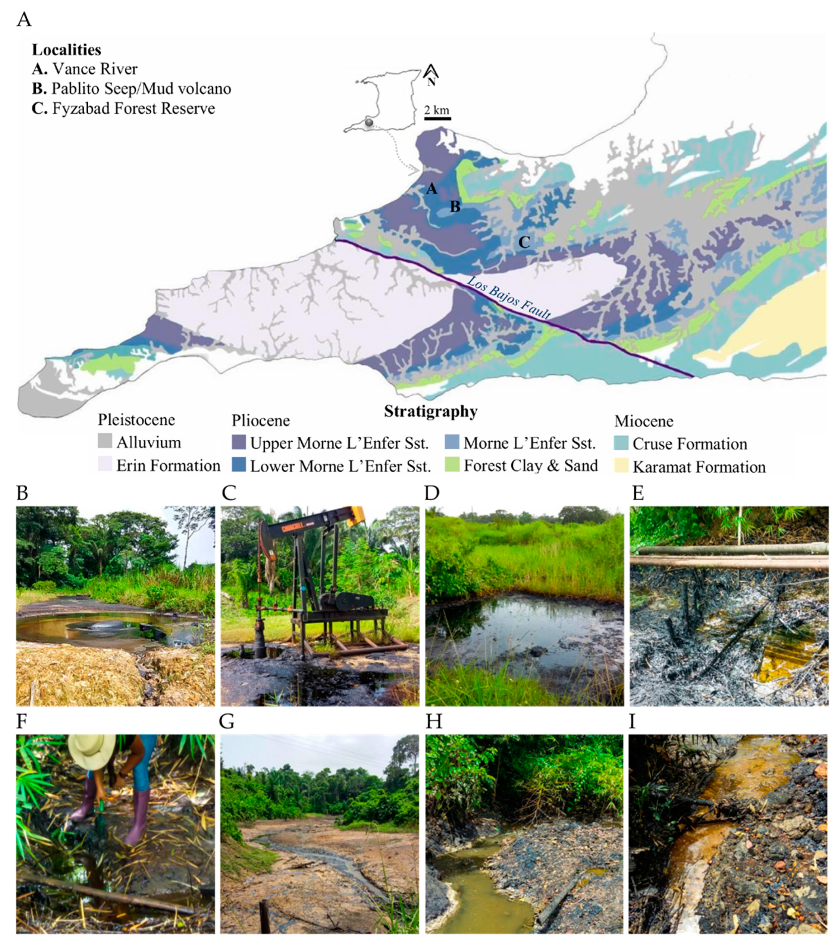
2.1. Site Descriptions

Eight study sites in three geographically separated localities in southern Trinidad were selected (Figure 1 and Table 1). These sites are associated with oil accumulations in the Miocene and Pliocene reservoirs, where crude oil has a historical occurrence. The first location, Pablito (P), was selected within the grassland area of a tropical evergreen forest (10° 11′ 37.2516″ N, 61° 37′ 13.8972″ W). One site in P containing a natural oil seep/mud volcano was sampled (Figure 1B). The second location, Fyzabad (F), is located in a tropical evergreen forest, the Fyzabad Forest Reserve (10° 10′ 0.01″ N, 61° 30′ 0″ W). Two geographically isolated sites in F, an abandoned well and a natural oil seep, were selected for sampling as an anthropogenic feature (Figure 1C,D). The third location was Vance River, Vance (V), (10° 12′ 06.2″ N, 61° 37′52.2″ W). Five geographically isolated sites were sampled at Vance River, including oil-contaminated sediments surrounding leaking pipelines, a natural oil seep where the surrounding land was heavily impregnated with oil, and sites along the river containing oil runoff accumulated in sediments (Figure 1E–I).
In Trinidad, significant asphaltic deposits are found in the southwestern region of the island, concentrated in Pilo-Pleistocene formations. For this study, we choose three unique asphaltic localities within these reservoirs. Pablito, Fyzabad, and Vance share their reservoirs with the same hydrocarbon source, the Pliocene-aged Morne L’Enfer formation sandstone and silt [2,59]. At the surface of this formation, several heavy oil and oil sand occurrences are common, and their exposure allowed for the opportunity to study the microbial communities present in heavy oil-contaminated sediments.
One site containing a natural seep is located in Pablito. This semi-evergreen seasonal forest locality is known to contain both asphalt seeps/mud volcanoes. There have been high levels of seep activity. There is no information, to our knowledge, about the physical environment in terms of soil composition, however, because it is a tropical forest, the soil pH would be slightly acidic to neutral [60]. The Fyzabad Forest Reserve is characterized as having a dense tropical semi-evergreen seasonal forest that has had minimal human disturbance since the private oilfield road spanning the reserve was gated off from the general public. The soil is the typical pH of tropical forest, slightly acidic to neutral [60]. Vance River lacks anthropogenic contamination since it is remote from settled areas.
Reservoir properties for these sites have been previously investigated [58] and are outlined in Table 1. The features and formation of these sites suggest that reservoirs are ideal for this study.

2.2. Soil Sampling and Isolation of Culturable Microbes

Samples of mud and oil within 0.5 m to crude oil sources were collected in September 2017. Soil and oil samples were collected at a depth of 0 to 10 cm using a stainless steel shovel and placed into Sterile Whirl-Pak bags that were sealed immediately after collection to avoid contamination. Soil samples were transported to the laboratory at 4 °C following collection, and microbial isolation was done within 24 h.
Microbes were isolated by means of serial dilution technique. For each soil sample, 1 g of soil was suspended in 9 mL sterile phosphate buffer pH 7.2 (KH2PO4 68 g/L) (Sigma-Aldrich, St. Louis, MO, USA), and a 100 μL aliquot from dilutions 10−1 to 10−3 were spread on a petri dish containing potato dextrose agar (PDA, HiMedia Laboratories LLC., West Chester, PA, USA) supplemented with 50 mg/L each of streptomycin and tetracycline (Sigma-Aldrich, St. Louis, MO, USA) to inhibit bacterial growth. All Petri dishes were incubated at 25 °C in the dark for a period of five days. Fungal, yeast, and antibiotic-resistant bacteria were then isolated in pure culture for further use. Inverted cultures were maintained on different media and incubated (27 °C for bacteria and 25 °C for fungi). Bacterial colonies were checked every 48 h for six days, and each morphotype was subcultured on PDA and Reasoner’s 2A agar (R2A, HiMedia Laboratories LLC., West Chester, PA, USA) for 48 h. R2A is designed to promote the growth of microorganisms that grow in low nutrient environments (Sigma-Aldrich, St. Louis, MO, USA). Fungal colonies were checked every day for two weeks and were subcultured on PDA for three weeks. Bacterial and fungal pure cultures were stored at 4 °C until gDNA extraction. In some cases, the isolates used in this study existed as co-cultures since successive subculturing to obtain pure isolates failed, and fungal–bacterial and yeast–bacterial co-cultures were included in the study.

2.3. Isolation of Crude Oil-Degrading Microbes

All isolated fungi, bacteria, and yeast were screened for their ability to utilize crude oil as a carbon source in vitro. Candidate crude oil metabolizing microbes were isolated using a growth bioassay on crude-oil-amended plates. Assays were prepared using Bushnell-Haas agar (BHA, Sigma-Aldrich, St. Louis, MO, USA) to provide the proper microelement amounts for microbial growth, except a carbon source. BHA is recommended for the examination of fuels for microbial contamination and for studying hydrocarbon deterioration by microorganisms (Sigma-Aldrich, St. Louis, MO, USA). BHA media was supplemented with crude oil (2% v/v; CARIRI Laboratories, St. Augustine, Trinidad and Tobago) and 50 mg/L each of streptomycin and tetracycline. Duplicate assays were prepared under aseptic conditions. In order to obtain homogenous distribution of oil, the media was thoroughly mixed by hand before it was added to each plate. All plates were inoculated with a 4 mm3 block of mycelium taken from the advancing edge of a colony of an actively growing axenic culture. The isolates showing growth on these assays were then characterized after two to five days of incubation at 25 °C in the dark.

2.4. Characterization of Growth on Crude Oil In vitro

Isolates showing growth on crude oil assays were characterized for their ability to grow on crude oil. Fungi and yeast (existing as co-cultures) were characterized in vitro on BHA (as described above), and, for luxuriant growth, isolates were also screened on PDA. Bacterial isolates (existing as co-cultures or pure isolates) were screened using R2A media. The media used was supplemented with crude oil (2% v/v), 50 mg/L each of streptomycin and tetracycline for fungal and yeast assays, and with and without these antibiotics for the bacterial assays.
To characterize the crude-oil-utilizing microbes, growth assays were conducted by comparing the growth rates of colonies after 6 days in addition to the appearance of the colony on the plates for fungi and yeast, i.e., (i) the zone of clearance of oil around the colony, and (ii) the disappearance of oil seen on the reverse view of the colony. The top performers were considered the most efficient hydrocarbon-utilizing microbes, and these were selected for identification and further study.

2.5. Separation of Yeast–Bacteria Co-Cultures

Isolates F1 6, F1 7, and F1 9, existing in co-cultures, were separated to obtain pure cultures to perform lipase assays. Isolation of the pure bacteria Chryseobacterium oranimense (F1 6), existing in bacteria–yeast co-culture, was achieved by successive subculturing by streaking the isolate onto specialized media consisting of MacConkey agar (HiMedia Laboratories LLC., West Chester, PA, USA) containing 0.1 mg/L cycloheximide (Sigma-Aldrich, St. Louis, MO, USA) and adjusted to pH 8.5 with 1 M NaOH (Sigma-Aldrich, St. Louis, MO, USA). After several weeks, the pure bacterial colony was plated on MacConkey plates with no fungicide, and DNA was extracted, amplified, sequenced, and identified, as described below, to confirm the presence of the bacteria alone. Isolation of Rhodotorula mucilaginosa (F1 7) and Lecythophora aff. decumbens (F1 9) yeast as pure cultures from the bacterial–yeast co-cultures was achieved by successive subculturing of isolates onto specialized media that was designed using key factors that inhibit bacterial growth, including low pH, high salinity, the use of multiple antibiotics, and a high concentration of glucose, to aid separation since initial attempts with common methods failed. Modified yeast malt agar (YM, HiMedia Laboratories LLC., West Chester, PA, USA) and glucose (Sigma-Aldrich, St. Louis, MO, USA) were prepared. Briefly, NaCl (7% w/v) was dissolved in double-distilled water; YM was then added, and the solution was sterilized. Once cooled, the pH was adjusted to approximately 3.5 using 1 N HCl (Sigma-Aldrich, St. Louis, MO, USA), and 50 mg/L each of streptomycin and tetracycline was added to the media. Plates were streaked with a co-culture colony, and, after successive subculturing, the pure yeast colony was plated on modified YM agar, and DNA was extracted, amplified, sequenced, and identified, as described below, to ensure the culture was indeed pure.

2.6. Selection of Efficient Hydrocarbon-Utilizing Microbes

Isolates showing maximum potential and top performance in vitro in crude oil assays were selected for molecular identification. The criteria for selection included: (i) minimum diameter of growth on BHA of at least 2/3 of the growth on PDA for fungal isolates, (ii) isolates must show a level of oil-degrading ability, i.e., significant zone of clearance, disappearance of crude oil, and (iii) novel species not previously reported in the literature. Isolates were maintained on appropriate agar plates (for fungi and yeast: PDA and BHA; for bacterial co-cultures: PDA and R2A) and in 1.5 mL centrifuge tubes (Eppendorf, Hamburg, Germany) in sterile distilled water at 4 °C for short term storage and in 15% glycerol at −20 °C for long term storage.

2.7. DNA Extraction, PCR, Sequencing, and Identification of Microbes

Fungal and yeast isolates (including those in co-culture) were grown on PDA supplemented with 50 mg/L each of streptomycin and tetracycline at 25 °C in the dark for two days (for fast-growing isolates) and up to five days (for slower-growing isolates). Total genomic DNA (gDNA) from fungal isolates was extracted using a MoBio PowerSoil® DNA extraction kit (Mo-Bio Laboratories, Carlsbad, CA, USA) according to the manufacturer’s protocol. DNA extracts were diluted 1:4, and this served as the working DNA concentration for polymerase chain reaction (PCR) amplification. The ITS rDNA gene region (expected PCR product size ~650 bp) was amplified by PCR using universal primer pair ITS5/4 [61]. PCR reaction conditions consisted of an initial denaturation of 5 min at 94 °C, followed by 35 cycles of 1 min of denaturation at 94 °C, 1 min of annealing at 55 °C, 1 min primer extension at 72 °C, followed by a final extension of 5 min at 72 °C.
Bacterial isolates (pure isolates and those in co-culture) were grown on R2A supplemented with 50 mg/L each of streptomycin and tetracycline in the dark for 16 h or longer until growth was sufficient for extraction. Plates were flooded with 500–700 μL of TE buffer (10 mM Tris HCl, 1 mM EDTA, pH8; Sigma-Aldrich, St. Louis, MO, USA). The wash was collected and transferred to a 1.5 mL centrifuge tube, and 100 μL of 50 mg/L each of lysozyme and proteinase K (Sigma-Aldrich, St. Louis, MO, USA) was added. The samples were incubated at 37 °C for 2 h in a water bath, with occasional mixing by inversion. Immediately after incubation, the entire sample content was transferred to Maxwell® 16 Cell DNA Purification kits (Promega, Madison, WI, USA) and gDNA was extracted according to the manufacturer’s protocol. DNA extracts were diluted 1:4, and this served as the working DNA concentration for PCR amplification. The 16S rRNA gene region (expected PCR product size ~1750 bp) was amplified by PCR with universal primer pairs 8F [62] and 1492R [63]. PCR conditions consisted of an initial denaturation of 5 min at 96 °C, followed by 33 cycles of 30 s of denaturation at 95 °C, 30 s of annealing at 55 °C, 2 min of primer extension at 72 °C, followed by a final extension of 5 min at 72 °C.
The PCR mixture (25 μL total volume) contained 12.5 μL of GoTaq® Green Master Mix (Promega, Madison, WI, USA), 0.5 μL (10 μM) of each primer (Integrated DNA Technologies, Coralville, IA, USA), 6.5 μL of Nuclease-Free water (Promega, Madison, WI, USA), and 5 μL of DNA template. All PCRs were performed on a Thermal Cycler 2720 (Thermo Fisher Scientific, Bedford, MA, USA). PCR products were visualized on a 1.5% agarose gel stained with ethidium bromide (Sigma-Aldrich, St. Louis, MO, USA) and visualized under a MiniBIS Pro System (DNR Bio Imaging System, Neve Yamin, Israel). Where amplification failed, samples were processed once again from the first PCR. Samples producing amplicons were sent for purification and sequencing (MCLAB, San Francisco, CA, USA). Due to incorrect identifications and/or incomplete ITS sequences that were submitted to GenBank, additional approved markers for molecular identification through sequence comparisons of different housekeeping genes were required for accurate species identification, i.e., the β-tubulin (βTUB) gene for Aspergillus spp. [64] and translation elongation factor 1α (TEF1α) for Fusarium spp. [65,66,67]. Currently, there is no consensus about these supplementary barcodes since, for many taxa, they are genus-specific.
DNA sequences were manually edited using BioEdit software version 7.1.9 [68] to resolve nucleotide sequence ambiguities. Identification of sequences was performed using the BLASTn (Basic Local Alignment Search Tool) algorithm against the National Centre for Biotechnology Information (NCBI) GenBank database [69]. To confirm sequence identity, at least to the genus level, phylogenetic inference was carried out using the best fit nucleotide substitution model in MEGAX [70] using the maximum likelihood (ML) algorithm with 1000 bootstrapped replications. The trees were unrooted, and the 75% consensus trees (bs > 75%) are presented. Reference sequences are available as supplementary material.

2.8. Extracellular Lipase Assay

The enzyme activity of lipase was detected by using a modified Rhodamine agar plate method [71]. Briefly, each of the selected isolates was inoculated on nutrient agar (NA, HiMedia Laboratories LLC., West Chester, PA, USA) consisting of olive oil (3% v/v) (Sigma-Aldrich, St. Louis, MO, USA), Rhodamine 6G solution (0.001% w/v) (Sigma-Aldrich, St. Louis, MO, USA), pH 7, supplemented with 50 mg/L each of streptomycin and tetracycline and incubated at 25 °C in the dark. A 4 mm3 block from the advancing edge of a colony from an actively growing axenic culture was used for inoculation. As a control, uninoculated plates were also prepared. Assays were performed in triplicate and repeated. This assay was also carried out on plates with crude oil (1% v/v). The plates were examined under UV light, and activity was determined by visual inspection for yellow- to orange-colored fluorescence [71,72,73].

2.9. Statistical Analyses

Mean growth rates and the percentage of growth inhibition of all isolates on oil-amended media were analyzed using Minitab (version 17, Minitab LLC, Pennsylvania State University, University Park, PA, USA). Percentage growth inhibition values were arcsine-transformed prior to analysis. The least significant difference (LSD) test was used to separate means that were statistically significant at p ≤ 0.05.
The RStudio software version 1.3.1093 [74] statistical programming environment was used to perform statistical analyses. Abundance statistics, standardization (conversion to relative abundances), and patterns via rank-abundance dominance (RAD) analysis were performed using the package ‘BiodiversityR’, where the best fitting model was chosen by applying the Akaike information criterion (AIC), as described by Johnson and Omland [75]. Alpha diversity metrics [76] were generated using the package ‘BiodiversityR’, the Chao 1 richness estimator [77] and absolute dominance and Simpson’s dominance (D2) [78] indices using the package ‘microbiome’, and Simpson’s evenness (E1/D) [76] using the package ‘codyn’. Nonmetric multidimensional scaling (NMDS) ordination and ward clustering based on the Bray-Curtis dissimilarity matrix were used to analyze site differences based on the abundance of microbes and extrinsic factors. Adequacy of NMDS representation was assessed by simulation of Shepard’s plot and the goodness of fit plot. A similarity percentage (SIMPER) was used to identify the major contributing genera associated. One-way analysis of similarity (ANOSIM) was also carried out to determine the level of statistical variation between locations. Figures were produced using the packages ‘ggplot2’, ‘reshape’, ‘scales’, ‘viridisLite’, ‘viridis’, and ‘extrafont’, and package ‘dendextend’ was used to construct dendrograms. Statistical differences were calculated using one-way analysis of variance (ANOVA).

3. Results

3.1. Isolate Recovery

A total of 358 microbes were isolated from the eight sites (Table 2). From site P, 29 isolates were obtained; in Fyzabad, 128 isolates were obtained from F1 and 33 isolates from F2; from Vance, 16 isolates were obtained from V1, 17 isolates from V2, 52 isolates from V3, 33 isolates from V4, and 50 isolates from V5 (Table 2). These isolates included filamentous fungi, yeast, and coisolated bacteria, which were identified using sequence comparisons.

3.2. Isolation of Microbes from Contaminated Sites

All isolates were tested for their ability to grow in the presence of crude oil as a carbon source. From the total of 358 isolates recovered, 161 isolates demonstrated the capability to grow on 2% crude-oil-amended media (BHA, PDA, and R2A) and were retained in the final dataset (Table 2). Of the 161 isolates, 128 filamentous fungi and 33 bacterial co-cultures consisting of fungal–bacterial and yeast–bacterial co-cultures grew on oil and were proven to be crude oil utilizers based on their significant survival and growth in crude-oil-enriched conditions. The bacteria recovered were resistant to streptomycin and tetracycline (50 mg/L each), were present in co-culture with fungi/yeast, and performed well on oil-amended media. Thus, they were included in the study despite the intended selection of pure filamentous fungi.

3.3. Identification of Microbes with Demonstrated Oil-Degrading Capability in Vitro

Growth rates (diameter/mm/day) and visual observations of performance were recorded for each isolate (Figure 2, Tables S1 and S2). For fungal isolates grown on PDA and BHA with 2% crude oil, Fisher’s pairwise comparisons revealed no significant difference in growth on PDA and BHA (Table 3). The result indicated that all microbes recovered were able to utilize crude oil on both media types in vitro.
ITS and 16S sequence comparisons revealed the identities of the isolates in this final dataset (GenBank Accession Nos. MW633287 to MW633318 and MW670464 to MW670580). The identities of the Trinidad isolates were confirmed at the genus level for all sequences and at the species level for the majority of the sequences (Tables S3–S5). The microbes identified were compared to other fungi and bacteria previously identified in the literature for Trinidad. Of all the genera detected, only four were detected in Trinidad in previous studies (Table S6). A global check revealed that novel oil-degrading microbes, including Oudemansiella sp. and Paraconiothyrium sp., were, for the first time, shown as utilizers of petroleum hydrocarbons, and Chaetomella sp., Neoascochyta sp., Sydowia sp., Lecythophora sp., and Sakaguchia sp. were isolated from crude-oil-contaminated soil for the first time in Trinidad and globally.

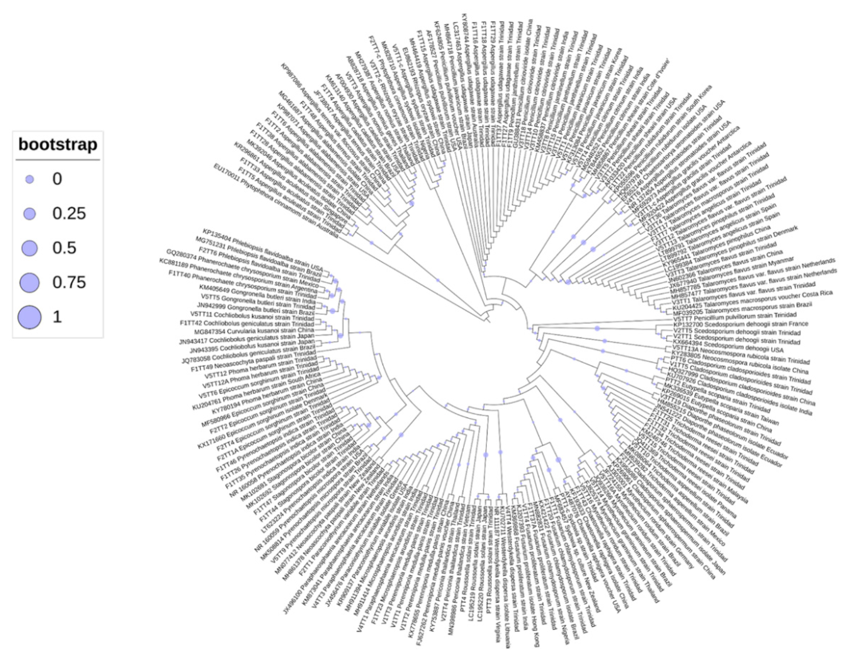
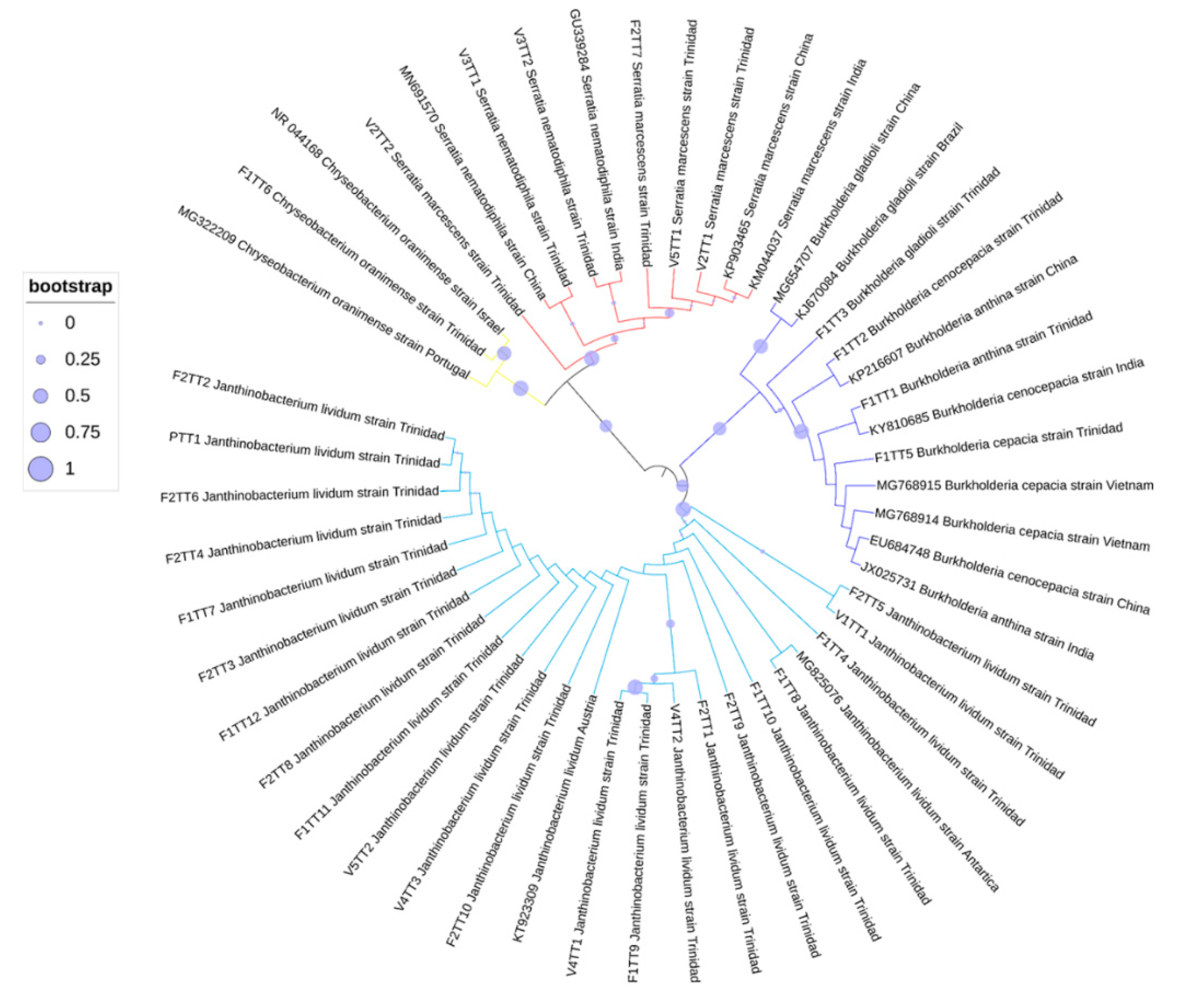
3.4. Phylogenetic Analyses of Oil-Degrading Microbes

Identification of microbes was based on ITS sequence comparisons with cognate sequences available in the GenBank database. Additional markers supported the identities indicated by ITS sequences, and, as such, unrooted phylogenetic trees were constructed based on ITS sequences using the inference of maximum likelihood with 1000 bootstrapped replicates (Figure 3, Figure 4 and Figure 5). The best fit model was also used to determine the placement of taxa, and the 50% consensus trees are presented. In each alignment, taxa were positioned according to genus, with high bootstrap support (bs > 75%), and the phylogenetic placement confirmed the identities of the isolated microbes.

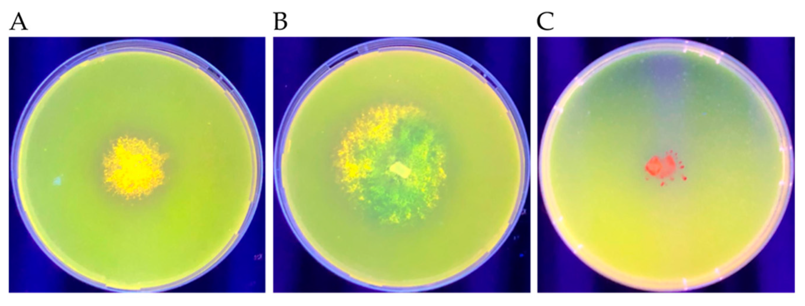
3.5. Extracellular Lipase Production

Microbes with the highest oil-degrading ability based on the parameters outlined in the previous section were selected for this extracellular lipase production assay. They were selected to determine whether the secreted lipase activity formed part of their mode(s) of oil utilization. After seven days of incubation on Rhodamine 6G plates at 25 °C, the presence of a yellow- to orange-colored fluorescence under UV light indicated secreted lipase activity. The fluorescence obtained for each isolate is described in Table 4, and examples of positive and negative results are shown in Figure 6.

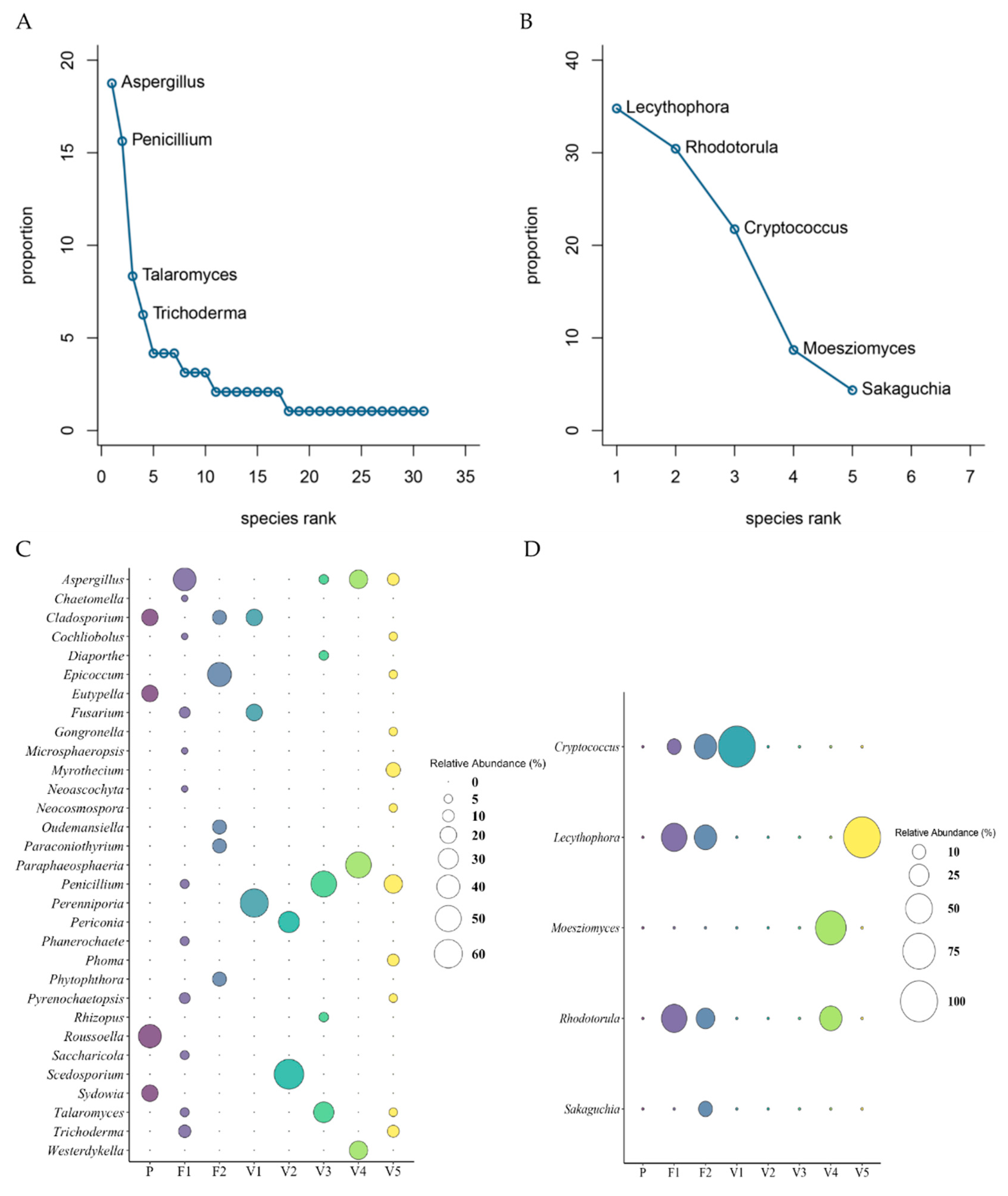
3.6. Fungi and Yeast Composition Among all Sites

Of the 119 isolates that fulfilled the selection criteria, 96 filamentous fungal isolates were detected and 23 yeast isolates were recovered with proven oil-degrading capability in vitro. The culturable oil-degrading microbial communities were microbes identified in each site, and their composition and abundance were recorded and ranked in Table 5 and Table 6. The composition and absolute abundance of genera were highly heterogeneous (Figure S1, Table 5). The overall abundance of fungi and yeast was 80.67% and 19.33%, respectively.
The filamentous fungi identified belonged to 4 phyla, 7 classes, and 31 genera. The Ascomycota (90.63%) phylum dominated the culturable diversity. Other isolates belonged to the phyla Basidiomycota (6.25%), Mucoromycota (2.08%), and Oomycota (1.04%). At a class level, Eurotiomycetes dominated (46.88%) the dataset; Dothideomycetes (28.13%) and Sordariomycetes (14.58%) were detected, while Agaricomycetes, Mucoromycetes, Leotiomycetes, and Oomycetes were present in smaller numbers (6.25%, 2.08%, 1.04%, and 1.04%, respectively). The culturable fungal genera were dominated by Aspergillus and Penicillium, accounting for 18.75% (18 individuals) and 15.63% (15 individuals) of the entire fungal community, respectively. The less abundant genera identified were Chaetomella, Cladosporium, Cochliobolus, Diaporthe, Epicoccum, Eutypella, Fusarium, Gongronella, Microsphaeropsis, Myrothecium, Neoascochyta, Neocosmospora, Oudemansiella, Paraconiothyrium, Paraphaeosphaeria, Perenniporia, Periconia, Phanerochaete, Phoma, Phytophthora, Pyrenochaetopsis, Rhizopus, Roussoella, Saccharicola, Scedosporium, Sydowia, Talaromyces, Trichoderma, and Westerdykella, accounting for 1.04–8.33% (1 to 8 individuals) of the dataset. The abundance of the different fungal classes and genera detected was significantly different (α = 0). All data are presented in Table 6 and Figure 7.
The yeast recovered belonged to 2 phyla, Basidiomycota (65.23%) and Ascomycota (34.78%). At the class level, 6 taxa were identified. Sordariomycetes, Microbotryomycetes, Tremellomycetes, Ustilaginomycetes, Agaricomycetes, and Cystobasidiomycetes were detected (34.78%, 30.44%, 17.39%, 8.70%, 4.35%, and 4.35%, respectively). A total of 5 genera of yeast were identified. Lecythophora 34.78% (8 individuals), Rhodotorula 30.43% (7 individuals), and Cryptococcus 21.74% (5 individuals) were the more dominant genera detected. Moesziomyces and Sakaguchia accounted for 8.7% (2 individuals) and 4.35% (1 individual), respectively, of the yeast dataset. There was a highly significant difference in abundance of classes and genera found (α = 0.001). All data are presented in Table 6 and Figure 7.
The RAD or dominance/diversity plot [79,80] for filamentous fungal distribution was fitted to the lognormal Zipf model (Figure S2A). Lognormal communities are common in nature, especially in tropical regions where many factors act together and environmental factors keep some stability, thus representing a more balanced community. This was seen in the fungal abundance data, where many genera were present in very close abundance values, except for the two highly abundant genera, Aspergillus and Penicillium. The yeast community RAD plot showed the best fit to be the broken stick model, which suggests that individuals are randomly distributed among the observed genera and that there are no fitted parameters (Figure S2B). Broken stick distribution usually applies to small and homogenous communities, while lognormal distribution applies to large and heterogeneous communities [81]. This common trend was reflected in this study, as represented by the smaller total abundance for yeast versus filamentous fungi as well as the number of different genera detected for filamentous fungi versus yeast. The AIC values for the RAD plots can be found in Table S7.

3.7. Microbial Composition of Fungi and Yeast per Site

In this study, we used the relative abundance, which is the percent composition of a given microbe relative to the total number of microbes in the site, to examine the abundances of the genera detected. The composition and abundance of genera were highly heterogeneous in each site, even for samples retrieved from the same locality, and the dominant genera in each site are highlighted in Table 5. The relative abundances of filamentous fungi and yeast in the 8 sites can be seen in Figure 7 and Table S8.
For the filamentous fungal isolates, Fisher’s pairwise comparisons revealed that there is a significant difference in the abundance of fungi in F1 and V5 compared to the other sites. For the yeast isolates, Fisher’s pairwise comparisons revealed that there was a significant difference in the abundance of yeast, where sites F1, F2, and V5 were significantly different from sites P, V1, V2, and V3. There was no detection of yeast in three sites (P, V2, and V3). Fisher’s pairwise comparison data can be found in Table 3.

3.8. Diversity of Fungi and Yeast

The filamentous fungi and yeast community α-diversity estimates, including the Shannon’s and Simpson’s indices, exhibited associations among the sites. In the fungal community, two sites, F1 and V5, grouped together by Fisher’s pairwise comparisons (Table 3), were the most diverse (F1: H = 2.07, D1 = 0.81; V5: H = 2.22, D1 = 0.87). These sites were multispecies communities (F1: S = 12; V5: S = 11). Site V2 was the least diverse (H = 0.64, D1 = 0.44) and was the sparsest community, consisting of only two species (S = 2). The diversity between the sites was highly significantly different (α = 0). Overall, the α-diversity estimates revealed that Vance was the most diverse location (S = 19, H = 2.52, D1 = 0.88), closely followed by Fyzabad (S = 17, H = 2.41, D1 = 0.86), while Pablito was the least diverse (S = 4, H = 1.33, D1 = 0.72). In all cases, the Chao 1 richness estimator indicated that the theoretical richness for each site was more than the observed richness. All diversity indices for the filamentous fungal community are given in Table S9.
In the yeast community, F1 and F2 were grouped in Fisher’s pairwise comparisons (Table 3) and were the most diverse (F1: H = 0.97, D1 = 0.60; F2: H = 1.31, D1 = 0.72), having the most species variation (F1: S = 3; F2: S = 4). Sites V1 and V5 were single-species communities and, thus, lacked diversity (H = 0, D1 = 0). Overall, Vance was the more diverse location (H = 1.33, D1 = 0.72) compared to Fyzabad (H = 1.22, D1 = 0.69). The Chao 1 richness estimator for Fyzabad showed that the observed richness equaled the theoretical richness; however, Vance had a lower observed richness compared to the theoretical one. All diversity indices for the yeast community are shown in Table S10.

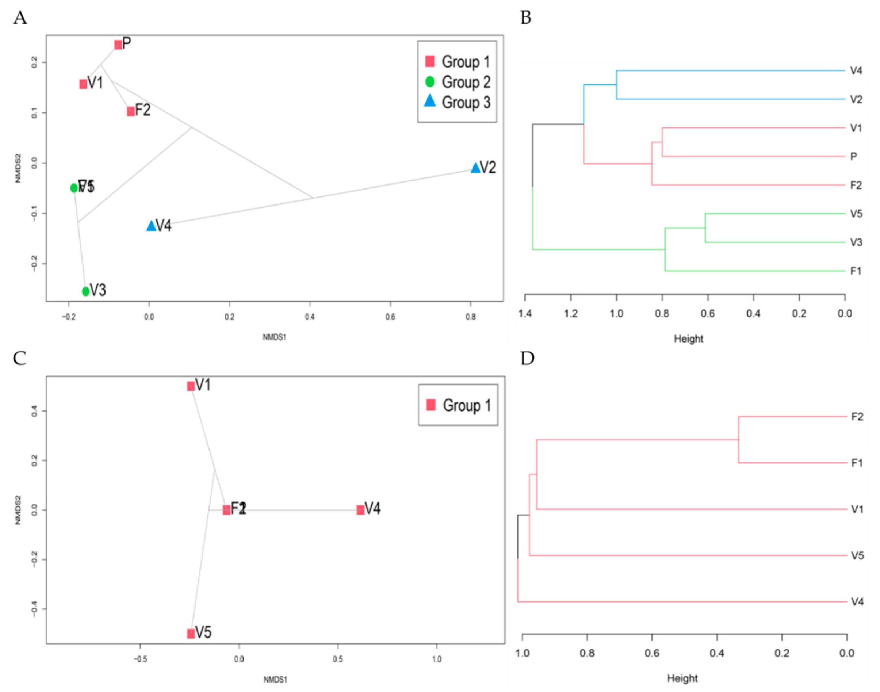
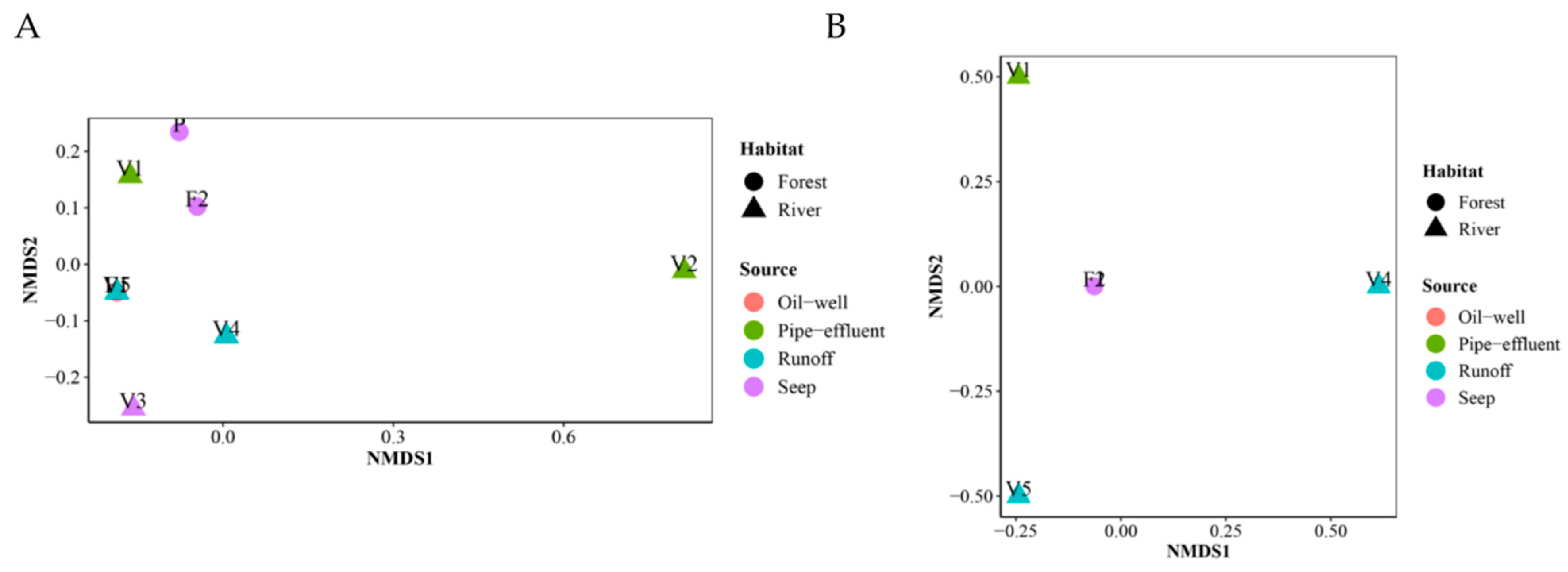
3.9. Community Ordination and Clustering

Through further investigation into the distribution of the sites’ microbial communities, the NMDS and ward clustering performed indicated that distinct microbial communities inhabited each site. RAD lognormal fungal distribution suggested large and heterogeneous communities (recall abundance of 96 filamentous fungi belonging to 31 genera was detected, with varying abundances and occurrences in each site), and broken stick yeast distribution suggested small homogenous communities (abundance of 23 yeast belonging to 5 genera was detected, with more homogenous occurrences among the sites). NMDs and Ward clustering confirmed this, as indicated by the order in which the clusters are joined and the variation in heights between these clusters (Figure 8). Adequacy of the NMDS representation was assessed by the low stress values obtained (<0.01) and the simulation of Shepard’s plot and the goodness of fit plot (Figure S3). SIMPER analysis indicated that specific genera were driving the differences between the sites (Table S11). ANOSIM analysis conveyed that similarities between locations are higher than within them but with no significant difference (Figure S4). There was no distinction in the filamentous fungal and yeast communities in the various sites based on the habitat type or source, apart from sites F1 and F2 in the yeast community (Figure 9).

4. Discussion

Indigenous microorganisms become highly adapted to survival in hydrocarbon-contaminated terrestrial environments through selective enrichment and genetic modifications that enable them to catabolize xenobiotic chemicals [82,83,84,85,86]. The potential of indigenous bacteria to degrade petroleum hydrocarbons isolated from Pitch Lake at La Brea in Trinidad has been reported [87,88,89,90]. Ramoutar et al. [91] described fungi isolated from Pitch Lake, including Aspergillus, Fusarium, Penicillium, Curvularia, and Mucorales, that were capable of PAH-degradation rates of 10–65% for single isolates and 64–76% for select consortia. Microbial populations indigenous to the many other naturally occurring hydrocarbon seeps and chronically polluted areas in Trinidad should, therefore, be more fully explored for their bioremediation and industrial potential. Stefani et al. [35] concluded that effective bioremediation remains a function of those microbes that are indigenous to a given polluted site. This investigation focused on eight such sites located in the southern peninsula of the island, and our findings indicate novel oil-degrading filamentous fungi and yeast as single-isolate degraders and naturally occurring consortia, with specific bacterial species not previously reported in the literature. Because of the different sources and means of identification of oil-degrading strains and isolates reported in the literature, the terms ‘strains’ and ‘isolates’ are used interchangeably in comparison to our isolates to retain the accuracy of the original reporting. A comparison of species reported in the literature with those isolated in this study revealed similarities in microbial composition; however, novel indigenous species with demonstrated oil-degrading ability were also revealed.

4.1. Fungi

The fungal community in the oil-contaminated soils consisted of species belonging to four phyla, where Ascomycota dominated (90.63%), followed by Basidiomycota (6.25%). The dominance of Ascomycota, followed by Basidiomycota, in polluted soils has been acknowledged: Siles and Margesin [92] reported that of the 87 operational taxonomic units (OTUs) that comprised the fungal community, up to 95.3% of the classified sequences belonged to the Ascomycota phylum and up to 62.7% belonged to the Basidiomycota phylum; Spini et al. [93] reported that of 94 unique fungal colonies, 95% belonged to Ascomycota and only 5% to Basidiomycota. PAHs include a group of priority pollutants that pose a serious threat to the health of humans and ecosystems [18]. Multiple reviews have highlighted the biotransformation of PAHs with Ascomycota and Basidiomycota fungi [43,94,95].
The fungal community was affiliated with 31 fungal genera, including Aspergillus (18.75%), Penicillium (15.63%), Talaromyces (8.33%), Trichoderma (6.25%), Epicoccum, Fusarium, Pyrenochaetopsis, Cladosporium, Myrothecium, Perenniporia, Cochliobolus, Paraphaeosphaeria, Phanerochaete, Phoma, Roussoella, Saccharicola, Scedosporium, Chaetomella, Diaporthe, Eutypella, Gongronella, Microsphaeropsis, Neoascochyta, Neocosmospora, Oudemansiella, Paraconiothyrium, Periconia, Phytophthora, Rhizopus, Sydowia, and Westerdykella, which were less abundant and represented 1% to 4% of the recovered isolates.
Hydrocarbon-degrading strains of Aspergillus and Penicillium are frequently isolated from soil [6]. In a recent study, Aspergillus and Trichoderma were reported as the most abundant genera in soils polluted with mixtures of aliphatic and polycyclic hydrocarbons [93], and, more recently, Aspergillus, Penicillium, and Trichoderma were among the culturable petroleum-degrading fungi isolated from soil samples [96]. Aspergillus and Penicillium have been reported as remediation agents of PAHs, using enzymes such as oxygenases during transformation [95,97,98].
Ligninolytic fungi such as Phanerochaete chrysosporium are well-known degraders of PAHs, including benzo[a]pyrene, due to the secretion of oxidative enzymes [99]. Members of the genus Cladosporium, Fusarium, Scedosporium, Eutypella, Talaromyces, and Cochliobolus [93,96,100,101,102], isolated from a variety of oil-contaminated sources, are also well-known biodegraders that are capable of metabolizing a range of compounds. Morales et al. [103] characterized a strain of Scedosporium that could degrade several petroleum hydrocarbons and identified 11,195 protein-coding genes that may be involved in hydrocarbon degradation pathways.
With respect to rarer genera, Epicoccum, Pyrenochaetopsis, Rhizopus, and Phoma have been reported as oil-degrading microbes [104,105,106]. Myrothecium, as a petroleum-utilizing microbe [107], has also been found to immobilize toxic metals, which may be relevant to specific bioremediation strategies as these fungi are capable of mediating metal precipitation [108]. Paraphaeosphaeria has been isolated from an asphalt seep but not investigated for its bioremediation potential [109]. Roussoella isolated from contaminated soil grows in the presence of toluene, hexadecane, and polychlorinated biphenyls (PCB) and utilizes ligninolytic enzymes for wood degradation [110]. Perenniporia [111], Saccharicola, and Diaporthe were reported as petroleum hydrocarbon degraders [112] with potential bioremediation functions, but the latter strains were isolated from leaf and stem tissues from plants that were growing in soil contaminated with crude oil and were not isolated from the contaminated soil itself [113]. Gongronella is capable of degrading hydrocarbons [114], including pyrene [115]. Microsphaeropsis is a promising genus for microbe-assisted phytoremediation [116,117]. Microsphaeropsis and Westerdykella exhibit the ability to degrade poly-ethylene terephthalate (PET depolymerization), and have shown upregulated expression of lipase and esterase activities [118]. Westerdykella also utilizes polycyclic PAHs [119] with tolerance to pyrene [120] and the depletion of fluorene [121]. Periconia, which is isolated from oil-contaminated soil, grows on petrol and kerosene, and can degrade oil [122], is one of the many microbes detected in soil for a phytoremediation strategy to remediate crude-oil-polluted soil [123]; it has also been isolated from a marine site with frequent oil spills but not investigated further for crude oil degradative capabilities [124]. Phytophthora has been reported as an aromatic degrader [125], specifically for naphthalene [126], and Neocosmospora isolated from contaminated soil has demonstrated hydrocarbonoclastic abilities [127].
Rarely isolated genera such as Oudemansiella has shown enzymatic activity similar to that of oil-degrading microbes, which also aids in its ability to decolorize polymeric dyes and can be correlated with xenobiotic degradation, just as petroleum hydrocarbon molecules are similar to lignin molecules [128]. It has also been used to promote the dissipation of pyrene from soil [129]. Paraconiothyrium has been isolated from mangroves that are impacted by PAH contamination [130]. In this study, Oudemansiella and Paraconiothyrium were, for the first time, shown to be utilizers of petroleum hydrocarbons. Additionally, new oil-degrading fungi, Chaetomella, Neoascochyta, and Sydowia, were isolated from contaminated soil and have not been reported elsewhere in any known bioremediation study. Since these fungi degrade crude oil, the possibility for their use exists in the development of microbial technology for the remediation of crude-oil-contaminated sites.

4.2. Yeast

All yeast isolates were affiliated to Basidiomycota (65.23%) and Ascomycota (34.78%) phyla, which is the most reported taxonomic phylum in terrestrial environments (see [131,132]). The yeast detected in this study belonged to the genera Lecythophora (34.78%), Rhodotorula (30.45%), Cryptococcus (21.74%), Moesziomyces (synonym of the asexually typified genus Pseudozyma) (8.70%), and Sakaguchia (4.35%).
Rhodotorula is a commonly isolated yeast from oil-contaminated environments [6,133]. In a recent study by Mikolasch et al. [134], Rhodotorula was isolated from oil-contaminated soils using a variety of hydrocarbons and could break down cyclohexanone. In a study by Hashem et al. [135] of the 67 yeast strains tested, Rhodotorula was among the top six isolates for its ability to degrade both aliphatic and aromatic hydrocarbons, and further investigation revealed two strains as the best degraders of octane and pyrene, with other research reporting similar findings [136]. Species of Rhodotorula have also been shown as degraders of decane and nonane [137]. Rhodotorula and Cryptococcus were found to catabolize benzene compounds [131] and are potent degraders of diesel oil, utilizing a range of enzymes including aminopyrine-N-demethylase, alcohol dehydrogenase, aldehyde dehydrogenase, catalase, and glutathione S transferase. The cytochrome P450 monooxygenases were especially important to diesel oil degradation [138], and isolates of these genera from petroleum-contaminated environments could produce secreted lipases [133].
The yeast Moesziomyces (previously cited as Pseudozyma but now reassigned based on molecular phylogenetic analysis [139,140] has rarely been described as hydrocarbon-degrading but was recently shown to be efficient tetradecane- [134] and diesel-fuel-degraders [141]. Furthermore, the yeast Lecythophora and Sakaguchia have, to our knowledge, never been reported in the literature as known oil degraders isolated from oil-contaminated soils. Lecythophora species have been shown to degrade PAHs such as pyrene [142,143]. Sakaguchia has been previously isolated from a marine environment, and it was surmised that its coexistence with another oil-degrading yeast species suggests hydrocarbon utilization capability [144].

4.3. Co-isolated Bacteria

At the genus level, Janthinobacterium (66.67%), Serratia (18.18%), Burkholderia (12.12%), and Chryseobacterium (3.03%) were isolated from crude-oil-contaminated soil in this study and had a similar occurrence with previously reported hydrocarbon-degrading members of the same genera [46,93,145,146,147]. Since only a few bacteria can grow on high molecular weight PAHs and the metabolism of PAHs by bacteria is limited due to the poor bioavailability of these compounds, the importance of research on bacteria capable of degrading petroleum hydrocarbons, especially PAHs, has been highlighted [43]. In addition, there is a lack of degradation studies on the complexity of PAHs present in multicomponent mixtures in natural environments, which influences the rate and extent of biodegradation [84]. In agreement with a previous study [148], the dominant bacterium isolated in this study was Janthinobacterium. Janthinobacterium has been isolated from oil-contaminated soil [148] and can degrade carbazole [149] and persistent organic pollutants, explicitly polychlorinated biphenyls (PCBs) [150]. Chryseobacterium has been isolated from oil-contaminated soil [145], and its use in consortia has shown potential for its application in the bioremediation of soils [151], including PAHs [152]. Serratia has shown a relatively high capacity to degrade a wide spectrum of hydrocarbons [93,146,153] and demonstrated degradation rates of up to 80.4% for benzo[a]pyrene [154]. Burkholderia has been reported as one of the limited taxonomic groups that can degrade PAHs in soil [46,147] and via bacterial consortium [155] after engaging aromatic degradation pathways [156]. Pseudomonas, one of the most popular and dominant bacteria, was, surprisingly, not isolated in this study [147].

4.4. Extracellular Lipase Production

Extracellular microbial lipases and their wide range of applications represent commercial value for such microbes [157,158,159]. Enhanced biodegradation of petroleum hydrocarbons is maintained by biocatalysts such as lipases [158]. Among the seven isolates screened, the most promising lipase producer appeared to be F1 40: Phanerochaete chrysosporium. This species is well-documented for its ability to produce lipase and its role in degrading xenobiotics, including PAHs [99,160]. Isolates V2 5 (Scedosporium dehoogii), F1 1 (Burkholderia anthina), V2 1 (Serratia marcescens), and F1 9 (Lecythophora aff. decumbens) also showed lipase production but to a lesser extent compared with P. chrysosporium. Two isolates, F1 6 (Chryseobacterium oranimense) and F1 7 (Rhodotorula mucilaginosa), showed no secreted lipase production. The media composition and physiochemical factors (temperature, pH, and dissolved oxygen) can influence the production of extracellular lipases [158,161]. Lipases are inducible enzymes, and one of the most influential factors is the carbon source [161,162]. Further studies on the induction of extracellular lipase activity are warranted.

4.5. Novel Oil-Degraders from Trinidad

This is the first report of representatives of fungal genera Penicillium, Talaromyces, Trichoderma, Epicoccum, Pyrenochaetopsis, Cladosporium, Myrothecium, Perenniporia, Cochliobolus, Paraphaeosphaeria, Phanerochaete, Phoma, Roussoella, Saccharicola, Scedosporium, Chaetomella, Diaporthe, Eutypella, Gongronella, Microsphaeropsis, Neoascochyta, Neocosmospora, Oudemansiella, Paraconiothyrium, Periconia, Phytophthora, Rhizopus, Sydowia, and Westerdykella; bacterial genera Janthinobacterium, Serratia, and Chryseobacterium; and yeast genera Lecythophora, Rhodotorula, Cryptococcus, Moesziomyces, and Sakaguchia in Trinidad with demonstrated hydrocarbon-degrading ability in vitro. This finding is especially important due to the deficit of published data on the use of common commercial products in tropical environments and their effectiveness and suitability in local conditions [87]. Such indigenous microorganisms, which have been established and developed through selective enrichment and genetic modifications to survive and thrive in their hydrocarbon-polluted environment [163], can serve as a more appropriate product source compared to other commercial counterparts [87].
Methods of detection are critical to the analysis of microbial diversity. Utilization of culture-dependent and culture-independent approaches can provide two exceedingly different microbial compositions [40]. While culture-based methods are the classical approach to isolated hydrocarbon-degrading microbes, it must be noted that less than 1% of the microbes present in soil are cultivable [164]. However, understanding the contribution of these microbes to the complex and cooperative processes of survival in chronically contaminated soil requires their cultivation [165]. This study was a culture-dependent one, and the results suggested the activation and growth of rarer species during culture. In a study by M’rassi et al. [37], 40% of the strains isolated from oil-contaminated soil could not be detected because microbial assemblages are influenced by several different factors [6,29]. The microenvironment, geographical location, reservoir temperatures, soil physico-chemical properties, and oil composition all affect the microbial community structure and abundance [29,164,166,167,168]. In accordance with previous research, the specific conditions of the culture-based approach for microbe recovery impacted the observed microbial diversity in this study and the detection of rarer taxa [29,40]. Culture-dependent methods can also elicit the favorable growth of microbes that are experiencing antagonistic effects and those that no longer have to compete for limited resources in their natural environment. Conversely, co-metabolism between fungi and yeast with bacteria, as found in this study, must also be considered and investigated [169].
The isolates in this study were identified by sequence comparisons and phylogenetic inference, and the results indicated that taxa placement was genus-specific in all cases, irrespective of the origin of the sequence, i.e., the sequence of an isolate or strain from a source that may or may not be contaminated with crude oil. Further studies on the mapping and profiling of single genes and biochemical gene clusters to PAH-degradative pathways of generalist oil degraders and those which may be specific to indigenous microbes in chronically contaminated soil that promote the enrichment of specialist oil degraders need to be carried out.

5. Conclusions

This study focused on the unique culturable microbial composition of soil chronically contaminated with crude oil. Several fungal and yeast species not previously reported have been identified here as potential crude oil degraders with demonstrated lipase production and secretion that aid in oil break-down. The latter aspect may also be important to several industrial applications. Co-metabolism between specific yeast and bacteria was also detected, which supports the consortium approach to understanding in situ degradation. These co-cultures were difficult to separate in vitro, and it is likely that survival is codependent in the natural environment. Biodiversity analysis indicated that specific fungal species of Aspergillus and Penicillium dominated the culturable microbial landscape, and these findings were similar to other studies. While yeast composition and species diversity are limited, the yeast–bacteria co-cultures demonstrated a very high potential for crude oil degradation. Cumulatively, the findings of this study are especially important for exploitation in tropical environments and for understanding the use of indigenous microbes in sustainable bioremediation and biodeterioration efforts.

Supplementary Materials

The following are available online at https://www.mdpi.com/article/10.3390/microorganisms9061167/s1, Figure S1: Absolute abundances of microbes detected. Abundance of (A) filamentous fungi and (B) yeast per genus among the 8 sites in southwestern Trinidad, Table S1: Quantitative and qualitative characteristics of fungal isolates on 2% oil-amended media. Growth rate is given as diameter/mm/day. * represents an overgrown culture, and no measurement was taken. Visual observations key: eod = excellent oil degradation, lod = little/minor oil degradation, god = good oil degradation, pod = poor oil degradation, nod = no oil degradation, d = oil droplets, r = ring of clearance around fungi, b = oil blobs, p = oil pusher, w = waves in oil, and r of oil = ring of oil. Isolate being selected as a top performer for identification is indicated by Y = yes. Isolates not selected as top performers are indicated by N = no. Figure S2: Rank-abundance dominance (RAD) plots for genera distribution of (A) filamentous fungi and (B) yeast. The best model is represented by the bolded line. Null represents the MacArthur model; Preemption represents the geometric series of the Motomura model; Lognormal represents the lognormal distribution of Preston; Zipf represents the Zipf Model; Mandelbrot represents the Zipf-Mandelbrot Model, Table S2: Visual observations of bacterial co-culture isolates on 2% oil-amended media. R2A media supported bacterial growth, and PDA supported yeast growth. Visual characterization key: z = zones of clearance by bacteria, nz = no zone clearance by bacteria, sz = small zone of clearance by bacteria, bz = big zone of clearance by bacteria, NOB = no oil behind bacteria, and r of oil = ring of oil. Zones of clearance key: * = small zone of clearance, ** = medium zone of clearance, *** = massive zone of clearance, **** = entire plate cleared, negative sign = no zone of clearance but growth was greater in this media, and x = no zone of clearance and growth was similar, Figure S3: Diagnostics for NMDS. (A) and (C) Shepard plot for filamentous fungi and yeast, respectively, showing the success of the NMDS since individual blue points do not deviate from fitted nonparametric regression (red line); R2 values of 1 indicate that the points match the monotonically increasing line perfectly. (B) and (D) Goodness of fit plot for filamentous fungi and yeast, respectively, shows no genera contributing to stress, Table S3: Identification of fungi in the final dataset from 8 sites in southwestern Trinidad. GenBank accession information is provided, where two references are provided per species, Figure S4: ANOSIM analysis for (A) filamentous fungi and (B) yeast among the locations, Table S4: Identification of bacterial dataset from 8 sites in southwestern Trinidad. GenBank accession information is provided where two references are provided per species. Table S5: Identification of yeast dataset from 8 sites in southwestern Trinidad. GenBank accession information is provided, where two references are provided per species. Table S6: Hydrocarbon degrading microbes previously identified in Trinidad. Microbes in bold were identified in this study. Table S7: The Akaike information criterion (AIC) value for the genera rank abundance distribution models of the filamentous fungi and yeast communities. Table S8: Relative abundances of filamentous fungi and yeast in the 8 sites in southwestern Trinidad. Table S9: Diversity indices for filamentous fungal communities in the 8 sites in southwestern Trinidad. Table S10: Diversity indices for yeast communities in the 8 sites in southwestern Trinidad. Table S11: SIMPER analysis. Cumulative and overall contributions of key contributing genera responsible for the observed patterns between sites. For example, for the filamentous fungal community, differences between Pablito and Fyzabad are driven by Aspergillus, Epicoccum, Roussoella, Eutypella, Sydowia, Trichoderma, Oudemansiella, and Paraconiothyrium in descending order; together, these eight genera drive more than 70% of the difference between the communities in Pablito and Fyzabad; overall, the fungal communities in Pablito and Fyzabad are 91.7% different from each other.

Author Contributions

Conceptualization, A.C.R. and S.N.R.; methodology, A.C.R. and S.N.R.; R software, A.C.R.; formal biodiversity analysis, A.C.R.; writing—original draft preparation, A.C.R. and S.N.R.; writing—review and editing, S.N.R.; supervision, S.N.R. All authors have read and agreed to the published version of the manuscript.

Funding

This research was funded by a UNIVERSITY OF THE WEST INDIES CAMPUS RESEARCH AND PUBLICATIONS GRANT (grant number CRP.5.OCT18.77) to A. C. Ramdass and a STAFF BENEFIT GRANT to S. N. Rampersad.

Institutional Review Board Statement

Not applicable.

Informed Consent Statement

Not applicable.

Data Availability Statement

Sequence reads were deposited in the National Center for Biotechnology Information Sequence GenBank (NCBI GenBank; Accession Nos. MW633287 to MW633318 and MW670464 to MW670580). The R data that supports the conclusions of this article will be made available by the authors upon reasonable request.

Acknowledgments

The authors wish to thank Vijai Ramdhan for laboratory assistance and Stephen Narine and Brent Daniel for field assistance. We also wish to thank Neil Jeffers for his assistance with producing the map and Sherwin Louis for his assistance with editing the figures.

Conflicts of Interest

The authors declare no conflict of interest. The funders had no role in the design of the study; in the collection, analyses, or interpretation of data; in the writing of the manuscript, or in the decision to publish the results.

References

  1. Macready, G.A. Petroleum industry of Trinidad. Trans. AIME 1921, 65, 58–68. [Google Scholar] [CrossRef]
  2. Mychajliw, A.M.; Mohammed, R.S.; Rice, K.A.; Farrell, A.B.; Rincón, A.D.; McAfee, R.; McDonald, H.G.; Lindsey, E.L. The biogeography of “breas”: Contextualizing the taphonomy, ecology, and diversity of Trinidad’s asphaltic fossil record. Quat. Sci. Rev. 2020, 232, 106179. [Google Scholar] [CrossRef]
  3. Persad, K.M.; Archie, C. History of petroleum exploration in Trinidad and Tobago. In Proceedings of the AAPG/SEG International Conference and Exhibition, Cancun, Mexico, 6–9 September 2016. [Google Scholar]
  4. Scribd. Offshore Seeps Study: Trinidad and Tobago 2018 Seepage Study. Available online: https://www.scribd.com/document/378203277/3693-17FY-NP-478-V1-Trinidad-Tobago-2017-300-v2 (accessed on 25 February 2021).
  5. Sutton, N.B.; Maphosa, F.; Morillo, J.A.; Al-Soud, W.A.; Langenhoff, A.A.M.; Grotenhuis, T.; Rijnaarts, H.H.M.; Smidt, H. Impact of long-term diesel contamination on soil microbial community structure. Appl. Environ. Microbiol. 2013, 79, 619–630. [Google Scholar] [CrossRef] [PubMed] [Green Version]
  6. Leahy, J.G.; Colwell, R.R. Microbial degradation of hydrocarbons in the environment. Microbiol. Mol. Biol. Rev. 1990, 54, 305–315. [Google Scholar] [CrossRef]
  7. Head, I.M.; Jones, D.M.; Röling, W.F.M. Marine microorganisms make a meal of oil. Nat. Rev. Microbiol. 2006, 4, 173–182. [Google Scholar] [CrossRef]
  8. Laffon, B.; Pásaro, E.; Valdiglesias, V. Effects of exposure to oil spills on human health: Updated review. J. Toxicol. Environ. Health Part B 2016, 19, 105–128. [Google Scholar] [CrossRef]
  9. Michel, J.; Fingas, M. Oil spills: Causes, consequences, prevention, and countermeasures. In Fossil Fuels: Current Status and Future Directions; Crawley, G.M., Ed.; World Scientific: Singapore, 2016; pp. 159–201. [Google Scholar]
  10. Al-Hawash, A.B.; Dragh, M.A.; Li, S.; Alhujaily, A.; Abbood, H.A.; Zhang, X.; Ma, F. Principles of microbial degradation of petroleum hydrocarbons in the environment. Egypt. J. Aquat. Res. 2018, 44, 71–76. [Google Scholar] [CrossRef]
  11. Duffy, J.J.; Peake, E.; Mohtadi, M.F. Oil spills on land as potential sources of groundwater contamination. Environ. Int. 1980, 3, 107–120. [Google Scholar] [CrossRef]
  12. Nadim, F.; Hoag, G.E.; Liu, S.; Carley, R.J.; Zack, P. Detection and remediation of soil and aquifer systems contaminated with petroleum products: An overview. J. Pet. Sci. Eng. 2000, 26, 169–178. [Google Scholar] [CrossRef]
  13. Doherty, V.F.; Otitoloju, A.A. Monitoring of soil and groundwater contamination following a pipeline explosion and petroleum product spillage in Ijegun, Lagos Nigeria. Environ. Monit. Assess. 2013, 185, 4159–4170. [Google Scholar] [CrossRef]
  14. Gross, S.A.; Avens, H.J.; Banducci, A.M.; Sahmel, J.; Panko, J.M.; Tvermoes, B.E. Analysis of BTEX groundwater concentrations from surface spills associated with hydraulic fracturing operations. J. Air Waste Manag. Assoc. 2013, 63. [Google Scholar] [CrossRef] [Green Version]
  15. Shores, A.; Laituri, M.; Butters, G. Produced water surface spills and the risk for BTEX and naphthalene groundwater contamination. Water Air Soil Pollut. 2017, 228, 1–13. [Google Scholar] [CrossRef]
  16. Rochman, F.F.; Sheremet, A.; Tamas, I.; Saidi-Mehrabad, A.; Kim, J.-J.; Dong, X.; Sensen, C.W.; Gieg, L.M.; Dunfield, P.F. Benzene and naphthalene degrading bacterial communities in an oil sands tailings pond. Front. Microbiol. 2017, 8, 1845. [Google Scholar] [CrossRef] [PubMed]
  17. Andersson, J.T.; Achten, C. A critical look at the 16 EPA PAHs. Polycycl. Aromat. Compd. 2015, 35, 143–146. [Google Scholar] [CrossRef]
  18. Ghosal, D.; Ghosh, S.; Dutta, T.K.; Ahn, Y. Current state of knowledge in microbial degradation of polycyclic aromatic hydrocarbons (PAHs): A review. Front. Microbiol. 2016, 7, 1369. [Google Scholar] [CrossRef] [Green Version]
  19. Alegbeleye, O.O.; Opeolu, B.O.; Jackson, V.A. Polycyclic aromatic hydrocarbons: A critical review of environmental occurrence and bioremediation. Environ. Manag. 2017, 60, 758–783. [Google Scholar] [CrossRef]
  20. Brown, L.D.; Ulrich, A.C.; Fingas, M. Bioremediation of oil spills on land. In Handbook of Oil Spill Science and Technology; Fingas, M., Ed.; John Wiley & Sons, Inc.: Hoboken, NJ, USA, 2015; Volume 724, pp. 395–406. [Google Scholar]
  21. Baniasadi, M.; Mousavi, S.M. A comprehensive review on the bioremediation of oil spills. In Microbial Action on Hydrocarbons; Kumar, V., Kumar, M., Prasad, R., Eds.; Springer: Singapore, 2018; pp. 223–254. [Google Scholar]
  22. Ali, N.; Dashti, N.; Khanafer, M.; Al-Awadhi, H.; Radwan, S. Bioremediation of soils saturated with spilled crude oil. Sci. Rep. 2020, 10, 1116. [Google Scholar] [CrossRef] [Green Version]
  23. Austin, R.; Callaghan, A. Microbial enzymes that oxidize hydrocarbons. Front. Microbiol. 2013, 4, 338. [Google Scholar] [CrossRef] [Green Version]
  24. Naing, S.-H.; Parvez, S.; Pender-Cudlip, M.; Groves, J.T.; Austin, R.N. Substrate specificity and reaction mechanism of purified alkane hydroxylase from the hydrocarbonoclastic bacterium Alcanivorax borkumensis (AbAlkB). J. Inorg. Biochem. 2013, 121, 46–52. [Google Scholar] [CrossRef] [Green Version]
  25. Obire, O.; Anyanwu, E.C. Impact of various concentrations of crude oil on fungal populations of soil. Int. J. Environ. Sci. Technol. 2009, 6, 211–218. [Google Scholar] [CrossRef] [Green Version]
  26. Makula, R.A.; Lockwood, P.J.; Finnerty, W.R. Comparative analysis of the lipids of Acinetobacter species grown on hexadecane. J. Bacteriol. 1975, 121, 250–258. [Google Scholar] [CrossRef] [Green Version]
  27. Wang, L.; Li, F.; Zhan, Y.; Zhu, L. Shifts in microbial community structure during in situ surfactant-enhanced bioremediation of polycyclic aromatic hydrocarbon-contaminated soil. Environ. Sci. Pollut. Res. 2016, 23, 14451–14461. [Google Scholar] [CrossRef]
  28. Bundy, J.G.; Paton, G.I.; Campbell, C.D. Microbial communities in different soil types do not converge after diesel contamination. J. Appl. Microbiol. 2002, 92, 276–288. [Google Scholar] [CrossRef]
  29. Powell, S.M.; Bowman, J.P.; Ferguson, S.H.; Snape, I. The importance of soil characteristics to the structure of alkane-degrading bacterial communities on sub-Antarctic Macquarie Island. Soil Biol. Biochem. 2010, 42, 2012–2021. [Google Scholar] [CrossRef]
  30. Hobbie, J.E.; Daley, R.J.; Jasper, S. Use of nuclepore filters for counting bacteria by fluorescence microscopy. Appl. Environ. Microbiol. 1977, 33, 1225–1228. [Google Scholar] [CrossRef] [Green Version]
  31. Roszak, D.B.; Colwell, R.R. Survival strategies of bacteria in the natural environment. Microbiol. Rev. 1987, 51, 365. [Google Scholar] [CrossRef] [PubMed]
  32. Yang, Y.; Wang, J.; Liao, J.; Xie, S.; Huang, Y. Distribution of naphthalene dioxygenase genes in crude oil-contaminated soils. Microb. Ecol. 2014, 68, 785–793. [Google Scholar] [CrossRef] [PubMed]
  33. Jiao, S.; Liu, Z.; Lin, Y.; Yang, J.; Chen, W.; Wei, G. Bacterial communities in oil contaminated soils: Biogeography and co-occurrence patterns. Soil Biol. Biochem. 2016, 98, 64–73. [Google Scholar] [CrossRef] [Green Version]
  34. Shin, B.; Kim, M.; Zengler, K.; Chin, K.-J.; Overholt, W.A.; Gieg, L.M.; Konstantinidis, K.T.; Kostka, J.E. Anaerobic degradation of hexadecane and phenanthrene coupled to sulfate reduction by enriched consortia from northern Gulf of Mexico seafloor sediment. Sci. Rep. 2019, 9, 1239. [Google Scholar] [CrossRef]
  35. Stefani, F.O.P.; Bell, T.H.; Marchand, C.; de la Providencia, I.E.; El Yassimi, A.E.; St-Arnaud, M.; Hijri, M. Culture-dependent and-independent methods capture different microbial community fractions in hydrocarbon-contaminated soils. PLoS ONE 2015, 10, e0128272. [Google Scholar] [CrossRef] [Green Version]
  36. Bhattacharya, D.; Sarma, P.M.; Krishnan, S.; Mishra, S.; Lal, B. Evaluation of genetic diversity among Pseudomonas citronellolis strains isolated from oily sludge-contaminated sites. Appl. Environ. Microbiol. 2003, 69, 1435–1441. [Google Scholar] [CrossRef] [Green Version]
  37. M’rassi, A.G.; Bensalah, F.; Gury, J.; Duran, R. Isolation and characterization of different bacterial strains for bioremediation of n-alkanes and polycyclic aromatic hydrocarbons. Environ. Sci. Pollut. Res. 2015, 22, 15332–15346. [Google Scholar] [CrossRef] [PubMed]
  38. El Mahdi, A.M.; Aziz, H.A.; Abu Amr, S.S.; El-Gendy, N.S.; Nassar, H.N. Isolation and characterization of Pseudomonas sp. NAF1 and its application in biodegradation of crude oil. Environ. Earth Sci. 2016, 75, 380. [Google Scholar] [CrossRef]
  39. Bell, T.H.; Stefani, F.O.P.; Abram, K.; Champagne, J.; Yergeau, E.; Hijri, M.; St-Arnaud, M. A diverse soil microbiome degrades more crude oil than specialized bacterial assemblages obtained in culture. Appl. Environ. Microbiol. 2016, 82, 5530. [Google Scholar] [CrossRef] [PubMed] [Green Version]
  40. Sun, J.-Q.; Xu, L.; Liu, X.-Y.; Zhao, G.-F.; Cai, H.; Nie, Y.; Wu, X.-L. Functional genetic diversity and culturability of petroleum-degrading bacteria isolated from oil-contaminated soils. Front. Microbiol. 2018, 9, 1332. [Google Scholar] [CrossRef]
  41. Gieg, L.M.; Fowler, S.J.; Berdugo-Clavijo, C. Syntrophic biodegradation of hydrocarbon contaminants. Curr. Opin. Biotechnol. 2014, 27, 21–29. [Google Scholar] [CrossRef] [PubMed]
  42. Juhasz, A.L.; Naidu, R. Bioremediation of high molecular weight polycyclic aromatic hydrocarbons: A review of the microbial degradation of benzo[a]pyrene. Int. Biodeterior. Biodegrad. 2000, 45, 57–88. [Google Scholar] [CrossRef]
  43. Harms, H.; Schlosser, D.; Wick, L.Y. Untapped potential: Exploiting fungi in bioremediation of hazardous chemicals. Nat. Rev. Microbiol. 2011, 9, 177–192. [Google Scholar] [CrossRef]
  44. Abbasian, F.; Lockington, R.; Mallavarapu, M.; Naidu, R. A comprehensive review of aliphatic hydrocarbon biodegradation by bacteria. Appl. Biochem. Biotechnol. 2015, 176, 670–699. [Google Scholar] [CrossRef]
  45. Booth, J.M.; Fusi, M.; Marasco, R.; Michoud, G.; Fodelianakis, S.; Merlino, G.; Daffonchio, D. The role of fungi in heterogeneous sediment microbial networks. Sci. Rep. 2019, 9, 7537. [Google Scholar] [CrossRef] [PubMed] [Green Version]
  46. Johnsen, A.R.; Wick, L.Y.; Harms, H. Principles of microbial PAH-degradation in soil. Environ. Pollut. 2005, 133, 71–84. [Google Scholar] [CrossRef] [PubMed]
  47. Saul, D.J.; Aislabie, J.M.; Brown, C.E.; Harris, L.; Foght, J.M. Hydrocarbon contamination changes the bacterial diversity of soil from around Scott Base, Antarctica. FEMS Microbiol. Ecol. 2005, 53, 141–155. [Google Scholar] [CrossRef] [PubMed] [Green Version]
  48. Al-Awadhi, H.; Dashti, N.; Khanafer, M.; Al-Mailem, D.; Ali, N.; Radwan, S. Bias problems in culture-independent analysis of environmental bacterial communities: A representative study on hydrocarbonoclastic bacteria. SpringerPlus 2013, 2, 1–11. [Google Scholar] [CrossRef] [Green Version]
  49. Bell, T.H.; Yergeau, E.; Maynard, C.; Juck, D.; Whyte, L.G.; Greer, C.W. Predictable bacterial composition and hydrocarbon degradation in Arctic soils following diesel and nutrient disturbance. ISME J. 2013, 7, 1200–1210. [Google Scholar] [CrossRef] [PubMed]
  50. dos Santos, H.F.; Cury, J.C.; do Carmo, F.L.; dos Santos, A.L.; Tiedje, J.; van Elsas, J.D.; Rosado, A.S.; Peixoto, R.S. Mangrove bacterial diversity and the impact of oil contamination revealed by pyrosequencing: Bacterial proxies for oil pollution. PLoS ONE 2011, 6, e16943. [Google Scholar] [CrossRef] [PubMed] [Green Version]
  51. Röling, W.F.M.; Milner, M.G.; Jones, D.M.; Lee, K.; Daniel, F.; Swannell, R.J.P.; Head, I.M. Robust hydrocarbon degradation and dynamics of bacterial communities during nutrient-enhanced oil spill bioremediation. Appl. Environ. Microbiol. 2002, 68, 5537–5548. [Google Scholar] [CrossRef] [Green Version]
  52. Kostka, J.E.; Prakash, O.; Overholt, W.A.; Green, S.J.; Freyer, G.; Canion, A.; Delgardio, J.; Norton, N.; Hazen, T.C.; Huettel, M. Hydrocarbon-degrading bacteria and the bacterial community response in Gulf of Mexico beach sands impacted by the Deepwater Horizon oil spill. Appl. Environ. Microbiol. 2011, 77, 7962–7974. [Google Scholar] [CrossRef] [Green Version]
  53. Vartoukian, S.R.; Palmer, R.M.; Wade, W.G. Strategies for culture of ‘unculturable’ bacteria. FEMS Microbiol. Lett. 2010, 309, 1–7. [Google Scholar] [CrossRef] [Green Version]
  54. Kugler, H.G. Geological Map of Trinidad; Petroleum Association of Trinidad: Port of Spain, Trinidad and Tobago, 1959. [Google Scholar]
  55. Chen, S.; Steel, R.J.; Dixon, J.F.; Osman, A. Facies and architecture of a tide-dominated segment of the late Pliocene Orinoco Delta (Morne L’Enfer Formation) SW Trinidad. Mar. Pet. Geol. 2014, 57, 208–232. [Google Scholar] [CrossRef]
  56. Dasgupta, S.; Buatois, L.A.; Zavala, C.; Mángano, M.G.; Törő, B. Ichnology of a subaqueously prograding clastic wedge, late Pliocene Morne L’Enfer Formation, Fullarton, Trinidad: Implications for recognition of autogenic erosional surfaces and delineation of stress factors on irregular echinoids. Palaeogeogr. Palaeoclimatol. Palaeoecol. 2016, 459, 365–380. [Google Scholar] [CrossRef]
  57. Baboolal, A.A.; Littke, R.; Wilson, B.; Stock, A.T.; Knight, J. Petrographical and geochemical characterization of lignites, sub-bituminous coals and carbonaceous sediments from the Erin Formation, Southern Basin, Trinidad: Implications on microfacies, depositional environment and organic matter alteration. Int. J. Coal Geol. 2016, 163, 112–122. [Google Scholar] [CrossRef]
  58. Hosein, R.; Bertrand, W.; Dawe, R. TRINIDAD EOR-1: More than 1 billion bbl of heavy oil remain onshore. Oil Gas J. 2011, 109, 62–69. [Google Scholar]
  59. Wach, G.D.; Vincent, H. Reservoir Heterogeneity and Characterization in Deltaic Depositional Systems-Outcrop Analogs for Heavy Oil and Oil Sand Developments. Available online: https://www.searchanddiscovery.com/pdfz/documents/2008/08019wach/images/wach.pdf.html (accessed on 1 May 2021).
  60. Khan, F.; Sastoo, J.; Ramlogan, V.; Ramnath, K. Mitigating the Health, Safety and Environmental Risks of an Enhanced Oil Recovery Project in a Tropical Forest. In Proceedings of the 11th Annual International Petroleum Environmental Conference, Albuquerque, NM, USA, 11–15 October 2004. [Google Scholar]
  61. White, T.J.; Bruns, T.; Lee, S.J.W.T.; Taylor, J.L. Amplification and direct sequencing of fungal ribosomal RNA genes for phylogenetics. In PCR Protocols: A Guide to Methods and Applications; Innis, M.A., Gelfand, D.H., Sninsky, J.J., White, T.J., Eds.; Academic Press: San Diego, CA, USA, 1990; Volume 18, pp. 315–322. [Google Scholar]
  62. Turner, S.; Pryer, K.M.; Miao, V.P.; Palmer, J.D. Investigating deep phylogenetic relationships among cyanobacteria and plastids by small subunit rRNA sequence analysis 1. J. Eukaryot. Microbiol. 1999, 46, 327–338. [Google Scholar] [CrossRef]
  63. Lane, D. 16S/23S rRNA sequencing. In Nucleic Acid Techniques in Bacterial Systematics; Stackebrandt, E., Goodfellow, M., Eds.; John Wiley and Sons: Chichester, NY, USA, 1991; pp. 115–175. [Google Scholar]
  64. Samson, R.A.; Visagie, C.M.; Houbraken, J.; Hong, S.B.; Hubka, V.; Klaassen, C.H.W.; Perrone, G.; Seifert, K.A.; Susca, A.; Tanney, J.B.; et al. Phylogeny, identification and nomenclature of the genus Aspergillus. Stud. Mycol. 2014, 78, 141–173. [Google Scholar] [CrossRef] [Green Version]
  65. Robert, L.V.; Szöke, S.; Eberhardt, U.; Cardinali, G.; Meyer, W.; Seifert, K.A.; Lévesque, C.A.; Lewis, C.T. The quest for a general and reliable fungal DNA barcode. Open Appl. Informat. J. 2011, 5, 45–61. [Google Scholar] [CrossRef] [Green Version]
  66. Short, D.P.G.; O’Donnell, K.; Thrane, U.; Nielsen, K.F.; Zhang, N.; Juba, J.H.; Geiser, D.M. Phylogenetic relationships among members of the Fusarium solani species complex in human infections and the descriptions of F. keratoplasticum sp. nov. and F. petroliphilum stat. nov. Fungal Genet. Biol. 2013, 53, 59–70. [Google Scholar] [CrossRef] [PubMed]
  67. Stielow, J.B.; Lévesque, C.A.; Seifert, K.A.; Meyer, W.; Iriny, L.; Smits, D.; Renfurm, R.; Verkley, G.J.M.; Groenewald, M.; Chaduli, D.; et al. One fungus, which genes? Development and assessment of universal primers for potential secondary fungal DNA barcodes. Persoonia 2015, 35, 242–263. [Google Scholar] [CrossRef] [Green Version]
  68. Hall, T.A. BioEdit: A user-friendly biology sequenc alignment editor and analysis program for Windows 95/98/NT. In Nucleic Acids Symposium Series; Information Retrieval Ltd.: London, UK, 1999; Volume 41, pp. 95–98. [Google Scholar]
  69. Altschul, S.F.; Gish, W.; Miller, W.; Myers, E.W.; Lipman, D.J. Basic local alignment search tool. J. Mol. Biol. 1990, 215, 403–410. [Google Scholar] [CrossRef]
  70. Kumar, S.; Stecher, G.; Li, M.; Knyaz, C.; Tamura, K. MEGA X: Molecular evolutionary genetics analysis across computing platforms. Mol. Biol. Evol. 2018, 35, 1547–1549. [Google Scholar] [CrossRef]
  71. Kouker, G.; Jaeger, K.E. Specific and sensitive plate assay for bacterial lipases. Appl. Environ. Microbiol. 1987, 53, 211–213. [Google Scholar] [CrossRef] [PubMed] [Green Version]
  72. Corbellini, V.A.; Scroferneker, M.L.; Carissimi, M.; Stopiglia, C.D.O.; Souza, T.F.D. Comparision of lipolytic activity of Sporothrix schenckii strains utilizing olive oil-rhodamine B and tween 80. Tecno-lógica Santa Cruz do Sul RS 2007, 11, 33–36. [Google Scholar]
  73. Kuzkova, N.; Popenko, O.; Yakunov, A. Application of temperature-dependent fluorescent dyes to the measurement of millimeter wave absorption in water applied to biomedical experiments. Int. J. Biomed. Imaging 2014, 2014, 243564. [Google Scholar] [CrossRef] [PubMed]
  74. RStudioTeam. RStudio: Integrated Development Environment for R, 1.3.1093 RStudio; PBC: Boston, MA, USA, 2020. [Google Scholar]
  75. Johnson, J.B.; Omland, K.S. Model selection in ecology and evolution. Trends Ecol. Evol. 2004, 19, 101–108. [Google Scholar] [CrossRef] [PubMed]
  76. Magurran, A.E. An index of diversity. In Measuring Biological Diversity; Blackwell Publishing: Malden, MA, USA, 2004; pp. 100–130. [Google Scholar]
  77. Chao, A. Nonparametric estimation of the number of classes in a population. Scand. J. Stat. 1984, 265–270. [Google Scholar]
  78. Simpson, E.H. Measurement of diversity. Nature 1949, 163, 688. [Google Scholar] [CrossRef]
  79. Whittaker, R.H. Dominance and diversity in land plant communities. Science 1965, 147, 250–260. [Google Scholar] [CrossRef] [PubMed]
  80. Wilson, J.B. Methods for fitting dominance/diversity curves. J. Veg. Sci. 1991, 2, 35–46. [Google Scholar] [CrossRef]
  81. Ngo-Massou, V.M.; Din, N.; Kenne, M.; Dongmo, A.B. Brachyuran crab diversity and abundance patterns in the mangroves of Cameroon. Reg. Stud. Mar. Sci. 2018, 24, 324–335. [Google Scholar] [CrossRef]
  82. Margesin, R.; Schinner, F. Effect of temperature on oil degradation by a psychrotrophic yeast in liquid culture and in soil. FEMS Microbiol. Ecol. 1997, 24, 243–249. [Google Scholar] [CrossRef]
  83. Atlas, R.M.; Bartha, R. Microbial Ecology: Fundamentals and Applications, 4th ed.; Benjamin Cummings: Menlo Park, CA, USA, 1998. [Google Scholar]
  84. Dean-Ross, D.; Moody, J.; Cerniglia, C.E. Utilization of mixtures of polycyclic aromatic hydrocarbons by bacteria isolated from contaminated sediment. FEMS Microbiol. Ecol. 2002, 41, 1–7. [Google Scholar] [CrossRef] [PubMed]
  85. Bundy, J.G.; Paton, G.I.; Campbell, C.D. Combined microbial community level and single species biosensor responses to monitor recovery of oil polluted soil. Soil Biol. Biochem. 2004, 36, 1149–1159. [Google Scholar] [CrossRef]
  86. Marchand, C.; St-Arnaud, M.; Hogland, W.; Bell, T.H.; Hijri, M. Petroleum biodegradation capacity of bacteria and fungi isolated from petroleum-contaminated soil. Int. Biodeterior. Biodegrad. 2017, 116, 48–57. [Google Scholar] [CrossRef]
  87. Mohammed, D.; Ramsubhag, A.; Beckles, D.M. An assessment of the biodegradation of petroleum hydrocarbons in contaminated soil using non-indigenous, commercial microbes. Water Air Soil Pollut. 2007, 182, 349–356. [Google Scholar] [CrossRef]
  88. Hosein, A. Isolation and Characterisation of Polycyclic Aromatic Hydrocarbon (PAH) Degrading Bacteria from the Soils Adjacent of the La Brea Pitch Lake Seepage. Master’s Thesis, University of the West Indies, St. Augustine, Trinidad and Tobago, West Indies, 2008. [Google Scholar]
  89. Meckenstock, R.U.; von Netzer, F.; Stumpp, C.; Lueders, T.; Himmelberg, A.M.; Hertkorn, N.; Schmitt-Kopplin, P.; Harir, M.; Hosein, R.; Haque, S.; et al. Water droplets in oil are microhabitats for microbial life. Science 2014, 345, 673. [Google Scholar] [CrossRef]
  90. Schulze-Makuch, D.; Haque, S.; de Sousa Antonio, M.R.; Ali, D.; Hosein, R.; Song, Y.C.; Yang, J.; Zaikova, E.; Beckles, D.M.; Guinan, E.; et al. Microbial life in a liquid asphalt desert. Astrobiology 2011, 11, 241–258. [Google Scholar] [CrossRef] [PubMed]
  91. Ramoutar, S.; Mohammed, A.; Ramsubhag, A. Laboratory-scale bioremediation potential of single and consortia fungal isolates from two natural hydrocarbon seepages in Trinidad, West Indies. Bioremediation J. 2019, 23, 131–141. [Google Scholar] [CrossRef]
  92. Siles, J.A.; Margesin, R. Insights into microbial communities mediating the bioremediation of hydrocarbon-contaminated soil from an alpine former military site. Appl. Microbiol. Biotechnol. 2018, 102, 4409–4421. [Google Scholar] [CrossRef] [Green Version]
  93. Spini, G.; Spina, F.; Poli, A.; Blieux, A.-L.; Regnier, T.; Gramellini, C.; Varese, G.C.; Puglisi, E. Molecular and microbiological insights on the enrichment procedures for the isolation of petroleum degrading bacteria and fungi. Front. Microbiol. 2018, 9, 2543. [Google Scholar] [CrossRef] [PubMed] [Green Version]
  94. Marco-Urrea, E.; García-Romera, I.; Aranda, E. Potential of non-ligninolytic fungi in bioremediation of chlorinated and polycyclic aromatic hydrocarbons. New Biotechnol. 2015, 32, 620–628. [Google Scholar] [CrossRef] [PubMed]
  95. Aranda, E. Promising approaches towards biotransformation of polycyclic aromatic hydrocarbons with Ascomycota fungi. Curr. Opin. Biotechnol. 2016, 38, 1–8. [Google Scholar] [CrossRef]
  96. Taha, H.; Shivanand, P.; Khoo, D.H.; Mohammad, Y.H.; Matussin, N.B.A.; Metali, F. Identification of culturable petroleum-degrading bacteria and fungi from petroleum-contaminated sites in Brunei Darussalam. J. Environ. Sci. Health Part A 2020, 1–6. [Google Scholar] [CrossRef] [PubMed]
  97. Zhang, J.H.; Xue, Q.H.; Gao, H.; Ma, X.; Wang, P. Degradation of crude oil by fungal enzyme preparations from Aspergillus spp. for potential use in enhanced oil recovery. J. Chem. Technol. Biotechnol. 2016, 91, 865–875. [Google Scholar] [CrossRef]
  98. Al-Hawash, A.B.; Zhang, X.; Ma, F. Removal and biodegradation of different petroleum hydrocarbons using the filamentous fungus Aspergillus sp. RFC-1. MicrobiologyOpen 2019, 8, e00619. [Google Scholar] [CrossRef] [PubMed] [Green Version]
  99. Wang, C.; Sun, H.; Li, J.; Li, Y.; Zhang, Q. Enzyme activities during degradation of polycyclic aromatic hydrocarbons by white rot fungus Phanerochaete chrysosporium in soils. Chemosphere 2009, 77, 733–738. [Google Scholar] [CrossRef]
  100. Chaillan, F.; Le Flèche, A.; Bury, E.; Phantavong, Y.-H.; Grimont, P.; Saliot, A.; Oudot, J. Identification and biodegradation potential of tropical aerobic hydrocarbon-degrading microorganisms. Res. Microbiol. 2004, 155, 587–595. [Google Scholar] [CrossRef]
  101. Al-Nasrawi, H. Biodegradation of crude oil by fungi isolated from Gulf of Mexico. J. Bioremediation Biodegrad. 2012, 3, 147. [Google Scholar] [CrossRef]
  102. Reyes-César, A.; Absalón, Á.E.; Fernández, F.J.; González, J.M.; Cortés-Espinosa, D.V. Biodegradation of a mixture of PAHs by non-ligninolytic fungal strains isolated from crude oil-contaminated soil. World J. Microbiol. Biotechnol. 2014, 30, 999–1009. [Google Scholar] [CrossRef] [PubMed]
  103. Morales, L.T.; González-García, L.N.; Orozco, M.C.; Restrepo, S.; Vives, M.J. The genomic study of an environmental isolate of Scedosporium apiospermum shows its metabolic potential to degrade hydrocarbons. Stand. Genom. Sci. 2017, 12. [Google Scholar] [CrossRef] [PubMed] [Green Version]
  104. Gałązka, A.; Grządziel, J.; Gałązka, R.; Gawryjołek, K.; Ukalska-Jaruga, A.; Smreczak, B. Fungal community, metabolic diversity, and glomalin-related soil proteins (grsp) content in soil contaminated with crude oil after long-term natural bioremediation. Front. Microbiol. 2020, 11, 572314. [Google Scholar] [CrossRef]
  105. Atlas, R.M. Microbial degradation of petroleum hydrocarbons: An environmental perspective. Microbiol. Rev. 1981, 45, 180–209. [Google Scholar] [CrossRef]
  106. Hughes, K.A.; Bridge, P.; Clark, M.S. Tolerance of Antarctic soil fungi to hydrocarbons. Sci. Total Environ. 2007, 372, 539–548. [Google Scholar] [CrossRef] [PubMed]
  107. Azarowicz, E.N. Microbial Degradation of Petroleum. U.S. Patent US3769164A, 30 October 1973. [Google Scholar]
  108. Li, Q.; Liu, J.; Gadd, G.M. Fungal bioremediation of soil co-contaminated with petroleum hydrocarbons and toxic metals. Appl. Microbiol. Biotechnol. 2020, 104, 8999–9008. [Google Scholar] [CrossRef] [PubMed]
  109. Nagano, Y.; Miura, T.; Nishi, S.; Lima, A.O.; Nakayama, C.; Pellizari, V.H.; Fujikura, K. Fungal diversity in deep-sea sediments associated with asphalt seeps at the Sao Paulo Plateau. Deep Sea Res. Part II Top. Stud. Oceanogr. 2017, 146, 59–67. [Google Scholar] [CrossRef]
  110. Poyntner, C.; Prem, M.; Mann, O.; Blasi, B.; Sterflinger, K. Selective screening: Isolation of fungal strains from contaminated soils in Austria. Die Bodenkult. J. Land Manag. Food Environ. 2018, 68, 157–169. [Google Scholar] [CrossRef] [Green Version]
  111. Lee, A.H.; Lee, H.; Heo, Y.M.; Lim, Y.W.; Kim, C.-M.; Kim, G.-H.; Chang, W.; Kim, J.-J. A proposed stepwise screening framework for the selection of polycyclic aromatic hydrocarbon (PAH)-degrading white rot fungi. Bioprocess Biosyst. Eng. 2020, 43, 767–783. [Google Scholar] [CrossRef]
  112. Marín, F.; Navarrete, H.; Narvaez-Trujillo, A. Total petroleum hydrocarbon degradation by endophytic fungi from the Ecuadorian Amazon. Adv. Appl. Microbiol. 2018, 8, 1029–1053. [Google Scholar] [CrossRef] [Green Version]
  113. Sim, C.S.F.; Chen, S.H.; Ting, A.S.Y. Endophytes: Emerging tools for the bioremediation of pollutants. In Emerging and Eco-Friendly Approaches for Waste Management; Bharagava, R.N., Chowdhary, P., Eds.; Springer: Singapore, 2019; pp. 189–217. [Google Scholar]
  114. Chaıneau, C.; Morel, J.; Dupont, J.; Bury, E.; Oudot, J. Comparison of the fuel oil biodegradation potential of hydrocarbon-assimilating microorganisms isolated from a temperate agricultural soil. Sci. Total Environ. 1999, 227, 237–247. [Google Scholar] [CrossRef]
  115. Ravelet, C.; Krivobok, S.; Sage, L.; Steiman, R. Biodegradation of pyrene by sediment fungi. Chemosphere 2000, 40, 557–563. [Google Scholar] [CrossRef]
  116. Li, H.-Y.; Wei, D.-Q.; Shen, M.; Zhou, Z.-P. Endophytes and their role in phytoremediation. Fungal Divers. 2012, 54, 11–18. [Google Scholar] [CrossRef]
  117. Krishnamurthy, Y.L.; Naik, B.S. Endophytic fungi bioremediation. In Endophytes: Crop Productivity and Protection; Maheshwari, D.K., Annapurna, K., Eds.; Springer: Cham, Germany, 2017; pp. 47–60. [Google Scholar]
  118. Malafatti-Picca, L.; de Barros Chaves, M.R.; de Castro, A.M.; Valoni, É.; de Oliveira, V.M.; Marsaioli, A.J.; de Angelis, D.D.F.; Attili-Angelis, D. Hydrocarbon-associated substrates reveal promising fungi for poly (ethylene terephthalate) (PET) depolymerization. Braz. J. Microbiol. 2019, 50, 633–648. [Google Scholar] [CrossRef]
  119. Cerniglia, C.E.; Sutherland, J.B. Degradation of polycyclic aromatic hydrocarbons by fungi. In Handbook of Hydrocarbon and Lipid Microbiology; Timmis, K.N., Ed.; Springer: Berlin/Heidelberg, Germany, 2010; pp. 2079–2110. [Google Scholar]
  120. da Silva, M.; Umbuzeiro, G.A.; Pfenning, L.H.; Canhos, V.P.; Esposito, E. Filamentous fungi isolated from estuarine sediments contaminated with industrial discharges. Soil Sediment Contam 2003, 12, 345–356. [Google Scholar] [CrossRef]
  121. Garon, D.; Sage, L.; Seigle-Murandi, F. Effects of fungal bioaugmentation and cyclodextrin amendment on fluorene degradation in soil slurry. Biodegradation 2004, 15, 1–8. [Google Scholar] [CrossRef]
  122. Sandhu, S.S.; Shakya, M.; Deshmukh, L.; Aharwal, R.P.; Kumar, S. Determination of hydrocarbon degrading potentiality of indigenous fungal isolates. Int. J. Environ. Sci. 2016, 6, 1163–1172. [Google Scholar] [CrossRef]
  123. Anoliefo, G.O.; Ikhajiagbe, B.; Berena, A.T.; Okoro, R.E. Bioremediation of crude oil-polluted soil by using Vernonia amagdylina and manure. Int. Res. J. Biotechnol. 2010, 1, 37–43. [Google Scholar]
  124. Bovio, E.; Gnavi, G.; Prigione, V.; Spina, F.; Denaro, R.; Yakimov, M.; Calogero, R.; Crisafi, F.; Varese, G.C. The culturable mycobiota of a Mediterranean marine site after an oil spill: Isolation, identification and potential application in bioremediation. Sci. Total Environ. 2017, 576, 310–318. [Google Scholar] [CrossRef]
  125. Fan, C.-Y.; Krishnamurthy, S. Enzymes for enhancing bioremediation of petroleum-contaminated soils: A brief review. J. Air Waste Manag. Assoc. 1995, 45, 453–460. [Google Scholar] [CrossRef] [Green Version]
  126. Mueller, J.G.; Cerniglia, C.E.; Pritchard, P.H. Bioremediation of environments contaminated by polycyclic aromatic hydrocarbons. In Bioremediation: Principles and Applications; Crawford, R.L., Crawford, D.L., Eds.; Cambridge University Press: Cambridge, UK, 1996; Volume 6, pp. 125–194. [Google Scholar]
  127. Naranjo-Briceño, L.; Pernía, B.; Perdomo, T.; González, M.; Inojosa, Y.; De Sisto, Á.; Urbina, H.; León, V. Potential role of extremophilic hydrocarbonoclastic fungi for extra-heavy crude oil bioconversion and the sustainable development of the petroleum industry. In Fungi in Extreme Environments: Ecological Role and Biotechnological Significance; Tiquia-Arashiro, S.M., Grube, M., Eds.; Springer: Cham, Germany, 2019; pp. 559–586. [Google Scholar]
  128. Okino, L.K.; Machado, K.M.G.; Fabris, C.; Bononi, V.L.R. Ligninolytic activity of tropical rainforest basidiomycetes. World J. Microbiol. Biotechnol. 2000, 16, 889–893. [Google Scholar] [CrossRef]
  129. Chen, R.; Zhou, Z.; Liu, Y.; Jiang, J.; Li, Q.; Song, H.; Pei, D.; Xu, H. Mycoremediation potential and tolerance responses of Oudemansiella radicata in cadmium-pyrene co-contaminated soil. J. Soils Sediments. 2015, 15, 1083–1093. [Google Scholar] [CrossRef]
  130. Ghizelini, A.M.; Martins, K.G.; Gießelmann, U.C.; Santoro, E.; Pasqualette, L.; Mendonça-Hagler, L.C.; Rosado, A.S.; Macrae, A. Fungal communities in oil contaminated mangrove sediments–who is in the mud? Mar. Pollut. Bull. 2019, 139, 181–188. [Google Scholar] [CrossRef]
  131. Middlehoven, W.J. Catabolism of benzene compounds by ascomycetous and basidiomycetous yeasts and yeast-like fungi. Antonie van Leeuwenhoek 1993, 63, 125–144. [Google Scholar] [CrossRef]
  132. Prenafeta-Boldú, F.X.; de Hoog, G.S.; Summerbell, R.C. Fungal communities in hydrocarbon degradation. In Microbial Communities Utilizing Hydrocarbons and Lipids: Members, Metagenomics and Ecophysiology. Handbook of Hydrocarbon and Lipid Microbiology; McGenity, T.J., Ed.; Springer: Cham, Germany, 2018; pp. 1–36. [Google Scholar]
  133. Yalçın, H.T.; Ergin-Tepebaşı, G.; Uyar, E. Isolation and molecular characterization of biosurfactant producing yeasts from the soil samples contaminated with petroleum derivatives. J. Basic Microbiol. 2018, 58, 782–792. [Google Scholar] [CrossRef] [PubMed]
  134. Mikolasch, A.; Donath, M.; Reinhard, A.; Herzer, C.; Zayadan, B.; Urich, T.; Schauer, F. Diversity and degradative capabilities of bacteria and fungi isolated from oil-contaminated and hydrocarbon-polluted soils in Kazakhstan. Appl. Microbiol. Biotechnol. 2019, 103, 7261–7274. [Google Scholar] [CrossRef]
  135. Hashem, M.; Alamri, S.A.; Al-Zomyh, S.S.A.A.; Alrumman, S.A. Biodegradation and detoxification of aliphatic and aromatic hydrocarbons by new yeast strains. Ecotoxicol. Environ. Saf. 2018, 151, 28–34. [Google Scholar] [CrossRef]
  136. Romero, M.C.; Salvioli, M.L.; Cazau, M.C.; Arambarri, A.M. Pyrene degradation by yeasts and filamentous fungi. Environ. Pollut. 2002, 117, 159–163. [Google Scholar] [CrossRef]
  137. Miranda, R.d.C.; Souza, C.S.d.; Gomes, E.d.B.; Lovaglio, R.B.; Lopes, C.E.; Sousa, M.d.F.V.d.Q. Biodegradation of diesel oil by yeasts isolated from the vicinity of Suape Port in the state of Pernambuco-Brazil. Braz. Arch. Biol. Technol. 2007, 50, 147–152. [Google Scholar] [CrossRef]
  138. Chandran, P.; Das, N. Role of plasmid in diesel oil degradation by yeast species isolated from petroleum hydrocarbon-contaminated soil. Environ. Technol. 2012, 33, 645–652. [Google Scholar] [CrossRef] [PubMed]
  139. Wang, Q.-M.; Begerow, D.; Groenewald, M.; Liu, X.-Z.; Theelen, B.; Bai, F.-Y.; Boekhout, T. Multigene phylogeny and taxonomic revision of yeasts and related fungi in the Ustilaginomycotina. Stud. Mycol. 2015, 81, 55–83. [Google Scholar] [CrossRef] [PubMed] [Green Version]
  140. Liu, Y.; Zou, Z.; Hu, Z.; Wang, W.; Xiong, J. Morphology and molecular analysis of Moesziomyces antarcticus isolated from the blood samples of a Chinese patient. Front. Microbiol. 2019, 10, 254. [Google Scholar] [CrossRef]
  141. Martin-Sanchez, P.M.; Becker, R.; Gorbushina, A.A.; Toepel, J. An improved test for the evaluation of hydrocarbon degradation capacities of diesel-contaminating microorganisms. Int. Biodeterior. Biodegrad. 2018, 129, 89–94. [Google Scholar] [CrossRef]
  142. Cerniglia, C.E. Fungal metabolism of polycyclic aromatic hydrocarbons: Past, present and future applications in bioremediation. J. Ind. Microbiol. Biotechnol. 1997, 19, 324–333. [Google Scholar] [CrossRef]
  143. Cerniglia, C.E.; Sutherland, J.B. Bioremediation of polycyclic aromatic hydrocarbons by ligninolytic and non-ligninolytic fungi. In Fungi in Bioremediation; Gadd, G.M., Ed.; Cambridge University Press: Cambridge, UK, 2001; Volume 23, pp. 136–187. [Google Scholar]
  144. Francis, M. A Preliminary Investigation of Marine Yeast Biodiversity in New Zealand Waters. Master’s Thesis, Victoria University of Wellington, Wellington, New Zealand, 2013. [Google Scholar]
  145. Chaudhary, D.K.; Kim, J. Chryseobacterium nepalense sp. nov., isolated from oil-contaminated soil. Int. J. Syst. Evol. Microbiol. 2017, 67, 646–652. [Google Scholar] [CrossRef]
  146. Borah, D.; Agarwal, K.; Khataniar, A.; Konwar, D.; Gogoi, S.B.; Kallel, M. A newly isolated strain of Serratia sp. from an oil spillage site of Assam shows excellent bioremediation potential. 3 Biotech 2019, 9, 283. [Google Scholar] [CrossRef]
  147. Gałązka, A.; Grządziel, J.; Gałązka, R.; Ukalska-Jaruga, A.; Strzelecka, J.; Smreczak, B. Genetic and functional diversity of bacterial microbiome in soils with long term impacts of petroleum hydrocarbons. Front. Microbiol. 2018, 9, 1923. [Google Scholar] [CrossRef]
  148. Borowik, A.; Wyszkowska, J. Remediation of soil contaminated with diesel oil. J. Elem. 2018, 23, 767–788. [Google Scholar] [CrossRef]
  149. Inoue, K.; Habe, H.; Yamane, H.; Omori, T.; Nojiri, H. Diversity of carbazole-degrading bacteria having the car gene cluster: Isolation of a novel gram-positive carbazole-degrading bacterium. FEMS Microbiol. Lett. 2005, 245, 145–153. [Google Scholar] [CrossRef] [PubMed] [Green Version]
  150. Weiland-Bräuer, N.; Fischer, M.A.; Schramm, K.-W.; Schmitz, R.A. Polychlorinated biphenyl (pcb)-degrading potential of microbes present in a cryoconite of Jamtalferner Glacier. Front. Microbiol. 2017, 8. [Google Scholar] [CrossRef] [PubMed] [Green Version]
  151. Saimmai, A.; Kaewrueng, J.; Maneerat, S. Used lubricating oil degradation and biosurfactant production by SC-9 consortia obtained from oil-contaminated soil. Ann. Microbiol. 2012, 62, 1757–1767. [Google Scholar] [CrossRef]
  152. Guo, W.; Li, D.; Tao, Y.; Gao, P.; Hu, J. Isolation and description of a stable carbazole-degrading microbial consortium consisting of Chryseobacterium sp. NCY and Achromobacter sp. NCW. Curr. Microbiol. 2008, 57, 251. [Google Scholar] [CrossRef]
  153. Wongsa, P.; Tanaka, M.; Ueno, A.; Hasanuzzaman, M.; Yumoto, I.; Okuyama, H. Isolation and characterization of novel strains of Pseudomonas aeruginosa and Serratia marcescens possessing high efficiency to degrade gasoline, kerosene, diesel oil, and lubricating oil. Curr. Microbiol. 2004, 49, 415–422. [Google Scholar] [CrossRef] [PubMed]
  154. Kotoky, R.; Singha, L.P.; Pandey, P. Draft genome sequence of heavy metal-resistant soil bacterium Serratia marcescens S2I7, which has the ability to degrade polyaromatic hydrocarbons. Genome Announc. 2017, 5. [Google Scholar] [CrossRef] [Green Version]
  155. Guo, G.; Tian, F.; Ding, K.; Wang, L.; Liu, T.; Yang, F. Effect of a bacterial consortium on the degradation of polycyclic aromatic hydrocarbons and bacterial community composition in Chinese soils. Int. Biodeterior. Biodegrad. 2017, 123, 56–62. [Google Scholar] [CrossRef]
  156. Denef, V.J.; Klappenbach, J.A.; Patrauchan, M.A.; Florizone, C.; Rodrigues, J.L.M.; Tsoi, T.V.; Verstraete, W.; Eltis, L.D.; Tiedje, J.M. Genetic and genomic insights into the role of benzoate-catabolic pathway redundancy in Burkholderia xenovorans LB400. Appl. Environ. Microbiol. 2006, 72, 585–595. [Google Scholar] [CrossRef] [Green Version]
  157. Jaeger, K.-E.; Reetz, M.T. Microbial lipases form versatile tools for biotechnology. Trends Biotechnol. 1998, 16, 396–403. [Google Scholar] [CrossRef]
  158. Margesin, R.; Zimmerbauer, A.; Schinner, F. Soil lipase activity-a useful indicator of oil biodegradation. Biotechnol. Tech. 1999, 13, 859–863. [Google Scholar] [CrossRef]
  159. Gupta, R.; Gupta, N.; Rathi, P. Bacterial lipases: An overview of production, purification and biochemical properties. Appl. Microbiol. Biotechnol. 2004, 64, 763–781. [Google Scholar] [CrossRef] [PubMed]
  160. Magan, N.; Fragoeiro, S.; Bastos, C. Environmental factors and bioremediation of xenobiotics using white rot fungi. Mycobiology 2010, 38, 238–248. [Google Scholar] [CrossRef] [Green Version]
  161. Treichel, H.; de Oliveira, D.; Mazutti, M.A.; Di Luccio, M.; Oliveira, J.V. A review on microbial lipases production. Food Bioprocess Technol. 2010, 3, 182–196. [Google Scholar] [CrossRef]
  162. Sharma, R.; Chisti, Y.; Banerjee, U.C. Production, purification, characterization, and applications of lipases. Biotechnol. Adv. 2001, 19, 627–662. [Google Scholar] [CrossRef] [Green Version]
  163. Patowary, K.; Patowary, R.; Kalita, M.C.; Deka, S. Development of an efficient bacterial consortium for the potential remediation of hydrocarbons from contaminated sites. Front. Microbiol. 2016, 7, 1092. [Google Scholar] [CrossRef] [PubMed]
  164. Fuentes, S.; Méndez, V.; Aguila, P.; Seeger, M. Bioremediation of petroleum hydrocarbons: Catabolic genes, microbial communities, and applications. Appl. Microbiol. Biotechnol. 2014, 98, 4781–4794. [Google Scholar] [CrossRef]
  165. Zengler, K.; Toledo, G.; Rappé, M.; Elkins, J.; Mathur, E.J.; Short, J.M.; Keller, M. Cultivating the uncultured. Proc. Natl. Acad. Sci. USA 2002, 99, 15681–15686. [Google Scholar] [CrossRef] [PubMed] [Green Version]
  166. Liao, J.; Wang, J.; Huang, Y. Bacterial community features are shaped by geographic location, physicochemical properties, and oil contamination of soil in main oil fields of China. Microb. Ecol. 2015, 70, 380–389. [Google Scholar] [CrossRef] [PubMed]
  167. Gao, P.; Tian, H.; Wang, Y.; Li, Y.; Li, Y.; Xie, J.; Zeng, B.; Zhou, J.; Li, G.; Ma, T. Spatial isolation and environmental factors drive distinct bacterial and archaeal communities in different types of petroleum reservoirs in China. Sci. Rep. 2016, 6, 20174. [Google Scholar] [CrossRef] [Green Version]
  168. Marchand, C.; Jani, Y.; Kaczala, F.; Hijri, M.; Hogland, W. Physicochemical and ecotoxicological characterization of petroleum hydrocarbons and trace elements contaminated soil. Polycycl. Aromat. Compd. 2020, 40, 967–978. [Google Scholar] [CrossRef]
  169. McGenity, T.J.; Folwell, B.D.; McKew, B.A.; Sanni, G.O. Marine crude-oil biodegradation: A central role for interspecies interactions. Aquat. Biosyst. 2012, 8, 10. [Google Scholar] [CrossRef] [PubMed] [Green Version]
Figure 1. Sampling site description. (A): Locations of the sampling sites in southwestern Trinidad. Sites were concentrated in Plio-Pleistocene formations, with surrounding active oil fields and seeps. Letters on the map represent the localities mentioned in the text, and the colors represent various geological formations. Geological formations are redrawn from Kugler [54], Chen et al. [55], Dasgupta et al. [56], and Baboolal et al. [57]. The 8 sampling sites in the 3 geographically separated locations in southwestern Trinidad: (B): Pablito P1; (C): Fyzabad F1; (D): Fyzabad F2; (E): Vance river V1; (F): Vance river V2; (G): Vance river V3; (H): Vance river V4; (I): Vance river V5. Photographs by AC Ramdass and SN Rampersad.
Microorganisms 09 01167 g001
Figure 2. Bioassay on crude oil-amended plates. Examples of oil-degradation/utilization for (A) Lecythophora sp. (yeast) at 0 h; (B) Lecythophora sp. at 24 h; (C) Lecythophora sp. at 72 h; (D) Lecythophora sp. at 120 h; (E) Aspergillus sp. (filamentous fungus) front view at 7 days; (F) Aspergillus sp. with the disappearance of oil seen on the reverse view of the colony at 7 days; (G) Serratia sp. (bacterium) at 72 h; (H) Janthinobacterium sp. (bacterium) at 72 h.
Microorganisms 09 01167 g002
Figure 3. Unrooted ML phylogenetic tree for filamentous fungi.
Microorganisms 09 01167 g003
Figure 4. Unrooted ML phylogenetic tree for yeast.
Microorganisms 09 01167 g004
Figure 5. Unrooted ML phylogenetic tree for co-isolated bacteria based on 16S sequence comparisons.
Microorganisms 09 01167 g005
Figure 6. Lipase assay with Rhodamine 6G under UV light. (A,B) Yellow- to orange-colored fluorescence as a positive result. (C) Growth with no fluorescence as a negative result.
Microorganisms 09 01167 g006
Figure 7. Rank abundance curves of (A) filamentous fungi and (B) yeast genus distributions are illustrated. The y-axis shows the proportional abundance (species abundance/total abundance) for a given genus, and the rank of genera from highest to lowest are represented on the x-axis. In the case of (A), several genera are dominant and appear written on the graph, while the long tail represents several rare genera in the fungal community. In the case of (B), the graph has no tail and, thus, shows that only a couple genera are rare. Based on the slopes observed, the filamentous fungal curve, which is steep, indicates lower evenness (i.e., a less homogeneous community is in terms of the abundances of genera present) than the yeast curve, which is less steep. The relative abundance of (C) filamentous fungi and (D) yeast genera is illustrated. In (C), 31 filamentous fungal genera were detected among all the sites; (D) 5 yeast genera were observed in 5 of the 8 sites.
Microorganisms 09 01167 g007
Figure 8. NMDS ordination plots based on the Bray-Curtis distances and ward cluster dendrograms. (A): NMDS plot of the filamentous fungal community; note that V5 and F1 are stacked together. (B,D) Ward cluster of filamentous fungal and yeast community, respectively. (C) NMDS plot of yeast community; note that F1 and F2 are stacked together.
Microorganisms 09 01167 g008
Figure 9. NMDS ordination plots based on the Bray-Curtis distances for environmental factors habitat and source. (A) NMDS plot of the filamentous fungal community; note that V5 and F1 are stacked together, where the V5 symbol is a blue triangle and F1 is a red circle. (B) NMDS plot of the yeast community; note that F1 and F2 are stacked together, where F1 is a red circle and F2 is a purple circle.
Microorganisms 09 01167 g009
Table 1. Sampling data of the 8 geographically separated sites and the physical properties [58] of the 3 geographically separated localities in southwestern Trinidad.
LocalityFormationDepth (ft)Porosity (%)Permeability (md)Oil GravityViscosityOil SaturationSite Type
Pablito (P)Morne L’Enfer1200–17002825013–21160–55065–75Forest
Fyzabad Forest Reserve (F)Morne L’Enfer1200–17012825013–22160–55065–75Forest
Vance River (V)Morne L’Enfer1200–17022825013–23160–55065–75River
SiteDescription
P1Natural seep/mud volcano
F1Manmade seep—abandoned oil well
F2Natural oil seep
V1Heavily oiled soil—pipeline contamination
V2Heavily oiled soil—pipeline contamination
V3Heavily oiled soil—natural seep
V4Heavily oiled soil—effluent contamination
V5Heavily oiled soil—effluent contamination
Table 2. Isolate recovery from the 8 sites in southwestern Trinidad. A total of 152 microbes were retained in the final dataset (96 filamentous fungi + 33 bacteria + 23 yeast).
LocalitySiteNo. of Soil Microbes RecoveredCrude-Oil-Utilizing MicrobesSelected Microbes
FungiBacteria Co-culturesFungiBacteria Co-culturesYeast *Fungi *
Pablito (P)P296141-1
Fyzabad (F)F11285012351291
F233111061091
Vance (V)V11651511-
V2179232--
V352192142-2
V43383433-
V55020219211
Total 35812833903323/336/33
Total 161 isolates123 isolates
152 microbes
236
* The breakdown of the number of yeast and fungi that were in co-culture with a bacterial species.
Table 3. Minimum, maximum, mean absolute abundance, and Fisher’s pairwise comparison according to media type and sites.
NMeanSE MeanStd. DevMinimumMaximumGrouping *
Media type
PDA1145.3950.2452.6211.34013.803A
BHA1224.7020.2682.9550.40914.91A
Fungi
P310.1610.0820.45402B
F1311.1610.4732.634014A
F2310.2260.1110.61703B
V1310.1610.1050.58303B
V2310.0970.0710.39602B
V3310.5160.3001.67108B
V4310.1290.0770.42802B
V5310.6450.2051.14205A
Yeast
P50.0000.0000.00000B
F151.8000.9172.04904A
F251.8000.5831.30403A
V150.2000.2000.44701B
V250.0000.0000.00000B
V350.0000.0000.00000B
V450.6000.4000.89402A B
V550.2000.2000.44701A
* Fisher’s LSD method and 95% confidence; means that do not share the same letter are significantly different.
Table 4. Lipase activity of selected isolates cultured on olive oil and Rhodamine 6G agar plates.
MicrobeIsolateRhodamine Intensity *
FungiF1 40+++
V2 5+
BacteriaF1 1++
F1 6-
V2 1++
YeastF1 7-
F1 9+
* Fluorescence intensity: (+) low; (++) medium; (+++) high; (-) no lipase produced.
Table 5. Top crude-oil-degrading genera in the 8 sites in southwestern Trinidad.
SitePF1F2V1V2V3V4V5Sum
Aspergillus0140001▪12▪18
Chaetomella010000001
Cladosporium101100003
Cochliobolus010000012
Diaporthe000001001
Epicoccum003000014
*** Eutypella100000001
Fusarium030100004
Gongronella000000011
Microsphaeropsis010000001
Myrothecium000000033
Neoascochyta010000001
Neocosmospora000000011
Oudemansiella001000001
Paraconiothyrium001000001
Paraphaeosphaeria000000202
Penicillium0200080515
Perenniporia000300003
Periconia000010001
Phanerochaete02▪0000002
Phoma000000022
Phytophthora001▪000001
Pyrenochaetopsis030000014
Rhizopus000001▪001
*** Roussoella200000002
Saccharicola020000002
Scedosporium000020002
*** Sydowia1▪00000001
Talaromyces020005018
Trichoderma040000026
Westerdykella000000101
Fungi Sum5367531642096
Cryptococcus013100005
* Lecythophora043000018
Moesziomyces000000202
Rhodotorula042000107
Sakaguchia001000001
Yeast Sum0991003123
Burkholderia040000004
Chryseobacterium010000001
Janthinobacterium1791003122
Serratia001022016
Bacteria Sum112101223233
Total Microbial Sum 152
▪ Represents an abundance of 1 for that genera existing in co-culture; • abundance of genera among localities is significant (α = 0.05; p ≤ 0.05); * abundance of genera among localities is highly significant (α = 0.01; p ≤ 0.01); *** abundance of genera among localities is extremely significant (α = 0). Shaded boxes represent the dominant genus (absolute dominance) in each site. Since filamentous fungi and yeast were selected for the study, bacteria were not subject to these statistics and are included here to show distribution among sites alone.
Table 6. Ranked abundance of fungi and yeast.
TaxonomyRankA%TaxonomyRankA%
PHYLA GENUS
Filamentous Fungi Filamentous Fungi 9680.672
Ascomycota18790.625Aspergillus11818.750
Basidiomycota266.250Penicillium21515.625
Mucoromycota322.083Talaromyces388.333
Oomycota411.042Trichoderma466.250
Total 96 Epicoccum544.167
ANOVA 0.195 Fusarium644.167
Yeast Pyrenochaetopsis744.167
Basidiomycota11565.217Cladosporium833.125
Ascomycota2834.783Myrothecium933.125
Total 23 Perenniporia1033.125
CLASS Cochliobolus1122.083
Filamentous Fungi Paraphaeosphaeria1222.083
Eurotiomycetes14546.875Phanerochaete1322.083
Dothideomycetes22728.125Phoma1422.083
Sordariomycetes31414.583Roussoella1522.083
Agaricomycetes466.250Saccharicola1622.083
Mucoromycetes522.083Scedosporium1722.083
Oomycetes611.042Chaetomella1811.042
Leotiomycetes711.042Diaporthe1911.042
Total 96 Eutypella2011.042
ANOVA 0.005 ** Gongronella2111.042
Yeast Microsphaeropsis2211.042
Sordariomycetes1834.783Neoascochyta2311.042
Microbotryomycetes2730.435Neocosmospora2411.042
Tremellomycetes3417.391Oudemansiella2511.042
Ustilaginomycetes428.696Paraconiothyrium2611.042
Agaricomycetes514.348Periconia2711.042
Cystobasidiomycetes614.348Phytophthora2811.042
Total 23 Rhizopus2911.042
ANOVA 0.002 *** Sydowia3011.042
Westerdykella3111.042
Total 96100
ANOVA1.5 × 10−5 ***
Yeast 2319.328
Lecythophora1834.783
Rhodotorula2730.435
Cryptococcus3521.739
Moesziomyces428.696
Sakaguchia514.348
Total 23100
ANOVA0.002 **
‘A’ represents the abundance of a given genera detected. ‘%’ is the percent that a genus represents of the total fungal or yeast community. ** abundance of genera among localities is highly significant (α = 0.001; p ≤ 0.001); *** abundance of genera among localities is extremely significant (α = 0).
Publisher’s Note: MDPI stays neutral with regard to jurisdictional claims in published maps and institutional affiliations.

