Article

Characterization of Erysipelothrix rhusiopathiae Isolates from Diseased Pigs in 15 Chinese Provinces from 2012 to 2018

1
State Key Laboratory of Agricultural Microbiology, Huazhong Agricultural University, Wuhan 430070, China
2
College of Veterinary Medicine, Huazhong Agricultural University, Wuhan 430070, China
3
College of Biomedicine and Health, Huazhong Agricultural University, Wuhan 430070, China
*
Authors to whom correspondence should be addressed.
Microorganisms 2021, 9(12), 2615; https://doi.org/10.3390/microorganisms9122615
Submission received: 15 November 2021 / Revised: 14 December 2021 / Accepted: 15 December 2021 / Published: 17 December 2021
(This article belongs to the Topic Infectious Diseases)

Abstract

:
Erysipelothrix rhusiopathiae can cause erysipelas in animals and erysipeloid in humans. Since its recurrence in 2012, swine erysipelas has caused serious losses within the pig industry in China. The aim of this study was to perform multilocus sequence typing and understand the virulence and antimicrobial susceptibility of E. rhusiopathiae isolates in China. Multilocus sequence typing (MLST) of a total of 120 strains was performed, and as a result, three different sequence types were identified, of which ST48 was the main one. Five isolates of each MLST type were randomly selected to be used to challenge mice. ST48 was associated with a higher virulence. Antimicrobial susceptibility was tested using a microdilution technique and, to analyze the resistance mechanism, six strains were selected for genome sequencing. A comparison of the six genomes indicated the presence of a suspected macrolide resistance gene, namely, Erm(A)-like, in erythromycin-resistant strains, which increased the minimum inhibitory concentration (MIC) of erythromycin against E. coli C600 at least four-fold. In addition, three mutations (gyrA86T-I, gyrA90D-N, and parC81S-I) were observed in the quinolone resistance-determining regions (QRDRs) of gyrA and parC in quinolone-resistant strains. After the gyrA gene with the 86T-I mutation or the parC gene with the 81S-I mutation was transfected into E. coli C600, the MIC of enrofloxacin against this strain increased at least two-fold. Our findings provide a theoretical basis for developing antibacterial drugs and may contribute to the clinical prevention and control of E. rhusiopathiae.

1. Introduction

The genus Erysipelothrix is divided into four categories: Erysipelothrix rhusiopathiae (serotype 1a, 1b, 2, 4, 5, 6, 8, 9, 11, 12, 15, 16, 17, 19, 21, and N), Erysipelothrix tonsillarum (serotype 3, 7, 10, 14, 20, 22, and 25), Erysipelothrix sp. strain 1 (serotype 13), and Erysipelothrix sp. strain 2 (serotype 18) [1,2]. E. rhusiopathiae is a small, Gram-positive, slender, straight, rod-shaped bacterium that causes erysipelas in swine and many other animals [3,4,5,6,7,8]. Swine erysipelas can cause pyrexia, lameness, characteristic diamond skin lesions, and even sudden death in growing and adult swine [9]. It occurs worldwide, causing huge economic loss [10,11,12]. In addition, E. rhusiopathiae is also a zoonotic pathogen that can cause a skin disease called erysipeloid in humans.
In recent years, methods such as Spa typing, multilocus sequence typing (MLST), pulse-field gel electrophoresis (PFGE), and whole-genome analysis have been widely used to analyze the genotype of E. rhusiopathiae [2,12,13,14,15]. MLST was established by amplifying and sequencing the seven housekeeping genes (gpsA, recA, purA, pta, prsA, galK, and ldhA) of 165 E. rhusiopathiae strains isolated in Europe and the U.S.A. However, no isolates from China were involved; thus, MLST of E. rhusiopathiae in China has not been performed yet.
Studies in recent years have revealed the serious drug resistance problems of E. rhusiopathiae on a global scale [16,17,18]. The tet(M) gene found in tetracycline-resistant E. rhusiopathiae has 99% similarity to the tet(M) gene in Enterococcus faecalis [19]. Some clinical isolates are highly resistant to pleuromutilin, lincosamide, and streptogramin A (called the PLSA type). A large number of drug resistance genes have been detected in PLSA-type strains, and sequencing analysis has found a multi-drug resistance gene cluster (orf1–aadE–apt–spw–lsa(E)–lnu(B)–rec–orf2–orf1–aadE–sat4–aphA3) [20]. In addition, a 3749 bp plasmid, pER29, was identified in E. rhusiopathiae isolated in China, and this plasmid carries the macrolide resistance gene erm(T) [21].
Since the 1990s, due to the widespread use of antibiotics and the improvements made in pig farm management, swine erysipelas has been effectively controlled in China. However, since 2012, swine erysipelas has broken out in pig farms in many provinces, which has posed a serious threat to the Chinese pig industry [22]. Considering this, the present study performed MLST and analyzed the antimicrobial susceptibility of 120 E. rhusiopathiae clinical strains isolated from diseased pigs from 2012 to 2018 in China. Certain strains were selected for mouse infection experiments to prove the association between virulence and MLST sequence types, as well as to determine the resistance mechanisms using genome sequencing.

2. Materials and Methods

2.1. Bacterial Isolates

A total of 120 E. rhusiopathiae clinical strains were isolated from diseased pigs suspected to be infected with swine erysipelas (septicemia, urticaria, arthritis, endocarditis, and lymphadenitis). These pigs were from unrelated large-scale pig farms in 15 provinces of China and were analyzed between 2012 and 2018 (Table S1). Some of these farms used swine erysipelas-associated vaccines, but none of the pigs were previously infected with E. rhusiopathiae. Disease material such as blood, spleen, lymph node, and joint fluid was collected from the diseased pigs suspected of swine erysipelas infection, inoculated in tryptic soy broth (BD, Franklin Lake, NJ, USA) medium containing 10% bovine serum (Sijiqing, Hangzhou, Zhejiang, China), and incubated for 24 h at 37 °C [23]. The isolates were identified as E. rhusiopathiae by 16srDNA sequencing.

2.2. MLST Analysis

The total DNA of the clinical isolates was extracted as described previously (18). Using the previously reported amplification method [2], the extracted DNA was used as a template to amplify 7 housekeeping genes (gpsA, recA, purA, pta, prsA, galK, and ldhA) of E. rhusiopathiae. The sequencing results were compared with the reported sequence types to determine the sequence types of the 120 clinical isolates. If the sequencing result was inconsistent with the reported sequence types, a new sequence type was identified.

2.3. Mouse Infection Experiment

Five isolates of each MLST type were randomly selected to be used to challenge mice at 7 gradient doses (320, 160, 80, 40, 20, 10 CFU, and 5 CFU), and each dose was tested in five 4-week-old ICR mice via subcutaneous injection, with PBS as the negative control. The clinical signs and death of the mice were observed once a day for 14 days. The 50% lethal dose (LD50) was calculated by the Reed–Muench method [24]. The LD50 among the different MLST isolates was compared using one-way ANOVA by GraphPad Prism 8 [25]. A p value of 0.05 was considered to be statistically significant. The protocol for the in vivo experiments was approved by the Scientific Ethics Committee of Huazhong Agricultural University (Approval Number: HZAUMO-2021-0178).

2.4. Antimicrobial Susceptibility Assay

MICs of tetracycline, erythromycin, clindamycin, ciprofloxacin, enrofloxacin, meropenem, and cefotaxime were calculated by the microdilution technique to determine the resistance phenotypes of 120 clinical isolates. Streptococcus pneumoniae ATCC 49619 served as the quality control strain. The breakpoint of the minimum inhibitory concentration (MIC) was established by the Clinical and Laboratory Standards Institute (CLSI, 2010).

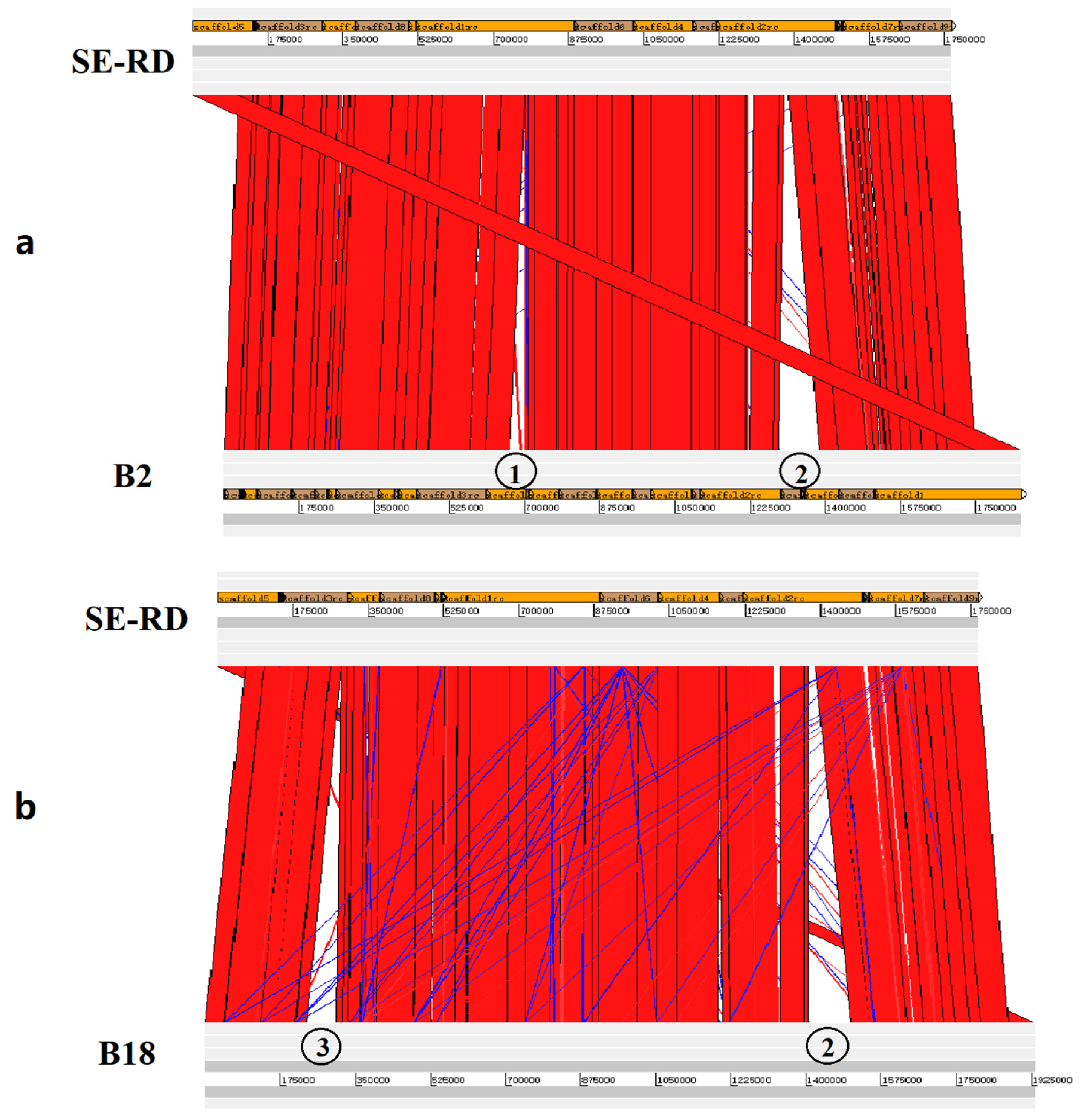
2.5. Genome Sequencing and Comparative Genomic Analysis

Based on the results of the antimicrobial susceptibility assay, 6 strains (B18, B52, B2, SE25, SE27, and SE-RD strains) with representative drug resistance spectra were selected for whole-genome sequencing (Table S2). High-quality genomic DNA was extracted using a modified CTAB method. Qualified genomic DNA was fragmented using G-tubes (Covaris) and then end-repaired to prepare SMRTbell DNA template libraries (with a fragment size > 10 kb selected using a bluepippin system) according to the manufacturer’s instructions (Pacific Biosciences). Library quality was analyzed by Qubit, and the average fragment size was estimated using an Agilent 2100 Bioanalyzer (Agilent, Santa Clara, CA, USA). Whole-genome sequencing was performed on the PacBio Sequel II system (Frasergen Bioinformatics Co., Ltd., Wuhan, China). The PacBio reads were de novo assembled using Microbial Assembly (smrtlink8), HGAP4 [26], and Canu (v1.6) [27]. The depth of genome coverage was analyzed using the pbalign (BLASR, v0.4.1) tool [28]. The genomes of strains B18, B52, B2, SE25, SE27, and SE-RD were annotated using Glimmer (v3.02) [29].
BLASTN (default parameters) was used for genome comparison, and the obtained results were imported into the Artemis Comparison Tool for visualization [30]. The sequences were ordered according to the visualization results. After ordering, the sequences were revisualized with ACT software. Six genome sequences were aligned with each other, and BLAST (basic local alignment search tool) was applied to the genes in the mismatched areas to search for suspected antibiotic resistance genes.

2.6. Detection of the Distribution of Drug Resistance Genes and Mutants

The total DNA of each strain was extracted as a template for PCR amplification [31]. To analyze the distributions of drug resistance genes and mutants in 120 isolates, the suspected macrolide resistance gene Erm(A)-like, the reported macrolide resistance gene Erm(T), the tetracycline resistance gene tet(M), and lsa(E) (representing the lsa(E)-carrying multiresistance gene cluster) were amplified by PCR, and the QRDRs of gyrA and parC were amplified and sequenced. The primers are shown in Table S3. The 50 µL PCR reaction solution contained 1 µL of DNA, 1 µL of each primer, 25 µL of PrimeSTAR Max Premix (2×) (Takara Biomedical Technology (Beijing) Co., Ltd., Beijing, China), and 22 µL of deionized distilled water.

2.7. Analysis of the Contribution of Suspected Drug Resistance Genes and Mutants

The wild-type genes gyrA and parC from the sensitive strain SE-RD, the suspected macrolide resistance gene Erm(A)-like, the mutated gyrA (90D-N) and parC (81S-I) from the resistant strain B18, and gyrA (86T-I) from the resistant strain B2, were separately cloned into pSET2 plasmids. The recombinant plasmids were transfected into E. coli C600, as described previously [32]. The primers used are presented in Table S3. The recombinant strains were subjected to an antimicrobial susceptibility assay by the method mentioned in Section 2.3. The C600 (pSET2) strain (transformed with the pSET2 plasmid) and wild-type E. coli C600 were used as controls.

2.8. Accession Numbers

The genome sequences of strains B18, B52, B2, SE25, SE27, and SE-RD were uploaded to NCBI GenBank with the accession numbers PRJNA750282, PRJNA750613, PRJNA750854, PRJNA750858, PRJNA750871, and PRJNA750617.

2.9. Statistical Analysis

The LD50 of the different MLST isolates was compared using one-way ANOVA by GraphPad Prism 8; a p value of 0.05 was considered to be statistically significant.

3. Results

3.1. MLST and Virulence o thef Isolates

In total, 7 housekeeping genes (gpsA, recA, purA, pta, prsA, galK, and ldhA) from 120 clinical strains were amplified and sequenced. One new allele was discovered separately for gpsA and recA. Based on new alleles, two new sequence types were identified and named ST73 and ST74. Moreover, 3 sequence types (two new types and one known type) were identified from the 120 strains. The largest number of strains (n = 103) belonged to ST48, followed by ST74 (n = 12) and ST73 (n = 5). The distribution of MLST of the 120 isolates is shown in Figure 1.
The LD50 of E. rhusiopathiae ranged from 11CFU to 135CFU. The LD50 of ST48 isolates (11CFU, 14CFU, 15CFU, 16CFU, and 26CFU) was significantly different from that of ST73 (106CFU, 108CFU, 113CFU, 135CFU, and 135CFU) and ST74 (113CFU, 115CFU, 121CFU, 127CFU, and 131CFU) (ST48 vs. ST73: p < 0.0001; ST48 vs. ST74: p < 0.0001), whereas there was no significant difference in LD50 between ST73 and ST74 isolates (ST73 vs. ST74: p = 0.9467). Compared to ST73 and ST74, ST48 was associated with a higher prevalence of highly virulent isolates (Figure 2).

3.2. Antimicrobial Susceptibility

The MICs of 7 antibiotics against the 120 strains are listed in Table 1. All strains were sensitive to meropenem and cefotaxime, and the resistance rates to tetracycline, erythromycin, clindamycin, ciprofloxacin, and enrofloxacin were 50.8%, 53.3%, 70.0%, 91.7%, and 82.5%, respectively. The drug resistance spectra of the 7 antibiotics against the 120 strains are shown in Table S4. One strain was sensitive to all seven antibiotics, and 44.2% of the strains were resistant to five antibiotics, namely, ciprofloxacin, enrofloxacin, clindamycin, tetracycline, and erythromycin.

3.3. Genome Sequencing and Comparative Genomic Analysis

Genome and assembly statistics of each strain are summarized in Table S5. The number of predicted coding sequences (CDSs) of the six sequenced genomes ranged from 1661 to 1840. The genome of strain B18 carried the greatest number of rRNA loci. The guanine and cytosine (GC) content ranged from 36.21% to 36.37%. Several genome islands and prophages were identified, with the number of genome islands ranging from three to seven, and that of prophages from zero to two.
Six genome sequences were aligned with each other (Figures S1–S15), and three mismatched areas were found in the genomes (Figure 3). The first one (about 35 Kb) was found only in strain B2, and no suspected drug resistance genes were found. The second one (about 70Kb) was found in B18 and B52, and in this area, a suspected macrolide antibiotic resistance gene, Erm(A)-like, was also found. The amino acid sequence of Erm(A)-like exhibited 59% similarity to that of Erm(T) (the only macrolide antibiotic resistance gene reported in E. rhusiopathiae) [21]. The third one (about 77Kb) was found in B18, B52, B2, and SE25, and it was the reported lsa(E)-carrying multiresistance gene cluster (ACCESSION: KP339868) containing lsa (E), spw, lnu (B), aadE, aphA3, and other drug resistance genes [20].

3.4. Distribution of Drug Resistance Genes and Resistance-Conferring Mutants

The distribution of Erm(A)-like and the known Erm(T) in clinical isolates is presented in Table S6. Erm(A)-like was detected in all 64 erythromycin-resistant strains, but it was undetected in all 56 erythromycin-sensitive strains. Erm(T) was not detected in the bacteria. These results indicated that the Erm(A)-like gene was widely distributed in the erythromycin-resistant strains of E. rhusiopathiae isolated in China.
The 120 E. rhusiopathiae isolates fell into 3 gyrAparC genotypes according to the QRDRs of gyrA and parC (Tables S7 and S8). Of these 120 isolates, 5 strains, including the sequenced quinolone-sensitive strain SE-RD, belonged to the genotype gyrAT86–gyrAD90–parCS81. The MIC of ciprofloxacin for these five strains was 0.06 μg/mL, and that of enrofloxacin was 0.06 μg/mL or 0.125 μg/mL. The 110 isolates, including the sequenced B18, B52, SE25, and SE27, belonged to the genotype gyrAT86–gyrAN90–parCI81. Five isolates, including the sequenced B2, belonged to genotype gyrAI86–gyrAN90–parCI81. The MIC of ciprofloxacin for the last two genotype strains was 1–8 μg/mL, and that of enrofloxacin was 0.5–16 μg/mL. Our results indicate that the three mutants, namely, gyrA86T-I, gyrA90D-N, and parC81S-I, were widely distributed in the quinolone resistance-determining regions (QRDRs) of gyrA and parC in Erysipelothrix rhusiopathiae isolated in China.
The tet(M) gene was detected in 45 out of the 120 isolates, with a detection rate of 37.5%. These 45 positive isolates included 35 tetracycline-resistant strains and 10 sensitive strains (Table S9). The las(E) gene was detected in 71 of the 120 isolates, with a detection rate of 59.2%. These 71 positive isolates included 68 clindamycin-resistant strains and three sensitive strains (Table S10).
The correlation between the antibiotic resistance genotypes and the phenotypes is shown in Table S11. The results indicated that the relationship between drug resistance phenotype and genotype was very complex.

3.5. Contribution of the Newly Identified Drug Resistance Genes and Resistance-Conferring Mutants

The antimicrobial susceptibility of recombinant E. coli containing the suspected macrolide resistance gene Erm(A)-like, C600 transformed with the pSET2 plasmid, and wild-type E. coli C600, was tested. As shown in Table 2, Erm(A)-like increased the MIC of erythromycin against E. coli C600 at least four-fold.
The antimicrobial susceptibility of recombinant E. coli (containing the mutant gyrA (90D-N), parC (81S-I), and gyrA (86T-I) and wild-type gyrA and parC) C600 transformed with the pSET2 plasmid and that of wild-type E. coli C600 was tested. As shown in Table 2, the mutant gyrA 86T-I and mutant parC 81S-I increased the MIC of enrofloxacin at least two-fold.

4. Discussion

E. rhusiopathiae is an important zoonotic pathogen that can cause erysipelas in many animals, including pigs, and erysipeloid in humans [3,12]. However, currently, there is insufficient analysis regarding MLST and the mechanisms of virulence and drug resistance of E. rhusiopathiae, though swine erysipelas recurred in 2012 in China.
MLST is an important method for bacterial genotyping [24,33,34]. The sequence types of E. rhusiopathiae isolated in Europe, Australia, and the United States are mainly ST3, ST9, and ST19 [2]. Our results showed that the sequence type of Chinese isolates is highly conservative and is mainly ST48, which is associated with a higher prevalence of highly virulent isolates.
The abuse of antibiotics can lead to an increase in bacterial resistance [35]. Bacterial resistance is a major global issue that can cause the failure of antibiotic treatments of bacterial diseases; more importantly, a large number of antibiotic-resistant bacteria will eventually endanger human health [36]. Since China has large-scale pig breeding farms, the analysis of drug resistance in E. rhusiopathiae isolated from large-scale pig farms throughout the country is conducive to a more scientific and reasonable use of antibiotics to prevent and control E. rhusiopathiae. There are many mechanisms of antibiotic resistance, including the alteration of the targets, the inactivation of drugs by hydrolysis or modification, the creation of alternative pathways, efflux pumps, and the inhibition of drug entry into cells [37]. Through comparative genomic analysis of E. rhusiopathiae isolates with different drug resistance phenotypes, some new drug resistance genes and mutants were identified. As it is impossible to directly verify the contribution of resistance genes or mutants to bacterial resistance due to the imperfect genetic manipulation of E. rhusiopathiae, an alternative model organism is necessary. The nucleotide sequences of gyrA and parC in Escherichia coli are highly similar to those in E. rhusiopathiae. Furthermore, E. coli C600 is sensitive to erythromycin and quinolone drugs (ciprofloxacin and enrofloxacin). Considering these findings, we chose E. coli C600 to further verify the contribution of three mutants of gyrA and parC to quinolone resistance and of Erm(A)-like to erythromycin resistance. Our results indicated that Erm(A)-like increased the MIC of erythromycin against E. coli C600 at least four-fold and that the mutation of gyrA 86T-I or parC 81S-I also increased E. coli C600 resistance to enrofloxacin at least two-fold. As the MICs for both recombinant strains and wild-type strains were lower than the minimum dilution concentration, the influence of the above two mutants on ciprofloxacin, or that of mutation gyrA90D-N on ciprofloxacin or enrofloxacin, could not be determined, indicating that ciprofloxacin and enrofloxacin must be further diluted to determine the MICs for the recombinant and wild-type strains or that a genetic operating system suitable for E. rhusiopathiae direct verification should be developed.
The total DNA extraction method can be used to obtain the bacterial genome and possible plasmids together; thus, total DNA can be used as a template for the amplification of Erm(T) in plasmids. However, Erm(T) was not amplified in the 120 clinical isolates, whereas Erm(A)-like was amplified in the erythromycin-resistant strains, indicating that Erm(A)-like is more widely distributed in clinical erythromycin-resistant isolates from China than Erm(T). Although Erm(A)-like was detected in all of the 64 erythromycin-resistant strains, the MIC of erythromycin against these bacteria varied greatly, ranging from 2 to 256 μg/mL, which may be attributed to other unknown erythromycin resistance mechanisms. The tet(M) gene was detected in most of the tetracycline-resistant strains rather than in all the strains, and lsa(E) was detected in the clindamycin-resistant strains, implying that there might be other resistance mechanisms to tetracycline and clindamycin. Interestingly, tet(M) or lsa(E) was also detected in some strains sensitive to tetracycline or clindamycin, in spite of a low detection rate, suggesting that there might be some differences in the expression of these resistance genes in clinical isolates. The correlation analysis results indicated that the relationship between drug resistance phenotype and genotype is very complex.
In conclusion, our data revealed the epidemic characteristics of E. rhusiopathiae isolates in China from 2012 to 2018 and showed that ST48 was the main sequence type and had the strongest virulence. Furthermore, we also tested the resistance phenotypes of these isolates. The results indicated that E. rhusiopathiae isolates from China were highly resistant to tetracycline, erythromycin, clindamycin, and quinolone, suggesting that these classes of antibiotics could be over- or misused in swine production in China. Through further analyzing the resistance mechanism, we not only confirmed that mutations in gyrA and parC were involved in quinolone resistance of E. rhusiopathiae strains, but also first discovered the wide distribution of the Erm(A)-like gene in E. rhusiopathiae strains. These findings could be beneficial to the clinical prevention and control of E. rhusiopathiae and the development of antibacterial drugs.

Supplementary Materials

The following are available online at https://www.mdpi.com/article/10.3390/microorganisms9122615/s1. Table S1: Background of E. rhusiopathiae used in this study. Table S2: A summary of the antimicrobial resistance of whole genome sequencing isolates. Table S3: Primers used in this study. Table S4: Distribution of drug resistant spectra of 120 strains isolated from 2012 to 2018 in China. Table S5: General genome features and assembly statistics of 6 isolates. Table S6: Distribution of Erm(A)-like and Erm(T) in 120 isolates. Table S7: MICs of ciprofloxacin against 120 isolates and distribution of sequence types of gyrA-parC. Table S8: MICs of enrofloxacin against 120 isolates and distribution of sequence types of gyrA-parC. Table S9: Distribution of tet(M) in 120 isolates. Table S10: Distribution of lsa(E) gene in 120 isolates. Table S11: Correlation between the antibiotic resistance genotypes and phenotypes of the 120 E. rhusiopathiae strains isolated from 2012 to 2018 in China. Figure S1: Alignment between B2 and SE25. Figure S2: Alignment between B2 and SE27. Figure S3: Alignment between B2 and SE-RD. Figure S4: Alignment between B18 and B2. Figure S5: Alignment between B18 and B52. Figure S6: Alignment between B18 and SE25. Figure S7: Alignment between B18 and SE27.Figure S8: Alignment between B18 and SE-RD. Figure S9: Alignment between B52 and B2. Figure S10: Alignment between B52 and SE25. Figure S11: Alignment between B52 and SE27. Figure S12: Alignment between B52 and SE-RD. Figure S13: Alignment between SE25 and SE27. Figure S14: Alignment between SE25 and SE-RD. Figure S15: Alignment between SE27 and SE-RD.

Author Contributions

Conceptualization, Q.Z. and M.J.; methodology, C.W., Q.Z. and M.J.; validation, C.L.; formal analysis, C.L.; investigation, Y.Z.; resources, W.Z.; data curation, L.L.; software, T.W.; visualization, C.K.; writing—original draft, C.W.; writing—review and editing, C.W. and M.J.; supervision, Y.Y.; project administration, X.S. and M.J. All authors have read and agreed to the published version of the manuscript.

Funding

This work was supported by Major special projects of technological innovation in Hubei Province (2020ABA016).

Institutional Review Board Statement

The study was conducted according to the guidelines of the Declaration of Helsinki, and approved by the Ethics Committee of Huazhong Agricultural University (protocol code: HZAUMO-2021-0178, 2021).

Informed Consent Statement

Not applicable.

Data Availability Statement

Publicly available datasets were analyzed in this study. This data can be found here: PRJNA750282, PRJNA750613, PRJNA750854, PRJNA750858, PRJNA750871, and PRJNA750617.

Acknowledgments

We thank Ping Liu from Huazhong Agriculture University, Wuhan, China, for her work on the English editing and language polishing.

Conflicts of Interest

The authors declare no conflict of interest.

References

  1. Pal, N.; Bender, J.S.; Opriessnig, T. Rapid detection and differentiation of Erysipelothrix spp. by a novel multiplex real-time PCR assay. J. Appl. Microbiol. 2010, 108, 1083–1093. [Google Scholar] [CrossRef]
  2. Janssen, T.; Voss, M.; Kuhl, M.; Semmler, T.; Philipp, H.C.; Ewers, C. A combinational approach of multilocus sequence typing and other molecular typing methods in unravelling the epidemiology of Erysipelothrix rhusiopathiae strains from poultry and mammals. Vet. Res. 2015, 46, 84. [Google Scholar] [CrossRef] [PubMed] [Green Version]
  3. Eriksson, H.; Brannstrom, S.; Skarin, H.; Chirico, J. Characterization of Erysipelothrix rhusiopathiae isolates from laying hens and poultry red mites (Dermanyssus gallinae) from an outbreak of erysipelas. Avian Pathol. 2010, 39, 505–509. [Google Scholar] [CrossRef]
  4. Ersdal, C.; Jorgensen, H.J.; Lie, K.I. Acute and Chronic Erysipelothrix rhusiopathiae Infection in Lambs. Vet. Pathol. 2015, 52, 635–643. [Google Scholar] [CrossRef] [PubMed]
  5. Jaeger, A.; Lebarbenchon, C.; Bourret, V.; Bastien, M.; Lagadec, E.; Thiebot, J.B.; Boulinier, T.; Delord, K.; Barbraud, C.; Marteau, C.; et al. Avian cholera outbreaks threaten seabird species on Amsterdam Island. PLoS ONE 2018, 13, e0197291. [Google Scholar] [CrossRef]
  6. Lee, J.J.; Kim, D.H.; Lim, J.J.; Kim, D.G.; Chang, H.H.; Lee, H.J.; Kim, S.H.; Rhee, M.H.; Endale, M.; Imada, Y.; et al. Characterization and identification of Erysipelothrix rhusiopathiae isolated from an unnatural host, a cat, with a clinical manifestation of depression. J. Vet. Med. Sci. 2011, 73, 149–154. [Google Scholar] [CrossRef] [Green Version]
  7. Marshall, K.R.; Walton, S.A.; Boyd, M.; Bishop, B.; Wellehan, J.; Craft, W.; Santoro, D. Erysipeloid lesions caused by Erysipelothrix rhusiopathiae in a dog: Clinical and histopathological findings, molecular diagnosis and treatment. Vet. Dermatol. 2019, 30, 434-e134. [Google Scholar] [CrossRef] [PubMed]
  8. Shimoji, Y.; Osaki, M.; Ogawa, Y.; Shiraiwa, K.; Nishikawa, S.; Eguchi, M.; Yamamoto, T.; Tsutsui, T. Wild boars: A potential source of Erysipelothrix rhusiopathiae infection in Japan. Microbiol. Immunol. 2019, 63, 465–468. [Google Scholar] [CrossRef] [PubMed]
  9. Cabrera-Garcia, A.I.; Muller, F.; Rodler, F.S.; Traub, F.; Heilmann, R.M. Infective endocarditis due to Erysipelothrix rhusiopathiae in a dog—A case report. BMC Vet. Res. 2020, 16, 328. [Google Scholar] [CrossRef] [PubMed]
  10. Cano-Manuel, F.J.; Lopez-Olvera, J.; Fandos, P.; Soriguer, R.C.; Perez, J.M.; Granados, J.E. Long-term monitoring of 10 selected pathogens in wild boar (Sus scrofa) in Sierra Nevada National Park, southern Spain. Vet. Microbiol. 2014, 174, 148–154. [Google Scholar] [CrossRef] [Green Version]
  11. McNeil, M.; Gerber, P.F.; Thomson, J.; Williamson, S.; Opriessnig, T. Serotypes and Spa types of Erysipelothrix rhusiopathiae isolates from British pigs (1987 to 2015). Vet. J. 2017, 225, 13–15. [Google Scholar] [CrossRef] [Green Version]
  12. To, H.; Sato, H.; Tazumi, A.; Tsutsumi, N.; Nagai, S.; Iwata, A.; Nagano, T. Characterization of Erysipelothrix rhusiopathiae strains isolated from recent swine erysipelas outbreaks in Japan. J. Vet. Med. Sci. 2012, 74, 949–953. [Google Scholar] [CrossRef] [PubMed] [Green Version]
  13. Ding, Y.; Zhu, D.; Zhang, J.; Yang, L.; Wang, X.; Chen, H.; Tan, C. Virulence determinants, antimicrobial susceptibility, and molecular profiles of Erysipelothrix rhusiopathiae strains isolated from China. Emerg. Microbes Infect. 2015, 4, e69. [Google Scholar] [CrossRef] [PubMed]
  14. Kwok, A.H.; Li, Y.; Jiang, J.; Jiang, P.; Leung, F.C. Complete genome assembly and characterization of an outbreak strain of the causative agent of swine erysipelas—Erysipelothrix rhusiopathiae SY1027. BMC Microbiol. 2014, 14, 176. [Google Scholar] [CrossRef] [PubMed] [Green Version]
  15. Yang, L.; Zhu, Y.; Peng, Z.; Ding, Y.; Jie, K.; Wang, Z.; Peng, Y.; Tang, X.; Wang, X.; Chen, H.; et al. Comparative Genome Analysis of a Pathogenic Erysipelothrix rhusiopathiae Isolate WH13013 from Pig Reveals Potential Genes Involve in Bacterial Adaptions and Pathogenesis. Vet. Sci. 2020, 7, 74. [Google Scholar] [CrossRef] [PubMed]
  16. Chuma, T.; Kawamoto, T.; Shahada, F.; Fujimoto, H.; Okamoto, K. Antimicrobial susceptibility of Erysipelothrix rhusiopathiae isolated from pigs in Southern Japan with a modified agar dilution method. J. Vet. Med. Sci. 2010, 72, 643–645. [Google Scholar] [CrossRef] [Green Version]
  17. Yamamoto, K.; Kijima, M.; Yoshimura, H.; Takahashi, T. Antimicrobial susceptibilities of Erysipelothrix rhusiopathiae isolated from pigs with swine erysipelas in Japan, 1988–1998. J. Vet. Med. B Infect. Dis. Vet. Public Health 2001, 48, 115–126. [Google Scholar] [CrossRef] [PubMed]
  18. Zhang, B.; Ku, X.; Yu, X.; Sun, Q.; Wu, H.; Chen, F.; Zhang, X.; Guo, L.; Tang, X.; He, Q. Prevalence and antimicrobial susceptibilities of bacterial pathogens in Chinese pig farms from 2013 to 2017. Sci. Rep. 2019, 9, 9908. [Google Scholar] [CrossRef]
  19. Yamamoto, K.; Sasaki, Y.; Ogikubo, Y.; Noguchi, N.; Sasatsu, M.; Takahashi, T. Identification of the tetracycline resistance gene, tet(M), in Erysipelothrix rhusiopathiae. J. Vet. Med. B Infect. Dis. Vet. Public Health 2001, 48, 293–301. [Google Scholar] [CrossRef]
  20. Zhang, A.; Xu, C.; Wang, H.; Lei, C.; Liu, B.; Guan, Z.; Yang, C.; Yang, Y.; Peng, L. Presence and new genetic environment of pleuromutilin-lincosamide-streptogramin A resistance gene lsa(E) in Erysipelothrix rhusiopathiae of swine origin. Vet. Microbiol. 2015, 177, 162–167. [Google Scholar] [CrossRef]
  21. Xu, C.W.; Zhang, A.Y.; Yang, C.M.; Pan, Y.; Guan, Z.B.; Lei, C.W.; Peng, L.Y.; Li, Q.Z.; Wang, H.N. First report of macrolide resistance gene erm(T) harbored by a novel small plasmid from Erysipelothrix rhusiopathiae. Antimicrob. Agents Chemother. 2015, 59, 2462–2465. [Google Scholar] [CrossRef] [PubMed] [Green Version]
  22. Wang, Y.; Li, J.; Zhang, A.; Zhu, W.; Zhang, Q.; Xu, Z.; Yan, S.; Sun, X.; Chen, H.; Jin, M. iTRAQ-based quantitative proteomic analysis reveals potential virulence factors of Erysipelothrix rhusiopathiae. J. Proteom. 2017, 160, 28–37. [Google Scholar] [CrossRef] [PubMed]
  23. Zhu, W.; Wang, Y.; Cai, C.; Li, J.; Wu, C.; Kang, C.; Jin, M. Erysipelothrix rhusiopathiae recruits host plasminogen via the major protective antigen SpaA. FEMS Microbiol. Lett. 2017, 364, 5. [Google Scholar] [CrossRef] [Green Version]
  24. Zhu, W.; Fan, Z.; Qiu, R.; Chen, L.; Wei, H.; Hu, B.; Chen, M.; Wang, F. Characterization of Pasteurella multocida isolates from rabbits in China. Vet. Microbiol. 2020, 244, 108649. [Google Scholar] [CrossRef] [PubMed]
  25. Cen, X.; Huang, Y.; Lu, Z.; Shao, W.; Zhuo, C.; Bao, C.; Feng, S.; Wei, C.; Tang, X.; Cen, L.; et al. LncRNA IGFL2-AS1 Promotes the Proliferation, Migration, and Invasion of Colon Cancer Cells and is Associated with Patient Prognosis. Cancer Manag. Res. 2021, 13, 5957–5968. [Google Scholar] [CrossRef]
  26. Chin, C.S.; Alexander, D.H.; Marks, P.; Klammer, A.A.; Drake, J.; Heiner, C.; Clum, A.; Copeland, A.; Huddleston, J.; Eichler, E.E.; et al. Nonhybrid, finished microbial genome assemblies from long-read SMRT sequencing data. Nat. Methods 2013, 10, 563–569. [Google Scholar] [CrossRef]
  27. Koren, S.; Walenz, B.P.; Berlin, K.; Miller, J.R.; Bergman, N.H.; Phillippy, A.M. Canu: Scalable and accurate long-read assembly via adaptive k-mer weighting and repeat separation. Genome Res. 2017, 27, 722–736. [Google Scholar] [CrossRef] [Green Version]
  28. Chaisson, M.J.; Tesler, G. Mapping single molecule sequencing reads using basic local alignment with successive refinement (BLASR): Application and theory. BMC Bioinform. 2012, 13, 238. [Google Scholar] [CrossRef] [Green Version]
  29. Delcher, A.L.; Bratke, K.A.; Powers, E.C.; Salzberg, S.L. Identifying bacterial genes and endosymbiont DNA with Glimmer. Bioinformatics 2007, 23, 673–679. [Google Scholar] [CrossRef]
  30. Carver, T.J.; Rutherford, K.M.; Berriman, M.; Rajandream, M.A.; Barrell, B.G.; Parkhill, J. ACT: The Artemis Comparison Tool. Bioinformatics 2005, 21, 3422–3423. [Google Scholar] [CrossRef] [PubMed] [Green Version]
  31. Makino, S.; Okada, Y.; Maruyama, T.; Ishikawa, K.; Takahashi, T.; Nakamura, M.; Ezaki, T.; Morita, H. Direct and rapid detection of Erysipelothrix rhusiopathiae DNA in animals by PCR. J. Clin. Microbiol. 1994, 32, 1526–1531. [Google Scholar] [CrossRef] [Green Version]
  32. Zhu, W.; Wu, C.; Kang, C.; Cai, C.; Wang, Y.; Li, J.; Zhang, Q.; Sun, X.; Jin, M. Evaluation of the protective efficacy of four newly identified surface proteins of Erysipelothrix rhusiopathiae. Vaccine 2018, 36, 8079–8083. [Google Scholar] [CrossRef] [PubMed]
  33. Estrada, A.A.; Gottschalk, M.; Rossow, S.; Rendahl, A.; Gebhart, C.; Marthaler, D.G. Serotype and Genotype (Multilocus Sequence Type) of Streptococcus suis Isolates from the United States Serve as Predictors of Pathotype. J. Clin. Microbiol. 2019, 57, e00377-19. [Google Scholar] [CrossRef] [Green Version]
  34. Zhu, W.; Wu, C.; Sun, X.; Zhang, A.; Zhu, J.; Hua, Y.; Chen, H.; Jin, M. Characterization of Streptococcus suis serotype 2 isolates from China. Vet. Microbiol. 2013, 166, 527–534. [Google Scholar] [CrossRef]
  35. Jani, K.; Srivastava, V.; Sharma, P.; Vir, A.; Sharma, A. Easy Access to Antibiotics; Spread of Antimicrobial Resistance and Implementation of One Health Approach in India. J. Epidemiol. Glob. Health 2021, 11, 444–452. [Google Scholar] [CrossRef] [PubMed]
  36. Ge, H.; Wang, Y.; Zhao, X. Research on the drug resistance mechanism of foodborne pathogens. Microb. Pathog. 2022, 162, 105306. [Google Scholar] [CrossRef]
  37. Putman, M.; van Veen, H.W.; Konings, W.N. Molecular properties of bacterial multidrug transporters. Microbiol. Mol. Biol. Rev. MMBR 2000, 64, 672–693. [Google Scholar] [CrossRef] [PubMed] [Green Version]
Figure 1. Distribution of multilocus sequence types of 120 strains isolated in China. Three sequence types were identified, with the number of Sequence Type 48 (ST48), Sequence Type 73 (ST73), and Sequence Type 74 (ST74) strains being 103, 5, and 12 respectively.
Microorganisms 09 02615 g001
Figure 2. Lethality dose 50 (LD50) of isolates and their association with Multilocus Sequence Type (MLST) (one-way ANOVA by GraphPad Prism 8; a p value of 0.05 was considered to be statistically significant).
Microorganisms 09 02615 g002
Figure 3. Comparative genomic analysis of different strains. Alignment between SE-RD and B2 (a) and between SE-RD and B18 (b). Alignment against blastn (default parameters); visualization with Artemis Comparison Tool (http://sanger-pathogens.github.io/Artemis/ACT/,accessed on 11 August 2020). ① A 35Kb mismatched area (no suspected drug resistance genes) found in B2; ② a 77 Kb mismatched area (lsa(E)-carrying multiresistance gene cluster) found in B2 and B18; ③ a 70 Kb mismatched area (containing the Erm(A)-like gene) found in B18.
Microorganisms 09 02615 g003
Table 1. Distribution of Minimum Inhibitory Concentrations (MICs) of antibiotics against the 120 E. rhusiopathiae strains isolated from 2012 to 2018 in China.
AntibioticsNumber of Strains with MIC (μg/mL)MIC(μg/mL) on the Breakpoint a of ResistanceNumber of Resistant Strains (%)
≤0.015≤0.03≤0.06≤0.1250.250.512481632≥64≥128≥256
TE 126291713 46132 1661 (50.8%)
ERY 5321 1192044664264 (53.3%)
CLI 31 57931 1351 0.584 (70.0%)
CIP 5 58966 2110 (91.7%)
EFX 32 24109243 499 (82.5%)
MEM1119 10 (0%)
CTX 187725 20 (0%)
The dark area shows the antibiotic resistance area. a Breakpoint of MIC was from the Clinical and Laboratory Standards Institute (CLSI, 2010). TE, tetracycline; ERY, erythromycin; CLI, clindamycin; CIP, ciprofloxacin; EFX, enrofloxacin; MEM, meropenem; CTX, cefotaxime.
Table 2. Contribution to resistance of suspected drug resistance gene and mutants.
Antibiotic aGene or MutantsMIC (μg/mL)Times Increase in MIC e
C600 bC600(pSET2) cC600 (Recombinant pSET2) d
ERYerm(A)-like6464>256>4
CIPwild-type gyrA<0.06<0.06<0.06f
wild-type parC<0.06
gyrA(90D-N)<0.06
gyrA(86T-I)<0.06
parC(81S-I)<0.06
EFXwild-type gyrA<0.03<0.03<0.03
wild-type parC<0.03
gyrA(90D-N)<0.03
gyrA(86T-I)0.06>2
parC(81S-I)0.06>2
a ERY, erythromycin; CIP, ciprofloxacin; EFX, enrofloxacin. b Wild-type E. coli C600. c C600 transformed with the pSET2 plasmid. d C600 transformed with the recombinant pSET2 plasmid containing Erm(A)-like, gyrA (90D-N), gyrA (86T-I), or parC (81S-I). e MIC against C600 (recombinant pSET2) divided by that of C600 (pSET2). f Since the MICs against the recombinant and wild-type strains were both lower than the minimum dilution concentration, the difference in MIC coulf not be determined.
Publisher’s Note: MDPI stays neutral with regard to jurisdictional claims in published maps and institutional affiliations.

