Article

ABO Blood Groups Do Not Predict Schistosoma mansoni Infection Profiles in Highly Endemic Villages of Uganda

1
Institute of Biodiversity, Animal Health and Comparative Medicine, University of Glasgow, Glasgow G12 8QQ, UK
2
Welcome Centre for Integrative Parasitology, University of Glasgow, Glasgow G12 8QQ, UK
3
Faculty of Science and Engineering, Department of Biological Sciences, University of Chester, Chester CH1 4BJ, UK
4
Vector Control Division, Ministry of Health, Kampala P.O. Box 1661, Uganda
*
Authors to whom correspondence should be addressed.
Deceased.
Microorganisms 2021, 9(12), 2448; https://doi.org/10.3390/microorganisms9122448
Submission received: 6 October 2021 / Revised: 16 November 2021 / Accepted: 23 November 2021 / Published: 27 November 2021
(This article belongs to the Special Issue Schistosoma and Schistosomiasis)

Abstract

:
Schistosoma mansoni is a parasite which causes significant public-health issues, with over 240 million people infected globally. In Uganda alone, approximately 11.6 million people are affected. Despite over a decade of mass drug administration in this country, hyper-endemic hotspots persist, and individuals who are repeatedly heavily and rapidly reinfected are observed. Human blood-type antigens are known to play a role in the risk of infection for a variety of diseases, due to cross-reactivity between host antibodies and pathogenic antigens. There have been conflicting results on the effect of blood type on schistosomiasis infection and pathology. Moreover, the effect of blood type as a potential intrinsic host factor on S. mansoni prevalence, intensity, clearance, and reinfection dynamics and on co-infection risk remains unknown. Therefore, the epidemiological link between host blood type and S. mansoni infection dynamics was assessed in three hyper-endemic communities in Uganda. Longitudinal data incorporating repeated pretreatment S. mansoni infection intensities and clearance rates were used to analyse associations between blood groups in school-aged children. Soil-transmitted helminth coinfection status and biometric parameters were incorporated in a generalised linear mixed regression model including age, gender, and body mass index (BMI), which have previously been established as significant factors influencing the prevalence and intensity of schistosomiasis. The analysis revealed no associations between blood type and S. mansoni prevalence, infection intensity, clearance, reinfection, or coinfection. Variations in infection profiles were significantly different between the villages, and egg burden significantly decreased with age. While blood type has proven to be a predictor of several diseases, the data collected in this study indicate that it does not play a significant role in S. mansoni infection burdens in these high-endemicity communities.

1. Introduction

Schistosomiasis is a debilitating, neglected tropical disease with over 240 million people infected globally [1]. Despite over a decade of mass drug administration (MDA) in many sub-Saharan African countries, hyperendemic hotspots persist, where individuals are repeatedly heavily and rapidly reinfected [2,3]. A range of factors may contribute to persistent hotspots relating to the environment, such as sanitation or snail habitat suitability [4,5]; and to parasite or host influences, including variation in genetic background, immune responses [6], and/or drug efficacy [7]. Individuals can vary in their predisposition to helminth infections [8], and, in particular, blood group has been hypothesised to contribute to schistosome susceptibility [9].
Associations between blood type and disease susceptibility or severity have been described [10]. A and/or B antigens are thought to be advantageous in conferring resistance to some pathogens [11]. For example, the prevalence of Ancylostoma duodenale (Hookworm) was found to be higher in individuals with an O blood type than in those with A or B antigens [12]. However, in some instances, A and/or B antigens increase susceptibility to some diseases. For example, non-O groups have been associated with increased risk and severity of acute respiratory syndrome coronavirus 2 (SARS-CoV-2) infection [13]. Individuals with blood group O appear to have a lower susceptibility to the virus [14], and group A shows a higher susceptibility to infection [15]. Susceptibility to both malaria and tuberculosis is highest in individuals with blood group B, due to the structurally similar α-Gal surface proteins on blood-cell and pathogen surfaces [16]. Variance in disease susceptibility amongst the blood types is thought to be mediated by cross-reacting responses, where anti-A or anti-B antibodies detect foreign proteins expressing A and B motifs or conversely do not detect them if they do not possess these antibodies [10,17]. Pathogens expressing A- and/or B-type epitope sequences are more likely to be detected by the host immune system of individuals with an O-type blood group [18]. Geographical variation among blood-type groups illustrates that pathogen selection can influence co-evolution between blood-group antigens and pathogens [19].
There are few studies solely investigating the impact of Rhesus (Rh) blood groups on disease. Variation in surface proteins on blood cells in Rh-negative individuals have been found to facilitate viral binding in West Nile Virus and thereby increase susceptibility of contracting West Nile Virus [20]. A recent study has found a correlation between Rh positivity and a predisposition to SARS-CoV-2 [21]. The risk of contracting dengue haemorrhagic fever is increased in Rh-positive individuals [22]. These studies suggest that there is a correlation between Rh blood types and infectious disease, but the effect is disease-specific.
One mechanism parasites use to evade immune defence is through the adhesion to host proteins. Several studies have reported a relationship between antigen acquisition and evasion of host immune responses [9]. Early work in this area has described the occurrence of adsorption of host antigens containing glycoproteins and glycolipids by schistosomes. This is also thought to include blood groups [23,24,25]. Adhesion of host proteins is understood to occur within the membranocalyx or inter-membrane zones of the schistosome tegument, wherein studies have detected internalised host-derived immunological proteins [26,27]. Further, structural similarities occur between human-blood proteins in A blood groups (GalNAc) and schistosome polysaccharides [28,29], potentially leading to increased susceptibility to schistosome infections and greater infection intensities. Meanwhile, individuals with B or O blood types may be more efficient at eliciting an anti-A immune response [29]. Therefore, the occurrence of blood-group protein adhesion by schistosomes may contribute to the magnitude of immunological detection by the host and might explain variation in prevalence, intensity, and severity among endemic populations.
Of the few studies that have attempted to establish a relationship between blood group and prevalence, intensity or severity of S. mansoni infections, the results have been conflicting [9]. A recent study found that individuals with an AB blood type are four times more likely to have a high egg burden than those with an O group [30]. This study focused on infection intensity based on one stool sample and did not look at the impact of blood group on clearance after treatment. Several studies have found that patients with A blood groups display an increased schistosomiasis disease severity, and O groups exhibit the lowest prevalence and intensity of schistosomes [31,32,33]. These studies also focused on infection status at the time of sampling and did not consider the longitudinal impact of blood type on schistosome infection dynamics over time or with drug treatment, and subsequent clearance, and reinfection. One study that did look at longitudinal data assessed the annual incidence of schistosome infection and found that individuals with A-type blood had the highest intensities and annual incidence of both S. mansoni and S. haematobium [34]. Conversely, another study found that the group with most prevalent S. mansoni infections at a single time point was type B [35]. Earlier work conducted by Katz et al. found no significant difference between blood groups and S. mansoni prevalence amonst patients with hepatosplenic schistosomiasis [36]. A study by Kassim and Eiezie (1982) examined the rate of Schistosoma haematobium and malaria infection and found that the frequencies of blood groups were not associated with infection rates [37]. However, diagnostic methods and sensitivities, and host–pathogen interactions, including the potential effect of host blood type, differ between S. mansoni and S. haematobium [38]. The above discrepancies indicate a need to expand knowledge on the interaction between blood type and S. mansoni infection dynamics in an endemic setting [9]. There are currently no published studies looking at the role blood type plays on treatment efficacy in schistosome infections or any longitudinal studies to determine whether blood type is a risk factor for repeated or rapid re/infection. Furthermore, no studies have investigated the effect host blood group may have on coinfection with soil-transmitted helminths (STHs) and schistosomes, despite how they commonly co-occur and are often co-treated at the same time through many national control programmes.
The purpose of this study was to evaluate the association between host blood type and its effect on S. mansoni infection dynamics, with repeated observations over time in school-aged children (SAC). Specifically, the aim was to analyse repeat monitoring data about infection/coinfection predisposition, prevalence, intensity, clearance, and reinfection post-treatment, so that a comprehensive representation could be assessed in relation to the impact that blood group may have on these parameters in areas where these S. mansoni dynamics remain high despite over 15 years of MDA.

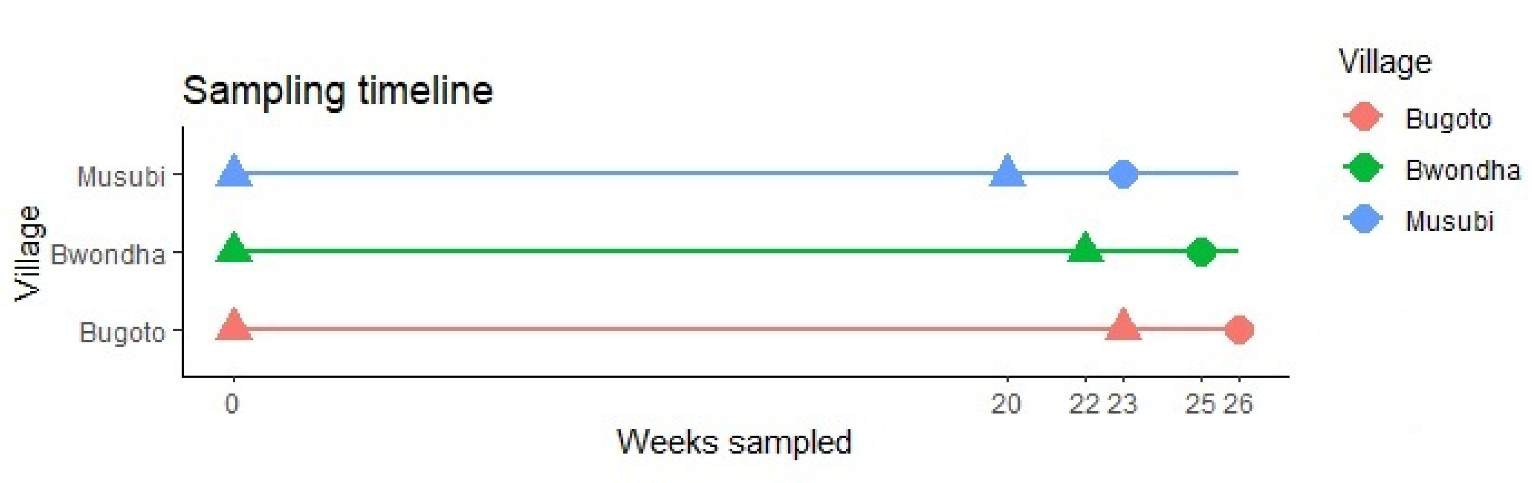
2. Materials and Methods
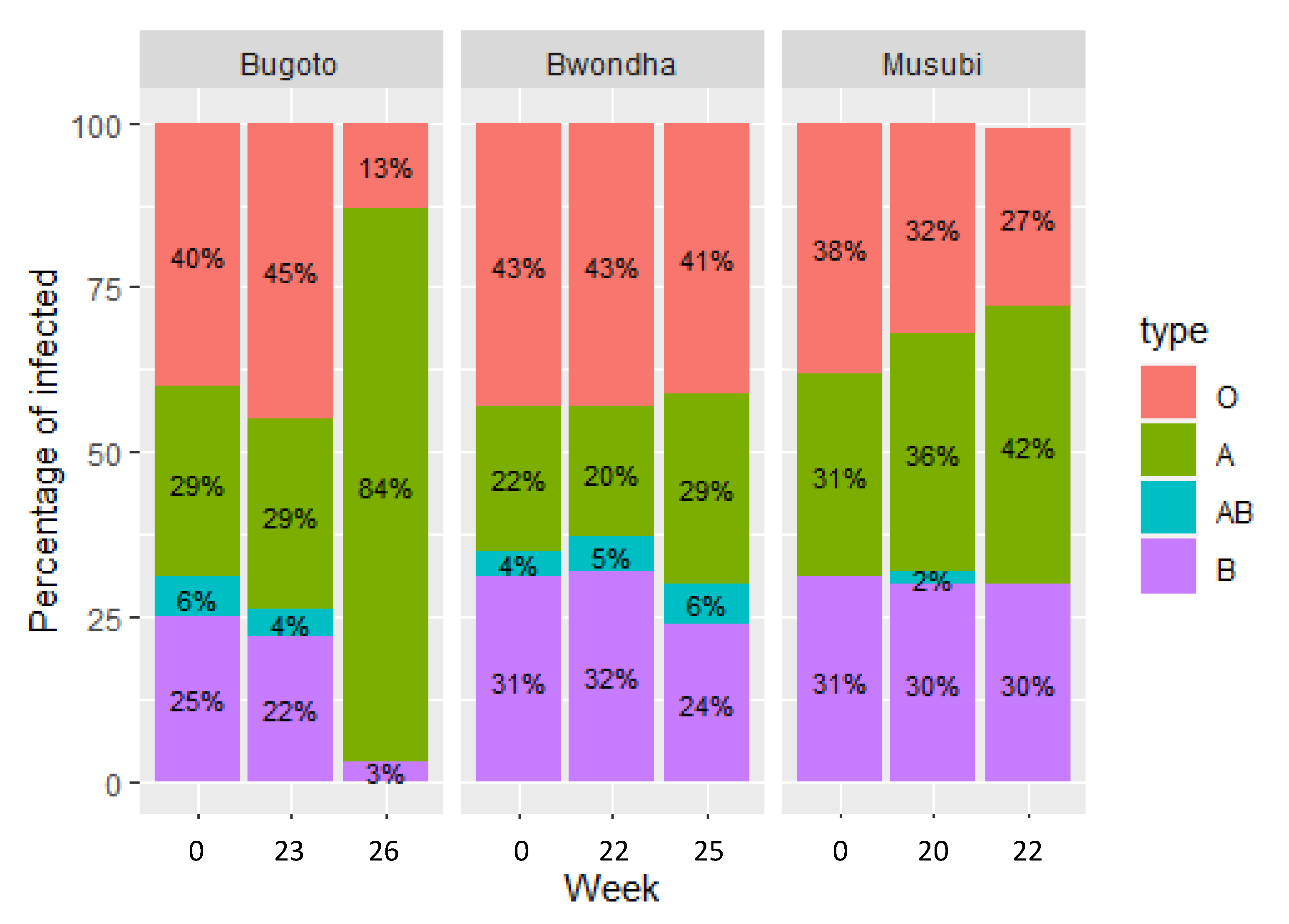
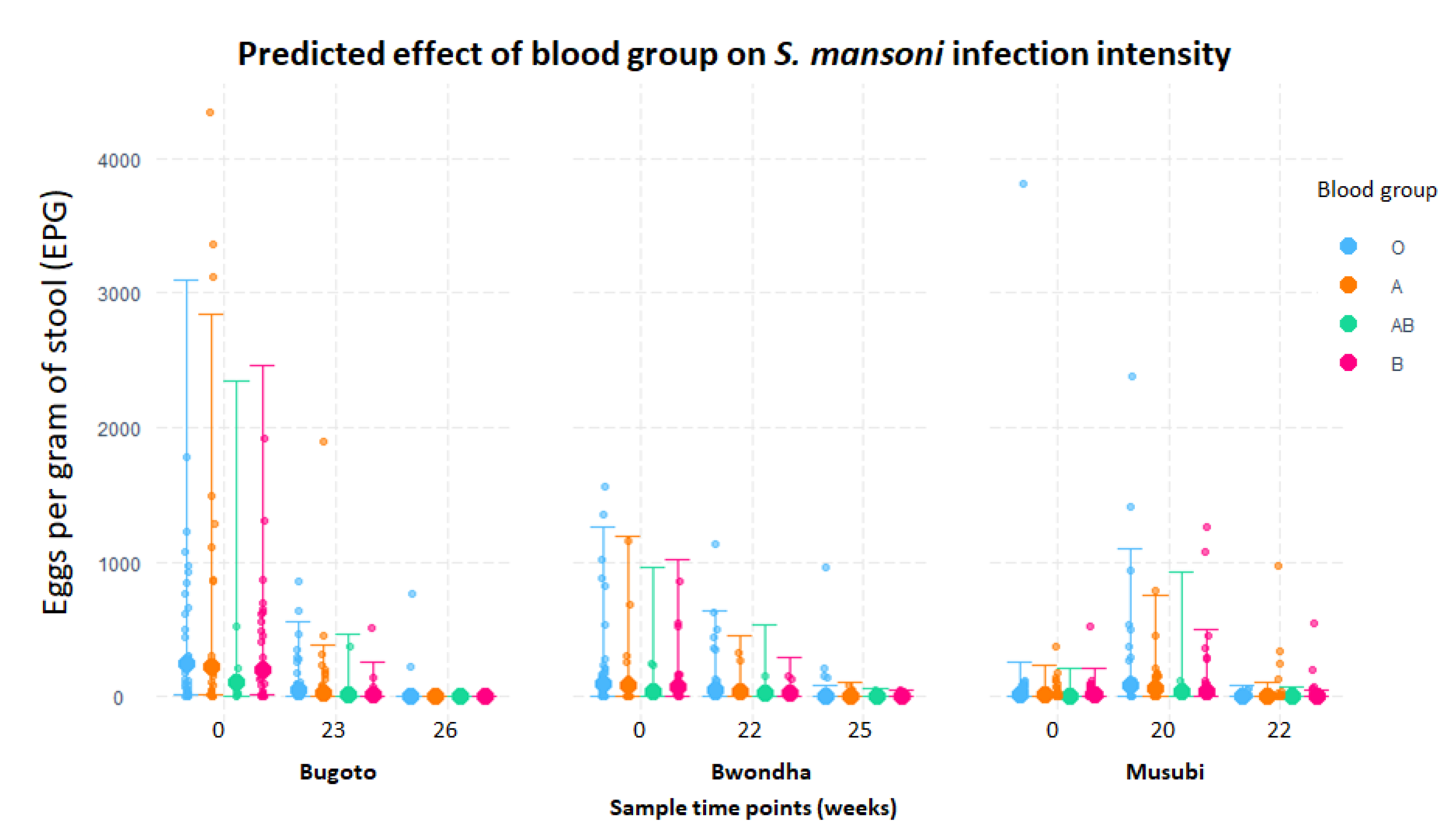
A total of 630 school children between the ages of 6 and 14 were recruited from three primary schools in Mayuge district Uganda, all in high S. mansoni endemicity villages along the shores of Lake Victoria. An equal ratio of boys and girls were recruited and evenly distributed between year age groups. Two hundred and seventy children were recruited from Bugoto Lake View Primary School (0° 1924.09′ N, 33°3741.40′ E) in September 2017. One hundred and eighty children were recruited from both Bwondha Primary School (0°117.15′ N, 33°3339.87′ E) and Musubi Church of Uganda God Primary School (0°1849.45′ N, 33°3947.50′ E) in September 2017 and October 2017, respectively.
In each school, three days of duplicate Kato–Katz thick smears were prepared, in accordance with established protocol [39], from three stool samples provided by participants. On the third day, after sample collection, the participants were all given a meal of porridge and butter an hour prior to observed treatments with 40 mg/kg of praziquantel and 400 mg of albendazole. Individuals from the three schools were all followed up approximately six months later to quantify reinfection rates; they were then retreated with 40 mg/kg praziquantel and followed up with three weeks later to quantify clearance post-treatment. Sample and treatment dates are shown in Figure 1.
In September 2019, participants provided a finger-prick blood sample to identify blood type. A small drop of blood was spotted onto glass slides and mixed with anti-A, anti-B monoclonal sera and an anti-D blend (Alpha Laboratories, Ltd., Hampshire, UK). The slides were checked for agglutination by trained technicians within 10 min of mixing blood and antisera.
Criteria for inclusion in the blood group study included parental consent, child assent, and the provision of a finger-prick blood sample and a stool sample at one or more time point/s.
Because the schools were not all sampled on the same dates, the number of weeks at follow-up was controlled for in the analysis. Participants were excluded from the study if weight and height information collected at baseline or blood-group data were missing. Loss to follow-up was high; therefore, only 265 individuals were included in the final analysis.
To assess the role that blood type plays in predisposition to infection and to determine its effect on clearance, prevalence, and intensity, a generalised linear mixed-effects model was performed, using the glmmTMB [40] package in R ver. 3.5.1 [41]. Negative binomial model assumptions were assessed by using several error structures, including both nbinom1 and nbinom2 parameters, as well as Poisson distributions [40]. The nbinom2 error distribution had minimal residual variance compared with the other error structures assessed and was therefore chosen as the best fit for the data. Residuals were assessed for uniformity, using the DHARMa package [42]. Briefly, a bootstrapped simulation of residuals was performed, along with Kolmogorov–Smirnov uniformity tests; quantile regression of residuals versus fitted model values and Q–Q plots of expected versus observed residual values were computed in order to validate the model.
The initial model included the age of the participants (6–14 years), status of co-infection with STH, gender, and BMI values as fixed effects, because these parameters are variables known to affect schistosome infection intensity and prevalence. The time point at which Kato–Katz samples were obtained and the school in which the child attends were also included in the model to assess the effect of blood type on S. mansoni intensity, clearance, and variability between the schools.
Model selection was performed by using the drop1 function in R, which compares all possible models that can be constructed by each time, removing the lowest estimate variable, which included the removal of BMI, gender, and Rh type, to obtain a parsimonious final model. ABO blood group was not a significant predictor, but because of its biological importance, it was retained during model selection.

3. Results

In the Ugandan villages of Bugoto, Bwondha, and Musubi, which have experienced over 15 years of regular praziquantel administration (either annually or bi-annually), S. mansoni infection was detected in 190 (72%) individuals (n = 265) across the schools, with prevalences of 86% in Bugoto, 71% in Bwondha, and 57% in Musubi and mean infection intensities of 164.6 EPG (95% CI: 113.9–215.3), 108.4 EPG (95% CI: 73.4–143.5), and 92.6 EPG (95% CI: 53.6–131.7), respectively, at baseline. The most commonly occurring blood group was O, with 39% of individuals in Bugoto, 49% in Bwondha, and 39% in Musubi. There was no significant difference between proportion of blood groups observed between the schools (p = 0.390). Type AB occurred significantly less often (p < 0.001) than all other groups, with a prevalence of 6%, 3%, and 1% in Bugoto, Bwondha, and Musubi, respectively. Only 4.8% of individuals from all three schools had an Rh- blood group, and no individuals were Rh- in Musubi. There was no significant association between blood groups and S. mansoni prevalence (p = 0.811). However, significant variation in prevalence occurred between the schools at each time point (p < 0.001) (Figure 2).
Infection intensity varied between the schools at baseline and each follow-up (p < 0.001), but there was no association between blood group and infection intensity (p = 0.818) (Figure 3).
The incidence of Rh- individuals was low, at 4.8%, but slightly higher than other studies conducted in similar regions [43,44]. However, because of its low estimate value, Rh was removed from the final model.
Body mass index (BMI) was calculated based on the World Health Organization’s (WHO) growth standards [45]; however, the best-fit model did not include this parameter, due to the low estimate of this variable.
At baseline, 27 (10%) individuals across all three schools had a STH coinfection, while only three (1%) were mono-infected with STHs. There was no significant relationship between blood group and mono or coinfection with STHs (p = 0.101).

4. Discussion

The Ugandan Ministry of Health has been conducting community-wide MDAs to control schistosomiasis in the Mayuge district since 2004. As with many areas in sub-Saharan Africa, despite these efforts, the burden remains high. This may be partly explained by low MDA coverage [46], but even when children are given observed praziquantel treatment, clearance rates are low [47], many are very rapidly reinfected [2,47], and several remain heavily infected over repeated time points. This could indicate a behavioural or a biological predisposition to rapid and heavy reinfections. This investigation, part of a larger host-factor study, focused on blood group as a potential biological factor which may be driving the high S. mansoni prevalence, intensities, low clearance, and rapid reinfections seen in this area of Uganda. However, no association was found between host blood-group type and S. mansoni infection prevalence or intensity at baseline or over time with two praziquantel treatments, and there was no association with clearance rates, egg reduction rates or co-infection with STHs.
The examination of the impact of blood type on disease incidence is important to help further our understanding of host factors driving the persistence of infection transmission. Research looking specifically at S. mansoni has produced contradictory results regarding the influence that blood type has on schistosomiasis. The results of this study showed no correlation between blood type and S. mansoni infection dynamics: prevalence, intensity, clearance, or reinfection rates. In the majority of other publications, as outlined above, schistosomiasis infection status was also determined using the Kato–Katz method; however, most of the other Kato–Katz/blood-type studies only obtained one stool sample at one time point. A more robust depiction of the infection status of the cohort is provided by using duplicate Kato–Katz slides over three days and repeating at follow-up time points [48,49], as was conducted in this study. Obtaining only one slide per time point increases the risk of false negatives [50]. Berhe et al. noted that false negatives occurred when using either a single Kato–Katz reading or three slides at a rate of 68% and 21%, respectively [51]. To mitigate limitations of the Kato–Katz method, the participants in this study provided stool samples at three time points, for a total of 18 (six per time point) Kato–Katz slides, thus enabling a better depiction of infection status and changes exhibited over time. Expanding on previous studies by assessing the effect of blood group longitudinally, this fully powered study indicates that it does not play a role in the risk of reinfection and clearance rates in these highly endemic villages in Uganda. It may be that the force of infection is so high in these villages that individuals become infected despite the presence or absence of blood-group antigens. However, there was also no association between blood group and infection intensities over time, which is a more sensitive measure for a predisposition to infections in such high force of infection foci and would be expected to demonstrate an effect if it existed here.
The incidence of Rh- individuals at 4.8% is slightly higher than other studies conducted in similar regions [43,44]. However, its role in S. mansoni infection dynamics could not be determined in this study, due to the disproportionate number of individuals with Rh- blood.
The association between age and infection prevalence and intensity suggests that the risk of infection decreases with age. A relationship between age and infection intensity has been described in numerous studies examining schistosomiasis and is an important risk factor for infection [52,53,54,55]. The driving factor behind this occurrence is thought to be due to repeated exposure to S. mansoni causing an increased immunity toward the pathogen [53,56]. It has also been suggested that an increase in age relates to changes in water contact behaviour, thereby reducing the opportunity for reinfection [55,57]. The association with age would suggest that continued work investigating age-related immunity may be imperative to advancing current knowledge to improve treatment and prevention measures.
The relationship between the school an individual attended and S. mansoni infection dynamics had a significant impact on infection profile. Several factors may be driving this intra-divergence in infection dynamics, including variances in MDA history, MDA coverage, water-contact behaviour, accessibility to sanitation and/or safe water, parasite or host genetics, and host immunity. While blood group is not associated with S. mansoni infection dynamics in this hyper-endemic region, further work is required to assess the influence of other within-host factors that may play a role in the risk of S. mansoni infection, in order to develop more targeted control programmes.

Author Contributions

Conceptualization, R.F., A.A., M.A. (Moses Arinaitwe), S.A.B. and P.H.L.L.; methodology, R.F., A.A., M.A. (Moses Arinaitwe), S.A.B. and P.H.L.L.; software, R.F. and S.A.B.; validation, R.F., A.A., M.A. (Moses Arinaitwe) and S.A.B.; formal analysis, R.F.; investigation, R.F., A.A., M.A. (Moses Arinaitwe), S.A.B. and P.H.L.L.; resources, R.F., A.A., M.A. (Moses Arinaitwe), M.A. (Moses Adriko), D.A., A.N., S.A.B. and P.H.L.L.; data curation, R.F. and P.H.L.L.; writing—original draft preparation, R.F.; writing—review and editing, R.F., A.A., M.A. (Moses Arinaitwe), M.A. (Moses Adriko), D.A., A.N., S.A.B. and P.H.L.L.; supervision, R.F., P.H.L.L. and S.A.B.; project administration, R.F. and P.H.L.L.; funding acquisition, P.H.L.L. All authors have read and agreed to the published version of the manuscript.

Funding

The research was funded by P.H.L.L.’s European Research Council Starting Grant (680088 SCHISTO_PERSIST). P.H.L.L. is also funded by Wellcome (grant number 204820/Z/16/Z); Engineering and Physical Sciences Research Council (EPSRC) (grant numbers EP/T003618/1 and EP/R01437X/1); and Medical Research Council (grant number MR/P025447/1).

Institutional Review Board Statement

The study protocol was approved by the College of Medical, Veterinary and Life Sciences Ethics Committee at the University of Glasgow (200160068), the Vector Control Division Research Ethics Committee (VCDREC/062) and the Executive Secretary for the Ugandan National Council for Science and Technology (UNCST-HS 2193).

Informed Consent Statement

Informed consent was obtained from all subjects involved in the study.

Data Availability Statement

Data are available in a publicly accessible repository. The data presented in this study are openly available in Enlighten: Research Data at [http://dx.doi.org/10.5525/gla.researchdata.1219], reference number [1219].

Acknowledgments

Support that was not covered by the author contribution was received from Paul Johnson at the University of Glasgow for advice on statistical analysis and Nabonge Juma the Mayuge District Vector Control Officer for assistance with interacting with the communities.

Conflicts of Interest

The authors declare no conflict of interest.

References

  1. Wang, W.; Yang, K. The Global Status and Control of Human Schistosomiasis: An Overview. In Sino-African Cooperation for Schistosomiasis Control in Zanzibar; Springer: Berlin/Heidelberg, Germany, 2021; pp. 43–51. [Google Scholar]
  2. Trienekens, S.C.; Faust, C.L.; Meginnis, K.; Pickering, L.; Ericsson, O.; Nankasi, A.; Moses, A.; Tukahebwa, E.M.; Lamberton, P.H. Impacts of host gender on Schistosoma mansoni risk in rural Uganda—A mixed-methods approach. PLoS Negl. Trop. Dis. 2020, 14, e0008266. [Google Scholar] [CrossRef] [PubMed]
  3. Kittur, N.; Campbell, C.H., Jr.; Binder, S.; Shen, Y.; Wiegand, R.E.; Mwanga, J.R.; Kinung’hi, S.M.; Musuva, R.M.; Odiere, M.R.; Matendechero, S.H. Discovering, defining, and summarizing persistent hotspots in SCORE studies. Am. J. Trop. Med. Hyg. 2020, 103, 24–29. [Google Scholar] [CrossRef]
  4. Assaré, R.K.; N’Tamon, R.N.; Bellai, L.G.; Koffi, J.A.; Mathieu, T.-B.I.; Ouattara, M.; Hürlimann, E.; Coulibaly, J.T.; Diabaté, S.; N’Goran, E.K. Characteristics of persistent hotspots of Schistosoma mansoni in western Côte d’Ivoire. Parasites Vectors 2020, 13, 1–16. [Google Scholar] [CrossRef] [PubMed]
  5. Walker, J.W.; Kittur, N.; Binder, S.; Castleman, J.D.; Drake, J.M.; Campbell, C.H., Jr.; King, C.H.; Colley, D.G. Environmental predictors of schistosomiasis persistent hotspots following mass treatment with praziquantel. Am. J. Trop. Med. Hyg. 2020, 102, 328. [Google Scholar] [CrossRef] [PubMed] [Green Version]
  6. Mawa, P.A.; Kincaid-Smith, J.; Tukahebwa, E.M.; Webster, J.P.; Wilson, S. Schistosomiasis morbidity hotspots: Roles of the human host, the parasite and their interface in the development of severe morbidity. Front. Immunol. 2021, 12, 751. [Google Scholar] [CrossRef]
  7. Wiegand, R.E.; Mwinzi, P.N.; Montgomery, S.P.; Chan, Y.L.; Andiego, K.; Omedo, M.; Muchiri, G.; Ogutu, M.O.; Rawago, F.; Odiere, M.R. A persistent hotspot of Schistosoma mansoni infection in a five-year randomized trial of praziquantel preventative chemotherapy strategies. J. Infect. Dis. 2017, 216, 1425–1433. [Google Scholar] [CrossRef] [PubMed] [Green Version]
  8. Wright, J.E.; Werkman, M.; Dunn, J.C.; Anderson, R.M. Current epidemiological evidence for predisposition to high or low intensity human helminth infection: A systematic review. Parasites Vectors 2018, 11, 65. [Google Scholar] [CrossRef] [PubMed] [Green Version]
  9. Tiongco, R.E.; Paragas, N.A.; Dominguez, M.J.; Lasta, S.L.; Pandac, J.K.; Pineda-Cortel, M.R. ABO blood group antigens may be associated with increased susceptibility to schistosomiasis: A systematic review and meta-analysis. J. Helminthol. 2018, 94, e21. [Google Scholar] [CrossRef]
  10. Jefferys, S.; Kenneth, C. Transfusion biology and therapy. In Mandell, Douglas, and Bennett’s Principles and Practice of Infectious Diseases, 6th ed.; Churchill Livingston: Philadephia, PA, USA, 2005; Volume 46, p. 708. [Google Scholar]
  11. Degarege, A.; Animut, A.; Medhin, G.; Legesse, M.; Erko, B. The association between multiple intestinal helminth infections and blood group, anaemia and nutritional status in human populations from Dore Bafeno, southern Ethiopia. J. Helminthol. 2014, 88, 152–159. [Google Scholar] [CrossRef] [PubMed]
  12. Wokem, G.; Abah, A.; Iwuaba, P. Hookworm Infection and ABO Blood Group among Children in Aba Metropolis, Abia State, Nigeria. World Appl. Sci. J. 2016, 34, 1216–1221. [Google Scholar]
  13. AbdelMassih, A.F.; Mahrous, R.; Taha, A.; Saud, A.; Osman, A.; Kamel, B.; Yacoub, E.; Menshawey, E.; Ismail, H.-A.; Aita, L. The potential use of ABO blood group system for risk stratification of COVID-19. Med. Hypotheses 2020, 145, 110343. [Google Scholar] [CrossRef] [PubMed]
  14. Pendu, J.L.; Breiman, A.; Rocher, J.; Dion, M.; Ruvoën-Clouet, N. ABO blood types and COVID-19: Spurious, anecdotal, or truly important relationships? A reasoned review of available data. Viruses 2021, 13, 160. [Google Scholar] [CrossRef] [PubMed]
  15. Zeng, X.; Fan, H.; Lu, D.; Huang, F.; Meng, X.; Li, Z.; Tang, M.; Zhang, J.; Liu, N.; Liu, Z. Association between ABO blood groups and clinical outcome of coronavirus disease 2019: Evidence from two cohorts. medRxiv 2020. [Google Scholar] [CrossRef]
  16. Cabezas-Cruz, A.; Mateos-Hernández, L.; Alberdi, P.; Villar, M.; Riveau, G.; Hermann, E.; Schacht, A.-M.; Khalife, J.; Correia-Neves, M.; Gortazar, C. Effect of blood type on anti-α-Gal immunity and the incidence of infectious diseases. Exp. Mol. Med. 2017, 49, e301. [Google Scholar] [CrossRef]
  17. Daniels, G. Human Blood Groups; John Wiley & Sons: Hoboken, NJ, USA, 2008. [Google Scholar]
  18. Calafell, F.; Roubinet, F.; Ramírez-Soriano, A.; Saitou, N.; Bertranpetit, J.; Blancher, A. Evolutionary dynamics of the human ABO gene. Hum. Genet. 2008, 124, 123. [Google Scholar] [CrossRef] [PubMed]
  19. Fumagalli, M.; Cagliani, R.; Pozzoli, U.; Riva, S.; Comi, G.P.; Menozzi, G.; Bresolin, N.; Sironi, M. Widespread balancing selection and pathogen-driven selection at blood group antigen genes. Genome Res. 2009, 19, 199–212. [Google Scholar] [CrossRef] [PubMed] [Green Version]
  20. Dürrbach, A.; Baple, E.; Preece, A.F.; Charpentier, B.; Gustafsson, K. Virus recognition by specific natural antibodies and complement results in MHC I cross-presentation. Eur. J. Immunol. 2007, 37, 1254–1265. [Google Scholar] [CrossRef] [PubMed]
  21. Esref, A.; Solmaz, I.; Akkoc, H.; Donmezdil, S.; Karahan, Z.; Safak, K.; Mertsoy, Y.; Yildirim, M.S.; Nazim, E.; Songul, A. Association between the Rh blood group and the Covid-19 susceptibility. Int. J. Hematol. Oncol. 2020, 31, 81–86. [Google Scholar]
  22. Hashan, M.R.; Ghozy, S.; El-Qushayri, A.E.; Pial, R.H.; Hossain, M.A.; Al Kibria, G.M. Association of dengue disease severity and blood group: A systematic review and meta-analysis. Rev. Med. Virol. 2021, 31, 1–9. [Google Scholar] [CrossRef] [PubMed]
  23. Ali, P.O.; Smithers, S.; Bickle, Q.; Phillips, S.; Harn, D.; Simpson, A. Analysis of the anti-Schistosoma mansoni surface antibody response during murine infection and its potential contribution to protective immunity. J. Immunol. 1988, 140, 3273–3279. [Google Scholar]
  24. Hokke, C.H.; Deelder, A.M. Schistosome glycoconjugates in host-parasite interplay. Glycoconj. J. 2001, 18, 573–587. [Google Scholar] [CrossRef] [PubMed]
  25. Goldring, O.; Clegg, J.; Smithers, S.; Terry, R. Acquisition of human blood group antigens by Schistosoma mansoni. Clin. Exp. Immunol. 1976, 26, 181. [Google Scholar]
  26. Mulvenna, J.; Moertel, L.; Jones, M.K.; Nawaratna, S.; Lovas, E.M.; Gobert, G.N.; Colgrave, M.; Jones, A.; Loukas, A.; McManus, D.P. Exposed proteins of the Schistosoma japonicum tegument. Int. J. Parasitol. 2010, 40, 543–554. [Google Scholar] [CrossRef] [PubMed]
  27. Braschi, S.; Wilson, R.A. Proteins exposed at the adult schistosome surface revealed by biotinylation. Mol. Cell. Proteom. 2006, 5, 347–356. [Google Scholar] [CrossRef] [PubMed] [Green Version]
  28. Dean, D.A. Schistosoma mansoni: Adsorption of human blood group A and B antigens by schistosomula. J. Parasitol. 1974, 60, 260–263. [Google Scholar] [CrossRef] [PubMed]
  29. Haseeb, M.; Thors, C.; Linder, E.; Eveland, L. Schistosoma mansoni: Chemoreception through N-acetyl-D-galactosamine-containing receptors in females offers insight into increased severity of schistosomiasis in individuals with blood group A. Exp. Parasitol. 2008, 119, 67–73. [Google Scholar] [CrossRef]
  30. Addisu, A.; Tekeste, Z.; Teshome, A.; Alemu, A.; Tesfa, H. The Association between Abo Blood Groups and Intestinal Schistosomiasis among Masero Primary School Children in Sanja, Northwest Ethiopia. J. Trop. Dis. 2016, 4, 2. [Google Scholar] [CrossRef]
  31. Camus, D.; Bina, J.; Carlier, Y.; Santoro, F. A, B, O blood groups and clinical forms of schistosomiasis mansoni. Trans. R. Soc. Trop. Med. Hyg. 1977, 71, 182. [Google Scholar] [CrossRef]
  32. Khattab, M.; El-Gengehy, M.; Sharaf, M. ABO blood groups in bilharzial hepatic fibrosis. J. Egypt. Med. Assoc. 1968, 51, 245–250. [Google Scholar] [PubMed]
  33. Wang, C.; Gong, W.C.; Qun, W.Q.; Wen, Z.Y.; Yu, H.P. ABO blood group and late schistosomiasis japonica. Chin. Med. J. 1983, 96, 370. [Google Scholar] [PubMed]
  34. Ndamba, J.; Gomo, E.; Nyazema, N.; Makaza, N.; Kaondera, K. Schistosomiasis infection in relation to the ABO blood groups among school children in Zimbabwe. Acta Trop. 1997, 65, 181–190. [Google Scholar] [CrossRef]
  35. Trangle, K.; Goluska, M.; O’Leary, M.; Douglas, S. Distribution of blood groups and secretor status in schistosomiasis. Parasite Immunol. 1979, 1, 133–140. [Google Scholar] [CrossRef] [PubMed]
  36. Katz, N.; Tavares, J.; Aerantes, W. ABO and Rh. blood groups from patients with hepatosplenic schistosomiasis mansoni. J. Parasitol. 1967, 53, 99. [Google Scholar] [CrossRef]
  37. Kassim, O.; Ejezie, G. ABO blood groups in malaria and schistosomiasis haematobium. Acta Trop. 1982, 39, 179–184. [Google Scholar]
  38. Feldmeier, H.; Poggensee, G. Diagnostic techniques in schistosomiasis control. A review. Acta Trop. 1993, 52, 205–220. [Google Scholar] [CrossRef]
  39. Katz, N.; Chaves, A.; Pellegrino, J. A simple device for quantitative stool thick-smear technique in schistosomiasis mansoni. Rev. Inst. Med. Trop. Sao Paulo 1972, 14, 397–400. [Google Scholar]
  40. Brooks, M.E.; Kristensen, K.; van Benthem, K.J.; Magnusson, A.; Berg, C.W.; Nielsen, A.; Skaug, H.J.; Bolker, B.M. glmmTMB Balances Speed and Flexibility Among Packages for Zero-inflated Generalized Linear Mixed Modeling. R J. 2017, 9, 378–400. [Google Scholar] [CrossRef] [Green Version]
  41. R Development Core Team. R: A Language and Environment for Statistical Computing; Version 3.5.2; R Foundation for Statistical Computing: Vienna, Austria, 2018. [Google Scholar]
  42. Hartig, F. DHARMa: Residual Diagnostics for Hierarchical (Multi-Level/Mixed) Regression Models. R Package Version 0.2. 2019. Available online: https://cran.r-project.org/web/packages/DHARMa/vignettes/DHARMa.html (accessed on 26 November 2021).
  43. Jahanpour, O.; Pyuza, J.J.; Ntiyakunze, E.O.; Mremi, A.; Shao, E.R. ABO and Rhesus blood group distribution and frequency among blood donors at Kilimanjaro Christian Medical Center, Moshi, Tanzania. BMC Res. Notes 2017, 10, 738. [Google Scholar] [CrossRef] [Green Version]
  44. Apecu, R.O.; Mulogo, E.M.; Bagenda, F.; Byamungu, A. ABO and Rhesus (D) blood group distribution among blood donors in rural south western Uganda: A retrospective study. BMC Res. Notes 2016, 9, 513. [Google Scholar] [CrossRef] [PubMed] [Green Version]
  45. De Onis, M. WHO Child Growth Standards; WHO: Geneva, Switzerland, 2006; pp. 1–336. [Google Scholar]
  46. Adriko, M.; Faust, C.L.; Carruthers, L.V.; Moses, A.; Tukahebwa, E.M.; Lamberton, P.H. Low praziquantel treatment coverage for Schistosoma mansoni in Mayuge District, Uganda, due to the absence of treatment opportunities, rather than systematic non-compliance. Trop. Med. Infect. Dis. 2018, 3, 111. [Google Scholar] [CrossRef] [PubMed] [Green Version]
  47. Clark, J.; Arinaitwe, M.; Nankasi, A.; Faust, C.L.; Moses, A.; Ajambo, D.; Besigye, F.; Atuhaire, A.; Wamboko, A.; Carruthers, L.; et al. Reconciling egg-and antigen-based estimates of Schistosoma mansoni clearance and reinfection: A modelling study. Clin. Infect. Dis. 2021, ciab679. [Google Scholar] [CrossRef]
  48. Liu, C.; Lu, L.; Zhang, L.; Bai, Y.; Medina, A.; Rozelle, S.; Smith, D.S.; Zhou, C.; Zang, W. More poop, more precision: Improving epidemiologic surveillance of soil-transmitted helminths with multiple fecal sampling using the Kato–Katz technique. Am. J. Trop. Med. Hyg. 2017, 97, 870–875. [Google Scholar] [CrossRef] [PubMed]
  49. Abla, N.; Keiser, J.; Vargas, M.; Reimers, N.; Haas, H.; Spangenberg, T. Evaluation of the pharmacokinetic-pharmacodynamic relationship of praziquantel in the Schistosoma mansoni mouse model. PLoS Negl. Trop. Dis. 2017, 11, e0005942. [Google Scholar] [CrossRef] [PubMed] [Green Version]
  50. Enk, M.J.; Lima, A.C.L.; Drummond, S.C.; Schall, V.T.; Coelho, P.M.Z. The effect of the number of stool samples on the observed prevalence and the infection intensity with Schistosoma mansoni among a population in an area of low transmission. Acta Trop. 2008, 108, 222–228. [Google Scholar] [CrossRef] [PubMed] [Green Version]
  51. Berhe, N.; Medhin, G.; Erko, B.; Smith, T.; Gedamu, S.; Bereded, D.; Moore, R.; Habte, E.; Redda, A.; Gebre-Michael, T. Variations in helminth faecal egg counts in Kato–Katz thick smears and their implications in assessing infection status with Schistosoma mansoni. Acta Trop. 2004, 92, 205–212. [Google Scholar] [CrossRef] [PubMed]
  52. Milner, T.; Reilly, L.; Nausch, N.; Midzi, N.; Mduluza, T.; Maizels, R.; Mutapi, F. Circulating cytokine levels and antibody responses to human Schistosoma haematobium: IL-5 and IL-10 levels depend upon age and infection status. Parasite Immunol. 2010, 32, 710–721. [Google Scholar] [CrossRef] [PubMed] [Green Version]
  53. Mutapi, F.; Burchmore, R.; Mduluza, T.; Midzi, N.; Turner, C.M.R.; Maizels, R.M. Age-related and infection intensity-related shifts in antibody recognition of defined protein antigens in a schistosome-exposed population. J. Infect. Dis. 2008, 198, 167–175. [Google Scholar] [CrossRef] [PubMed] [Green Version]
  54. Mohammed, M.K.; Halaly, S.; Awadalla, H.; Abdelrahman, A.; Balla, S. Prevalence, risk factors and effect of urinary schistosomiasis on academic performance of school children Age 6–15 years in Asalaya locality, White Nile State, Sudan 2017. J. Adv. Med. Med. Res. 2018, 1–7. [Google Scholar] [CrossRef]
  55. Gryseels, B.; Polman, K.; Clerinx, J.; Kestens, L. Human schistosomiasis. Lancet 2006, 368, 1106–1118. [Google Scholar] [CrossRef]
  56. Mitchell, K.M.; Mutapi, F.; Mduluza, T.; Midzi, N.; Savill, N.J.; Woolhouse, M.E.J. Predicted impact of mass drug administration on the development of protective immunity against Schistosoma haematobium. PLoS Negl. Trop. Dis. 2014, 8, e3059. [Google Scholar] [CrossRef] [Green Version]
  57. Bala, A.; Ladan, M.; Mainasara, M. Prevalence and intensity of urinary schistosomiasis in Abarma village, Gusau, Nigeria: A preliminary investigation. Sci. World J. 2012, 7, 1–4. [Google Scholar]
Figure 1. Kato–Katz thick smear sampling (three days of duplicate Kato–Katz at each time point) and praziquantel treatment timeline, indicating the weeks sampling occurred in each school. Triangles indicate praziquantel administration given, after stool samples were collected, on that week, and circles denote treatment was only administered to children with Schistosoma mansoni detected infections.
Microorganisms 09 02448 g001
Figure 2. Schistosoma mansoni infection prevalence, split by school, host blood type, pretreatment, six months post-treatment, and then three weeks later after re-treatment. Blood groups are denoted by colour.
Microorganisms 09 02448 g002
Figure 3. Relationship between blood group and infection intensity (eggs per gram of stool (EPG)) categorised by village school. The colour-coded effect lines underlaid by raw data points illustrate the model predictions that there is no significant association between blood group and EPG.
Microorganisms 09 02448 g003
Publisher’s Note: MDPI stays neutral with regard to jurisdictional claims in published maps and institutional affiliations.

