Review

Microbiomics in Collusion with the Nervous System in Carcinogenesis: Diagnosis, Pathogenesis and Treatment

1
SAMRC Precision Oncology Research Unit (PORU), Pan African Cancer Research Institute (PACRI), University of Pretoria, Hatfield 0028, South Africa
2
Department of Medicine, National & Kapodistrian University of Athens, 11527 Athens, Greece
3
FALCONBIO PTE, Ltd., 32 Carpenter Street, SGInnovate, Singapore 059911, Singapore
4
Division of Cardiovascular Medicine, Department of Health, Medicine and Caring Sciences, Faculty of Medicine, Linköping University, 581 83 Linköping, Sweden
5
Department of Neurosurgery, University of Pretoria, Hatfield 0028, South Africa
6
Centre for Health Innovation and Policy (CHIP) Foundation, Noida 201301, India
7
Datar Cancer Genetics, Nashik 422010, India
8
Centre for Quality of Health and Living, Faculty of Health and Environmental Sciences, Central University of Technology, Bloemfontein 9300, South Africa
9
Department of Surgery, Faculty of Health Sciences, Steve Biko Academic Hospital, University of Pretoria, Pretoria 0007, South Africa
*
Author to whom correspondence should be addressed.
Microorganisms 2021, 9(10), 2129; https://doi.org/10.3390/microorganisms9102129
Submission received: 19 August 2021 / Revised: 23 September 2021 / Accepted: 27 September 2021 / Published: 11 October 2021
(This article belongs to the Special Issue Microbiota: From the Environment to Humans)

Abstract

:
The influence of the naturally occurring population of microbes on various human diseases has been a topic of much recent interest. Not surprisingly, continuously growing attention is devoted to the existence of a gut brain axis, where the microbiota present in the gut can affect the nervous system through the release of metabolites, stimulation of the immune system, changing the permeability of the blood–brain barrier or activating the vagus nerves. Many of the methods that stimulate the nervous system can also lead to the development of cancer by manipulating pathways associated with the hallmarks of cancer. Moreover, neurogenesis or the creation of new nervous tissue, is associated with the development and progression of cancer in a similar manner as the blood and lymphatic systems. Finally, microbes can secrete neurotransmitters, which can stimulate cancer growth and development. In this review we discuss the latest evidence that support the importance of microbiota and peripheral nerves in cancer development and dissemination.

1. Introduction

It has been estimated that the microbiota in a human body consists of approximately 3000 different species of microbes with 40,000,000,000,000 (40 trillion) individual microbial cells. The vast majority of these are gastrointestinal bacteria [1]. One of the first microbial based treatments proposed for cancer was found in the Eber’s Papyrus, written by the Egyptian physician Imhotep around the year 2600 BCE [2]. He proposed that infection of the area affected by the cancer through an open wound covered by a poultice could have therapeutic potential. At that time Germ theory was unknown [3]. This treatment was revived in the 1800s by William Coley who “vaccinated” cancer patients with live or heat-killed Streptococcus and Serratia species [2]. Coley documented 4 cases of sarcoma where the patients were completely cured by erysipelas infection. The medical establishment, however, discounted his treatment as fatal. We now know, erysipelas causes an inflammatory reaction that most probably are negating the sarcoma [4,5]. Coley’s infections with erysipelas showed for the first time that microbes influence the development and progression of cancer. The use of microbe metabolites, supplementing the diets with probiotics or by faecal transfers is now being explored to treat cancers. This new treatment strategy is the result of growing evidence that the microbiota can play an important role in cancer development and progression [6,7].
Each specific region of the human body has its specific microbiome. Those areas with the richest microbiome include the skin, airways, urogenital tract, eyes and gastrointestinal tract [1]. Microbes present in the human body can also interact with the nervous system in various ways. These include via the enteric nervous system, the vagus nerve, microbial metabolites and the immune system [8,9]. Recently, the fact that the microbiome is able to interact with the nervous system and the fact that changes in the microbiota can promote the development of cancer or indeed help prevent or treat cancer, has led to the suggestion that these pathways may be mechanistically connected. Their interaction can be facilitated through the effects of the microbiome on the immune system [10]. A downregulated immune response provides favourable conditions for the development and progression of cancer [11]. A growing body of research demonstrates that changes to the composition of the gastrointestinal microbiota is an initiating factor in numerous neurocognitive conditions, profoundly influencing both CNS immunity and the integrity of the blood–brain barrier (BBB) [12].
It is now well known that the growth and progression of cancer can occur through the support of nerve tissue (neoneurogenesis) in the same way that new blood vessels (angiogenesis), or lymph vessels (lymphangiogenesis) support cancer development and progression. Additionally, in the same way, nerves are able to provide a means for cancer to metastasize and invade new tissues. Here, nerves may provide a “pathway” along which cancer cells can migrate [13]. Nerves interact with multiple tissues throughout the body, through direct interaction or via chemokines, cytokines, hormones, and neurotransmitters. Tumour cells generally have receptors for these different molecules and can also secrete a wide array of them. This provides a means for the nervous system to interact with and promote tumour development [13].
This review will discuss the role that the microbiome plays in the development and progression of various cancers, through the effect of the microbiota on the nervous system.

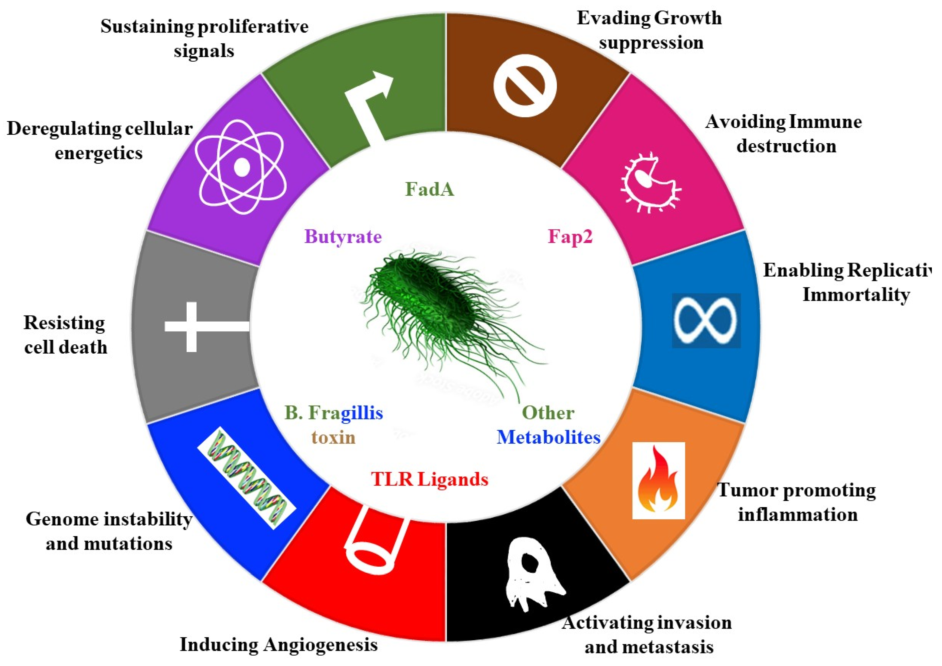
2. Contribution of Microbes to Hallmarks of Cancer

Microbes act either directly or indirectly to facilitate development and progression of cancer. The notable example of direct action are gamaproteobaceria, which express the cytidine deaminase, which metabolizes and thus inactivates the chemotherapeutic gemcetabine [14]. Examples of indirect action include the induction of DNA damage through the production of mutagens and the promotion of inflammation by reactive oxygen species (Figure 1) [15].
The pks+ strain of E. coli drives carcinogenic processes in a multitude of ways Including by induction of double-strand breaks, aneuploidy, cell-cycle arrest, and improper cellular division [16]. Mechanistically, the E.coli pks+ bacteria invade the colonic epithelial cells using the carcinoembryonic antigen related cell adhesion molecule 6 (CEACAM6) receptor, and once inside the cell, they release the toxin pks+ [17].
Microbes can also stimulate cancer promoting signalling pathways, such as the e-cadherin– Wnt–b-catenin signalling pathways [16,18]. Enterotoxigenic Bacteroides fragilis (ETBF), secretes the B. fragilis toxin (BFT) (Table 1), which accelerates the endogenous cleavage of e-cadherin [19]. ß-catenin, normally bound to e-cadherins, is then liberated, and translocates to the nucleus to promote transcription of c-Myc, leading to epithelial cell proliferation (Figure 1) [19].
Fusobacterium nucleatum achieves a similar interaction with e-cadherin via its adhesin FadA (Table 1) [20]. Since the e-cadherin–β catenin complex regulates cellular adhesion, any interference with the complex can lead to loss of cellular adhesion, increased movement of tumour cells, invasion and metastasis [21].
Gut microbiota have been shown to promote obesity-linked liver cancer through metabolites and microbial components, such as lipoteichoic acid (LTA)—a Gram-positive gut microbial component. LTA promotes cancer by increasing the senescence-associated secretory phenotype (SASP) of the hepatic stellate cells (HSC), secreting inflammatory cytokines and growth factors. LTA also increases the expression of cyclo-oxygenase-2 (COX2), through Toll-Like Receptor-2 activation [22].
Complicit microbes are microbes that promote cancer but are unable to directly cause cancer. These are often involved through their production of bioactive metabolites that modulate immune function. For example, microbes associated with lung cancer can stimulate the expression of interleukin 1β (IL-1β) and interleukin-23 (IL-23), leading to inflammation and tumour cell proliferation [21]. Jin et al. used the Sftpc-Cre;KrasLSL-G12D/+;p53fl/fl to drive the expression of Cre recombinase in lung epithelial cells, thereby inducing expression of oncogenic KrasG12D and knock out of the tumor-suppressor gene p53. Jin et al. harvested the lungs from 8 and 15 weeks old mice maintained under two different conditions: the first group was placed under germ-free conditions and the second group was placed in specific-pathogen free conditions. Jin et al. then stained the lungs for the proliferation marker Ki67 and showed that the lungs from mice grown in specific-pathogen free conditions were more heavily proliferating (i.e., Ki67-positive) compared to the lungs of the germ-free grown mice, demonstrating that the presence of commensal bacteria in the specific pathogen free grown mice exacerbated the proliferation of tumor cells. When Jin et al. examined the bronchoalveolar lavage fluid (BALF) from all mice and submitted BALF to 16sRNA qPCR, they identified the genuses Staphylococcus, Streptococcus, Lactobacillus and Pasteurellaceae. Furthermore, the specific pathogen free mice had elevated levels of the cytokines Il-1β and Il-23 compared to the germ-free mice, indicating the commensal bacteria were stimulating cytokine production. Using FACS, they showed the GF mice had elevated numbers of γδT cells and the elevated γδT cells localized specifically in the lungs, where the γδT cells were responsible for producing the majority of the Il-17 pro-inflammatory cytokine. When the SP-free mice were treated with the UC7-13D, which acts against the γδT cells, the number of neutrophils was decreased, showing the elevated γδT cells promote neutrophil infiltration. Jin et al. argue the commensal bacteria can exacerbate tumors via stimulating the proliferation of γδT cells, which also produce pro-inflammatory cytokines like CXC2 and IL17 and promote neutrophils to infiltrate lungs [23].
Another example is the induction of angiogenesis (Figure 1). Pathological angiogenesis is augmented by microbial products such as lipopolysaccharide. These components activate pathways leading to angiogenesis by binding to Toll-like receptors [24]. Suppression or modulation of the immune system and the initiation of the inflammatory response also lead to increased angiogenesis as well as invasiveness [25].
Figure 1. The effect of the microbiome on the hallmarks of cancer. This schematic represents the different effects that microbiota have within the body on different hallmarks of cancer. More specifically, the hallmarks of cancer are ten biological traits that define the transition from normal cells to cancer cells [26]. The bacteria can affect the induction of angiogenesis through secreting TLR ligands, deregulate cellular energetics through the secretion of the bacterial metabolite butyrate, alter immune system activation as well as inflammation, and initiate DNA damage and mutations.
Microorganisms 09 02129 g001

3. Gut Brain Axis

The correct composition of the populations of the microbiota is important for the maintenance of the correct homeostasis of any region of the body [27]. It has been well established from the way that an individual’s ‘emotional’ state can affect digestion, that the nervous system can play an important role on the gut and vice versa. This bidirectional interaction has been dubbed the gut–brain axis [28,29]. The microbiota–gut–brain axis consists of the brain, glands, gut, immune cells, and gastrointestinal microbiota (Figure 2) [30]. Both the central [31] and enteric nervous systems [32] regulate the communication between the gastrointestinal tract and the brain and apart from the nervous system, it is also regulated through hormones and immunological signalling [8,33]. Multiple lines of evidence confirm the existence of the gut–brain axis. Studies have shown that germ free (GF) animal models have altered cognitive function [34,35]. The behaviour of animals [36] and humans [37] can also be altered by feeding them specific strains of bacteria. The different populations of microbes in the human gut, have been classified into three different enterotypes, based on the dominant genus of the microbe present in each type: Bacteroides, Prevotella, and Ruminococcus [38]. Antibiotic treatment, influences the heterogeneity of microbiota, altering the activity of the enteric nervous system and the brain [39]. Additionally, radiotherapy is now known to affect the composition of the GI microbiome, leading to changes in the composition of metabolites produced and excreted by the microbiome [40]. Most importantly, the presence of certain microbial pathogens or the proportions of one species of bacteria in comparison to other bacteria in the gut can contribute to the etiopathogenesis of different types of cancer. Initially, changes in the composition of the microbiota were only known to be associated with the development of colon cancer. However, it is now known that changes in gut bacteria populations are related to not only gastrointestinal cancer but also prostate, pancreatic [41], blood (leukaemia) [42] and brain cancers [12].

3.1. Metabolites Released by Microbes

One of the most important ways that the microbiota is able to influence both the nervous system and the contribution of the nervous system to the development of cancer is through the release of metabolites (Table 1) (Figure 1 and Figure 2). Commensal gut bacteria contribute to the development of colon cancer through the acceleration of DNA damage and induction of chromosomal instability. This occurs through the ability of the gut bacteria to trigger macrophages leading them to produce clastogenic agents [43]. Another important class of metabolites are the Short Chain Fatty Acids (SCFAs) that are produced as a result of the fermentation of non-digestible carbohydrates by anaerobic commensal bacteria. SCFAs have been found to function as energetic substrates for epithelial cells. Among the SCFAs one of the most important is butyrate. Other SCFAs include acetate, propionate and the structurally related ketone bodies (e.g., acetoacetate and d-β-hydroxybutyrate) [44]. High doses of butyrate have neuropharmacological effects by influencing the brain indirectly via regulating the immune system and vagus nerve activity. Butyric acid is produced by many bacteria (Figure 3) [44]. Butyrate and other SCFAs exert their influence by binding to specific receptors. These include MCT1/SLC16A1; SMCT1/SLC5A8; GPR43/FFAR2; GPR41/FFAR3 and GPR109a/HCAR2. Butyric acid can also be used by tumor cells as an energy source via the β-oxidation pathway [44]. Cancer cell metabolism relies on a process known as the Warburg effect. This describes the way in which cancer cells depend mainly on glucose as a carbon source using the glycolytic cycle. The use of butyrate as an energy source means that it alters the metabolic pathway favoured by the cell. Therefore, the use of butyrate in this way exerts an anticancer effect by starving cancer cells [45]. Additionally, when butyrate is not metabolized rapidly it remains at a high concentration intracellularly. The end result of this is inhibition of histone deacetylases (HDACs) [44], which results in the promotion of apoptosis and inhibition of cellular proliferation mediated by epigenetic modifications [45]. Colon cancer patients were found to have lower numbers of many butyrate-producing bacteria in their stool thus resulting in lower levels of butyrate. The bacterial species Akkermansia muciniphila was found at higher levels in the stool of colon cancer patients. These bacteria are able to degrade mucin, which may result in changes in the levels of metabolites present in or absorbed from the intestinal tract [46]. In another study, Singh et al. showed that Gpr109a had an important effect in colonic chronic inflammation and carcinogenesis through its dual role as a receptor of both niacin (a well-known vitamin) and butyrate. [47].

3.2. The Vagus Nerve

The Vagus nerve (VN) functions as an important communications channel between the gut and the central nervous system [48]. Retrospective studies have indicated that increased activity in the vagus nerve is observed as cancer progresses or metastasizes [49]. The vagus nerve communicates with the gastrointestinal system through the release of neurotransmitters in response to changes in the environment or changes brought about by factors such as infection by different pathogens and cancer [50]. Over or under activation of the vagus nerve therefore leads to excessive or lowered release of neurotransmitters, respectively, resulting in aberrant digestion, gastric motility or signalling along the brain gut axis [51]. The vagus nerve is also regulating immune function [52] as well as the rate and ease of absorption of substances from the intestine [53].
During the processing of ingested food, the enteroendocrine cells lining the gut secrete various factors to communicate with nearby neurons and the microbiota [54]. The microbes also release metabolites that bind to receptors on the surface of the enteroendocrine cells, thereby regulating their function. Some of the hormones produced by the enteroendocrine cells include serotonin or 5-hydroxytryptamine (5-HT), cholecystokinin (CCK), and peptide YY (PYY) [55]. These responding enteric neurons then communicate with the afferent fibres of the vagus nerve, which then conduct the signals to the brain [56].

3.3. Blood–Brain Barrier

The blood–brain barrier (BBB) is a selective barrier that allows for the transport of numerous specific molecules. These include signalling molecules, nutrients, and minerals. The specific signalling molecules that are allowed to cross from the circulatory system, include those that are transported from the gut to the brain. The barrier arises from the endothelial cells of the brain capillaries and consists of multiple layers of tight junctions, adherence junctions, membranes and uncharged small lipid soluble molecules [57]. Some of these molecules include proteins such as occludin and claudin. The BBB also helps prevent the free movement of cells and therefore acts as a barrier to brain metastasis of most tumour types. The BBB has consistently been an impedance to the treatment of brain cancers as it limits the access of drugs to the brain, as many chemotherapeutic agents do not cross the blood–brain barrier BBB. Some drugs may even weaken the BBB allowing more rapid and extensive metastasis [58]. Many of the metabolites secreted by the gut microbiome have similar characteristics to the molecules that are transported across the blood–brain barrier; and are therefore able to cross [57]. The integrity of the blood–brain barrier, however, is also depending on the microbiota and the molecules they secrete. For instance, in germ free mice the blood–brain barrier becomes more permeable as the microbiota, that normally assist in the expression of molecules to form tight junctions, are absent [59]. This situation can be reversed by inoculating these germ-free mice with bacteria like Clostridium tyrobutyricum which produce high levels of butyrate [59]. The BBB also allows for communication between the nervous and immune systems. The BBB regulate the passage of immune cells and several other factors associated with immunity [60].

3.4. The Gut Microbiome and the Immune System

Based on studies initially performed on the role played by altered gut microbiomes in prostate cancer, it was established that certain pathogenic gut bacteria are able to trigger an immune inflammatory response (Table 1), which promotes the development of prostate cancer [61]. Gut bacteria may help to stimulate the immune response to other types of cancer as well (Figure 2). Treatment of mouse models using the chemotherapy drug cyclophosphamide, resulted in certain Gram-positive bacteria to move into secondary lymphoid organs resulting in the immune system responding to these bacteria by producing T helper 17 (pTh17) cells and memory Th1 immune responses. This resulted in decreased levels of cancer and increased treatment success. This response was not observed in mice treated with antibiotics to kill Gram-positive bacteria. Tumours in these antibiotic treated mice were also resistant to cyclophosphamide treatment [62].

4. The Microbiota and the Immune System in Nerve Related Cancer

The microbes in the various tissues activate the immune system in multiple ways. Much of the communication between microbes and the immune system, however, depends on the lymphatic system. It was long thought that the brain did not possess a lymphatic system. However, a substantial and immunologically significant lymphatic drainage system has since been discovered. This system functions to drain lymphatics from brain to cervical lymph nodes [72]. This system, also known as the glymphatic system, is another means by which the nervous and immune systems can engage in bi-directional communication. This system connects the peripheral lymphatic tissues to the CNS [73]. The pseudo-lymphatic role of the glymphatic system implies that it also plays a role in neuroinflammation.

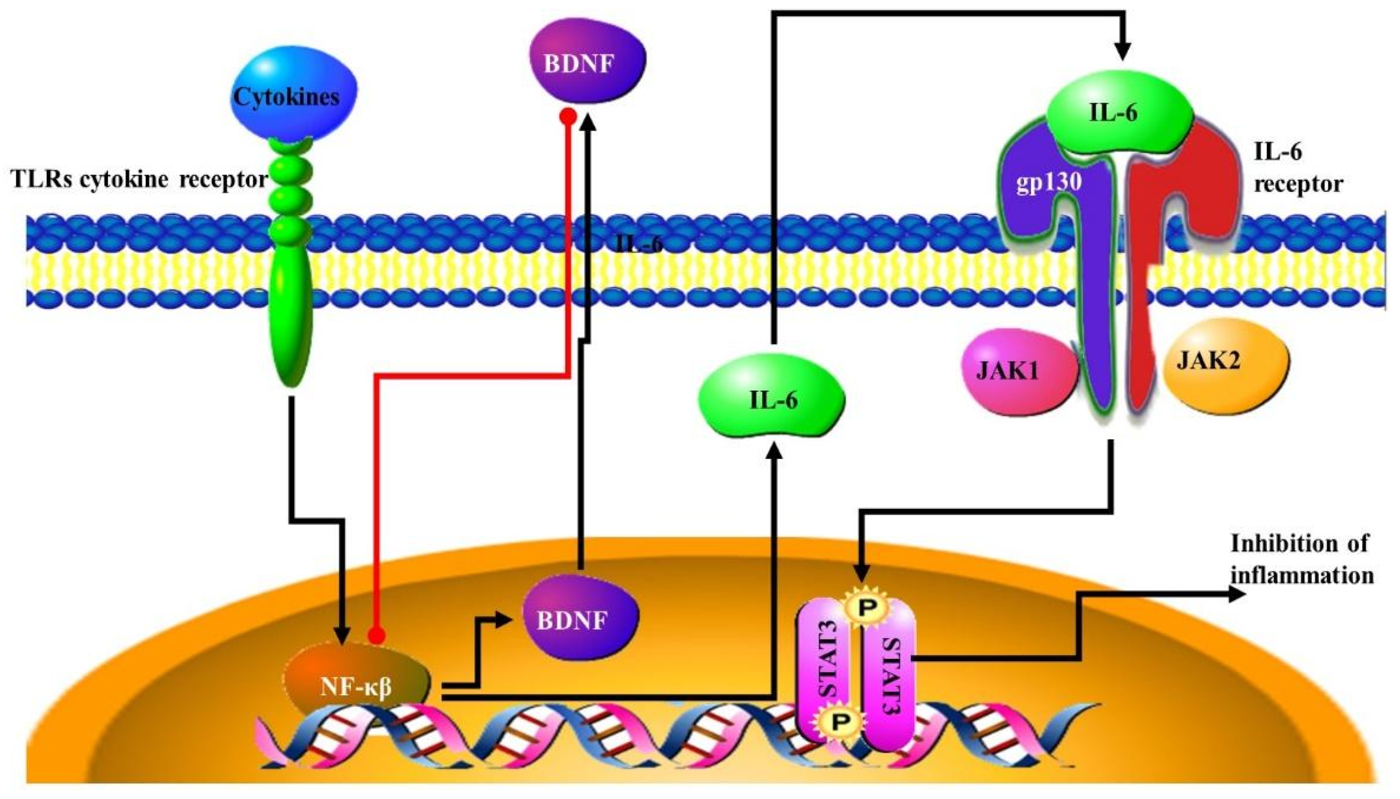
4.1. The Inflammatory Response

The inflammasome is triggered by the innate immune system through the recognition of pathogen recognition receptors. Once the inflammasome is activated it recruits both the apoptosis-associated speck-like protein, which contains a caspase recruitment domain, and the cysteine protease caspase 1. The caspase 1 cleaves pro-IL-1β and pro-IL-18 to make active IL-1β and IL-18 proteins [74]. Different gut microbiome populations have been shown to activate the inflammasome to different extents. In addition, the activation of the inflammasome has been shown to have different neuronal effectors on mouse models, resulting in behavioral and cognitive changes [75]. Certain pro-inflammatory cytokines are known to be involved in the development of brain cancer. These include IL1β, IL6, IL8, IL12, GM-CSF, and TNFα [76]. Some commensal gut bacteria are able to assist dendritic cells to initiate the development of Th1 and pro-tumorigenic T-helper-17 (Th17) cells, leading them to secrete pro-inflammatory cytokines (Figure 4) [77,78] The contribution of the microbiome to the inflammatory response was further demonstrated by the fact that germ free mice have a decreased inflammatory response and a lower level of pro-inflammatory cytokines [79]. In lung cancer, T cells in the lung tissue, γδ T cells, react to the presence of microbiota in the lung and activate an inflammatory response. IL-1β and IL-23 are inducing proliferation and activation of the γδ T cells, which subsequently produce IL-17, which in turn promotes more proliferation [23]. Another pro-tumorigenic response is initiated by the bacteria Fusobacterium nucleatum. This bacterium dampens the anti-tumour immune response. It achieves this through the use of the Fap2 (Table 1) adhesion protein to inhibit the ability of cytotoxic immune cells to kill tumours. It is also able to stimulate the T-cell immune-receptor, immunoglobulin and immunoreceptor tyrosine-based inhibitory motif domains (TIGIT) which functions as an immune inhibitor [80]. Increased levels of F. nucleatum in the microbiome is associated with higher numbers of tumour-associated macrophages which function to inhibit antitumor T-cell responses [81].
Anti-cancer effects initiated by the immune response involving the microbiota, include the response initiated by the intestinal bacteria of the genus Bifidobacterium. These bacteria increase the tumour-killing capabilities of cytotoxic T cells by assisting the function of dendritic cells [82]. The cytotoxic T-lymphocyte-associated protein 4 (CTLA4) is a negative regulator of T cell responses. Therapies such as anti-CTLA4 antibodies, that block CTLA4 function have an anti-tumour effect. The efficacy of these therapies depends on the presence of the bacteria Bacteroides thetaiotamicron and B. fragilis [83]. The immune response initiated by the presence of polysaccharides secreted by B. fragilis, such as polysaccharide A (PSA), can enhance antitumor immune responses. This function is achieved through Toll-like receptor 4 (TLR4) (Table 1)– and IL-12–dependent TH1 responses. This suggests that the immune activation facilitated by the presence of these bacteria also initiates an antitumor response that is enhanced by CTLA4 inhibition [83].

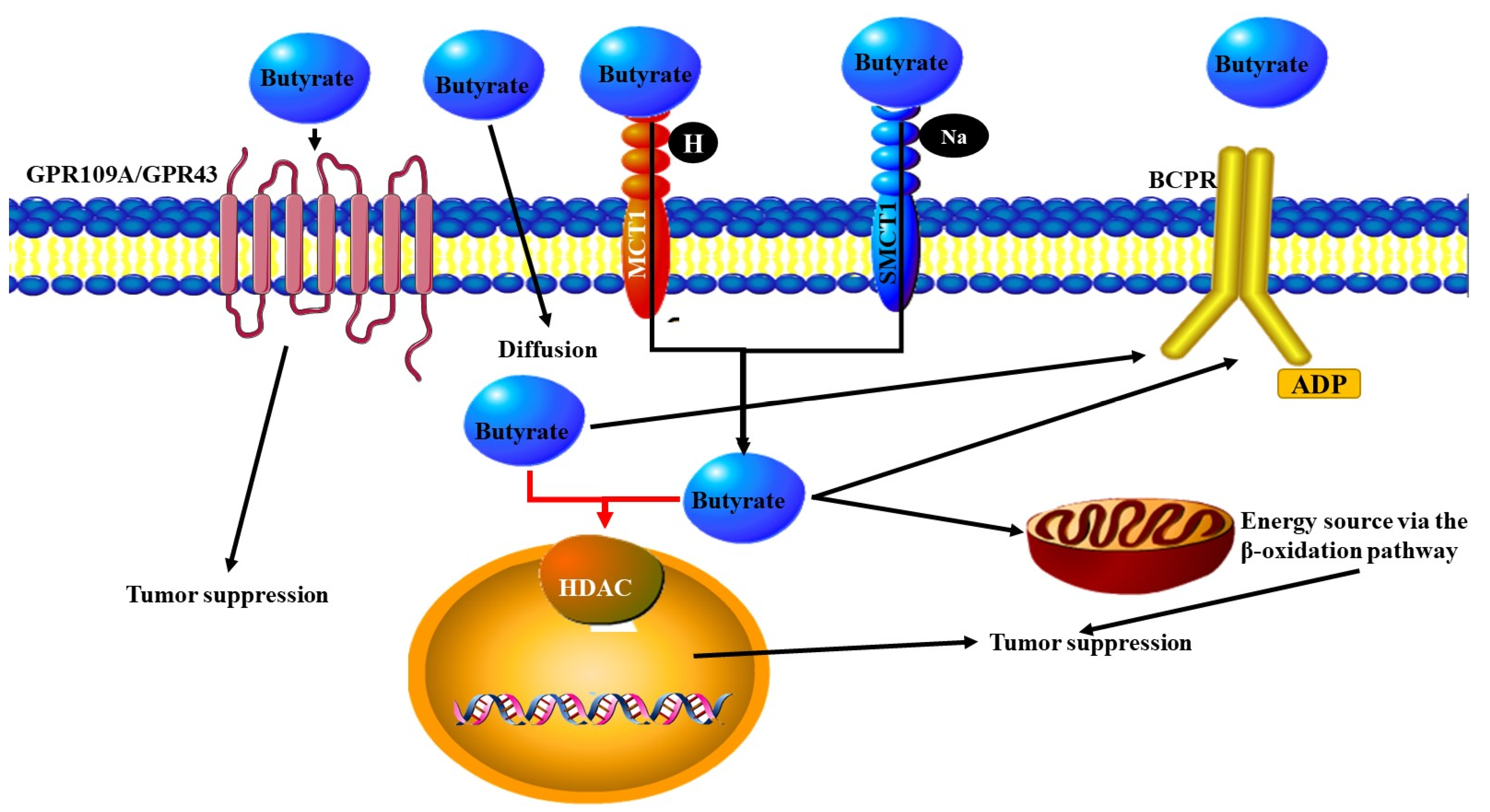
4.2. Bacterial Metabolites and the Immune Response

One of the mechanisms by which bacteria can affect the immune response and either promote or inhibit cancer development is through the production and secretion of secondary metabolites. Once secreted into the gut they can enter the circulatory or lymphatic system and circulate throughout the body [84]. Some of these metabolites secreted by bacteria are CNS-related neurotransmitters and neuromodulators [85]. Others are the previously mentioned SCFAs [86]. SCFAs decrease the levels of pro-inflammatory cytokines that are released as part of an immune response, by affecting Th1 cell populations. SCFAs promote Treg development, which in turn secrete IL10 secretion/ IL-10 impairs the anti-cancer activity of Th1 cells and therefore promotes cancer development [86]. The presence of high concentrations of the bacteria Bacteroides fragilis leads to increased formation of Tregs that secrete IL-10 [87].
Long chain fatty acids are another type of metabolite released by microbes. These increase the proinflammatory response by increasing the rates of differentiation of T cells to form increased numbers of Th1 and Th17 cells. The expression of pro-inflammatory factors such as TNF-α, IFN-γ, and Csf2 is also increased [88].

4.3. NF-κB Signalling Pathway

NF-κB signalling has been found to activate the immune system in response to altered gut microbiome populations. For example, higher levels of Campylobacter jejuni in the gut led to the secretion of cytokines which lead to the activation of NF-κB [89]. NF-κB signalling in response to changes in the gut microbiota can lead to CNS inflammation. This was observed in the neurons of mice. This increase in NF-κB activity is also associated with the inhibition of brain-derived neurotrophic factor BDNF (BDNF) expression (Figure 5) [90]. BDNF is important for the formation of new nervous tissue which assists in the development and progression of cancer as new nerve fibers assist in the expansion and migration of tumours [91]. In brain cancer, the transcription factor STAT3 pathway is activated through the IL6-NFκB pathway, which results in aggressive cancer that progresses faster and a poor prognosis for the patient. This is a result of the suppression of the inflammatory response (Figure 5) [92,93]. The STAT3 pathway can be blocked by blocking IL-17 signalling, resulting in decreased inflammation and tumorigenesis [94].
Bacteria from the genus Helicobacter play an important role in both prostate and colon cancer. Many unique Helicobacter species were isolated exclusively from patients with gastrointestinal cancers [46]. Mice infected with the bacteria Helicobacter hepaticus were found to be more commonly affected by prostate intraepithelial neoplasia and microinvasive adenocarcinoma lesions, without the accompanied presence of IBD or large adenomatous polyps in their bowel. When lymphoid node cells were extracted from these mice and injected into healthy mice, the majority of these mice developed neoplasias. A high concentration of mast cells secreting TNF-α and proteases were present, leading to elevated NF-κB signaling in the prostate of the Helicobacter hepaticus infected mice. It was assumed that these mast cell secretions contributed to carcinogenesis [95].

4.4. Immune Cells in the CNS

Immune cells within the brain not only defend it against infection and injury, but also assist with processes such as neural remodeling and plasticity. Due to it being partially separated from the rest of the body by the blood–brain barrier, the central nervous system must have its own native immune cells. These include glial cells, macrophages, CD8+ T cells, Tregs, other CD4+ T helper (Th), microglia and astrocytes. These cells are involved in both the adaptive and innate immune systems [96]. Gut microbiota can release antigens that stimulate immune signalling pathways through CD4+ T cell activation and resulting in Th17 cell activation [97]. The SCFAs butyric acid and propionic acid produced by microbes discussed previously can cross ΒΒΒ, transported through the blood and have the potential to regulate T cell differentiation in other tissue sites as well. This activation was accompanied by increased expression of the transcription factor Foxp3 through altering the foxp3 promoter activity [98]. Germ free mice have also been shown to have microglia with abnormal morphological characteristics. These microglia also have altered gene expression [99].
Microbial metabolites are able to activate astrocytes from their resting state. They achieve this by acting on aryl hydrocarbon receptors involved in IFN-I signalling, thereby restricting the recruitment and activity of neurotoxic immune cells to initiate anti-inflammatory activity [100]. In germ-free mice, microglia have altered structure and show higher levels of the receptors that are involved in the differentiation and proliferation of immune cells: colony stimulating factor 1 receptor (CSF1R), F4/80 and CD31, on their surfaces. These receptors are normally only found in high numbers on the surface of immature microglia cells. As microglia mature the expression of these receptors decrease. The activation of the GPR43 receptor on innate immune cells activates the inflammatory response. The same observations have been noted in mice that have been treated with antibiotics. In both germ free and antibiotic treated mice, the microglia numbers remain high [101]. Microglia from germ free mice also show increased expression of multiple genes, this increased gene expression is typical of younger microglia [102]. Germ free mice show defects in their microglia activity [100].

4.5. Type I Interferon Signalling Pathways

Type I interferon (IFN-I) is a cytokine induced by pathogen-associated molecular patterns (PAMPs) to prime the immune system to recognise various viral, bacterial and tumour cells. IFN-1 is also active in the CNS and is known to play a role in the protection against brain cancer in animal models [103], reviewed in [104]. IFN-I is associated with the maturation of dendritic cells and cytotoxic T cells, which are both involved in the immune response against cancer cells [105]. IFN-I also exerts an anti-cancer activity through its ability to regulate growth and induce apoptosis in haematological cancers [106]. IFN-1 expression can affect or be affected by the microbiome [107]. TLR3 can be activated by increased numbers of lactic acid bacteria in the gut. Once activated TLR3 increases the secretion of INF-β secretion from dendritic cells [108].

5. Neurotransmitters in Cancer and the Microbiome

Receptors for neurotransmitters are commonly expressed on the surface of tumour cells. These include receptors such the G protein coupled receptors (GPCRs), otherwise known as serpentine receptors. Once neurotransmitters bind to these receptors they can alter the behaviour and characteristics of tumour cells. This can result in increased proliferation, migration, and a more aggressive tumour [109]. Tumours can also produce and secrete neurotransmitters. An example of this is that prostate cancer cells behave like neuroendocrine cells in their ability to secrete neurotransmitters. This response is increased in tumour cells that have been exposed to therapeutic agents and the cells may have done this in response to these agents [110].
The monoamine neurotransmitter, Serotonin or 5-hydroxytryptamine (5-HT) is able to act on the central nervous system (CNS), neuroendocrine system (enteric nervous system) [111,112] and the immune system [113]. It is known that serotonin interacts with the microbiome and plays a role in the development and progression of various cancers [114]. Contradictory to this, lower levels of serotonin may also promote colon cancer development, as low levels of serotonin are accompanied by increased levels of DNA damage, increased inflammation, and consequently increased levels of CRC development [115]. The production of much of the serotonin within the body is regulated by the gut microbiota. The enterochromaffin cells located in the gut supply serotonin to the mucosa, lumen, and circulating platelets and these cells are stimulated to produce serotonin through the actions of spore-forming bacteria [112]. Germ free male mice were also found to have higher levels of serotonin in their hippocampi. This is preceded by an increase in the tryptophan in the blood of the male rats, which is the precursor of serotonin [116].
In addition, serotonin stimulates proliferation in various cancers such as gliomas (where it also plays a role in migration) [117], prostate cancer [118], bladder cancer [119], small cell lung carcinoma [120], colon cancer [121], breast cancer [122], and hepatocellular carcinoma [123]. One of the processes affected by serotonin that contributes to cancer development and progression is angiogenesis. Increased serotonin leads to increased blood vessel development and an increase in the size of blood vessels [124,125]. Studies have also focused on the use of altered serotonin or serotonin receptor [126] expression patterns as a diagnostic or prognostic biomarker in various cancers including urological cancers [126], and colon cancer [127]. The receptors most commonly associated with the development and progression of cancers are the 5-HT1 and 5-HT2 receptors [128,129,130]. Activation of these receptors alters the cell cycle progress, stimulates cell growth and results in improved cell viability this is due to the activation of genes such as MEK-ERK1/2 and JAK2-STAT3 signalling pathways [124]. Increased expression of these receptors has been identified in ovarian [131], and prostate cancer [132]. In some cases, antagonists of serotonin receptors, inhibitors of selective serotonin transporter and of serotonin synthesis have been successfully used to prevent cancer cell growth in prostate cancer [133].
Importantly, microbiota-dependent effects on gut 5-HT significantly impact host physiology, modulating gastrointestinal motility and platelet function. Metabolites from spore forming bacteria were isolated in higher amounts from the feces of patients with high colonic and blood levels of 5-HT, suggesting that gut microbes signal directly to neuroendocrine cells. This was further demonstrated by the fact that in germ free mice higher concentrations of certain metabolites increases the level of 5-HT in the colon and blood. Spore forming bacteria are therefore able to control 5-HT levels in the host [112].

5.1. Catecholamines, Norepinephrine and Dopamine

The migration of cancer cells was found to be stimulated by neurobiological signals, namely through the signalling of norepinephrine [134]. The correct levels of the neurotransmitter may depend on the correct populations of bacteria within the gut as germ free mice have significantly lower levels of norepinephrine [135]. In addition to dopamine stimulating dopaminergic neurons, they activate the innate and adaptive immune cells [136]. The implications of the activation of the immune systems in the development of cancer has already been discussed. Dopamine is also synthesized and secreted by various bacteria [137].

5.2. Gamma-Aminobutyric Acid (GABA)

Bacteria from the genera Lactobacillus and Bifidobacterium are able to produce the neurotransmitter gamma-aminobutyric acid (GABA) [138]. GABA binds to the serpentine receptor GABA(B) and the signal is internalised through a decrease in the level of the cyclic AMP [138]. GABA was found to reduce the migration of colon cancer cells in culture through modulating the activity of norepinephrine [134].

5.3. Acetylcholine

The neurotransmitter acetylcholine has been found to play a role in many different cancers. It induces cell growth and division in epithelial cells [139], and the increased expression of acetylcholine receptors has been identified in multiple cancer types in mouse models including acetylcholine receptor 3 (M3R3) in gastric cancer [140] and the acetylcholine receptor M (Chrm1) muscarinic receptors in prostate cancer on stromal cells [141]. Lactobacillus subspecies can produce acetylcholine [137]. Ganglia in both the sympathetic nervous system (SNS), consisting of ganglia which are parallel to the spinal cord, and the parasympathetic nervous system (PNS), consisting of the vagus nerve and some spinal nerves, respond to acetylcholine stimulation. However, only the PNS produces and secretes it (reviewed in [142]). This is important as the vagus nerve is one of the major connections between the brain and the gut microbiota.

6. Neurogenesis and miRNA Regulation by Microbiota

The creation of new nervous tissue (neurogenesis) is an important process for the progression of most cancers. Tumour cells produce factors that lead to the formation of new nerve tissue [143]. These newly formed nerves release neurotransmitters that stimulate tumour growth and migration [144]. Cancers can invade new tissue and migrate along nerves or nerve tissue. Like angiogenesis and lymphogenesis these new nerves also support the new tumour leading to the growth of cancers around these new nerves in a process known as perineural invasion (PNI) [145]. The microbiome is also able to initiate signalling cascades which stimulate neurogenesis by activating TLR2. The process of neurogenesis can be inhibited, delayed, or even counteracted by feeding animals a mixture of specific bacteria that changes the populations of their gut microbiota [146,147].
The regulation of gene expression through the actions of miRNA is known to play a role in neuronal proliferation, neurogenesis, and Brain Derived Neurotrophic Factor (BDNF) signalling. These processes as well as the expression of certain miRNAs is altered in germ free mice [148]. Studies involving the next generation sequencing of miRNA from normal, germ free and antibiotic treated mice indicate that miRNA expression in the amygdala and prefrontal cortex is regulated by the microbiota and changes in the microbiota populations result in changes in the expression of miRNA. The miRNA expression pattern of germ-free mice was altered once again following bacterial colonisation of the germ-free mice [149], One of the miRNAs whose expression is targeted by gut microbiota is miR-206-3p. This miRNA is known to regulate the expression of the neurotrophic BDNF [149,150]. BDNF is known to stimulate growth of neurons and is important for the cancer related neurogenesis that is also involved in the invasion, metastasis and support of cancer development and growth (Reviewed in [142]).

7. Treatments for Cancers Based on the Microbiome Neural Interactions

Inoculation of patients with specific commensal microbiota are now known to have beneficial effects in a variety of cancers [151,152]. For example, when the supplementation of the diet of mice with bacteria of the genus Bifidobacterium is part of a treatment strategy that also include PD-L1 blockade, it augments the anti-cancer growth inhibition elicited by PD-L1. This occurs through pathways that initiate the maturation of dendritic cells, those that stimulate tumour specific CD8+ T cells, signals that recruit immune cells and the activation of IFN 1 pathways [82]. The bacteria Bifidobacterium longum had an inhibitory effect on the development and progression of colon cancer. Studies have shown that the use of B. longum supplements on colon cancer suppressed colon cancer incidence. It is now known that these bacteria inhibit azoxymethane-induced cell proliferation as well as lower the activity of onco-proteins such as ras-p21 and ornithine decarboxylase [153].
However, there is a problem concerning the use of microbial inoculation as a treatment method for cancer. Traditional treatment such as chemotherapy and radiotherapy may have negative impacts on the microbial population. In addition to this, the use of antibiotics may also disturb the microbiota (both those that are already present as well as those that have been given to a patient as a treatment). This has been demonstrated by treating mice with tumours with the immunostimulatory drug, cyclophosphamide. When combined with antibiotics the drug was far less effective in treating the cancers. This was due to the lower levels of Th1 and Th17 cells [62].
In addition to these therapeutic techniques involving microbiota and the nervous system function in cancer, research has been conducted on the use of microbiota to lessen the side-effects of cancer treatment. Following chemotherapy, it is common for patients to experience post-chemotherapy abdominal pain. This pain seems to be as a result of microbial toxicity leading to changes in the microbial effects on nerves capable of sensing pain. A study has reported that this pain can be lessened through probiotic treatment of the patient [154]. This can reconstitute the microbiota that was lost following chemotherapeutic treatment [155]. Another complication of chemotherapy is known as chemotherapy-induced cognitive impairment (CICI). This disorder involves decreased memory, attention, and concentration as a result of chemotherapy and is related to cytotoxic effects on the CNS. It may also be compounded by neuroinflammation and BBB damage. Once again this is thought to be due to chemotherapy disrupting the gastrointestinal microbiota. Research has been conducted to show that probiotic supplementation of the microbiome can assist in treating CICI [12].
The change in the population of microbes may be used as a diagnostic tool [27]. Since the changes in the microbiome may be cancer specific [156], the changes may be used as a personalized diagnostic tool. This can be exploited by examining transcriptome or proteome profiles of cancer patients. Whole transcriptome or proteome analysis has been used to detect cancer specific pattern changes [157]. However, there are several problems using microbial populations as diagnostic biomarkers. Firstly, the microbial biomass is much lower than that of the host while secondly there is a high risk of contamination by the environment and other microbes not isolated from the patient.

8. Conclusions

The concept of the microbiome affecting the development and progression of cancer through interactions involving nerves, neurotransmitters, the immune system, and metabolites secreted by microorganisms (Figure 6) is most clearly seen in the example of the gut brain axis. However, this interaction occurs throughout the body and is influenced not only by the ability of the gut microbiome to release metabolites that can stimulate or inhibit nervous function, but also through the microbiome influencing the immune system and the production of cytokines resulting in altered neural function. These relationships are currently being investigated for their ability to provide future therapeutic targets, through the use of probiotics to alter the microbiota in a patient’s body and thereby increase the levels of certain microbial ‘species which excrete metabolites with an anti-tumour function. Additionally, these microbes may activate the immune response allowing a greater number of anti-tumour immune cells to be created. The changes in the populations of microbes in patients with various cancers are also being explored as new diagnostic or prognostic biomarkers.

Author Contributions

Conceptualization, editing and review, Z.D. and G.L.; writing—original draft preparation, R.H.; review and editing, S.M., G.E., L.D.J., K.N.S., L.P., C.E., M.O., R.M. and T.J.M.; funding acquisition, Z.D. All authors have read and agreed to the published version of the manuscript.

Funding

This research was funded by the South African Medical Research Council (SAMRC).

Institutional Review Board Statement

Not applicable.

Informed Consent Statement

Not applicable.

Conflicts of Interest

The authors declare no conflict of interest.

References

  1. Sender, R.; Fuchs, S.; Milo, R. Revised Estimates for the Number of Human and Bacteria Cells in the Body. PLoS Biol. 2016, 14, e1002533. [Google Scholar] [CrossRef] [Green Version]
  2. Coley, W.B., II. Contribution to the Knowledge of Sarcoma. Ann. Surg. 1891, 14, 199–220. [Google Scholar] [CrossRef] [PubMed]
  3. Hoption Cann, S.A.; van Netten, J.P.; van Netten, C. Dr William Coley and tumour regression: A place in history or in the future. Postgrad. Med. J. 2003, 79, 672–680. [Google Scholar] [PubMed]
  4. Bickels, J.; Kollender, Y.; Merinsky, O.; Meller, I. Coley’s toxin: Historical perspective. Isr. Med. Assoc. J. 2002, 4, 471–472. [Google Scholar] [PubMed]
  5. Starnes, C.O. Coley’s toxins in perspective. Nature 1992, 357, 11–12. [Google Scholar] [CrossRef]
  6. Xavier, J.B.; Young, V.B.; Skufca, J.; Ginty, F.; Testerman, T.; Pearson, A.T.; Macklin, P.; Mitchell, A.; Shmulevich, I.; Xie, L.; et al. The Cancer Microbiome: Distinguishing Direct and Indirect Effects Requires a Systemic View. Trends Cancer 2020, 6, 192–204. [Google Scholar] [CrossRef] [PubMed] [Green Version]
  7. Sepich-Poore, G.D.; Zitvogel, L.; Straussman, R.; Hasty, J.; Wargo, J.A.; Knight, R. The microbiome and human cancer. Science 2021, 371, eabc4552. [Google Scholar] [CrossRef] [PubMed]
  8. Carabotti, M.; Scirocco, A.; Maselli, M.A.; Severi, C. The gut-brain axis: Interactions between enteric microbiota, central and enteric nervous systems. Ann. Gastroenterol. 2015, 28, 203–209. [Google Scholar] [PubMed]
  9. Breit, S.; Kupferberg, A.; Rogler, G.; Hasler, G. Vagus Nerve as Modulator of the Brain-Gut Axis in Psychiatric and Inflammatory Disorders. Front. Psychiatry 2018, 9, 44. [Google Scholar] [CrossRef] [PubMed] [Green Version]
  10. Kamada, N.; Seo, S.U.; Chen, G.Y.; Núñez, G. Role of the gut microbiota in immunity and inflammatory disease. Nat. Rev. Immunol. 2013, 13, 321–335. [Google Scholar] [CrossRef]
  11. Mangani, D.; Weller, M.; Roth, P. The network of immunosuppressive pathways in glioblastoma. Biochem. Pharmacol. 2017, 130, 1–9. [Google Scholar] [CrossRef] [PubMed] [Green Version]
  12. Subramaniam, C.B.; Bowen, J.M.; Gladman, M.A.; Lustberg, M.B.; Mayo, S.J.; Wardill, H.R. The microbiota-gut-brain axis: An emerging therapeutic target in chemotherapy-induced cognitive impairment. Neurosci. Biobehav. Rev. 2020, 116, 470–479. [Google Scholar] [CrossRef] [PubMed]
  13. Zahalka, A.H.; Frenette, P.S. Nerves in cancer. Nat. Rev. Cancer 2020, 20, 143–157. [Google Scholar] [CrossRef]
  14. Geller, L.T.; Barzily-Rokni, M.; Danino, T.; Jonas, O.H.; Shental, N.; Nejman, D.; Gavert, N.; Zwang, Y.; Cooper, Z.A.; Shee, K.; et al. Potential role of intratumor bacteria in mediating tumor resistance to the chemotherapeutic drug gemcitabine. Science 2017, 357, 1156–1160. [Google Scholar] [CrossRef] [Green Version]
  15. Wang, X.; Yang, Y.; Huycke, M.M. Commensal bacteria drive endogenous transformation and tumour stem cell marker expression through a bystander effect. Gut 2015, 64, 459–468. [Google Scholar] [CrossRef] [PubMed] [Green Version]
  16. Arthur, J.C.; Perez-Chanona, E.; Mühlbauer, M.; Tomkovich, S.; Uronis, J.M.; Fan, T.J.; Campbell, B.J.; Abujamel, T.; Dogan, B.; Rogers, A.B.; et al. Intestinal inflammation targets cancer-inducing activity of the microbiota. Science 2012, 338, 120–123. [Google Scholar] [CrossRef] [Green Version]
  17. Raisch, J.; Buc, E.; Bonnet, M.; Sauvanet, P.; Vazeille, E.; de Vallée, A.; Déchelotte, P.; Darcha, C.; Pezet, D.; Bonnet, R.; et al. Colon cancer-associated B2 Escherichia coli colonize gut mucosa and promote cell proliferation. World J. Gastroenterol. 2014, 20, 6560–6572. [Google Scholar] [CrossRef]
  18. Silva-García, O.; Valdez-Alarcón, J.J.; Baizabal-Aguirre, V.M. Wnt/β-Catenin Signaling as a Molecular Target by Pathogenic Bacteria. Front. Immunol. 2019, 10, 2135. [Google Scholar] [CrossRef]
  19. Rhee, K.J.; Wu, S.; Wu, X.; Huso, D.L.; Karim, B.; Franco, A.A.; Rabizadeh, S.; Golub, J.E.; Mathews, L.E.; Shin, J.; et al. Induction of persistent colitis by a human commensal, enterotoxigenic Bacteroides fragilis, in wild-type C57BL/6 mice. Infect. Immun. 2009, 77, 1708–1718. [Google Scholar] [CrossRef] [Green Version]
  20. Rubinstein, M.R.; Wang, X.; Liu, W.; Hao, Y.; Cai, G.; Han, Y.W. Fusobacterium nucleatum promotes colorectal carcinogenesis by modulating E-cadherin/β-catenin signaling via its FadA adhesin. Cell Host Microbe 2013, 14, 195–206. [Google Scholar] [CrossRef] [Green Version]
  21. Beavon, I.R. The E-cadherin-catenin complex in tumour metastasis: Structure, function and regulation. Eur. J. Cancer 2000, 36, 1607–1620. [Google Scholar] [CrossRef]
  22. Loo, T.M.; Kamachi, F.; Watanabe, Y.; Yoshimoto, S.; Kanda, H.; Arai, Y.; Nakajima-Takagi, Y.; Iwama, A.; Koga, T.; Sugimoto, Y.; et al. Gut Microbiota Promotes Obesity-Associated Liver Cancer through PGE(2)-Mediated Suppression of Antitumor Immunity. Cancer Discov. 2017, 7, 522–538. [Google Scholar] [CrossRef] [Green Version]
  23. Jin, C.; Lagoudas, G.K.; Zhao, C.; Bullman, S.; Bhutkar, A.; Hu, B.; Ameh, S.; Sandel, D.; Liang, X.S.; Mazzilli, S.; et al. Commensal Microbiota Promote Lung Cancer Development via γδ T Cells. Cell 2019, 176, 998–1013.e1016. [Google Scholar] [CrossRef] [Green Version]
  24. Osherov, N.; Ben-Ami, R. Modulation of Host Angiogenesis as a Microbial Survival Strategy and Therapeutic Target. PLoS Pathog. 2016, 12, e1005479. [Google Scholar] [CrossRef] [Green Version]
  25. Mehrian-Shai, R.; Reichardt, J.K.V.; Harris, C.C.; Toren, A. The Gut-Brain Axis, Paving the Way to Brain Cancer. Trends Cancer 2019, 5, 200–207. [Google Scholar] [CrossRef] [PubMed]
  26. Hanahan, D.; Weinberg, R.A. Hallmarks of cancer: The next generation. Cell 2011, 144, 646–674. [Google Scholar] [CrossRef] [PubMed] [Green Version]
  27. Wirbel, J.; Pyl, P.T.; Kartal, E.; Zych, K.; Kashani, A.; Milanese, A.; Fleck, J.S.; Voigt, A.Y.; Palleja, A.; Ponnudurai, R.; et al. Meta-analysis of fecal metagenomes reveals global microbial signatures that are specific for colorectal cancer. Nat. Med. 2019, 25, 679–689. [Google Scholar] [CrossRef] [PubMed] [Green Version]
  28. Heym, N.; Heasman, B.C.; Hunter, K.; Blanco, S.R.; Wang, G.Y.; Siegert, R.; Cleare, A.; Gibson, G.R.; Kumari, V.; Sumich, A.L. The role of microbiota and inflammation in self-judgement and empathy: Implications for understanding the brain-gut-microbiome axis in depression. Psychopharmacology 2019, 236, 1459–1470. [Google Scholar] [CrossRef] [PubMed] [Green Version]
  29. Rhee, S.H.; Pothoulakis, C.; Mayer, E.A. Principles and clinical implications of the brain-gut-enteric microbiota axis. Nat. Rev. Gastroenterol. Hepatol. 2009, 6, 306–314. [Google Scholar] [CrossRef] [PubMed] [Green Version]
  30. Cryan, J.F.; Dinan, T.G. Mind-altering microorganisms: The impact of the gut microbiota on brain and behaviour. Nat. Rev. Neurosci. 2012, 13, 701–712. [Google Scholar] [CrossRef]
  31. Ma, Q.; Xing, C.; Long, W.; Wang, H.Y.; Liu, Q.; Wang, R.F. Impact of microbiota on central nervous system and neurological diseases: The gut-brain axis. J. Neuroinflamm. 2019, 16, 53. [Google Scholar] [CrossRef] [PubMed] [Green Version]
  32. Kuwahara, A.; Matsuda, K.; Kuwahara, Y.; Asano, S.; Inui, T.; Marunaka, Y. Microbiota-gut-brain axis: Enteroendocrine cells and the enteric nervous system form an interface between the microbiota and the central nervous system. Biomed. Res. 2020, 41, 199–216. [Google Scholar] [CrossRef] [PubMed]
  33. Sherman, M.P.; Zaghouani, H.; Niklas, V. Gut microbiota, the immune system, and diet influence the neonatal gut-brain axis. Pediatr. Res. 2015, 77, 127–135. [Google Scholar] [CrossRef] [PubMed]
  34. Diaz Heijtz, R.; Wang, S.; Anuar, F.; Qian, Y.; Björkholm, B.; Samuelsson, A.; Hibberd, M.L.; Forssberg, H.; Pettersson, S. Normal gut microbiota modulates brain development and behavior. Proc. Natl. Acad. Sci. USA 2011, 108, 3047–3052. [Google Scholar] [CrossRef] [PubMed] [Green Version]
  35. Gareau, M.G.; Wine, E.; Rodrigues, D.M.; Cho, J.H.; Whary, M.T.; Philpott, D.J.; Macqueen, G.; Sherman, P.M. Bacterial infection causes stress-induced memory dysfunction in mice. Gut 2011, 60, 307–317. [Google Scholar] [CrossRef] [PubMed]
  36. Savignac, H.M.; Kiely, B.; Dinan, T.G.; Cryan, J.F. Bifidobacteria exert strain-specific effects on stress-related behavior and physiology in BALB/c mice. Neurogastroenterol. Motil. 2014, 26, 1615–1627. [Google Scholar] [CrossRef] [PubMed]
  37. Pinto-Sanchez, M.I.; Hall, G.B.; Ghajar, K.; Nardelli, A.; Bolino, C.; Lau, J.T.; Martin, F.P.; Cominetti, O.; Welsh, C.; Rieder, A.; et al. Probiotic Bifidobacterium longum NCC3001 Reduces Depression Scores and Alters Brain Activity: A Pilot Study in Patients with Irritable Bowel Syndrome. Gastroenterology 2017, 153, 448–459.e448. [Google Scholar] [CrossRef]
  38. Arumugam, M.; Raes, J.; Pelletier, E.; Le Paslier, D.; Yamada, T.; Mende, D.R.; Fernandes, G.R.; Tap, J.; Bruls, T.; Batto, J.M.; et al. Enterotypes of the human gut microbiome. Nature 2011, 473, 174–180. [Google Scholar] [CrossRef]
  39. O’Mahony, S.M.; Felice, V.D.; Nally, K.; Savignac, H.M.; Claesson, M.J.; Scully, P.; Woznicki, J.; Hyland, N.P.; Shanahan, F.; Quigley, E.M.; et al. Disturbance of the gut microbiota in early-life selectively affects visceral pain in adulthood without impacting cognitive or anxiety-related behaviors in male rats. Neuroscience 2014, 277, 885–901. [Google Scholar] [CrossRef]
  40. Jones, C.B.; Davis, C.M.; Sfanos, K.S. The Potential Effects of Radiation on the Gut-Brain Axis. Radiat. Res. 2020, 193, 209–222. [Google Scholar] [CrossRef]
  41. Riquelme, E.; Zhang, Y.; Zhang, L.; Montiel, M.; Zoltan, M.; Dong, W.; Quesada, P.; Sahin, I.; Chandra, V.; San Lucas, A.; et al. Tumor Microbiome Diversity and Composition Influence Pancreatic Cancer Outcomes. Cell 2019, 178, 795–806.e712. [Google Scholar] [CrossRef]
  42. Meisel, M.; Hinterleitner, R.; Pacis, A.; Chen, L.; Earley, Z.M.; Mayassi, T.; Pierre, J.F.; Ernest, J.D.; Galipeau, H.J.; Thuille, N.; et al. Microbial signals drive pre-leukaemic myeloproliferation in a Tet2-deficient host. Nature 2018, 557, 580–584. [Google Scholar] [CrossRef]
  43. Chu, F.F.; Esworthy, R.S.; Chu, P.G.; Longmate, J.A.; Huycke, M.M.; Wilczynski, S.; Doroshow, J.H. Bacteria-induced intestinal cancer in mice with disrupted Gpx1 and Gpx2 genes. Cancer Res. 2004, 64, 962–968. [Google Scholar] [CrossRef] [PubMed] [Green Version]
  44. Stilling, R.M.; van de Wouw, M.; Clarke, G.; Stanton, C.; Dinan, T.G.; Cryan, J.F. The neuropharmacology of butyrate: The bread and butter of the microbiota-gut-brain axis? Neurochem. Int. 2016, 99, 110–132. [Google Scholar] [CrossRef] [PubMed]
  45. Donohoe, D.R.; Holley, D.; Collins, L.B.; Montgomery, S.A.; Whitmore, A.C.; Hillhouse, A.; Curry, K.P.; Renner, S.W.; Greenwalt, A.; Ryan, E.P.; et al. A gnotobiotic mouse model demonstrates that dietary fiber protects against colorectal tumorigenesis in a microbiota- and butyrate-dependent manner. Cancer Discov. 2014, 4, 1387–1397. [Google Scholar] [CrossRef] [PubMed] [Green Version]
  46. Boleij, A.; Tjalsma, H. Gut bacteria in health and disease: A survey on the interface between intestinal microbiology and colorectal cancer. Biol. Rev. Camb. Philos. Soc. 2012, 87, 701–730. [Google Scholar] [CrossRef] [PubMed]
  47. Singh, N.; Gurav, A.; Sivaprakasam, S.; Brady, E.; Padia, R.; Shi, H.; Thangaraju, M.; Prasad, P.D.; Manicassamy, S.; Munn, D.H.; et al. Activation of Gpr109a, receptor for niacin and the commensal metabolite butyrate, suppresses colonic inflammation and carcinogenesis. Immunity 2014, 40, 128–139. [Google Scholar] [CrossRef] [PubMed] [Green Version]
  48. Forsythe, P.; Bienenstock, J.; Kunze, W.A. Vagal pathways for microbiome-brain-gut axis communication. Adv. Exp. Med. Biol. 2014, 817, 115–133. [Google Scholar] [CrossRef] [PubMed]
  49. De Couck, M.; Caers, R.; Spiegel, D.; Gidron, Y. The Role of the Vagus Nerve in Cancer Prognosis: A Systematic and a Comprehensive Review. J. Oncol. 2018, 2018, 1236787. [Google Scholar] [CrossRef] [PubMed]
  50. Browning, K.N.; Zheng, Z.; Gettys, T.W.; Travagli, R.A. Vagal afferent control of opioidergic effects in rat brainstem circuits. J. Physiol. 2006, 575, 761–776. [Google Scholar] [CrossRef]
  51. Travagli, R.A.; Anselmi, L. Vagal neurocircuitry and its influence on gastric motility. Nat. Rev. Gastroenterol. Hepatol. 2016, 13, 389–401. [Google Scholar] [CrossRef] [PubMed] [Green Version]
  52. Yuan, P.Q.; Taché, Y. Abdominal surgery induced gastric ileus and activation of M1-like macrophages in the gastric myenteric plexus: Prevention by central vagal activation in rats. Am. J. Physiol. Gastrointest. Liver Physiol. 2017, 313, G320–G329. [Google Scholar] [CrossRef] [PubMed] [Green Version]
  53. Wang, H.; Wang, L.; Shi, X.; Qi, S.; Hu, S.; Tong, Z.; Ma, Z.; Qian, Y.; Litscher, D.; Litscher, G. Electroacupuncture at Zusanli Prevents Severe Scalds-Induced Gut Ischemia and Paralysis by Activating the Cholinergic Pathway. Evid.-Based Complement. Altern. Med. 2015, 2015, 787393. [Google Scholar] [CrossRef] [PubMed] [Green Version]
  54. Gribble, F.M.; Reimann, F. Enteroendocrine Cells: Chemosensors in the Intestinal Epithelium. Annu. Rev. Physiol. 2016, 78, 277–299. [Google Scholar] [CrossRef] [PubMed]
  55. Palazzo, M.; Balsari, A.; Rossini, A.; Selleri, S.; Calcaterra, C.; Gariboldi, S.; Zanobbio, L.; Arnaboldi, F.; Shirai, Y.F.; Serrao, G.; et al. Activation of enteroendocrine cells via TLRs induces hormone, chemokine, and defensin secretion. J. Immunol. 2007, 178, 4296–4303. [Google Scholar] [CrossRef] [Green Version]
  56. Kaelberer, M.M.; Buchanan, K.L.; Klein, M.E.; Barth, B.B.; Montoya, M.M.; Shen, X.; Bohórquez, D.V. A gut-brain neural circuit for nutrient sensory transduction. Science 2018, 361, eaat5236. [Google Scholar] [CrossRef] [Green Version]
  57. Banks, W.A. Characteristics of compounds that cross the blood-brain barrier. BMC Neurol. 2009, 9 (Suppl. S1), S3. [Google Scholar] [CrossRef] [PubMed] [Green Version]
  58. Blecharz, K.G.; Colla, R.; Rohde, V.; Vajkoczy, P. Control of the blood-brain barrier function in cancer cell metastasis. Biol. Cell 2015, 107, 342–371. [Google Scholar] [CrossRef] [PubMed]
  59. Braniste, V.; Al-Asmakh, M.; Kowal, C.; Anuar, F.; Abbaspour, A.; Tóth, M.; Korecka, A.; Bakocevic, N.; Ng, L.G.; Kundu, P.; et al. The gut microbiota influences blood-brain barrier permeability in mice. Sci. Transl. Med. 2014, 6, 263ra158. [Google Scholar] [CrossRef] [PubMed] [Green Version]
  60. Banks, W.A. The blood-brain barrier in neuroimmunology: Tales of separation and assimilation. Brain Behav. Immun. 2015, 44, 1–8. [Google Scholar] [CrossRef] [PubMed] [Green Version]
  61. Poutahidis, T.; Cappelle, K.; Levkovich, T.; Lee, C.W.; Doulberis, M.; Ge, Z.; Fox, J.G.; Horwitz, B.H.; Erdman, S.E. Pathogenic intestinal bacteria enhance prostate cancer development via systemic activation of immune cells in mice. PLoS ONE 2013, 8, e73933. [Google Scholar] [CrossRef] [PubMed] [Green Version]
  62. Viaud, S.; Saccheri, F.; Mignot, G.; Yamazaki, T.; Daillère, R.; Hannani, D.; Enot, D.P.; Pfirschke, C.; Engblom, C.; Pittet, M.J.; et al. The intestinal microbiota modulates the anticancer immune effects of cyclophosphamide. Science 2013, 342, 971–976. [Google Scholar] [CrossRef] [PubMed] [Green Version]
  63. Huycke, M.M.; Abrams, V.; Moore, D.R. Enterococcus faecalis produces extracellular superoxide and hydrogen peroxide that damages colonic epithelial cell DNA. Carcinogenesis 2002, 23, 529–536. [Google Scholar] [CrossRef] [PubMed]
  64. Sierra, J.C.; Piazuelo, M.B.; Luis, P.B.; Barry, D.P.; Allaman, M.M.; Asim, M.; Sebrell, T.A.; Finley, J.L.; Rose, K.L.; Hill, S.; et al. Spermine oxidase mediates Helicobacter pylori-induced gastric inflammation, DNA damage, and carcinogenic signaling. Oncogene 2020, 39, 4465–4474. [Google Scholar] [CrossRef] [PubMed]
  65. Konishi, H.; Fujiya, M.; Tanaka, H.; Ueno, N.; Moriichi, K.; Sasajima, J.; Ikuta, K.; Akutsu, H.; Tanabe, H.; Kohgo, Y. Probiotic-derived ferrichrome inhibits colon cancer progression via JNK-mediated apoptosis. Nat. Commun. 2016, 7, 12365. [Google Scholar] [CrossRef]
  66. Vivarelli, S.; Salemi, R.; Candido, S.; Falzone, L.; Santagati, M.; Stefani, S.; Torino, F.; Banna, G.L.; Tonini, G.; Libra, M. Gut Microbiota and Cancer: From Pathogenesis to Therapy. Cancers 2019, 11, 38. [Google Scholar] [CrossRef] [PubMed] [Green Version]
  67. Rothhammer, V.; Mascanfroni, I.D.; Bunse, L.; Takenaka, M.C.; Kenison, J.E.; Mayo, L.; Chao, C.C.; Patel, B.; Yan, R.; Blain, M.; et al. Type I interferons and microbial metabolites of tryptophan modulate astrocyte activity and central nervous system inflammation via the aryl hydrocarbon receptor. Nat. Med. 2016, 22, 586–597. [Google Scholar] [CrossRef] [PubMed]
  68. Lara-Tejero, M.; Galán, J.E. A bacterial toxin that controls cell cycle progression as a deoxyribonuclease I-like protein. Science 2000, 290, 354–357. [Google Scholar] [CrossRef] [PubMed] [Green Version]
  69. He, Z.; Gharaibeh, R.Z.; Newsome, R.C.; Pope, J.L.; Dougherty, M.W.; Tomkovich, S.; Pons, B.; Mirey, G.; Vignard, J.; Hendrixson, D.R.; et al. Campylobacter jejuni promotes colorectal tumorigenesis through the action of cytolethal distending toxin. Gut 2019, 68, 289–300. [Google Scholar] [CrossRef] [PubMed] [Green Version]
  70. Weiss, G.; Rasmussen, S.; Zeuthen, L.H.; Nielsen, B.N.; Jarmer, H.; Jespersen, L.; Frøkiaer, H. Lactobacillus acidophilus induces virus immune defence genes in murine dendritic cells by a Toll-like receptor-2-dependent mechanism. Immunology 2010, 131, 268–281. [Google Scholar] [CrossRef] [PubMed]
  71. Iida, N.; Dzutsev, A.; Stewart, C.A.; Smith, L.; Bouladoux, N.; Weingarten, R.A.; Molina, D.A.; Salcedo, R.; Back, T.; Cramer, S.; et al. Commensal bacteria control cancer response to therapy by modulating the tumor microenvironment. Science 2013, 342, 967–970. [Google Scholar] [CrossRef] [PubMed]
  72. Dissing-Olesen, L.; Hong, S.; Stevens, B. New Brain Lymphatic Vessels Drain Old Concepts. EBioMedicine 2015, 2, 776–777. [Google Scholar] [CrossRef] [Green Version]
  73. Erickson, M.A.; Dohi, K.; Banks, W.A. Neuroinflammation: A common pathway in CNS diseases as mediated at the blood-brain barrier. Neuroimmunomodulation 2012, 19, 121–130. [Google Scholar] [CrossRef] [PubMed] [Green Version]
  74. Cui, J.; Chen, Y.; Wang, H.Y.; Wang, R.F. Mechanisms and pathways of innate immune activation and regulation in health and cancer. Hum. Vaccines Immunother. 2014, 10, 3270–3285. [Google Scholar] [CrossRef] [Green Version]
  75. Kaufmann, F.N.; Costa, A.P.; Ghisleni, G.; Diaz, A.P.; Rodrigues, A.L.S.; Peluffo, H.; Kaster, M.P. NLRP3 inflammasome-driven pathways in depression: Clinical and preclinical findings. BrainBehav. Immun. 2017, 64, 367–383. [Google Scholar] [CrossRef]
  76. Braganhol, E.; Kukulski, F.; Lévesque, S.A.; Fausther, M.; Lavoie, E.G.; Zanotto-Filho, A.; Bergamin, L.S.; Pelletier, J.; Bahrami, F.; Ben Yebdri, F.; et al. Nucleotide receptors control IL-8/CXCL8 and MCP-1/CCL2 secretions as well as proliferation in human glioma cells. Biochim. Biophys. Acta 2015, 1852, 120–130. [Google Scholar] [CrossRef] [Green Version]
  77. Bene, K.; Varga, Z.; Petrov, V.O.; Boyko, N.; Rajnavolgyi, E. Gut Microbiota Species Can Provoke both Inflammatory and Tolerogenic Immune Responses in Human Dendritic Cells Mediated by Retinoic Acid Receptor Alpha Ligation. Front. Immunol. 2017, 8, 427. [Google Scholar] [CrossRef] [PubMed] [Green Version]
  78. Grivennikov, S.I.; Wang, K.; Mucida, D.; Stewart, C.A.; Schnabl, B.; Jauch, D.; Taniguchi, K.; Yu, G.-Y.; Österreicher, C.H.; Hung, K.E.; et al. Adenoma-linked barrier defects and microbial products drive IL-23/IL-17-mediated tumour growth. Nature 2012, 491, 254–258. [Google Scholar] [CrossRef] [Green Version]
  79. Atarashi, K.; Nishimura, J.; Shima, T.; Umesaki, Y.; Yamamoto, M.; Onoue, M.; Yagita, H.; Ishii, N.; Evans, R.; Honda, K.; et al. ATP drives lamina propria T(H)17 cell differentiation. Nature 2008, 455, 808–812. [Google Scholar] [CrossRef]
  80. Gur, C.; Ibrahim, Y.; Isaacson, B.; Yamin, R.; Abed, J.; Gamliel, M.; Enk, J.; Bar-On, Y.; Stanietsky-Kaynan, N.; Coppenhagen-Glazer, S.; et al. Binding of the Fap2 protein of Fusobacterium nucleatum to human inhibitory receptor TIGIT protects tumors from immune cell attack. Immunity 2015, 42, 344–355. [Google Scholar] [CrossRef] [Green Version]
  81. Kostic, A.D.; Chun, E.; Robertson, L.; Glickman, J.N.; Gallini, C.A.; Michaud, M.; Clancy, T.E.; Chung, D.C.; Lochhead, P.; Hold, G.L.; et al. Fusobacterium nucleatum potentiates intestinal tumorigenesis and modulates the tumor-immune microenvironment. Cell Host Microbe 2013, 14, 207–215. [Google Scholar] [CrossRef] [Green Version]
  82. Sivan, A.; Corrales, L.; Hubert, N.; Williams, J.B.; Aquino-Michaels, K.; Earley, Z.M.; Benyamin, F.W.; Lei, Y.M.; Jabri, B.; Alegre, M.L.; et al. Commensal Bifidobacterium promotes antitumor immunity and facilitates anti-PD-L1 efficacy. Science 2015, 350, 1084–1089. [Google Scholar] [CrossRef] [PubMed] [Green Version]
  83. Vétizou, M.; Pitt, J.M.; Daillère, R.; Lepage, P.; Waldschmitt, N.; Flament, C.; Rusakiewicz, S.; Routy, B.; Roberti, M.P.; Duong, C.P.; et al. Anticancer immunotherapy by CTLA-4 blockade relies on the gut microbiota. Science 2015, 350, 1079–1084. [Google Scholar] [CrossRef] [PubMed] [Green Version]
  84. Sharon, G.; Garg, N.; Debelius, J.; Knight, R.; Dorrestein, P.C.; Mazmanian, S.K. Specialized metabolites from the microbiome in health and disease. Cell Metab. 2014, 20, 719–730. [Google Scholar] [CrossRef] [Green Version]
  85. Wikoff, W.R.; Anfora, A.T.; Liu, J.; Schultz, P.G.; Lesley, S.A.; Peters, E.C.; Siuzdak, G. Metabolomics analysis reveals large effects of gut microflora on mammalian blood metabolites. Proc. Natl. Acad. Sci. USA 2009, 106, 3698–3703. [Google Scholar] [CrossRef] [Green Version]
  86. Sun, M.; Wu, W.; Liu, Z.; Cong, Y. Microbiota metabolite short chain fatty acids, GPCR, and inflammatory bowel diseases. J. Gastroenterol. 2017, 52, 1–8. [Google Scholar] [CrossRef] [PubMed]
  87. Coyte, K.Z.; Schluter, J.; Foster, K.R. The ecology of the microbiome: Networks, competition, and stability. Science 2015, 350, 663–666. [Google Scholar] [CrossRef]
  88. Haghikia, A.; Jörg, S.; Duscha, A.; Berg, J.; Manzel, A.; Waschbisch, A.; Hammer, A.; Lee, D.H.; May, C.; Wilck, N.; et al. Dietary Fatty Acids Directly Impact Central Nervous System Autoimmunity via the Small Intestine. Immunity 2016, 44, 951–953. [Google Scholar] [CrossRef] [PubMed] [Green Version]
  89. Masanta, W.O.; Heimesaat, M.M.; Bereswill, S.; Tareen, A.M.; Lugert, R.; Groß, U.; Zautner, A.E. Modification of intestinal microbiota and its consequences for innate immune response in the pathogenesis of campylobacteriosis. Clin. Dev. Immunol. 2013, 2013, 526860. [Google Scholar] [CrossRef] [Green Version]
  90. Jang, H.M.; Lee, H.J.; Jang, S.E.; Han, M.J.; Kim, D.H. Evidence for interplay among antibacterial-induced gut microbiota disturbance, neuro-inflammation, and anxiety in mice. Mucosal Immunol. 2018, 11, 1386–1397. [Google Scholar] [CrossRef] [Green Version]
  91. Kumar, A.; Godwin, J.W.; Gates, P.B.; Garza-Garcia, A.A.; Brockes, J.P. Molecular basis for the nerve dependence of limb regeneration in an adult vertebrate. Science 2007, 318, 772–777. [Google Scholar] [CrossRef] [PubMed] [Green Version]
  92. McFarland, B.C.; Hong, S.W.; Rajbhandari, R.; Twitty, G.B., Jr.; Gray, G.K.; Yu, H.; Benveniste, E.N.; Nozell, S.E. NF-κB-induced IL-6 ensures STAT3 activation and tumor aggressiveness in glioblastoma. PLoS ONE 2013, 8, e78728. [Google Scholar] [CrossRef] [PubMed]
  93. Zanotto-Filho, A.; Gonçalves, R.M.; Klafke, K.; de Souza, P.O.; Dillenburg, F.C.; Carro, L.; Gelain, D.P.; Moreira, J.C. Inflammatory landscape of human brain tumors reveals an NFκB dependent cytokine pathway associated with mesenchymal glioblastoma. Cancer Lett. 2017, 390, 176–187. [Google Scholar] [CrossRef] [PubMed]
  94. Housseau, F.; Wu, S.; Wick, E.C.; Fan, H.; Wu, X.; Llosa, N.J.; Smith, K.N.; Tam, A.; Ganguly, S.; Wanyiri, J.W.; et al. Redundant Innate and Adaptive Sources of IL17 Production Drive Colon Tumorigenesis. Cancer Res. 2016, 76, 2115–2124. [Google Scholar] [CrossRef] [PubMed] [Green Version]
  95. Akaza, H. Prostate cancer chemoprevention by soy isoflavones: Role of intestinal bacteria as the “second human genome”. Cancer Sci. 2012, 103, 969–975. [Google Scholar] [CrossRef] [PubMed]
  96. Yin, J.; Valin, K.L.; Dixon, M.L.; Leavenworth, J.W. The Role of Microglia and Macrophages in CNS Homeostasis, Autoimmunity, and Cancer. J. Immunol. Res. 2017, 2017, 5150678. [Google Scholar] [CrossRef] [PubMed] [Green Version]
  97. Goto, Y.; Panea, C.; Nakato, G.; Cebula, A.; Lee, C.; Diez, M.G.; Laufer, T.M.; Ignatowicz, L.; Ivanov, I.I. Segmented filamentous bacteria antigens presented by intestinal dendritic cells drive mucosal Th17 cell differentiation. Immunity 2014, 40, 594–607. [Google Scholar] [CrossRef] [Green Version]
  98. Kim, C.H.; Park, J.; Kim, M. Gut microbiota-derived short-chain Fatty acids, T cells, and inflammation. Immune Netw. 2014, 14, 277–288. [Google Scholar] [CrossRef] [Green Version]
  99. Itzhaki, R.F.; Lathe, R.; Balin, B.J.; Ball, M.J.; Bearer, E.L.; Braak, H.; Bullido, M.J.; Carter, C.; Clerici, M.; Cosby, S.L.; et al. Microbes and Alzheimer’s Disease. J. Alzheimer’s Dis. 2016, 51, 979–984. [Google Scholar] [CrossRef] [Green Version]
  100. Fearon, K.; Arends, J.; Baracos, V. Understanding the mechanisms and treatment options in cancer cachexia. Nat. Rev. Clin. Oncol. 2013, 10, 90–99. [Google Scholar] [CrossRef]
  101. Erny, D.; Hrabě de Angelis, A.L.; Jaitin, D.; Wieghofer, P.; Staszewski, O.; David, E.; Keren-Shaul, H.; Mahlakoiv, T.; Jakobshagen, K.; Buch, T.; et al. Host microbiota constantly control maturation and function of microglia in the CNS. Nat. Neurosci. 2015, 18, 965–977. [Google Scholar] [CrossRef]
  102. Matcovitch-Natan, O.; Winter, D.R.; Giladi, A.; Vargas Aguilar, S.; Spinrad, A.; Sarrazin, S.; Ben-Yehuda, H.; David, E.; Zelada González, F.; Perrin, P.; et al. Microglia development follows a stepwise program to regulate brain homeostasis. Science 2016, 353, aad8670. [Google Scholar] [CrossRef] [PubMed]
  103. Budhwani, M.; Mazzieri, R.; Dolcetti, R. Plasticity of Type I Interferon-Mediated Responses in Cancer Therapy: From Anti-tumor Immunity to Resistance. Front. Oncol. 2018, 8, 322. [Google Scholar] [CrossRef] [Green Version]
  104. Schaefer, M.; Schwaiger, M.; Pich, M.; Lieb, K.; Heinz, A. Neurotransmitter changes by interferon-alpha and therapeutic implications. Pharmacopsychiatry 2003, 36 (Suppl. S3), S203–S206. [Google Scholar] [CrossRef] [PubMed]
  105. Zitvogel, L.; Galluzzi, L.; Kepp, O.; Smyth, M.J.; Kroemer, G. Type I interferons in anticancer immunity. Nat. Rev. Immunol. 2015, 15, 405–414. [Google Scholar] [CrossRef] [PubMed]
  106. Lee, S.E.; Li, X.; Kim, J.C.; Lee, J.; González-Navajas, J.M.; Hong, S.H.; Park, I.K.; Rhee, J.H.; Raz, E. Type I interferons maintain Foxp3 expression and T-regulatory cell functions under inflammatory conditions in mice. Gastroenterology 2012, 143, 145–154. [Google Scholar] [CrossRef] [Green Version]
  107. Giles, E.M.; Stagg, A.J. Type 1 Interferon in the Human Intestine-A Co-ordinator of the Immune Response to the Microbiota. Inflamm. Bowel Dis. 2017, 23, 524–533. [Google Scholar] [CrossRef]
  108. Metidji, A.; Rieder, S.A.; Glass, D.D.; Cremer, I.; Punkosdy, G.A.; Shevach, E.M. IFN-α/β receptor signaling promotes regulatory T cell development and function under stress conditions. J. Immunol. 2015, 194, 4265–4276. [Google Scholar] [CrossRef] [Green Version]
  109. Hutchings, C.; Phillips, J.A.; Djamgoz, M.B.A. Nerve input to tumours: Pathophysiological consequences of a dynamic relationship. Biochim. Biophys. Acta Rev. Cancer 2020, 1874, 188411. [Google Scholar] [CrossRef]
  110. Jiang, S.H.; Zhu, L.L.; Zhang, M.; Li, R.K.; Yang, Q.; Yan, J.Y.; Zhang, C.; Yang, J.Y.; Dong, F.Y.; Dai, M.; et al. GABRP regulates chemokine signalling, macrophage recruitment and tumour progression in pancreatic cancer through tuning KCNN4-mediated Ca2+ signalling in a GABA-independent manner. Gut 2019, 68, 1994–2006. [Google Scholar] [CrossRef]
  111. Chin, A.; Svejda, B.; Gustafsson, B.I.; Granlund, A.B.; Sandvik, A.K.; Timberlake, A.; Sumpio, B.; Pfragner, R.; Modlin, I.M.; Kidd, M. The role of mechanical forces and adenosine in the regulation of intestinal enterochromaffin cell serotonin secretion. Am. J. Physiol. Gastrointest. Liver Physiol. 2012, 302, G397–G405. [Google Scholar] [CrossRef] [PubMed] [Green Version]
  112. Yano, J.M.; Yu, K.; Donaldson, G.P.; Shastri, G.G.; Ann, P.; Ma, L.; Nagler, C.R.; Ismagilov, R.F.; Mazmanian, S.K.; Hsiao, E.Y. Indigenous bacteria from the gut microbiota regulate host serotonin biosynthesis. Cell 2015, 161, 264–276. [Google Scholar] [CrossRef] [PubMed] [Green Version]
  113. Kwon, Y.H.; Wang, H.; Denou, E.; Ghia, J.E.; Rossi, L.; Fontes, M.E.; Bernier, S.P.; Shajib, M.S.; Banskota, S.; Collins, S.M.; et al. Modulation of Gut Microbiota Composition by Serotonin Signaling Influences Intestinal Immune Response and Susceptibility to Colitis. Cell. Mol. Gastroenterol. Hepatol. 2019, 7, 709–728. [Google Scholar] [CrossRef] [PubMed] [Green Version]
  114. Singhal, M.; Turturice, B.A.; Manzella, C.R.; Ranjan, R.; Metwally, A.A.; Theorell, J.; Huang, Y.; Alrefai, W.A.; Dudeja, P.K.; Finn, P.W.; et al. Serotonin Transporter Deficiency is Associated with Dysbiosis and Changes in Metabolic Function of the Mouse Intestinal Microbiome. Sci. Rep. 2019, 9, 2138. [Google Scholar] [CrossRef] [PubMed]
  115. Sakita, J.Y.; Bader, M.; Santos, E.S.; Garcia, S.B.; Minto, S.B.; Alenina, N.; Brunaldi, M.O.; Carvalho, M.C.; Vidotto, T.; Gasparotto, B.; et al. Serotonin synthesis protects the mouse colonic crypt from DNA damage and colorectal tumorigenesis. J. Pathol. 2019, 249, 102–113. [Google Scholar] [CrossRef]
  116. Clarke, G.; Grenham, S.; Scully, P.; Fitzgerald, P.; Moloney, R.D.; Shanahan, F.; Dinan, T.G.; Cryan, J.F. The microbiome-gut-brain axis during early life regulates the hippocampal serotonergic system in a sex-dependent manner. Mol. Psychiatry 2013, 18, 666–673. [Google Scholar] [CrossRef] [PubMed] [Green Version]
  117. Merzak, A.; Koochekpour, S.; Fillion, M.P.; Fillion, G.; Pilkington, G.J. Expression of serotonin receptors in human fetal astrocytes and glioma cell lines: A possible role in glioma cell proliferation and migration. Brain Res. Mol. Brain Res. 1996, 41, 1–7. [Google Scholar] [CrossRef]
  118. Siddiqui, E.J.; Shabbir, M.; Mikhailidis, D.P.; Thompson, C.S.; Mumtaz, F.H. The role of serotonin (5-hydroxytryptamine1A and 1B) receptors in prostate cancer cell proliferation. J. Urol. 2006, 176, 1648–1653. [Google Scholar] [CrossRef]
  119. Siddiqui, E.J.; Shabbir, M.A.; Mikhailidis, D.P.; Mumtaz, F.H.; Thompson, C.S. The effect of serotonin and serotonin antagonists on bladder cancer cell proliferation. BJU Int. 2006, 97, 634–639. [Google Scholar] [CrossRef]
  120. Cattaneo, M.G.; Palazzi, E.; Bondiolotti, G.; Vicentini, L.M. 5-HT1D receptor type is involved in stimulation of cell proliferation by serotonin in human small cell lung carcinoma. Eur. J. Pharmacol. 1994, 268, 425–430. [Google Scholar] [CrossRef]
  121. Tutton, P.J.; Steel, G.G. Influence of biogenic amines on the growth of xenografted human colorectal carcinomas. Br. J. Cancer 1979, 40, 743–749. [Google Scholar] [CrossRef] [PubMed] [Green Version]
  122. Pai, V.P.; Marshall, A.M.; Hernandez, L.L.; Buckley, A.R.; Horseman, N.D. Altered serotonin physiology in human breast cancers favors paradoxical growth and cell survival. Breast Cancer Res. 2009, 11, R81. [Google Scholar] [CrossRef] [PubMed] [Green Version]
  123. Soll, C.; Jang, J.H.; Riener, M.O.; Moritz, W.; Wild, P.J.; Graf, R.; Clavien, P.A. Serotonin promotes tumor growth in human hepatocellular cancer. Hepatology 2010, 51, 1244–1254. [Google Scholar] [CrossRef] [PubMed]
  124. Oufkir, T.; Arseneault, M.; Sanderson, J.T.; Vaillancourt, C. The 5-HT 2A serotonin receptor enhances cell viability, affects cell cycle progression and activates MEK-ERK1/2 and JAK2-STAT3 signalling pathways in human choriocarcinoma cell lines. Placenta 2010, 31, 439–447. [Google Scholar] [CrossRef] [PubMed]
  125. Zamani, A.; Qu, Z. Serotonin activates angiogenic phosphorylation signaling in human endothelial cells. FEBS Lett. 2012, 586, 2360–2365. [Google Scholar] [CrossRef]
  126. Jungwirth, N.; Haeberle, L.; Schrott, K.M.; Wullich, B.; Krause, F.S. Serotonin used as prognostic marker of urological tumors. World J. Urol. 2008, 26, 499–504. [Google Scholar] [CrossRef]
  127. Ataee, R.; Ajdary, S.; Zarrindast, M.; Rezayat, M.; Shokrgozar, M.A.; Ataee, A. Y25130 hydrochloride, a selective 5HT3 receptor antagonist has potent antimitogenic and apoptotic effect on HT29 colorectal cancer cell line. Eur. J. Cancer Prev. 2010, 19, 138–143. [Google Scholar] [CrossRef]
  128. Ataee, R.; Ajdary, S.; Rezayat, M.; Shokrgozar, M.A.; Shahriari, S.; Zarrindast, M.R. Study of 5HT3 and HT4 receptor expression in HT29 cell line and human colon adenocarcinoma tissues. Arch. Iran. Med. 2010, 13, 120–125. [Google Scholar]
  129. Nishikawa, T.; Tsuno, N.H.; Shuno, Y.; Sasaki, K.; Hongo, K.; Okaji, Y.; Sunami, E.; Kitayama, J.; Takahashi, K.; Nagawa, H. Antiangiogenic effect of a selective 5-HT4 receptor agonist. J. Surg. Res. 2010, 159, 696–704. [Google Scholar] [CrossRef]
  130. Sonier, B.; Arseneault, M.; Lavigne, C.; Ouellette, R.J.; Vaillancourt, C. The 5-HT2A serotoninergic receptor is expressed in the MCF-7 human breast cancer cell line and reveals a mitogenic effect of serotonin. Biochem. Biophys. Res. Commun. 2006, 343, 1053–1059. [Google Scholar] [CrossRef]
  131. Henriksen, R.; Dizeyi, N.; Abrahamsson, P.A. Expression of serotonin receptors 5-HT1A, 5-HT1B, 5-HT2B and 5-HT4 in ovary and in ovarian tumours. Anticancer Res. 2012, 32, 1361–1366. [Google Scholar]
  132. Dizeyi, N.; Bjartell, A.; Hedlund, P.; Taskén, K.A.; Gadaleanu, V.; Abrahamsson, P.A. Expression of serotonin receptors 2B and 4 in human prostate cancer tissue and effects of their antagonists on prostate cancer cell lines. Eur. Urol. 2005, 47, 895–900. [Google Scholar] [CrossRef]
  133. Abdul, M.; Anezinis, P.E.; Logothetis, C.J.; Hoosein, N.M. Growth inhibition of human prostatic carcinoma cell lines by serotonin antagonists. Anticancer Res. 1994, 14, 1215–1220. [Google Scholar]
  134. Joseph, J.; Niggemann, B.; Zaenker, K.S.; Entschladen, F. The neurotransmitter gamma-aminobutyric acid is an inhibitory regulator for the migration of SW 480 colon carcinoma cells. Cancer Res. 2002, 62, 6467–6469. [Google Scholar]
  135. Neufeld, K.M.; Kang, N.; Bienenstock, J.; Foster, J.A. Reduced anxiety-like behavior and central neurochemical change in germ-free mice. Neurogastroenterol. Motil. 2011, 23, 255–264.e119. [Google Scholar] [CrossRef]
  136. Ben-Shaanan, T.L.; Azulay-Debby, H.; Dubovik, T.; Starosvetsky, E.; Korin, B.; Schiller, M.; Green, N.L.; Admon, Y.; Hakim, F.; Shen-Orr, S.S.; et al. Activation of the reward system boosts innate and adaptive immunity. Nat. Med. 2016, 22, 940–944. [Google Scholar] [CrossRef]
  137. Dinan, T.G.; Stanton, C.; Cryan, J.F. Psychobiotics: A novel class of psychotropic. Biol. Psychiatry 2013, 74, 720–726. [Google Scholar] [CrossRef] [PubMed]
  138. Barrett, E.; Ross, R.P.; O’Toole, P.W.; Fitzgerald, G.F.; Stanton, C. γ-Aminobutyric acid production by culturable bacteria from the human intestine. J. Appl. Microbiol. 2012, 113, 411–417. [Google Scholar] [CrossRef] [PubMed]
  139. Knox, S.M.; Lombaert, I.M.; Haddox, C.L.; Abrams, S.R.; Cotrim, A.; Wilson, A.J.; Hoffman, M.P. Parasympathetic stimulation improves epithelial organ regeneration. Nat. Commun. 2013, 4, 1494. [Google Scholar] [CrossRef] [PubMed] [Green Version]
  140. Hayakawa, Y.; Sakitani, K.; Konishi, M.; Asfaha, S.; Niikura, R.; Tomita, H.; Renz, B.W.; Tailor, Y.; Macchini, M.; Middelhoff, M.; et al. Nerve Growth Factor Promotes Gastric Tumorigenesis through Aberrant Cholinergic Signaling. Cancer Cell 2017, 31, 21–34. [Google Scholar] [CrossRef] [Green Version]
  141. Dobrenis, K.; Gauthier, L.R.; Barroca, V.; Magnon, C. Granulocyte colony-stimulating factor off-target effect on nerve outgrowth promotes prostate cancer development. Int. J. Cancer 2015, 136, 982–988. [Google Scholar] [CrossRef]
  142. Dlamini, Z.; Mathabe, K.; Padayachy, L.; Marima, R.; Evangelou, G.; Syrigos, K.N.; Bianchi, A.; Lolas, G.; Hull, R. Many Voices in a Choir: Tumor-Induced Neurogenesis and Neuronal Driven Alternative Splicing Sound Like Suspects in Tumor Growth and Dissemination. Cancers 2021, 13, 2138. [Google Scholar] [CrossRef] [PubMed]
  143. Entschladen, F.; Palm, D.; Lang, K.; Drell, T.L.t.; Zaenker, K.S. Neoneurogenesis: Tumors may initiate their own innervation by the release of neurotrophic factors in analogy to lymphangiogenesis and neoangiogenesis. Med. Hypotheses 2006, 67, 33–35. [Google Scholar] [CrossRef] [PubMed]
  144. Magnon, C.; Hall, S.J.; Lin, J.; Xue, X.; Gerber, L.; Freedland, S.J.; Frenette, P.S. Autonomic nerve development contributes to prostate cancer progression. Science 2013, 341, 1236361. [Google Scholar] [CrossRef] [PubMed] [Green Version]
  145. Amit, M.; Na’ara, S.; Gil, Z. Mechanisms of cancer dissemination along nerves. Nat. Rev. Cancer 2016, 16, 399–408. [Google Scholar] [CrossRef] [PubMed]
  146. Ait-Belgnaoui, A.; Colom, A.; Braniste, V.; Ramalho, L.; Marrot, A.; Cartier, C.; Houdeau, E.; Theodorou, V.; Tompkins, T. Probiotic gut effect prevents the chronic psychological stress-induced brain activity abnormality in mice. Neurogastroenterol. Motil. 2014, 26, 510–520. [Google Scholar] [CrossRef] [PubMed]
  147. Ogbonnaya, E.S.; Clarke, G.; Shanahan, F.; Dinan, T.G.; Cryan, J.F.; O’Leary, O.F. Adult Hippocampal Neurogenesis Is Regulated by the Microbiome. Biol. Psychiatry 2015, 78, e7–e9. [Google Scholar] [CrossRef]
  148. Fung, T.C.; Olson, C.A.; Hsiao, E.Y. Interactions between the microbiota, immune and nervous systems in health and disease. Nat. Neurosci. 2017, 20, 145–155. [Google Scholar] [CrossRef]
  149. Hoban, A.E.; Stilling, R.; Moloney, G.; Moloney, R.D.; Shanahan, F.; Dinan, T.G.; Cryan, J.F.; Clarke, G. Microbial regulation of microRNA expression in the amygdala and prefrontal cortex. Microbiome 2017, 5, 102. [Google Scholar] [CrossRef] [Green Version]
  150. Lee, S.T.; Chu, K.; Jung, K.H.; Kim, J.H.; Huh, J.Y.; Yoon, H.; Park, D.K.; Lim, J.Y.; Kim, J.M.; Jeon, D.; et al. miR-206 regulates brain-derived neurotrophic factor in Alzheimer disease model. Ann. Neurol. 2012, 72, 269–277. [Google Scholar] [CrossRef]
  151. Bashiardes, S.; Tuganbaev, T.; Federici, S.; Elinav, E. The microbiome in anti-cancer therapy. Semin. Immunol. 2017, 32, 74–81. [Google Scholar] [CrossRef]
  152. Routy, B.; Gopalakrishnan, V.; Daillère, R.; Zitvogel, L.; Wargo, J.A.; Kroemer, G. The gut microbiota influences anticancer immunosurveillance and general health. Nat. Rev. Clin. Oncol. 2018, 15, 382–396. [Google Scholar] [CrossRef]
  153. Singh, J.; Rivenson, A.; Tomita, M.; Shimamura, S.; Ishibashi, N.; Reddy, B.S. Bifidobacterium longum, a lactic acid-producing intestinal bacterium inhibits colon cancer and modulates the intermediate biomarkers of colon carcinogenesis. Carcinogenesis 1997, 18, 833–841. [Google Scholar] [CrossRef] [PubMed]
  154. Osterlund, P.; Ruotsalainen, T.; Korpela, R.; Saxelin, M.; Ollus, A.; Valta, P.; Kouri, M.; Elomaa, I.; Joensuu, H. Lactobacillus supplementation for diarrhoea related to chemotherapy of colorectal cancer: A randomised study. Br. J. Cancer 2007, 97, 1028–1034. [Google Scholar] [CrossRef] [PubMed] [Green Version]
  155. Ong, I.M.; Gonzalez, J.G.; McIlwain, S.J.; Sawin, E.A.; Schoen, A.J.; Adluru, N.; Alexander, A.L.; Yu, J.J. Gut microbiome populations are associated with structure-specific changes in white matter architecture. Transl. Psychiatry 2018, 8, 6. [Google Scholar] [CrossRef] [Green Version]
  156. Nejman, D.; Livyatan, I.; Fuks, G.; Gavert, N.; Zwang, Y.; Geller, L.T.; Rotter-Maskowitz, A.; Weiser, R.; Mallel, G.; Gigi, E.; et al. The human tumor microbiome is composed of tumor type-specific intracellular bacteria. Science 2020, 368, 973–980. [Google Scholar] [CrossRef]
  157. Poore, G.D.; Kopylova, E.; Zhu, Q.; Carpenter, C.; Fraraccio, S.; Wandro, S.; Kosciolek, T.; Janssen, S.; Metcalf, J.; Song, S.J.; et al. Microbiome analyses of blood and tissues suggest cancer diagnostic approach. Nature 2020, 579, 567–574. [Google Scholar] [CrossRef] [PubMed]
Figure 2. The Gut Brain axis: Schematic outlining the interactions between the gut–brain axis and cancer cells. The microbiota–gut–brain axis consists of the brain, glands, gut, immune cells, and gastrointestinal microbiota. Altered microbial populations can result in the development of cancer in multiple ways. These include the altered secretion of bacterial metabolites, the altered immune response due to the presence of different bacteria and the altered expression of neurotransmitters. These three factors can interact with each other to promote the development of cancer. The Vagus nerve connects the gut and the central nervous system. Increased Vagus nerve activity correlates with increased cancer progression and metastasis. The vagus nerve communicates with the gastrointestinal system through the release of neurotransmitters, excessive or lowered release of neurotransmitters, in response to changes in the microbiome. The selective blood–brain barrier regulates the transport of molecules such as neurotransmitters, or metabolites that are derived from the microbiota. This means the blood–brain barrier can regulate the influence of the microbiota upon the brain.
Microorganisms 09 02129 g002
Figure 3. The function of the secondary microbial metabolite butyrate. This figure represents a summary of butyrate entry into the cell and the effects it has on gene expression and tumorigenesis. Butyrate is a Short Chain Fatty Acid (SCFA). High levels of Butyrate have neuropharmacological effects and influences the brain indirectly via regulating the immune system and vagus nerve activity. This metabolite is produced by many bacteria through fermentation. Butyrate can enter cells by binding to MCT1/SLC16A1; SMCT1/SLC5A8, GPR43/FFAR2; GPR41/FFAR3 and GPR109a/HCAR2 receptors. It can also actively diffuse through the cell membrane. Once in the cell butyrate can be used by tumor cells as an energy source via the β-oxidation pathway and it can also alter gene expression by inhibiting histone deacetylases (HDACs). This results in butyrate promoting apoptosis and inhibit cellular proliferation through epigenetic modifications.
Microorganisms 09 02129 g003
Figure 4. The differentiation of T-cells in response to inflammatory cytokines. The schematic above demonstrates the signals from the microbiome resulting in the differentiation of T-cells into specific sub-populations. The secretion of the IL-12 and IL-14 cytokines result in dendritic cells initiating the development of Th1 and Th2 cells, respectively. Th1 cells are vital for the immune system to mount an effective response against tumour cells. T-helper-17 (Th17) cells arise from IL-6 and TGF-B signalling pathways. These cells secrete pro-inflammatory cytokines.
Microorganisms 09 02129 g004
Figure 5. NF-KB and IFN1 signalling in immune responses: NF-κB signalling has been found to activate the immune system and result in the secretion of cytokines into the CNS, and inflammation in the CNS. Increasing NF-κB activity is also associated with the inhibition of brain-derived neurotrophic factor (BDNF). BDNF is important for the formation of new nervous tissue which assists in the development and progression of cancer. The NF-KB pathway is activated by cytokines such as TNF. In brain cancer, the STAT3 pathway is activated through the IL6- NF-κB signalling which results in an aggressive cancer.
Microorganisms 09 02129 g005
Figure 6. Schematic illustrating the Nerve related contribution of the Microbiome to the development of cancer. The microbiome can influence the synthesis of neurotransmitters as well as some microorganisms having the ability to synthesize neurotransmitters of their own. This is related to the secretion of specific metabolites by the microorganisms making the microbiome that have the ability to promote or inhibit cancer in various ways. The presence of different microorganisms can also alter the immune specific response to these microorganisms. All these responses can be mediated through the specific response of the nervous system to the presence of neurotransmitters, metabolites, and the activation of the immune system.
Microorganisms 09 02129 g006
Table 1. Important bacteria and their influence on cancer progression.
Metabolites
Anti-Cancer
OrganismActivityMechanismRef
Enterococcus faecalisProduces extracellular superoxideCell damaging ROS[63]
Helicobacter pylori or Bacteroides fragilisActivate the host’s spermine oxidase, which, in turn, promotes gastric cancerGenerates hydrogen peroxide and reactive oxygen species[64]
Lactobacillus caseiFerrochromeTrigger apoptosis in tumor cells via JNK pathway[65]
Bacteria of the genus PropionibacteriaButyrate, propionateInhibit histone deacetylase[66]
Pro-oncogenic
OrganismActivityMechanismRef
Akkermansia muciniphilaDegrade mucin leading to changes in the levels of metabolitesHigher levels in colon cancer[67]
Enerococcus faecalisSuperoxideROS production[66]
B. acteroides
fragilis
MP toxinB catenin pathway activation ROS production[66]
Clostridium coccoidesΒ-glucuronidase-Deconjugates liver-catabolized
and plant-derived estrogen’s, enabling them to bind and activate the estrogen receptors expressed by target cells
[68]
Clostridium leptumΒ-glucuronidase-Deconjugates liver-catabolized
and plant-derived estrogen’s, enabling them to bind and activate the estrogen receptors expressed by target cells
[68]
Escherichia coliColibactin and cytolethal distending toxin (CDT)Generate DNA double-strand breaks genomic mutations[68]
Fusobacterium nucleatumFadAAmplify tumorigenesis through E-cadherin–Wnt–β-catenin signaling[18]
Helicobacter pyloriCagAp53 degradation, B catenin MAPK, AKT pathway activation, ROS production[66]
Salmonella strainsAvrAAmplify tumorigenesis through E-cadherin–Wnt–β-catenin signaling[20]
Salmonella entericoAvrAB catenin MAPK AKT pathway activation[66]
Shigella flexneriIpgD
VirA
p53 degradation[66]
Immune related activity
OrganismActivityMechanismRef
Clostridium orbiscindensTLR3 signalingTLR3-mediated
INF-β secretion by DCs in the intestine
[69]
Lactobacillus acidophilusTLR2 signalingAnti-viral responses via TLR2-dependent IFN-β in murine bone marrow-derived DCs[70]
Salmonella entericaMon phosphoryl lipid A (MPL)Adjuvant in anti-cancer vaccines[66]
Fusobacterium nucleatumbacterial virulence factor Fap2, able to bind and block the NK inhibitory receptorInhibits host’s Natural Killer (NK) cells stimulating cancer formation by blocking immune effectors that
normally inhibit tumorigenesis
[68]
Porphyromonas gingivalisActivation of TLR4 to produce increased levels of cytokinesStimulates astrocytes contributes to the formation of neuroinflammatory lesions[71]
Publisher’s Note: MDPI stays neutral with regard to jurisdictional claims in published maps and institutional affiliations.

