Article

The In Vitro Potential of 1-(1H-indol-3-yl) Derivatives against Candida spp. and Aspergillus niger as Tyrosinase Inhibitors

1
Department of Biomedical and Dental Sciences and Morphofunctional Imaging, University of Messina, 98125 Messina, Italy
2
Department of Chemical, Biological, Pharmaceutical and Environmental Science, University of Messina, 98166 Messina, Italy
*
Authors to whom correspondence should be addressed.
Microorganisms 2021, 9(10), 2070; https://doi.org/10.3390/microorganisms9102070
Submission received: 20 August 2021 / Revised: 27 September 2021 / Accepted: 28 September 2021 / Published: 1 October 2021
(This article belongs to the Section Antimicrobial Agents and Resistance)

Abstract

:
Given the increased antimicrobial resistance, global effort is currently focused on the identification of novel compounds, both of natural and chemical origin. The present study reports on the antifungal potential of 1-(1H-indol-3-yl) derivatives, previously known as tyrosinase inhibitors. The effect of seven compounds (indicated as 3ag) was determined against Candida albicans ATCC 10531, three clinical isolates of Candida albicans, two clinical isolates of Candida glabrata, two clinical isolates of Candida parapsilosis and Aspergillus niger ATCC 16404. The effect of these derivatives on tyrosinase enzymatic activity was also evaluated. Results showed a fungicidal activity of compounds 3b, 3c and 3e against all tested strains at concentrations ranging between 0.250 and 1 mg/mL. Furthermore, the association between 3c and fluconazole and between 3b and caspofungin showed a trend of indifference tending toward synergism. Compound 3c was also able to inhibit microbial tyrosinase up to ~28% at the concentration of 0.250 mg/mL. These data could help provide novel therapeutics for topical use to treat fungal infections and increase the potential effectiveness of the association between novel compounds and commercial antifungals in order to combat drug resistance.

1. Introduction

The current therapeutic drugs for Candida spp. infections are limited to five main classes of compounds, namely, polyenes, allylamines, azoles, fluoropyrimidines, and echinocandins, with amphotericin B, terbinafine, fluconazole, 5-fluorocytosine, and caspofungin as principal representatives [1]. The cell wall represents the main target for echinocandins and nikkomycins, whereas polyenes target the membrane phospholipid bilayer; azoles, allylamines and phenyl-morphololines block the synthesis of sterol, soldarin and flucytosine target the protein synthesis and nucleic acid synthesis, respectively, whereas griseofulvin acts on the microtubule assembly. However, given the increased incidence of Candida spp. community-based and hospital-acquired infections [2], more effort has recently focused on the identification of novel therapeutics. In immunocompetent subjects, Candida spp. are responsible for mucosal infections, including thrush and vaginitis, which could lead to invasive candidiasis in immunocompromised patients, especially with new species becoming multi-drug resistant [3]. Moreover, C. albicans could cause cerebritis and a mild memory impairment [4].
Tyrosinase inhibitors have been widely investigated, and their sources include plants and microbes, as well as semisynthetic and synthetic origins [5]. Tyrosinase is a multi-copper enzyme extensively distributed in different organisms, where it plays an important role in melanogenesis and enzymatic browning. Its role has also been studied to identify novel therapeutics preventing skin pigmentation and melanoma [6,7]. In addition to their antimycotic effect, azole inhibitors have been shown to have anti-inflammatory and anti-oxidant effects, as well as the potential to inhibit melanogenesis [8]. Typically, tyrosinase inhibitors present a monophenolic substrate, including tyrosine, or a diphenolic substrate, such as L-dopa, and activity is assessed based on dopachrome formation. Taslimi evaluated the anti-melanogenesis effect of several compounds obtained by natural sources, identifying natural phenols with inhibition against tyrosinase (IC50 values ranging between 2.37 and 7.90 µM) [9]. Cairone et al. have recently reported on the inhibitory activity towards acetylcholinesterase, butyrylcholinesterase, α-amylase, α-glucosidase and tyrosinase exerted by Clery strawberries, as well as their antifungal activity against Candida albicans [10].
The antifungal activity of an essential oil extracted from pistachio hulls against standard and clinical strains of C. albicans, C. glabrata and C. parapsilopsis, both alone and in combination with antifungal drugs, has been previously tested [11]. Furthermore, the 3-(4-benzylpiperidin-1-yl)-1-(1H-indol-3-yl)propan-1-one (1a) was identified as a promising mushroom tyrosinase inhibitor, and a number of new analogues were then synthesized [12,13]. The aim of the present work was to test a series of 1-(1H-indol-3-yl) derivatives against standard and clinical isolates of Candida spp. and Aspergillus niger. Their potential mechanisms of action as tyrosinase inhibitors were also evaluated.

2. Materials and Methods

2.1. Chemistry

All reagents were bought from common commercial suppliers and used without further purification. Microwave-assisted reactions were carried out in a Focused Microwave TM Synthesis System, Model Discover (CEM Technology Ltd. Buckingham, UK). Melting points were determined using Buchi B-545 apparatus (BUCHI Labortechnik AG Flawil, Switzerland) and were uncorrected. By combustion analysis (C, H, N) carried out on a Carlo Erba Model 1106-Elemental Analyser, the purity of synthesized compounds was determined; the results confirmed ≥ 95% purity. Merck Silica Gel 60 F254 plates were used for analytical TLC (Merck KGaA, Darmstadt, Germany). Flash Chromatography (FC) was carried out on a Biotage SP1 EXP (Biotage AB Uppsala, Sweden). 1H-NMR and 13C-NMR spectra were measured in dimethylsulfoxide-δ6 (DMSO-δ6) with a Varian Gemini 500 spectrometer (Varian Inc. Palo Alto, California USA); chemical shifts are expressed in δ (ppm) and coupling constants (J) in hertz. The spectral data for compounds 2ab, 2dg and 3a3g were consistent with those reported in the literature [12,13,14]. The 13C-NMR data for compound 3b are reported as a representative of the entire compound set.

2.2. General Synthetic Procedure for 1-(1H-indol-3-yl)ethanone Derivatives (2ab and 2dg)

The intermediates 2ab and 2dg were synthesized according to a previously reported experimental procedure [12,13]. Phosphorus oxychloride (POCl3) (1 molar equivalent) was added to dimethylacetamide (DMA) (3 molar equivalents) in an ice bath. Then, a suitable indole (1a1b, 1dg) (0.1 molar equivalents) was added, and the reaction mixture was stirred at room temperature for 24 h, then poured and basified with a NaOH solution (4N). Furthermore, the mixture was extracted with EtOAc (3 x 10mL) and dried over Na2SO4. The solvent was removed under reduced pressure, and the crude was treated with a solution of diethyl ether (Et2O) and dichloromethane (CH2Cl2) to produce the desired 1-(1H-indol-3-yl) ethanone derivatives (2ab and 2dg).

2.3. General Synthetic Procedure for Compounds 3ac, 3di, 3eg

To a stirred solution of the appropriate 1-(1H-indol-3-yl) ethanone derivatives 2a2b and 2dg (1 molar equivalent) in N,N-dimetilformamide (DMF) (3 mL), paraformaldehyde (1.3 molar equivalents) and 4-benzylpiperidine or 4-fluorobenzylpiperidine hydrochloride (1.1 molar equivalent) were added. Hydrochloric acid (37%) was used in catalytic amount. Then, the mixture was subjected to microwave irradiation at 250 W at 80 °C for 3 min. The progress of the reaction was monitored by thin layer chromatography (TLC) using a solution of CH2Cl2/CH3OH (9:1, v/v) as the eluent. The mixture was quenched with H2O (10 mL) and extracted with EtOAc (3 × 10 mL). The aqueous layer was alkalized with NaOH (2N) and extracted with EtOAc (3 × 10 mL). The combined organic layers were dried over Na2SO4 and concentrated in vacuo. The crude desired compound was purified by flash chromatography (CH2Cl2/CH3OH, 9:1, v/v) and crystallized by treatment with Et2O to afford compounds 3a3c, 3d and 3eg as white powders.
13C-NMR data for 3-(4-benzyl-1-piperidyl)-1-(6-methoxy-1H-indol-3-yl)propan-1-one, 3b.
Colourless solid. Yield: 42%. M.p.: 187–188 °C; 13C-NMR (126 MHz, DMSO-d6): 18.72, 27.23, 31.72, 37.02, 42.39, 50.47, 55.35, 55.54, 56.22, 95.32, 111.74, 116.06, 117.03, 119.39, 119.77, 125.86, 126.18, 128.23, 128.44, 127.07, 129.16, 137.75, 156.60, 191.60. Anal. calcd. for C24H28N2O2: C, 76.56; H, 7.50; N, 7.44. Found C, 76.59; H, 7.53; N, 7.47.

2.4. Synthetic Procedure for 3-(4-Benzylpiperidin-1-yl)-1-(6-hydroxy-1H-indol-3-yl) Propan-1-one (3d)

The precursor 3-(4-Benzylpiperidin-1-yl)-1-(6-methoxy-1H-indol-3-yl)propan-1-one (3di, 1 molar equivalent) was dissolved in CH2Cl2 (5 mL). Boron tribromide (BBr3, 1 M in DCM) (6 molar equivalents) was added to the mixture under a atmosphere, which was then stirred overnight at room temperature. Successively, the reaction crude was quenched with CH3OH (7 mL) in an ice bath and, therefore, the solvent was removed under reduced pressure. The solid residue was dissolved in EtOAc (10 mL), washed firstly with H2O (3 × 10 mL) and then with NaHCO3 saturated aqueous solution (2 × 10 mL). The organic layer was dried with Na2SO4 and evaporated in vacuo. The crude was purified by flash chromatography (CH2Cl2/CH3OH, 9:1, v/v) and recrystallized by treatment with EtOH and Et2O to afford the final compound as white powder (3d).

2.5. Microbial Strains and Culture Conditions

The following strains were used for the antifungal testing: C. albicans ATCC 10231, 3 clinical strains of C. albicans (12, 13, 16), 2 clinical strains of C. parapsilosis (30, 34), 2 clinical strains of C. glabrata (9, 33) and Aspergillus niger ATCC16404. All clinical Candida isolates were obtained at the IRCCS Centro Neurolesi “Bonino-Pulejo” hospital, Messina, Italy, and characterized as previously reported [11]. Candida strains were grown in RPMI 1640 (Sigma, Italy) at 30 °C for 24 h. For minimal fungicidal determination and killing curves, Sabouraud Dextrose Agar (Oxoid) was used. Aspergillus niger was grown in Sabouraud Dextrose Agar at 30 °C for 7 days as previously reported [15].

2.6. Susceptibility Studies

For the susceptibility studies, 1-(1H-indol-3-yl) derivatives (3ag) were dissolved in DMSO at the concentration of 10 mg/mL. The minimum inhibitory concentration (MIC) and the minimum fungicidal concentration (MFC) of 1-(1H-indol-3-yl) derivatives and the antifungal compounds fluconazole and caspofungin (Sigma Aldrich, Italy) were determined by following the CLSI guidelines [16]. Serial dilutions were performed in the growth medium at concentrations between 1.000 and 0.391 mg/mL for the 1-(1H-indol-3-yl) derivatives, 64 and 0.0625 μg/mL for fluconazole, 2 and 0.00195 μg/mL for caspofungin. A positive control was included in each assay. Minimal fungicidal concentration (MFC) was determined by transferring each clear sample (20 μL) to an agar plate incubated at 30 °C for 48 h. The MFC was defined as the lowest extract concentration that killed 99.9% of the final inocula after 24–48 h incubation. The MIC was defined as the lowest concentration inhibiting the visible growth of the tested strains after incubation.
In order to test the efficacy of the combination of 1-(1H-indol-3-yl) derivatives and antifungal compounds, the ‘checkerboard’ procedure was followed [17]. MIC data for the 1-(1H-indol-3-yl) derivatives and each antifungal compound were converted into fractional inhibitory concentration (FIC), defined as the ratio of the concentration of the antimicrobial in an inhibitory concentration with a second compound to the concentration of the antimicrobial by itself, as follows:
FICI = MIC of A with B/MIC of A.
where A and B are the 1-(1H-indol-3-yl) derivative and the antifungal compound, respectively. All experiments were performed in triplicate on three independent days.

2.7. Erythrocytes Isolation and Haemolysis Assay

Blood was obtained by the venepuncture of healthy male volunteers and collected in heparinized tubes. Erythrocytes were separated from plasma and buffy coat and washed three times with l0 volumes of 0.9% NaCl and centrifuged at 2500 rpm for 5 min. During the last washing, the packed cells were resuspended in the incubation buffer (phosphate saline buffer), at pH 7.4 and utilized for subsequent experiments. The amounts of erythrocytes were selected to obtain, following dilution with distilled water, a value of Abs at 576 nm of 1.0. Erythrocytes were incubated for 24 h at 37 °C, both in the absence and in the presence of 1.000, 0.500 0.250, 0.125 and 0.061 mg/mL final concentrations of compounds 3b and 3c. At the end of incubation time, cells were centrifuged at 2500 rpm for 5 min. The supernatant was analysed to detect the release of haemoglobin following absorbance changes at 576 nm. The data are expressed as a percentage (%) of the maximum haemolysis, obtained in the sample without additive and diluted to distilled water to obtain the maximum available haemolysis.

2.8. Partial Purification of Tyrosinase, Total Protein Content and Enzymes Activity

The fungal strains were grown in the appropriate medium, as above described, at 37 °C and adjusted with sterile medium to result in approximately 5 × 10⁶ CFU/mL. Fungal strains were then collected by centrifugation at 10,000× g rpm for 10 min at 4 °C, washed three times and re-suspended in 0.5 mL of phosphate buffered saline (0.1 M, pH 6.8) with 1 mM phenylmethylsulfonyl fluoride (PMSF) and 1 mM ethylenedinitrilotetracetic acid (EDTA). Cell suspensions were broken with ultrasound in an ice bath for 5 min (550 W, working at 10 s intervals), and cell debris was removed by centrifugation at 10,000 rpm for 10 min at 4 °C. The enzyme, present in the supernatant, was partially purified using a 20 kDa cut-off dialyzer (20 kDa, 546-00051 Wako Chemicals, Richmond, VA, USA) against the same buffer.
The number of international units presented was analysed according to the methods of Naraoka et al. [18]. One international unit (IU) of enzyme activity was described as the amount of enzyme catalysing the transformation of 1 µmole of substrate to product per min at pH 6.8 at 25 °C, using the molar absorption coefficient of dopachrome (3600 M−1 cm−1). The obtained sample was collected, stored in an ice bath and used for total protein content and enzyme activity determinations. Total protein content was determined according to Bradford [19] at 595 nm, using bovine serum albumin as a standard.
Tyrosinase inhibition was tested according to the method of Masamoto et al. with minor modifications [20]. Briefly, aliquots (0.05 mL) of the tested compounds at various concentrations (to reach the final concentration in the mix of 0.250–0.0615 mg/mL) were mixed with 0.5 mL of L-DOPA solution (1.25 mM), 0.9 mL of phosphate buffered saline (0.02 M, pH 6.8) and preincubated at 25 °C for 10 min. Then, 0.05 mL of an aqueous solution of the partial purified tyrosinase (~200 U/mL) was added to the samples. The reaction mixture was immediately monitored for the formation of dopachrome by measuring the linear increase in absorbance (Abs) at 475 nm. The extent of inhibition by the addition of samples is expressed as the inhibition percentage (%) and calculated as follows:
Inhibition% = [(A − B)/A] × 100
where A represents the difference in the incubation time between 0.5 and 1.0 min in the in the sample, while B is the difference in the incubation time between 0.5 and 1.0 min in the blank. Kojic acid was employed as a reference inhibitor. The non-specific absorbance of the reagents was subtracted from one of the corresponding samples before performing the calculation inactivating the enzyme by heat treatment before adding it to the reaction mix.

2.9. Partial Purification of Tyrosinase, Total Protein Content and Enzyme Activity

Data are presented as means ± standard deviations (S.D.). Data were analysed by one-way analysis of variance (ANOVA). The significance of the difference from the respective controls for each experimental test condition was assayed by using Tukey’s test for each paired experiment. A p < 0.05 was regarded as indicating a significant difference.

3. Results

3.1. Chemistry

The synthesis of the desired compounds 3ag was performed by following a procedure previously reported [12,13], and it is summarized in Figure 1.
Specifically, the acetylation of the appropriate indole derivatives (1ab, 1dg) was carried out by using N,N-dimethyl acetamide (DMA) or N,N-dimethylchloroacetamide (pathway i, A or B, respectively) and phosphorus oxychloride (POCl3), which afforded the corresponding intermediates 2ab and 2dg. The intermediates 2ab, 2e and 2g reacted with the suitable cycloalkylamines in the presence of paraformaldehyde and under microwave irradiation to produce the desired compounds 3a3b, 3e and 3g in high yields. Alternatively, the 2-chloro-1-(1H-indol-3-yl)ethan-1-one derivatives 2d and 2f were converted to the corresponding compounds 3di and 3f by coupling with the suitable benzylpiperidine derivatives. Finally, the demethylation of the methoxy group of the pioneer compound 3di was performed using boron tribromide (BBr3) to yield the hydroxyindole derivative 3d.

3.2. Antifungal Activity of 1-(1H-indol-3-yl) Derivatives

The MICs and MFCs values for the 1-(1H-indol-3-yl) derivatives (3a-g) are reported in Table 1 and Table 2, respectively. Results of negative controls using DMSO as a solvent indicated the complete absence of inhibition of all the strains tested (data not shown). All compounds, with the exception of 3a, 3d and 3f, were active against the tested strains, with MIC values between 0.125 and 1.000 mg/mL. Compound 3f was active against A. niger only. C. parapsilosis and C. glabrata strains were generally the most sensitive (complete inhibition achieved with a concentration of 0.125–0.250 mg/mL), followed by C. albicans strains (complete inhibition achieved with a concentration of 0.250–0.500 mg/mL). A. niger was overall more resistant compared to the yeast strains. Interestingly, the compound 3g was only effective against one strain of C. glabrata (MIC values between 0.125 and 0.250 mg/mL) and one strain of C. albicans (MIC values between 0.250 and 0.500 mg/mL). The observed effect was fungicidal against all tested strains. The MIC and MFC values of the antifungal compounds fluconazole and caspofungin were obtained as previously reported [11].
Table 3 reports the FIC index calculated for the two most effective compounds (3b and 3c) and each antifungal compound against C. albicans ATCC 10231, C. albicans strain 16, C. glabrata strain 33 and C. parapsilosis strain 34. Given that the FIC index interpretation depends on which definition is used, here, we report the value as synergistic if the FIC index is ≤0.5, additive or indifferent if >0.5 but ≤4 and antagonistic if >4 [21,22]. Indifference tending to synergism was observed between 3c and floconazole against C. albicans strain 16 and C. parapsilosis strain 34 and between 3b and caspofungin against C. albicans ATCC 20231 (FIC indices always <1). All the other combinations showed an indifferent effect, whereas no antagonistic interaction was observed against all tested strains.

3.3. Cytotoxicity Studies by Haemolytic Activity

The cytotoxicity of the two most promising compounds was analysed by checking haemolytic activity against human red blood cells. The lysis percentage was evaluated by comparing the absorbances of each sample to the sample treated with distilled water to obtain the complete erythrocytes haemolysis (Figure 2). The positive control showed about 100% lysis (E), whereas the spontaneous haemolysis was ~7% (B). Compounds 3b and 3c were also analysed for potential haemolytic activity on erythrocytes. As reported in Figure 2, the incubation of erythrocytes with the tested compounds resulted in a haemolytic activity ranging from ~20% (for the lowest concentration tested) to ~60% (for the highest one) for both compounds.

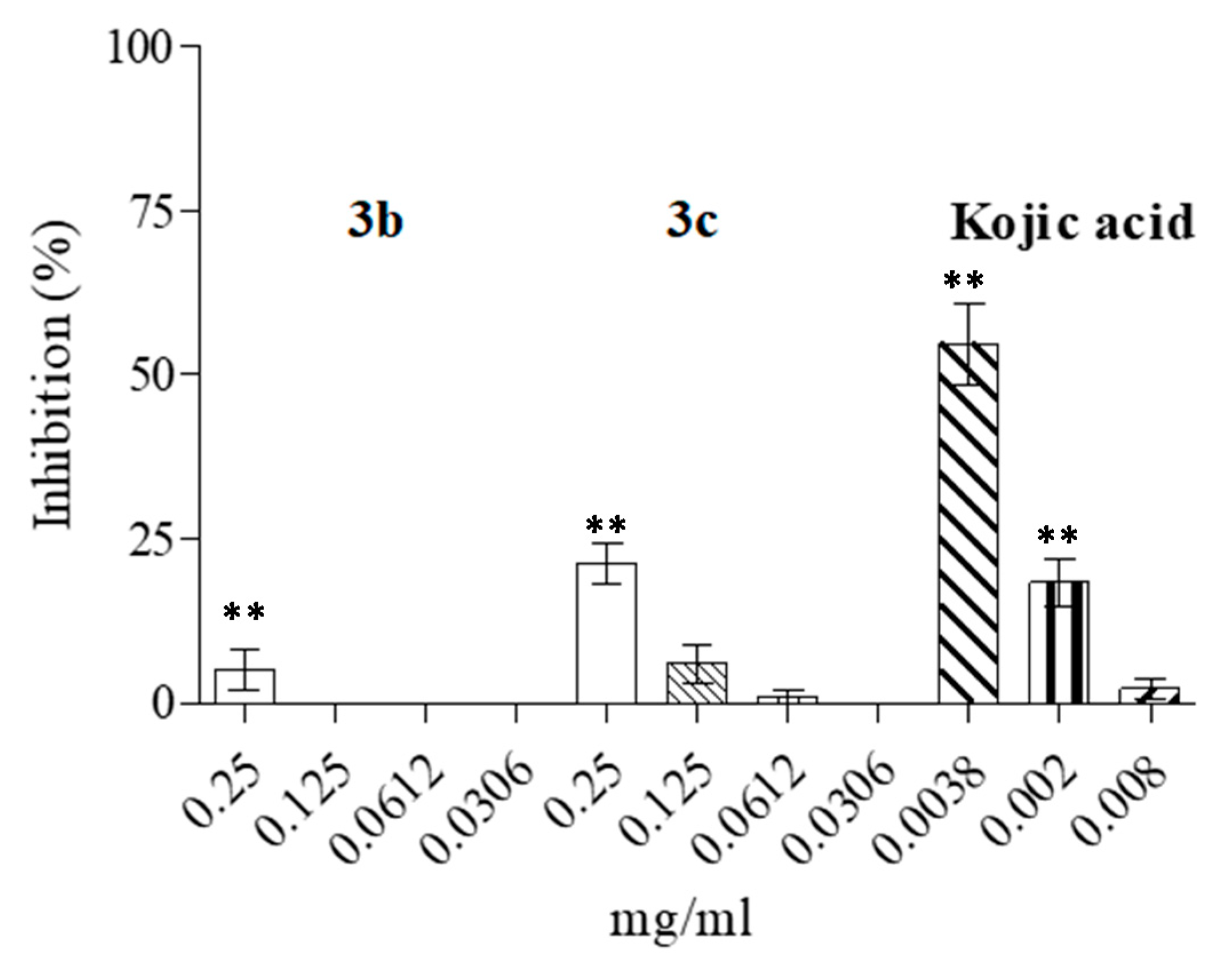
3.4. Effect of 1-(1H-indol-3-yl) Derivatives on Tyrosinase Enzymatic Activity

In order to shed some light on the possible mechanisms of action of the compounds, we performed a partial purification of tyrosinase from the tested microorganisms and evaluated the inhibitory effects of the most promising compounds (3b and 3c) at the concentrations of the experimental obtained MIC. The results showed an inhibition of the microorganism enzyme of up to about 28%, which may be, in part, responsible for the observed antimicrobial effects. As reported in Figure 3, compound 3c was able to inhibit the enzyme up to ~28% at the maximum tested concentration of 0.250 mg/mL, whereas the inhibitory effects were clearly less evident at lower concentrations. On the other hand, compound 3b was only able to perform a slight inhibition at the maximum tested concentration.

4. Discussion

The increase in antimicrobial resistance is considered a major health issue, and an estimated 700,000 people lose their life annually due to drug-resistant infections [23]. Therefore, global effort is currently focused on the development of novel therapeutics, both of natural and synthetic origin, to combat bacterial, fungal and viral resistance. Here, we have demonstrated that some 1-(1H-indol-3-yl) derivatives, known as tyrosinase inhibitors, have fungicidal activity against standard and clinical isolates of Candida spp. and the fungus Aspergillus niger and could potentially be used for topical formulations. Recently, a series of 2-aryl-3-azolyl-1-indolyl-propan-2-ol was synthetized and tested against Candida albicans and other Candida species: results showed a good antifungal potential of the compounds, with MIC values ranging between 0.005 and 1.25 μg/mL against C. albicans CA98001 [24]. The antimicrobial activity of novel indole derivatives containing 1,2,4-triazole, 1,3,4-thiadiazole and carbothioamide was evaluated against bacterial strains, including Staphylococcus aureus, methicillin-resistant Staphylococcus aureus, Escherichia coli, Bacillus subtilis, and the yeasts Candida albicans and Candida krusei: all compounds showed antimicrobial activity, with MIC values of 3.125–50 µg/mL [25]. Another study revealed that that 7-benzyloxyindole, 4-fluoroindole and 5-iodoindole effectively inhibited C. albicans biofilm formation compared to fluconazole [26].
Among tyrosinase inhibitors, a variety of different compounds, including polyphenols, benzaldehyde and benzoate derivatives, long-chain lipids and steroids, other natural (such as anthraquinones and dieckol) and synthetic (N-phenylthiourea, N-substituted-N-nitrosohydroxylamines, sildenafil, oxadiazole, oxazolones, and tetraketones types) inhibitors, as well as irreversible inactivators, have been identified [6]. Oliveira et al. [27] recently evaluated 16 analogues of coumaric and cinnamic acid as possible tyrosinase inhibitors; the results also indicated a growth inhibition in Cryptococcus neoformans, as well as an increase in amphotericin B antifungal activity (Coumaric acid analogues inhibit growth and melanin biosynthesis in Cryptococcus neoformans and potentialize amphotericin B antifungal activity).
A range of hydroxyl-substituted benzoic acid/cinnamic acid derivatives, whose antifungal potential has been widely investigated, have also been assessed for their tyrosinase inhibition and antimelanogenic activity [28]. The antifungal potential of two extracts obtained from millipede order Julida was recently evaluated against seven Fusarium species in combination with their acetylcholinesterase and tyrosinase inhibition activity [29].
Herein, we report the synthesis of derivatives 3a3g following a multistep process. All compounds were tested for antifungal activity against Candida spp. and Aspergillus niger. Among them, the best outcomes were obtained for 3b and 3c, which exhibited potent action against all the strains tested. Given the results obtained, it was found that the addition of substituents on the indole ring (e.g., compound 3b) or benzyl tail (e.g., compound 3c) play an important role in the modulation of antifungal activity. Indeed, moving from the unsubstituted derivative 3a to the analogues bearing substituents on indole or benzyl portion, an excellent improvement of biological activity was observed. However, the simultaneous presence of substituents on the two heads of the molecule led to inactive compounds (e.g., 3f). In addition, the analysis of the biological results pointed out the importance of the correct linker length between the indole ring and the benzylpiperidine moiety. Indeed, the two compounds, 3d and 3f, bearing a methylene bridge were found to be inactive against all the tested strains. The SAR information that emerged from this biological screening could be useful for further design aimed to identify optimizing antifungal candidates.
The use of novel antimicrobials in combination with traditional drugs could be useful to identify mechanisms of synergism and drug-resistant modulating properties. Here, we have shown that the association between compound 3c and fluconazole was effective against C. albicans and C. parapsilosis. The same compound produced an inhibition of the microbial tyrosinase at the concentrations tested. The synergistic enhancements of antimicrobial activity could also be explored with magainins antimicrobial peptides, which may occur by indirect interactions through the lipid phase of bacterial membranes [30]. Recently, Vriens et al. [31] demonstrated that the radish defensins RsAFP1 and RsAFP2 act synergistically with caspofungin against the formation of Candida albicans biofilms).

5. Conclusions

In conclusion, we have demonstrated the fungicidal potential of 1-(1H-indol-3-yl) derivatives, which can potentially be used for topical formulations. In particular, the presence of the indole ring or benzyl tail on this present structure plays a crucial role in the demonstrated antifungal activity. Further studies are warranted to evaluate the mechanisms of action of potentially synergistic association to combat drug resistance as well as their effect on the partial inhibition of microbial tyrosinase.

Author Contributions

Conceptualization, G.M., L.D.L. and D.B.; methodology, T.G., G.G. and F.M.; writing—original draft preparation, G.G.; writing—review and editing, T.G., L.D.L. and D.B.; funding acquisition, G.M. and D.B. All authors have read and agreed to the published version of the manuscript.

Funding

This research received no external funding.

Institutional Review Board Statement

Not applicable.

Informed Consent Statement

Not applicable.

Data Availability Statement

The data presented in this study are available on request from the corresponding author.

Conflicts of Interest

The authors declare no conflict of interest.

References

  1. Odds, F.C.; Brown, A.J.; Gow, N.A. Antifungal agents: Mechanisms of action. Trends Microbiol. 2003, 11, 272–279. [Google Scholar] [CrossRef]
  2. Yapar, N. Epidemiology and risk factors for invasive candidiasis. Ther. Clin. Risk Manag. 2014, 10, 95–105. [Google Scholar] [CrossRef] [PubMed] [Green Version]
  3. Colombo, A.L.; Júnior, J.N.A.; Guinea, J. Emerging multidrug-resistant Candida species. Curr. Opin. Infect. Dis. 2017, 30, 528–538. [Google Scholar] [CrossRef]
  4. Wu, Y.; Du, S.; Johnson, J.L.; Tung, H.Y.; Landers, C.T.; Liu, Y.; Seman, B.G.; Wheeler, R.T.; Costa-Mattioli, M.; Kheradmand, F.; et al. Microglia and amyloid precursor protein coordinate control of transient Candida cerebritis with memory deficits. Nat. Commun. 2019, 10, 58. [Google Scholar] [CrossRef]
  5. Zolghadri, S.; Bahrami, A.; Hassan Khan, M.T.; Munoz-Munoz, J.; Garcia-Molina, F.; Garcia-Canovas, F.; Saboury, A.A. A comprehensive review on tyrosinase inhibitors. J. Enzym. Inhib. Med. Chem. 2019, 34, 279–309. [Google Scholar] [CrossRef] [Green Version]
  6. Chang, T.S. An updated review of tyrosinase inhibitors. Int. J. Mol. Sci. 2009, 10, 2440–2475. [Google Scholar] [CrossRef] [Green Version]
  7. Garcia-Jimenez, A.; Teruel-Puche, J.A.; Garcia-Ruiz, P.A.; Berna, J.; Rodríguez-López, J.N.; Tudela, J.; Garcia-Canovas, F. Action of 2,2’,4,4’-tetrahydroxybenzophenone in the biosynthesis pathway of melanin. Int. J. Biol. Macromol. 2017, 98, 622–629. [Google Scholar] [CrossRef]
  8. Chung, B.Y.; Kim, S.Y.; Jung, J.M.; Won, C.H.; Choi, J.H.; Lee, M.W.; Chang, S.E. The antimycotic agent clotrimazole inhibits melanogenesis by accelerating ERK and PI3K-/Akt-mediated tyrosinase degradation. Exp. Dermatol. 2015, 24, 386–388. [Google Scholar] [CrossRef]
  9. Taslimi, P. Evaluation of in vitro inhibitory effects of some natural compounds on tyrosinase activity and molecular docking study: Antimelanogenesis potential. J. Biochem. Mol. Toxic. 2020, 34, e22566. [Google Scholar] [CrossRef] [PubMed]
  10. Cairone, F.; Simonetti, G.; Orekhova, A.; Casadei, M.A.; Zengin, G.; Cesa, S. Health Potential of Clery Strawberries: Enzymatic Inhibition and Anti-Candida Activity Evaluation. Molecules 2021, 26, 1731. [Google Scholar] [CrossRef] [PubMed]
  11. D’Arrigo, M.; Bisignano, C.; Irrera, P.; Smeriglio, A.; Zagami, R.; Trombetta, D.; Romeo, O.; Mandalari, G. In vitro evaluation of the activity of an essential oil from Pistacia vera L. variety Bronte hull against Candida sp. Bmc. Complem. Altern. Med. 2019, 19, 6. [Google Scholar] [CrossRef]
  12. Buemi, M.R.; De Luca, L.; Ferro, S.; Gitto, R. Targeting GluN2B-containing N-Methyl-D-aspartate receptors: Design, synthesis, and binding affinity evaluation of novel 3-substituted indoles. Arch. Pharm. (Weinheim) 2014, 347, 533–539. [Google Scholar] [CrossRef] [PubMed]
  13. Ferro, S.; De Luca, L.; Germanò, M.P.; Buemi, M.R.; Ielo, L.; Certo, G.; Kanteev, M.; Fishman, A.; Rapisarda, A.; Gitto, R. Chemical exploration of 4-(4-fluorobenzyl)piperidine fragment for the development of new tyrosinase inhibitors. Eur. J. Med. Chem. 2017, 125, 992–1001. [Google Scholar] [CrossRef]
  14. Gitto, R.; De Luca, L.; Ferro, S.; Buemi, M.R.; Russo, E.; De Sarro, G.; Costa, L.; Ciranna, L.; Prezzavento, O.; Arena, E.; et al. Synthesis and biological characterization of 3-substituted-1H-indoles as ligands of GluN2B-containing N-methyl-D-aspartate receptors. J. Med. Chem. 2011, 54, 8702–8706. [Google Scholar] [CrossRef] [PubMed]
  15. Bisignano, C.; Filocamo, A.; Faulks, R.M.; Mandalari, G. In vitro antimicrobial activity of pistachio (Pistacia vera L.) polyphenols. FEMS Microbiol. Lett. 2013, 341, 62–67. [Google Scholar] [CrossRef] [PubMed] [Green Version]
  16. Clinical and laboratory Standards Institute. CLSI. Reference Method for Broth Dilution Antifungal Susceptibility Testing of Yeasts, Approved Standard M27-A3. M27-A3, 3rd ed.; CLSI: Wayne, PA, USA, 2008. [Google Scholar]
  17. White, R.L.; Burgess, D.S.; Manduru, M.; Bosso, J.A. Comparison of three different in vitro methods of detecting synergy: Time-kill, checkerboard, and E test. Antimicrob. Agents Chemother. 1996, 40, 1914–1918. [Google Scholar] [CrossRef] [PubMed] [Green Version]
  18. Naraoka, T.; Uchisawa, H.; Mori, H.; Matsue, H.; Chiba, S.; Kimura, A. Purification, characterization and molecular cloning of tyrosinase from the cephalopod mollusk, Illex argentinus. Eur. J. Biochem. 2003, 270, 4026–4038. [Google Scholar] [CrossRef] [PubMed]
  19. Bradford, M.M. A rapid and sensitive method for the quantitation of microgram quantities of protein utilizing the principle of protein-dye binding. Anal. Biochem. 1976, 72, 248–254. [Google Scholar] [CrossRef]
  20. Masamoto, Y.; Ando, H.; Murata, Y.; Shimoishi, Y.; Tada, M.; Takahata, K. Mushroom tyrosinase inhibitory activity of esculetin isolated from seeds of Euphorbia lathyris L. Biosci. Biotechnol. Biochem. 2003, 67, 631–634. [Google Scholar] [CrossRef]
  21. Filocamo, A.; Bisignano, C.; D’Arrigo, M.; Ginestra, G.; Mandalari, G.; Galati, E.M. Norfloxacin and ursolic acid: In vitro association and postantibiotic effect against Staphylococcus aureus. Lett. Appl. Microbiol. 2011, 53, 193–197. [Google Scholar] [CrossRef]
  22. Visalli, M.A.; Jacobs, M.R.; Appelbaum, P.C. Activities of three quinolones, alone and in combination with extended-spectrum cephalosporins or gentamicin, against Stenotrophomonas maltophilia. Antimicrob. Agents Chemother. 1998, 42, 2002–2005. [Google Scholar] [CrossRef] [Green Version]
  23. O’Neill, J. Tackling Drug-Resistant Infections Globally: Final Report and Recommendations; HM Government and the Wellcome Trust: London, UK, 2016.
  24. Lebouvier, N.; Pagniez, F.; Na, Y.M.; Shi, D.; Pinson, P.; Marchivie, M.; Guillon, J.; Hakki, T.; Bernhardt, R.; Yee, S.W.; et al. Synthesis, Optimization, Antifungal Activity, Selectivity, and CYP51 Binding of New 2-Aryl-3-azolyl-1-indolyl-propan-2-ols. Pharmaceuticals 2020, 13, 186. [Google Scholar] [CrossRef] [PubMed]
  25. Shirinzadeh, H.; Süzen, S.; Altanlar, N.; Westwell, A.D. Antimicrobial Activities of New Indole Derivatives Containing 1,2,4-Triazole, 1,3,4-Thiadiazole and Carbothioamide. Turk. J. Pharm. Sci. 2018, 15, 291–297. [Google Scholar] [CrossRef] [PubMed]
  26. Manoharan, R.K.; Lee, J.H.; Lee, J. Efficacy of 7-benzyloxyindole and other halogenated indoles to inhibit Candida albicans biofilm and hyphal formation. Microb. Biotechnol. 2018, 11, 1060–1069. [Google Scholar] [CrossRef] [Green Version]
  27. Oliveira, L.; Ferrarini, M.; Dos Santos, A.P.; Varela, M.T.; Corrêa, I.; Tempone, A.G.; Melhem, M.; Vallim, M.A.; Fernandes, J.; Pascon, R.C. Coumaric acid analogues inhibit growth and melanin biosynthesis in Cryptococcus neoformans and potentialize amphotericin B antifungal activity. Eur. J. Pharm. Sci 2020, 153, 105473. [Google Scholar] [CrossRef] [PubMed]
  28. Nazir, Y.; Saeed, A.; Rafiq, M.; Afzal, S.; Ali, A.; Latif, M.; Zuegg, J.; Hussein, W.M.; Fercher, C.; Barnard, R.T.; et al. Hydroxyl substituted benzoic acid/cinnamic acid derivatives: Tyrosinase inhibitory kinetics, anti-melanogenic activity and molecular docking studies. Bioorg. Med. Chem. Lett. 2020, 30, 126722. [Google Scholar] [CrossRef] [PubMed]
  29. Ilić, B.; Unković, N.; Knežević, A.; Savković, Ž.; Ljaljević Grbić, M.; Vukojević, J.; Jovanović, Z.; Makarov, S.; Lučić, L. Multifaceted activity of millipede secretions: Antioxidant, antineurodegenerative, and anti-Fusarium effects of the defensive secretions of Pachyiulus hungaricus (Karsch, 1881) and Megaphyllum unilineatum (C. L. Koch, 1838) (Diplopoda: Julida). PLoS ONE 2019, 14, e0209999. [Google Scholar] [CrossRef] [Green Version]
  30. Glattard, E.; Salnikov, E.S.; Aisenbrey, C.; Bechinger, B. Investigations of the synergistic enhancement of antimicrobial activity in mixtures of magainin 2 and PGLa. Biophys. Chem. 2016, 210, 35–44. [Google Scholar] [CrossRef] [PubMed]
  31. Vriens, K.; Cools, T.L.; Harvey, P.J.; Craik, D.J.; Braem, A.; Vleugels, J.; De Coninck, B.; Cammue, B.P.; Thevissen, K. The radish defensins RsAFP1 and RsAFP2 act synergistically with caspofungin against Candida albicans biofilms. Peptides 2016, 75, 71–79. [Google Scholar] [CrossRef]
Figure 1. Reagents and conditions: (i) A, POCl3, DMA, rt, 24 h; B, ClCH2CON(CH3)2, POCl3, rt, 2.5 h; (ii) paraformaldehyde, 4-benzylpiperidine · HCl or 4-(4-fluorobenzyl)piperidine · HCl, HCl conc., DMF, MW, 80 °C, 3 min. (iii) BBr3, DCM, N2 atm., rt, overnight.
Microorganisms 09 02070 g001
Figure 2. Haemolytic activity of compounds 3b and 3c and the concentration utilized in the experiments. The total haemolysis was obtained by diluting erythrocytes with distilled water (E). A control without additives to monitor spontaneous haemolysis was also performed (B). The data are expressed as means ± S.D. (n = 4). Asterisks (**) indicate a significant difference with respect to control (p < 0.05).
Microorganisms 09 02070 g002
Figure 3. Inhibitory activity against partial purified tyrosinase obtained from C. albicans of the main compounds with antifungal activity and kojic acid (substrate:L-DOPA). Asterisks (**) indicate a significant difference with respect to control (p < 0.05).
Microorganisms 09 02070 g003
Table 1. MICs of 1-(1H-indol-3-yl) derivatives 3ag (expressed as mg/mL) against Candida spp. and Aspergillus niger.
STRAIN3a3b3c3d3e3f3g
Candida glabrata strain 9>1.0000.125–0.1250.125–0.250>1.0000.250–0.250>1.000>1.000
Candida glabrata strain 33>1.0000.125–0.1250.125–0.125>1.0000.250–0.250>1.0000.125–0.250
Candida parapsilosis strain 30>1.0000.125–0.1250.125–0.125>1.0000.250–0.250>1.000>1.000
Candida parapsilosis strain 34>1.0000.125–0.1250.125–0.125>1.0000.250–0.250>1.000>1.000
Candida albicans strain 12>1.0000.250–0.5000.250–0.500>1.0000.500–1.000>1.000>1.000
Candida albicans strain 13>1.0000.250–0.2500.250–0.250>1.0000.500–1.000>1.000>1.000
Candida albicans strain 16>1.0000.125–0.1250.125–0.250>1.0000.500–1.000>1.0000.250–0.500
Candida albicans ATCC10231>1.0000.250–0.2500.250–0.250>1.0000.500–1.000>1.000>1.000
Aspergillus niger ATCC16404>1.0000.500–0.5000.500–0.500>1.0001.000–1.0000.500–0.500>1.000
MIC, minimum inhibitory concentration.
Table 2. MFCs of 1-(1H-indol-3-yl) derivatives 3ag (expressed as mg/mL) against Candida spp. and Aspergillus niger.
STRAIN3a3b3c3d3e3f3g
Candida glabrata strain 9>1.0000.2500.250>1.0000.500>1.000>1.000
Candida glabrata strain 33>1.0000.2500.125>1.0000.500>1.000>1.000
Candida parapsilosis strain 30>1.0000.2500.125>1.0000.500>1.000>1.000
Candida parapsilosis strain 34>1.0000.1250.125>1.0000.500>1.000>1.000
Candida albicans strain 12>1.0001.0000.500>1.0000.500>1.000>1.000
Candida albicans strain 13>1.0001.0000.500>1.0000.500>1.000>1.000
Candida albicans strain 16>1.0000.2500.250>1.0000.500>1.000>1.000
Candida albicans ATCC10231>1.0000.5000.250>1.0000.500>1.000>1.000
Aspergillus niger ATCC16404>1.0001.0001.000>1.0001.000>1.000>1.000
MFC, minimum fungicidal concentration.
Table 3. FIC index of the association between 3b and 3c with reference antifungal (caspofungin and fluconazole) against ATCC and clinical Candida spp.
STRAIN3b/Caspofungin3b/Fluconazole3c/Caspofungin3c/Fluconazole
Candida glabrata strain 331.501.062.002.00
Candida parapsilosis strain 341.501.061.060.61
Candida albicans strain 161.472.621.210.62
Candida albicans ATCC102310.753.001.251.10
Publisher’s Note: MDPI stays neutral with regard to jurisdictional claims in published maps and institutional affiliations.

