Article

Development and Inter-Laboratory Validation of Diagnostics Panel for Detection of Biothreat Bacteria Based on MOL-PCR Assay

1
Department of Microbiology and Antimicrobial Resistance, Veterinary Research Institute, Hudcova 296/70, 621 00 Brno, Czech Republic
2
Department of Experimental Biology, Faculty of Science, Masaryk University, Kamenice 753/5, 625 00 Brno, Czech Republic
3
Military Health Institute, Military Medical Agency, Tychonova 1, 160 01 Prague 6, Czech Republic
4
Military Veterinary Institute, Opavska 29, 748 01 Hlucin, Czech Republic
5
Department of Meat Hygiene and Technology, Faculty of Veterinary Hygiene and Ecology, University of Veterinary and Pharmaceutical Sciences Brno, Palackeho tr. 1946/1, 612 42 Brno, Czech Republic
6
Collection of Animal Pathogenic Microorganisms, Department of Bacteriology, Veterinary Research Institute, Hudcova 296/70, 621 00 Brno, Czech Republic
7
Department of Botany and Zoology, Faculty of Science, Masaryk University, Kotlarska 2, 611 37 Brno, Czech Republic
*
Author to whom correspondence should be addressed.
Microorganisms 2021, 9(1), 38; https://doi.org/10.3390/microorganisms9010038
Submission received: 6 November 2020 / Revised: 16 December 2020 / Accepted: 22 December 2020 / Published: 24 December 2020
(This article belongs to the Section Microbial Biotechnology)

Abstract

:
Early detection of biohazardous bacteria that can be misused as biological weapons is one of the most important measures to prevent the spread and outbreak of biological warfare. For this reason, many instrument platforms need to be introduced into operation in the field of biological warfare detection. Therefore the purpose of this study is to establish a new detection panel for biothreat bacteria (Bacillus anthracis, Yersinia pestis, Francisella tularensis, and Brucella spp.) and confirm it by collaborative validation by using a multiplex oligonucleotide ligation followed by polymerase chain reaction and hybridization to microspheres by MagPix detection platform (MOL-PCR). Appropriate specific sequences in bacterial DNA were selected and tested to assemble the detection panel, and MOLigo probes (short specific oligonucleotides) were designed to show no cross-reactivity when tested between bacteria and to decrease the background signal measurement on the MagPix platform. During testing, sensitivity was assessed for all target bacteria using serially diluted DNA and was determined to be at least 0.5 ng/µL. For use as a diagnostic kit and easier handling, the storage stability of ligation premixes (MOLigo probe mixes) was tested. This highly multiplex method can be used for rapid screening to prevent outbreaks arising from the use of bacterial strains for bioterrorism, because time of analysis take under 4 h.

1. Introduction

Bioterrorism refers to the deliberate abuse of pathogenic microorganisms (bacteria, viruses or their toxins) to spread life threatening diseases on a large scale and thus devastating the population of the area. Bacterial strains can be misused as biological weapons not only as a threat to human health but also in terms of agricultural abuse and ecotoxicological risks [1]. The use of biological agents in comparison with conventional weapons is very attractive to terrorists because of their relatively low cost and relative availability [2]. The most efficient way for delivery of biological agents is in the form of aerosols. However, there are a number of other ways in which biological agents can be disseminated, e.g., via food, feed, or water contamination [3]. Bacillus anthracis—anthrax, Yersinia pestis—plague, Francisella tularensis—tularemia or Brucella spp.—brucellosis are among the most abused bacteria in biological weapons [4]. The Centre for Disease Control and Prevention (CDC) classifies biological agents into categories A–C, where the category A represents the most dangerous pathogens. All the biothreat bacteria listed above belong to group A, except for Brucella spp., which is classified in category B [5].
B. anthracis is a spore-forming bacterium and is classified as one of the most important pathogens for abuse as a bioterrorist weapon [6]. The respiratory route of infection caused by inhalation of spores is a minor issue in the context of global human anthrax cases but a serious issue when associated with bioterrorism [7]. Anthrax containing letters from October 2001 confirmed that only a small amount of B. anthracis spores are sufficient for an outbreak due to a terrorist action [8]. The infectious dose ranges from less than 10 to over 10,000 spores depending on factors like the route of the infection or the health status of the exposed individual [7,8,9,10]. Another destructive disease is the plague caused by Y. pestis. Person to person transmission of Y. pestis, i.e., primary pneumonic plague, is possible by way of airborne droplets or aerosols and if untreated, it is 100% fatal [11,12]. A short incubation period, very low infectious dose requirement of only 1 to 10 organisms, and the progressive nature of the infection classifies this bacterium into group A of pathogens at risk of bioterrorism abuse [13,14]. F. tularensis is able to cause a highly infectious disease called tularemia by as little as a few microbes aspirated from the surrounding air. Tularemia is a disease of wild animals (rodents, hares, and rabbits) that can be transmitted to humans [15]. F. tularensis is classified into four subspecies, but only two of them are the causative agents of disease in humans. Type A is subsp. tularensis (predominantly found in North America), and type B is subsp. holartica (predominantly found in Eurasia) [16]. Brucella spp. causes a serious contagious disease transmissible to humans, which results in reproductive failure of infected animals [17]. Genus Brucella includes several species exhibiting host adaptations. B. abortus (cattle), B. melitensis (sheep and goats), and B. suis (pigs) belong to the most common and virulent types not only for livestock but also for wildlife and humans [18,19]. Transmission of 100 to 1000 cells are sufficient for the development of the disease [20]. Although brucellosis has been eradicated in most developed countries, it is still found in many developing countries [21].
In order to prevent the spread of the agent after a bioterrorist attack and to apply efficient preventive measures to stop the spread of infectious diseases, it is important to quickly identify and specify bacterial species [22]. The method of choice for fast and reliable identification of pathogens in various matrices is polymerase chain reaction (PCR) [23]. However, analysis of numerous samples for the presence of multiple infectious agents by PCR requires modifications of the PCR workflow. One of the possibilities is to use PCR as a suspension arrays in which the fluorescently labelled PCR product is visualized by its attachment to a specific bead. A various modifications of suspension arrays exists [24]. The latter approach, multiple oligonucleotide ligation PCR (MOL-PCR), allows the use of only a single pair of universal primers which makes optimization of the whole assay easier. The first step is then accomplished by ligation of specific targets sequence which will create a template with specific complementary primer sequences for annealing of the universal primers. The PCR products can be labelled either by the post-PCR conjugation of streptavidin-phycoerythrin (SAPE) complex with e.g., biotinylated primer or by direct labelling of a PCR primer with fluorescent dyes (Alexa Fluor532, BODIPY-TMRX). The last step is the hybridization of the PCR product using a specific 24 base DNA sequence, which is already part of the probe and its complementary sequence located on the selected set of magnetic microspheres. Precise optimization of the MOL-PCR assay is crucial and key points in the optimization process were experimentally identified [25]. Since that time, MOL-PCR has been successfully adopted for parallel detection of many human, animal, or even insect pathogens [25,26,27,28,29,30,31]. All proposed detection panels show high specificity and sensitivity and are useful for screening a wide spectrum of samples in general.
To address the need of security bodies to be capable of simultaneously detecting the four major biothreat bacteria (B. anthracis, Brucella spp., Y. pestis and F. tularensis), a comprehensive and specific protocol for the MOL-PCR suspension array was developed in this study. The scheme of the whole MOL-PCR reaction is figuratively described in the our previous study [25]. The main aim of the study was to construct a panel of detection markers for all listed pathogens using at least two DNA markers (chromosomal markers and virulence genes for each pathogen). The whole multiplex detection panel comprised an internal control (IC) to exclude inhibition of the PCR by impurities potentially present in the sample. All the diagnostic parameters including analytical specificity (inclusivity and exclusivity) and sensitivity (limit of detection (LoD)) were determined and the multiplex panel was validated according to the FDA Guidelines for the Validation of Analytical Methods for the Detection of Microbial Pathogens in Foods and Feeds [32] by a ring trial among four laboratories to prove its diagnostic potential in routine practice.

2. Materials and Methods

2.1. Bacterial Strains and DNA Extraction

Bacillus anthracis, Francisella tularensis subsp. tularensis, Francisella tularensis subsp. holartica, Brucella abortus, Brucella suis and Brucella melitensis obtained from the Collection of Animal Pathogenic Microorganisms (CAPM) at the Veterinary Research Institute (Brno, Czech Republic) were used for testing inclusivity. Yersinia pestis was purchased from the National Collection of Type Cultures—NCTC (Public Health England, Salisbury, United Kingdom) and was also used for inclusivity testing. Other bacterial DNA from Staphylococcus aureus, Listeria monocytogenes, Bacillus cereus, Yersinia enterocolitica, Campylobacter jejuni, Salmonella enterica, Escherichia coli O26, Escherichia coli O157, Enterococcus faecalis, Enterococcus faecium, Vibrio parahaemoliticus, Yersinia pseudotuberculosis, Clostridium tetani, and Clostridium botulinum were also obtained from the CAPM and other collections and were used for testing exclusivity (Table 1). Genomic DNA (gDNA) was purified using the DNeasy® Blood & Tissue kit (Qiagen, Germany) according to the manufacturer’s protocol with a few modifications as described previously [33]. DNA concentrations were determined spectrophotometrically using a NanoDrop™ 2000/2000c Spectrophotometer (Thermo Fisher Scientific, Waltham, MA, USA) and diluted in sterile distilled water to the required concentrations, if appropriate. To test exclusivity and long-term storage effect of the ligation mix, DNA was diluted to a concentration of 10 ng/µL.

2.2. Internal Control

An internal control (IC) was added to each reaction to differentiate a false negative from truly negative results because of the inhibition of the MOL-PCR reaction. The IC was designed as a synthetic sequence based on the joined mitochondrial DNA sequences of two extinct species, Thylacine (Thylacinus cynocephalus, GenBank Acc. No. FJ515781.1) and Moa bird (Dinornis struthoides, GenBank Acc. No. AY326187.1). This 150 bp control synthetic sequence was synthesized de novo and cloned into a plasmid [34]. The essence of internal control is to be positive in all samples as well as in negative controls without a template—no template controls (NTC). It confirms that all reaction steps have taken place correctly.

2.3. Design of MOLigo Probes (Short Specific Oligonucleotides)

Pathogen-specific sequences (specific genes for detection of bacteria) were selected according to previously published data. The References are listened in Table 2. Most previous work, of which there were specific targets chosen, were based on qPCR method. Sequences of each species were extracted from the National Centre for Biotechnology Information (NCBI) database. For detection of a specific target sequence are designed MOLigo probes. Each pair of MOLigo probes is specific for a particular target sequence, but all MOLigo pairs contain the same sequence for annealing the universal primers (Reverse—Rw and Forward—Fw). One of the MOLigo probes also contains the unique 24 base DNA sequence called an xTAG (from Luminex corporation; https://www.luminexcorp.com/magplex-tag-microspheres/), by which PCR products hybridize to a magnetic microsphere with a covalently linked anti-TAG sequence. The specific parts of MOLigo probe sequences (specific sequence for a given target—part 1 and part 2) were tested with OligoAnalyzer 3.1 tool (https://eu.idtdna.com/calc/analyzer) to identify properties such as melting temperature, hairpins, dimer formation etc., of the individual target sequences used for probe design. Optimal probe sequences were finally checked by NCBI BLAST for possible non-target interactions. A more detailed scheme for designing probes for the MOL-PCR reaction is shown in Figure 1. MOLigo probe synthesis was performed by standard desalination purification (Generi-Biotech, Czech Republic). The final size of the ligation product ranged from 102 to 113 base pairs. All other parameters for MOLigo probe design were used according to the previous study [25].

2.4. MOL-PCR Assay

2.4.1. Coating the Magnetic Microspheres with Anti-xTAG Oligonucleotides

Magplex®—C Microspheres sets (6.5 µm magnetic, carboxylated and internally labeled with two fluorescent dyes for bead identification) were purchased from Luminex Corporation. Each set of microspheres (12.5 × 10⁶ microspheres/mL; Luminex Corp., Texas, USA) was vortexed and 200 µL was collected into a protein low bind DNA tube (Eppendorf, Germany). The tubes were placed on a DynaMag-2 magnetic separator (Thermo Fisher Scientific, Waltham, MA, USA) for approximately 1–2 min and then the supernatant was aspirated. The magnetic bead pellet was suspended in 22 µL of 0.1M MES buffer pH 4.5 (Sigma-Aldrich, St. Louis, MO, USA) and further vortexed and sonicated for 30 s in an ultrasonic cleaning bath (Desen Precision Instruments, Fuzhou, China). Subsequently, 2 µL of 100 µM antiTAG (C6-amino modifications at the 5′-end, specific modification to form a covalent bond with the surface of the microspheres) was added into the appropriate tube with beads and vortexed. After that, 1.25 µL of a freshly prepared 10 mg/mL 1-ethyl-3-(3-dimethylaminopropyl) carbodiimide solution (solved in ultrapure H2O) (Thermo Fisher Scientific) was added and tubes were incubated in the dark at room temperature for 30 min. This step was performed twice with a fresh preparation of the 1-ethyl-3-(3-dimethylaminopropyl) carbodiimide solution. The tubes were vortexed every 10 min during this time. Afterwards, the beads were washed with 1 mL of 0.02% Tween 20 (Alpha Diagnostic, Texas, USA) and 1 mL of 0.1% SDS (Sigma-Aldrich) and resuspended in 40 µL of TE buffer (pH 8.0; SERVA, Heidelberg, Germany) with vortexing and removal of the supernatant using a magnetic separator after each step. The coated beads were then stored in the dark at 4 °C. To validate bead coating, direct hybridization using xTAG oligonucleotide sequences fluorescently labelled with Bodipy dye (validation TAGs) was performed using the hybridization protocol described below. The signal intensity using validation tags and coated beads was around 1000–1200 median fluorescent intensity (MFI). Prior to any downstream reaction, the real number of beads was determined by enumeration on a hemocytometer and adjusted to 40,000 beads/µL.

2.4.2. Multiplex Oligonucleotide Ligation

Each multiplex ligation mix included 5 nM individual MOLigo pair probes (Table 2) with 2.5 μL10X Hifi Taq DNA ligase reaction buffer, 0.5 μL Hifi Taq DNA ligase (New England BioLabs, Massachusetts, USA), 0.1 μL of 0.05 ng/μL IC cloned into the plasmid and 2.5 μL template DNA. The reaction was brought to a final volume of 25 μL with H2O. Ligation protocol in the thermocycler (DNA Engine Dyad, Bio-Rad, Foster City, CA, USA) was set to: 10 min of denaturation at 95 °C followed by 20 cycles of 30 s at 95 °C and 1 min at 59 °C. Ligation products were stored at 10 °C until the next step of the MOL-PCR reaction.

2.4.3. Singleplex PCR

Amplification of the ligation products was performed in a final volume of 24 μL which consisted of 12 μL 2X EliZyme HS Robust MIX (Elisabeth Pharmacon, Brno-Židenice, Czech Republic), 0.0625 μM universal FW primer and 0.25 μM BODIPY-TMRX-labelled REV primer (Table 2). Ligation products were added to the already prepared tubes with PCR mix at a ratio of 1:3, i.e., a volume of 6 μL. PCR consisted of initial denaturation at 95 °C for 2 min, followed by 40 cycles of 95 °C for 15 s, 60 °C for 15 s, and 72 °C for 15 s.

2.4.4. Hybridization and MAGPIX Analysis

Hybridization of amplified PCR products to microspheres mediated by an xTAG sequence inside the amplified DNA and a complementary antixTAG sequence covalently bound to the surface of the magnetic beads, was performed in a bead mix formed by 1250 beads for each target (Table 2) per 1 sample; 2.5 µL of 800 mM NaCl (VWR, Stříbrná Skalice, Czech Republic) and 0.8 µL of 50 mM β-(N-Morpholino)ethanesulphonic acid (MES) monohydrate free acid ≥99%, ultrapure buffer (VWR) and adjusted to a final volume of 5 µL with 1xTE buffer at pH 8.0 (VWR). About 5 µL of the bead mix was pipetted into 0.2 mL tear-off strips (BIOplastics, Landgraaf, The Netherlands) followed by the addition of 10 µL of the PCR products from the previous step. The prepared strips with bead mix and PCR products were run on the thermocycler with the following protocol: denaturation 96 °C for 90 s, followed by 37 °C for 30 min and hold at 37 °C until further processing. After hybridization was completed, 45 µL of analysis buffer was added (10 mM Tris-Cl (pH 8.0); 0.1 mM EDTA; 90 mM NaCl and 0.2% Tween 20). Strips with hybridization products and analysis buffer were placed on a 0.2 mL Multi Rack bench (Bioplastics) and analyzed in a MAGPIX instrument (Bio-Plex MAGPIX from Bio-Rad) using xPONENT 4.2. ® SOFTWARE (Luminex, Austin, TX, USA).

2.5. Specificity and Sensitivity of MOL-PCR Assay

The specificity of the developed MOL-PCR panel was tested using selected pathogens (Table 1). A concentration gradient (5×; 10×; 40×) of extracted DNA from bacterial strains (B. anthracis, Y. pestis, F. tularensis and Brucella spp.) was performed to confirm inclusivity. Concentrations of stock bacterial DNA ranged from 20 ng/μL (B. anthracis DNA) to approx. 220 ng/μL (Y. pestis, F. tularensis and B. suis DNA). A ten-fold serial dilution was made from the lowest concentrations (40×) and used to determine sensitivity of the liquid array detection system. The diluted DNA was analyzed in quadruplicate for reproducibility testing. The LoD was determined as the lowest concentration of sample measured at 100 median fluorescence intensity (MFI). The detection limit was determined for each pathogen as well as for the two detection markers. In the evaluation, the appropriate no template control (NTC) was subtracted for the appropriate marker.

2.6. Variability in Preparation of Ligation Mix and Stability during Long-Term Storage

The effect of the ligation mix composition (various MOLigo probes) and the stability during long-term storage was investigated with two independent batches of 100 premixed ligation reactions. In the first batch, ligation buffer and only MOLigo probes 1 were mixed in separate tubes and stored separately (named Ligation premix 1 = LP1); the same was done with all MOLigo probes 2 (LP2). Ligation premix 1 and 2 were mixed together just before the ligation step. Performance of this protocol was compared with all the MOLigo probes mixed and stored together in ligation buffer. Ligase was always kept separated and added to the ligation premixes just before the ligation step. B. anthracis, Y. pestis, F. tularensis, and B. melitensis DNA at 10 ng/µL were used as template in the testing and the entire MOL-PCR protocol was performed on days 0, 3, 5, 8, and 10 to investigate the freeze-thawing effect of ligation premixes. The difference in storage during freezing was compared for each of the genes by using a paired t-test, i.e., the MOLigo probes separately (LP1 and LP2) and/or the ligation mix (MIX) prepared together. The analysis was performed using Statistica 13.2 software (StatSoft Inc., Tulsa, OK, USA) and differences with P values < 0.05 were considered as statistically significant.

2.7. Inter-Laboratory Validation of the Biothreat Detection Panel

Master stocks of all chemicals were prepared, aliquoted, and frozen at the Veterinary Research Institute, Brno (VRI). Thereafter, 16 designed MOLigo pairs of probes with 1 pair of IC probes were divided into two tubes, one with MOLigo probes 1 and the second with MOLigo probes 2. Ligase was supplied separately. Pure bacterial DNA was isolated at VRI and mixed with DNA isolated from soil to mimic a natural background. Each laboratory analyzed the concentration gradient of reference DNA—B. anthracis, Y. pestis, F. tularensis, B. melitensis, B. suis, B. abortus (4 samples for each pathogen—stock solution; 5×; 10×; 40×), non-target DNA (Excess) from S. aureus at 20 ng/uL concentrations and 4 NTC– water only. The prepared aliquots were shipped on dry ice to partner laboratories at University of Veterinary and Pharmaceutical Sciences, Brno, Military Veterinary Institute, Hlucin and Department of Biological Protection, Techonin. The raw data from testing in all laboratories were exported as Excel files. Each sample was analyzed in biological duplicate and technical duplicate.

2.8. Data Analysis and Interpretation

MFI values obtained from analysis of at least 50 microspheres of each target sequence per sample were used for test evaluations. NTC with no target DNA was used to determine the background MFI value for each microsphere region. The respective NTC MFI value for the microspheres region was subtracted from the measured MFI values. Samples with MFI values higher than 100 compared to background MFI were considered as positive.

3. Results and Discussion

3.1. Biothreat Bacteria Panel Optimization

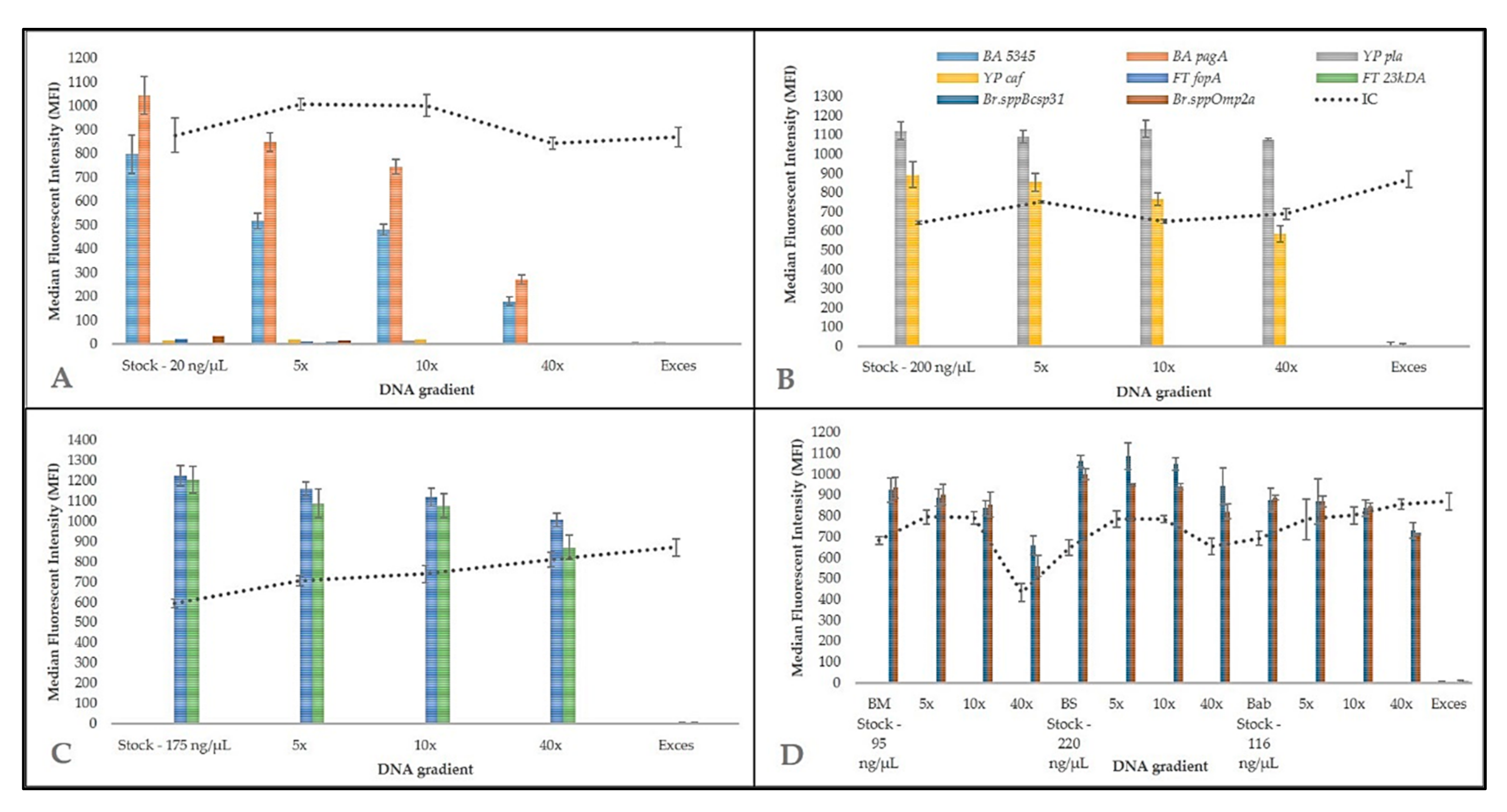
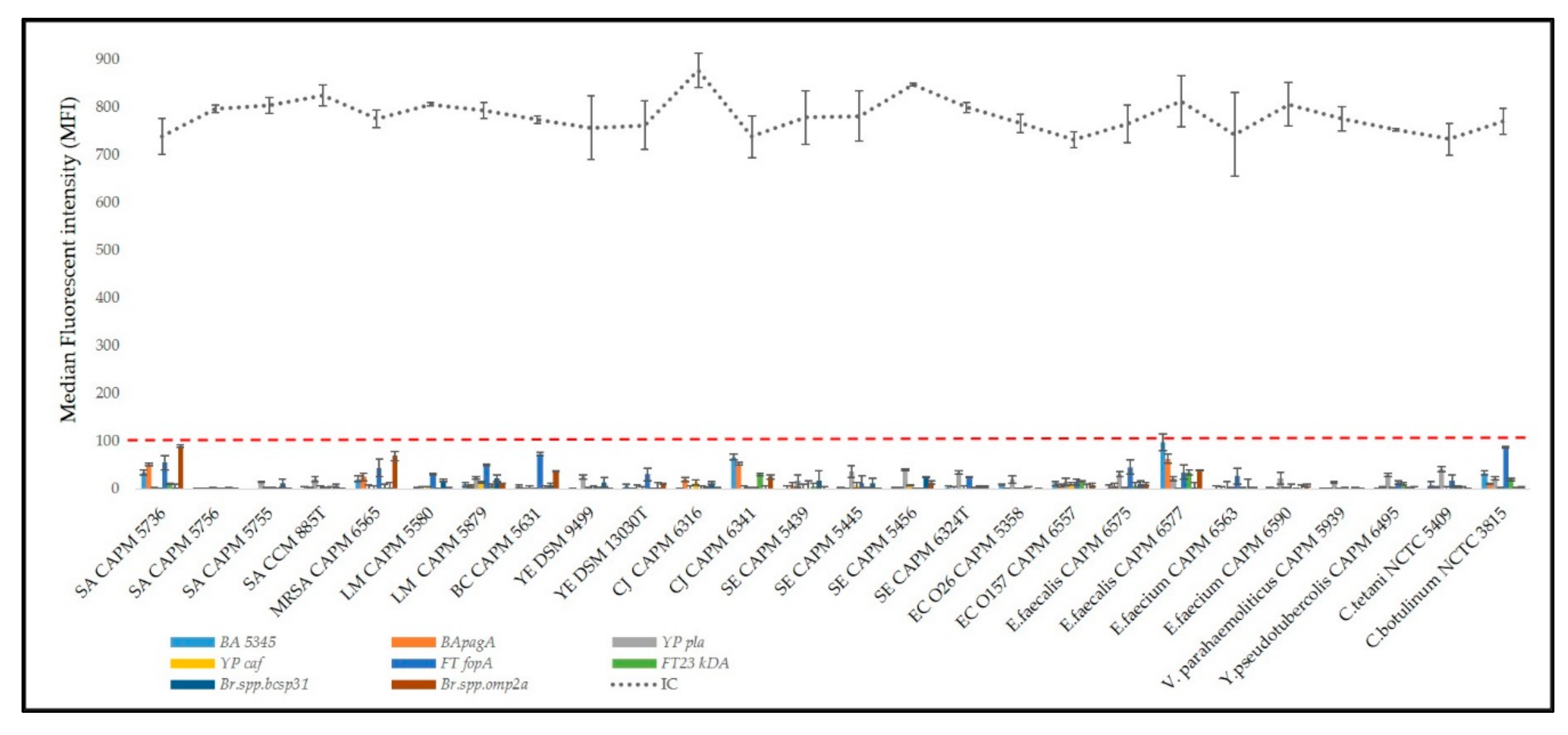
Specific genes have been selected for four bacteria that can be primarily abused as biological weapons, namely pagA, BA5345 (both B. anthracis), pla, caf1 (Y. pestis), 23kDA, fopA (F. tularensis), and omp2a and bcsp31 (Brucella spp.). These specific sequences in bacterial pathogens have been selected according to previous studies based on PCR methods, in particular real-time PCR. Relative references are listed in Table 2. Some of these are chromosomal markers, and other genes are placed on plasmids and cause a virulence of selected pathogen. Because of the gene deletion on the plasmid, two markers were preferably used for detection to confirm more identity. The assembled multiplex MOL-PCR containing all the MOLigos freshly prepared each time were tested with pure reference DNA of B. anthracis, Y. pestis, F. tularensis, and Brucella species (Figure 2) and unrelated bacterial species (Figure 3). The difference between positive and NTC samples was demonstrated when the threshold 100 MFI was applied. At the same time, the IC of MOL-PCR reaction was between 600 and 1000 MFI which shows low level of cross-reaction in amplification between target and IC. Specificity, particularly inclusivity (Figure 2) and exclusivity (Figure 3), did not show any cross-reactions or false positive or false negative interactions among all four bacterial species (Table 1). A similar detection panel for biothreat bacteria has already been assembled by Deshpande et al. 2010 [26]. Compared to Deshpande et al. study [26], Brucella species detection MOLigo probes and IC were added to reveal the reaction inhibition. The inclusion of IC is necessary in multiplexing for detection in diagnostic PCR assays to detect false negative results [44]. Frequently as prevention of false positive results is the most commonly used uracil DNA glycosylase [45]. A huge advantage of this method is its multiplexity and detection of a large number of pathogens in one reaction and therefore it is possible to extend the panel with other pathogens if necessary. Additionally, each bacterium was detected by two independent targets. Based on the previous findings, BODIPY-TMRX fluorescent dye was used for labelling the hybridization product instead of SAPE (time consuming) or Alexa Fluor 532 [25]. This approach enabled the omission of the separate step of PCR product labelling by phycoerythrin since BODIPY-TMRX dye successfully survives PCR amplification. As in most studies, the ligation step was separated from the PCR step to reduce the background signal and to allow better control over each step [27,43]. Compared to our previous study [25], the number of microspheres per sample was reduced to 1250 beads since this amount was sufficient to determine background and sample MFI but at reduced cost per reaction. Time generally plays a huge role in preventing the spread of the disease, epidemic or a biological warfare, so this method seems to be very suitable, as it is able to obtain results within 4 h and detect up to 50 targets and test a large number of samples at one time. All these things are important for the rapid implementation of measures to protect the population. Of course, the qPCR method (reaction time approximately 1.5 h) is most often used as the “gold standard” for the detection of bacteria, but unfortunately this method does not offer such a huge possibility of multiplexity in the same time.

3.2. Sensitivity Test of the MOL-PCR Reaction

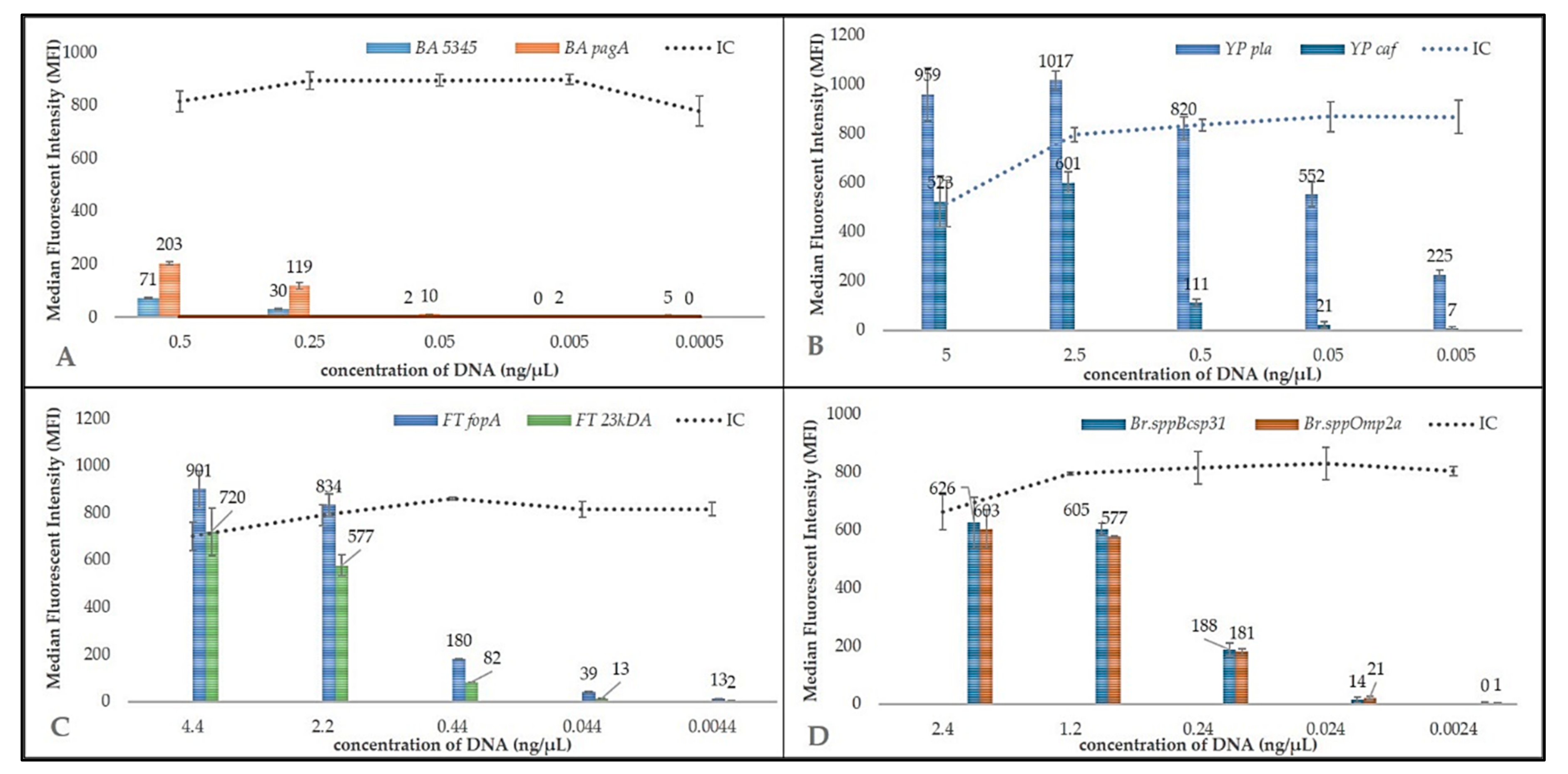
MOL-PCR sensitivity was determined using ten-fold serial dilution of B. anthracis, Y. pestis, F. tularensis, and B. melitensis DNA (Figure 4). LoD for B. anthracis was 0.5 ng/μL for BA5345 or 0.25–0.05 ng/μL for pagA. In the case of Y. pestis, pla target sequence could be detected at 0.0005 ng/μL and detection limit for caf ranged between 0.5 and 0.05 ng/μL. In the case of F. tularensis, fopA could be detected at 0.44–0.044 ng/μL and 23kDA had a detection limit of 2.2 to 0.44 ng/μL. In the case of the Brucella melitensis, the LoD was determined to be 0.29–0.0029 ng/μL for both target sequences (Table 3). In other studies, the LoD was determined for the Mycobacterial complex based on MOL-PCR and the detection limit was around 0.1 ng/µL. In another study, using a 13-plex for B. anthracis SNP typing, the LoD was 2 ng genomic DNA [27,28,29,30]. In conclusion, the biothreat panel reached a detection limit of at least 0.5 ng/uL for all pathogens and targets which corresponds with similar studies based on MOL-PCR methods. The results obtained after the evaluation of the limit of detection are that MOL-PCR method has a lower sensitivity than detection methods based on the use of DNA, thus qPCR achieves very low sensitivity. However, the MOL-PCR method is mainly a screening and semi-quantitative method, where it is very important to detect different types of pathogenic bacteria during one laboratory examination.

3.3. Long-Term Storage Effect of Ligation Mix

In a previous study, the effect of mixing and storing the ligation premix (MOLigo probes) was described [46]. There were up to 40 probe pairs divided into three premixes of which each premix contained 8, 5, and 3 pairs and the remaining 8 pairs were added prior to ligation mixture preparation. Based on these results, the impact of storage on background signals (MFI measured in the NTC) was confirmed [46]. For this reason, we performed two experiments (Figure 5) to compare the freezing effect of different MOLigo mix variants. In the first experiment, all MOLigo pair probes were frozen together with the ligation buffer. In the second experiment, MOLigo 1 probe and MOLigo 2 probe premixes were frozen separately. Immediately before use, premixes 1 and 2 were mixed together and ligase was added. From the point of view of stability, the separation of MOLigo probes (LP1 + LP2) resulted in better performance because none of the genes showed an increasing or decreasing trend in NTC MFI values during storage (Stability over time—trend). In contrast, the MOLigo mix method showed an increasing trend (p < 0.05; significance of slope of regression line) of NTC MFI values during storage of the three genes (BA pagA, YP caf, and BMspp omp2a). For seven of the eight genes, the MFI values for the mixed probes protocol were higher than for the LP1 + LP2 method, except for the FT 23kDA gene. The average differences between the two protocols were statistically significant in six cases (p < 0.05 at least). Only the BA pagA and BMspp bcsp31 genes did not show a statistically significant difference between LP1 + LP2 and mixed probes protocols (Figure 5). Our suggestion is that some undesirable cross-interactions between individual probes may occur. We therefore recommend to freeze the probe mix MOLigo 1 and MOLigo 2 separately.

3.4. Inter-Laboratory Validation of Biothreat Detection Panel

Inter-laboratory tests were performed to evaluate the reproducibility and robustness of the MOL-PCR biothreat panel in routine settings. Although reproducibility is defined as a quantitative parameter, it is possible to use it as a qualitative measure in validation of qPCR assays [44]. The results among all four laboratories showed agreement with one exception of one negative technical replicate of 40 × diluted sample analyzed at Military Veterinary Institute (Figure 2; Supplementary Figures S1–S3), however, the final interpretation of the results showed 100% agreement. Internal control (IC) was correct in all reactions and across all departments. The same applies to no template control (NTC) and the use of unrelated DNA (Exces) to evaluate the specificity of the probes. It was shown that the validation criteria postulated for qPCR assays by Food and Drug Administration (FDA) are applicable on MOL-PCR as well [47].

3.5. On-Site Usability of the Panel

To simplify the feasibility of the detection tool described here, its future conversion into a microfluidic chip or disposable cassette format is possible. A similar approach has already been introduced by Luminex corporation—ARIES®. This modification could not only simplify the analysis for point of care testing, but also significantly reduce the material costs and the possibility of technical error [48,49].
In conclusion, the presented protocol for the detection of four biothreat bacteria was developed and validated in this study. It connects the versatility of the MOL-PCR principle, single-tube detection of all bacteria, and increased sensitivity of detection/identification of each bacterium by at least two DNA targets. In addition, the IC of the process was included to exclude false negative results. The performance of the MOL-PCR assay was validated by a ring trial in four independent laboratories and the results showed that this assay can be implemented as a routine diagnostic for the most dangerous bacteria that could be misused as biological weapons. Of course, it is possible to enlarge the panel with detection targets for various other pathogens that could be misused as bioterrorist weapons. This method can be finished under 4 h, mainly due to its multiplexity and thus the detection (screening) of multiple amounts of pathogens in one reaction at the same time, which is indispensable in preventing bioterrorist action.

Supplementary Materials

The following are available online at https://www.mdpi.com/2076-2607/9/1/38/s1. Figure S1: Biothreat panel validated in University of Veterinary and Pharmaceutical Sciences Brno—collaborative validation of established biothreat detection panel based on MOL-PCR reaction. A concentration gradient of DNA isolated from biothreat bacteria (A) B. anthracis, (B) Y. pestis, (C) F. tularensis, (D) B. melitensis, B. suis and B. abortus was used to validate, verify, and to test the robustness of the method. IC confirmed the correct course of the whole reaction. The exclusivity of the detection probes was confirmed by unrelated DNA (S. aureus). Panel was evaluated by MFI after subtracting the NTC (H2O only) to the appropriate target. Figure S2: Biothreat panel validated in Centre of Biological Defence Techonin—collaborative validation of established biothreat detection panel based on MOL-PCR reaction. A concentration gradient of DNA isolated from biothreat bacteria (A) B. anthracis, (B) Y. pestis, (C) F. tularensis, (D) B. melitensis, B. suis and B. abortus was used to validate, verify, and to test the robustness of the method. IC confirmed the correct course of the whole reaction. The exclusivity of the detection probes was confirmed by unrelated DNA (S. aureus). Panel was evaluated by MFI after subtracting the NTC (H2O only) to the appropriate target. Figure S3: Biothreat panel validated in Military Veterinary Institute Hlucin—collaborative validation of established biothreat detection panel based on MOL-PCR reaction. A concentration gradient of DNA isolated from biothreat bacteria (A) B. anthracis, (B) Y. pestis, (C) F. tularensis, (D) B. melitensis, B. suis and B. abortus was used to validate, verify and to test the robustness of the method. IC confirmed the correct course of the whole reaction. The exclusivity of the detection probes was confirmed by unrelated DNA (S. aureus). Panel was evaluated by MFI after subtracting the NTC (H2O only) to the appropriate target.

Author Contributions

P.J. performed assay development, molecular work, analysis, created data outputs, and wrote the manuscript. J.M. wrote the manuscript, consulted on optimization. J.H. wrote the manuscript, consulted on design and optimization, and revised the manuscript. N.R. designed the study, consulted on data interpretation, and revised the manuscript. P.K. designed the study, secured the biological material, provided financial support, consulted on data interpretation, and revised the manuscript. O.P. performed sample validation. P.B. performed sample validation. G.B. performed sample validation. P.P. consulted on samples a development of BL3 panel. J.D. consulted on samples a development of BL3 panel. M.R. prepared samples in BL3 laboratory and performed sample validation. V.B. performed a statistical analysis of the obtained input data. All authors have read and agreed to the published version of the manuscript.

Funding

This work was supported by the project of Security Research of the Ministry of the Interior of the Czech Republic (VI 20152020044) and by the Ministry of Agriculture of the Czech Republic, institutional support MZE-RO0518. The testing part was also supported by funding from projects of Ministry of Interior Affairs, Czech Republic, [VH20172020012: Preparation of the collection of biologically significant toxins with the support of European biological European biodefence laboratory network, [VF20142015039] and by the Ministry of Defence of the Czech Republic through a long-term organization development plan 907930101413.

Informed Consent Statement

Not applicable.

Data Availability Statement

All data underlying the results are included as part of the published article and its Supplementary Materials.

Acknowledgments

Authors would like to thank Peter Eggenhuizen for language corrections. Authors also acknowledge the assistance of Monika Dufkova, Jiri Novotny, Tomas Mati for their technical assistance.

Conflicts of Interest

The authors declare no conflict of interest.

References

  1. Arun Kumar, R.; Nishanth, T.; Ravi Teja, Y.; Sathish Kumar, D. Bio threat s bacterial warfare agents. J. Bioterr. Biodef. 2011, 2, 2. [Google Scholar]
  2. Christopher, L.G.W.; Cieslak, L.T.J.; Pavlin, J.A.; Eitzen, E.M. Biological warfare: A historical perspective. JAMA 1997, 278, 412–417. [Google Scholar] [CrossRef] [PubMed]
  3. Shah, J.; Wilkins, E. Electrochemical biosensors for detection of biological warfare agents. Electroanal. Int. J. Devoted Fundam. Pract. Asp. Electroanal. 2003, 15, 157–167. [Google Scholar] [CrossRef]
  4. Flora, S.; Pachauri, V. Handbook on Biological Warfare Preparedness; Academic Press: Cambridge, MA, USA, 2019. [Google Scholar]
  5. Mölsä, M.; Hemmilä, H.; Katz, A.; Niemimaa, J.; Forbes, K.M.; Huitu, O.; Stuart, P.; Henttonen, H.; Nikkari, S. Monitoring biothreat agents (Francisella tularensis, Bacillus anthracis and Yersinia pestis) with a portable real-time PCR instrument. J. Microbiol. Methods 2015, 115, 89–93. [Google Scholar] [CrossRef]
  6. Peculi, A.; Campese, E.; Serrecchia, L.; Marino, L.; Boci, J.; Bijo, B. Genotyping of Bacillus anthracis strains circulating in Albania. J. Bioterror. Biodef. 2015, 6. [Google Scholar] [CrossRef]
  7. Carlson, C.J.; Kracalik, I.T.; Ross, N.; Alexander, K.A.; Hugh-Jones, M.E.; Fegan, M.; Elkin, B.T.; Epp, T.; Shury, T.K.; Zhang, W. The global distribution of Bacillus anthracis and associated anthrax risk to humans, livestock and wildlife. Nat. Microbiol. 2019, 4, 1337–1343. [Google Scholar] [CrossRef] [Green Version]
  8. Wilson, W.J.; Erler, A.M.; Nasarabadi, S.L.; Skowronski, E.W.; Imbro, P.M. A multiplexed PCR-coupled liquid bead array for the simultaneous detection of four biothreat agents. Mol. Cell. Probes 2005, 19, 137–144. [Google Scholar] [CrossRef]
  9. Fennelly, K.P.; Davidow, A.L.; Miller, S.L.; Connell, N.; Ellner, J.J. Airborne infection with Bacillus anthracis—From mills to mail. Emerg. Infect. Dis. 2004, 10, 996. [Google Scholar] [CrossRef]
  10. World Health Organization. International Health Regulations (2005); World Health Organization: Geneva, Switzerland, 2008. [Google Scholar]
  11. Kool, J.L.; Weinstein, R.A. Risk of person-to-person transmission of pneumonic plague. Clin. Infect. Dis. 2005, 40, 1166–1172. [Google Scholar] [CrossRef]
  12. Gur, D.; Glinert, I.; Aftalion, M.; Vagima, Y.; Levy, Y.; Rotem, S.; Zauberman, A.; Tidhar, A.; Tal, A.; Maoz, S. Inhalational gentamicin treatment is effective against pneumonic plague in a mouse model. Front. Microbiol. 2018, 9, 741. [Google Scholar] [CrossRef]
  13. Perry, R.D.; Fetherston, J.D. Yersinia pestis—Etiologic agent of plague. Clin. Microbiol. Rev. 1997, 10, 35–66. [Google Scholar] [CrossRef] [PubMed]
  14. Kolodziejek, M.A.; Hovde, C.J.; Minnich, S.A. Yersinia pestis Ail: Multiple roles of a single protein. Front. Cell. Infect. Microbiol. 2012, 2, 103. [Google Scholar] [CrossRef] [Green Version]
  15. Skládal, P.; Pohanka, M.; Kupská, E.; Šafář, B. Biosensors for Detection of Francisella tularensis and Diagnosis of Tularemia. In Biosensors; INTECH: Vienna, Austria, 2010; pp. 115–126. [Google Scholar]
  16. Gillard, J.J.; Laws, T.R.; Lythe, G.; Molina-Paris, C. Modeling early events in Francisella tularensis pathogenesis. Front. Cell. Infect. Microbiol. 2014, 4, 169. [Google Scholar] [CrossRef] [PubMed] [Green Version]
  17. Soares, C.d.P.O.C.; Teles, J.A.A.; dos Santos, A.F.; Silva, S.O.F.; Cruz, M.V.R.A.; da Silva-Júnior, F.F. Prevalence of Brucella spp in humans. Rev. Latino-Am. Enferm. 2015, 23, 919–926. [Google Scholar] [CrossRef] [PubMed] [Green Version]
  18. Scholz, H.C.; Nöckler, K.; Göllner, C.; Bahn, P.; Vergnaud, G.; Tomaso, H.; Al Dahouk, S.; Kämpfer, P.; Cloeckaert, A.; Maquart, M. Brucella inopinata sp. nov., isolated from a breast implant infection. Int. J. Syst. Evolut. Microbiol. 2010, 60, 801–808. [Google Scholar] [CrossRef] [Green Version]
  19. Wang, Q.; Zhao, S.; Wureli, H.; Xie, S.; Chen, C.; Wie, Q.; Cui, B.; Tu, C.; Wang, Y. Brucella melitensis and B. abortus in eggs, larvae and engorged females of Dermacentor marginatus. Ticks Tick-Borne Dis. 2018, 9, 1045–1048. [Google Scholar] [CrossRef]
  20. Abou Zaki, N.; Salloum, N.T.; Osman, M.; Rafei, R.; Hamze, M.; Tokajian, S. Typing and comparative genome analysis of Brucella melitensis isolated from Lebanon. FEMS Microbiol. Lett. 2017, 364. [Google Scholar] [CrossRef] [Green Version]
  21. Deng, H.; Zhou, H.J.; Gong, B.; Xiao, M.; Zhang, M.; Pang, Q.; Zhang, X.; Zhao, B.; Zhou, X. Screening and identification of a human domain antibody against Brucella abortus VirB5. Acta Trop. 2019, 197, 105026. [Google Scholar] [CrossRef]
  22. Yang, Y.; Wang, J.; Wen, H.; Liu, H. Comparison of Two Suspension Arrays for Simultaneous Detection of Five Biothreat Bacterial in Powder Samples. J. Biomed. Biotechnol. 2012, 2012, 831052. [Google Scholar] [CrossRef] [Green Version]
  23. Saiki, R.K.; Scharf, S.; Faloona, F.; Mullis, K.B.; Horn, G.T.; Erlich, H.A.; Arnheim, N. Enzymatic amplification of beta-globin genomic sequences and restriction site analysis for diagnosis of sickle cell anemia. Science 1985, 230, 1350–1354. [Google Scholar] [CrossRef]
  24. Reslova, N.; Michna, V.; Kasny, M.; Mikel, P.; Kralik, P. xMAP technology: Applications in detection of pathogens. Front. Microbiol. 2017, 8, 55. [Google Scholar] [CrossRef] [PubMed]
  25. Reslova, N.; Huvarova, V.; Hrdy, J.; Kasny, M.; Kralik, P. A novel perspective on MOL-PCR optimization and MAGPIX analysis of in-house multiplex foodborne pathogens detection assay. Sci. Rep. 2019, 9, 13. [Google Scholar]
  26. Deshpande, A.; Gans, J.; Graves, S.W.; Green, L.; Taylor, L.; Kim, H.B.; Kunde, Y.A.; Leonard, P.M.; Li, P.-E.; Mark, J.; et al. A rapid multiplex assay for nucleic acid-based diagnostics. J. Microbiol. Methods 2010, 80, 155–163. [Google Scholar] [CrossRef] [PubMed]
  27. Stucki, D.; Mall, B.; Hostettler, S.; Huna, T.; Feldmann, J.; Yeboah-Manu, D.; Borrell, S.; Fenner, L.; Comas, I.; Coscolla, M. Two new rapid SNP-typing methods for classifying Mycobacterium tuberculosis complex into the main phylogenetic lineages. PLoS ONE 2012, 7, e41253. [Google Scholar] [CrossRef] [PubMed] [Green Version]
  28. Wuyts, V.; Mattheus, W.; Roosens, N.H.; Marchal, K.; Bertrand, S.; De Keersmaecker, S.C. A multiplex oligonucleotide ligation-PCR as a complementary tool for subtyping of Salmonella Typhimurium. Appl. Microbiol. Biotechnol. 2015, 99, 8137–8149. [Google Scholar] [CrossRef] [Green Version]
  29. Ceyssens, P.-J.; Garcia-Graells, C.; Fux, F.; Botteldoorn, N.; Mattheus, W.; Wuyts, V.; De Keersmaecker, S.; Dierick, K.; Bertrand, S. Development of a Luminex xTAG® assay for cost-effective multiplex detection of β-lactamases in Gram-negative bacteria. J. Antimicrob. Chemother. 2016, 71, 2479–2483. [Google Scholar] [CrossRef] [Green Version]
  30. Woods, T.A.; Mendez, H.M.; Ortega, S.; Shi, X.; Marx, D.; Bai, J.; Moxley, R.A.; Nagaraja, T.G.; Graves, S.W.; Deshpande, A. Development of 11-Plex MOL-PCR assay for the rapid screening of samples for Shiga toxin-producing Escherichia coli. Front. Cell. Infect. Microbiol. 2016, 6, 92. [Google Scholar] [CrossRef] [Green Version]
  31. Wang, H.-Y.; Wu, S.Q.; Jiang, L.; Xiao, R.H.; Li, T.; Mei, L.; Lv, J.Z.; Liu, J.J.; Lin, X.M.; Han, X.Q. Establishment and optimization of a liquid bead array for the simultaneous detection of ten insect-borne pathogens. Parasites Vectors 2018, 11, 442. [Google Scholar] [CrossRef] [Green Version]
  32. US Food & Drug Administration Office. Guidelines for the Validation of Analytical Methods for the Detection of Microbial Pathogens in Foods and Feeds; US Food & Drug Administration Office of Foods and Veterinary Medicine: Silver Spring, MD, USA, 2015. [Google Scholar]
  33. Slana, I.; Kaevska, M.; Kralik, P.; Horvathova, A.; Pavlik, I. Distribution of Mycobacterium avium subsp. avium and M. a. hominissuis in artificially infected pigs studied by culture and IS901 and IS1245 quantitative real time PCR. Vet. Microbiol. 2010, 144, 437–443. [Google Scholar] [CrossRef]
  34. Vasickova, P.; Slany, M.; Chalupa, P.; Holub, M.; Svoboda, R.; Pavlik, I. Detection and phylogenetic characterization of human hepatitis E virus strains, Czech Republic. Emerg. Infect. Dis. 2011, 17, 917. [Google Scholar] [CrossRef]
  35. Van Tongeren, S.P.; Roest, H.I.J.; Degener, J.E.; Harmsen, J.M. Bacillus anthracis-Like Bacteria and Other B. cereus Group Members in a Microbial Community Within the International Space Station: A Challenge for Rapid and Easy Molecular Detection of Virulent B. anthracis. PLoS ONE 2014, 9, e98871. [Google Scholar] [CrossRef] [PubMed]
  36. Bell, C.A.; Uhl, J.R.; Hadfield, T.L.; David, J.C.; Meyer, R.F.; Smith, T.F.; Cockerill, F.R. Detection of Bacillus anthracis DNA by LightCycler PCR. J. Clin. Microbiol. 2002, 40, 2897–2902. [Google Scholar] [CrossRef] [PubMed] [Green Version]
  37. Tomaso, H.; Reisinger, E.C.; Al Dahouk, S.; Frangoulidis, D.; Rakin, A.; Landt, O.; Neubauer, H. Rapid detection of Yersinia pestis with multiplex real-time PCR assays using fluorescent hybridisation probes. FEMS Immunol. Med. Microbiol. 2003, 38, 117–126. [Google Scholar] [CrossRef] [Green Version]
  38. Hatkoff, M.; Runco, L.M.; Pujol, C.; Jayatilaka, I.; Furie, M.B.; Bliska, J.B.; Thanassi, D.G. Roles of chaperone/usher pathways of Yersinia pestis in a murine model of plague and adhesion to host cells. Infect. Immun. 2012, 80, 3490–3500. [Google Scholar] [CrossRef] [PubMed] [Green Version]
  39. Versage, J.L.; Severin, D.D.M.; Chu, M.C.; Petersen, J.M. Development of a multitarget real-time TaqMan PCR assay for enhanced detection of Francisella tularensis in complex specimens. J. Clin. Microbiol. 2003, 41, 5492–5499. [Google Scholar] [CrossRef] [Green Version]
  40. Fujita, O.; Tatsumi, M.; Tanabayashi, K.; Yamada, A. Development of a real-time PCR assay for detection and quantification of Francisella tularensis. Jpn. J. Infect. Dis. 2006, 59, 46–51. [Google Scholar]
  41. Probert, W.S.; Schrader, K.N.; Khuong, N.Y.; Bystrom, S.L.; Graves, M.H. Real-time multiplex PCR assay for detection of Brucella spp., B. abortus, and B. melitensis. J. Clin. Microbiol. 2004, 42, 1290–1293. [Google Scholar] [CrossRef] [Green Version]
  42. Bounaadja, L.; Albert, D.; Chénais, B.; Hénault, S.; Zygmunt, M.S.; Poliak, S.; Garin-Bastuji, B. Real-time PCR for identification of Brucella spp.: A comparative study of IS711, bcsp31 and per target genes. Vet. Microbiol. 2009, 137, 156–164. [Google Scholar] [CrossRef] [Green Version]
  43. Thierry, S.; Hamidjaja, R.A.; Girault, G.; Löfström, C.; Ruuls, R.; Sylviane, D. A multiplex bead-based suspension array assay for interrogation of phylogenetically informative single nucleotide polymorphisms for Bacillus anthracis. J. Microbiol. Methods 2013, 95, 357–365. [Google Scholar] [CrossRef]
  44. Kralik, P.; Ricchi, M. A Basic Guide to Real Time PCR in Microbial Diagnostics: Definitions, Parameters, and Everything. Front. Microbiol. 2017, 8, 108. [Google Scholar] [CrossRef] [Green Version]
  45. Longo, C.M.; Berninger, M.S.; Hartley, J.L. Use of uracil DNA glycosylase to control carry-over contamination in polymerase chain reactions. Gene 1990, 93, 125–128. [Google Scholar] [CrossRef]
  46. Wuyts, V.; Roosens, N.H.C.; Bertrand, S.; Marchal, K.; De Keersmaecker, S.C.J. Guidelines for Optimisation of a Multiplex Oligonucleotide Ligation-PCR for Characterisation of Microbial Pathogens in a Microsphere Suspension Array. BioMed Res. Int. 2015, 2015, 790170. [Google Scholar] [CrossRef] [PubMed]
  47. US Food and Drug Administration. Guidelines for the Validation of Microbiological Methods for the FDA Foods Program, 3rd ed.; US Food and Drug Administration: Silver Spring, MA, USA, 2019. [Google Scholar]
  48. Yan, Y.; Luo, J.Y.; Chen, Y.; Wang, H.H.; Zhu, G.Y.; He, P.Y.; Guo, J.L.; Lei, Y.L.; Chen, Z.W. A multiplex liquid-chip assay based on Luminex xMAP technology for simultaneous detection of six common respiratory viruses. Oncotarget 2017, 8, 96913. [Google Scholar] [CrossRef] [PubMed] [Green Version]
  49. Nguyen, T.; Chidambara, V.A.; Andreasen, S.Z.; Golabi, M.; Huynh, V.N.; Linh, Q.T.; Bang, D.D.; Wolff, A. Point-of-care devices for pathogen detections: The three most important factors to realise towards commercialization. TrAC Trends Anal. Chem. 2020, 131, 116004. [Google Scholar] [CrossRef]
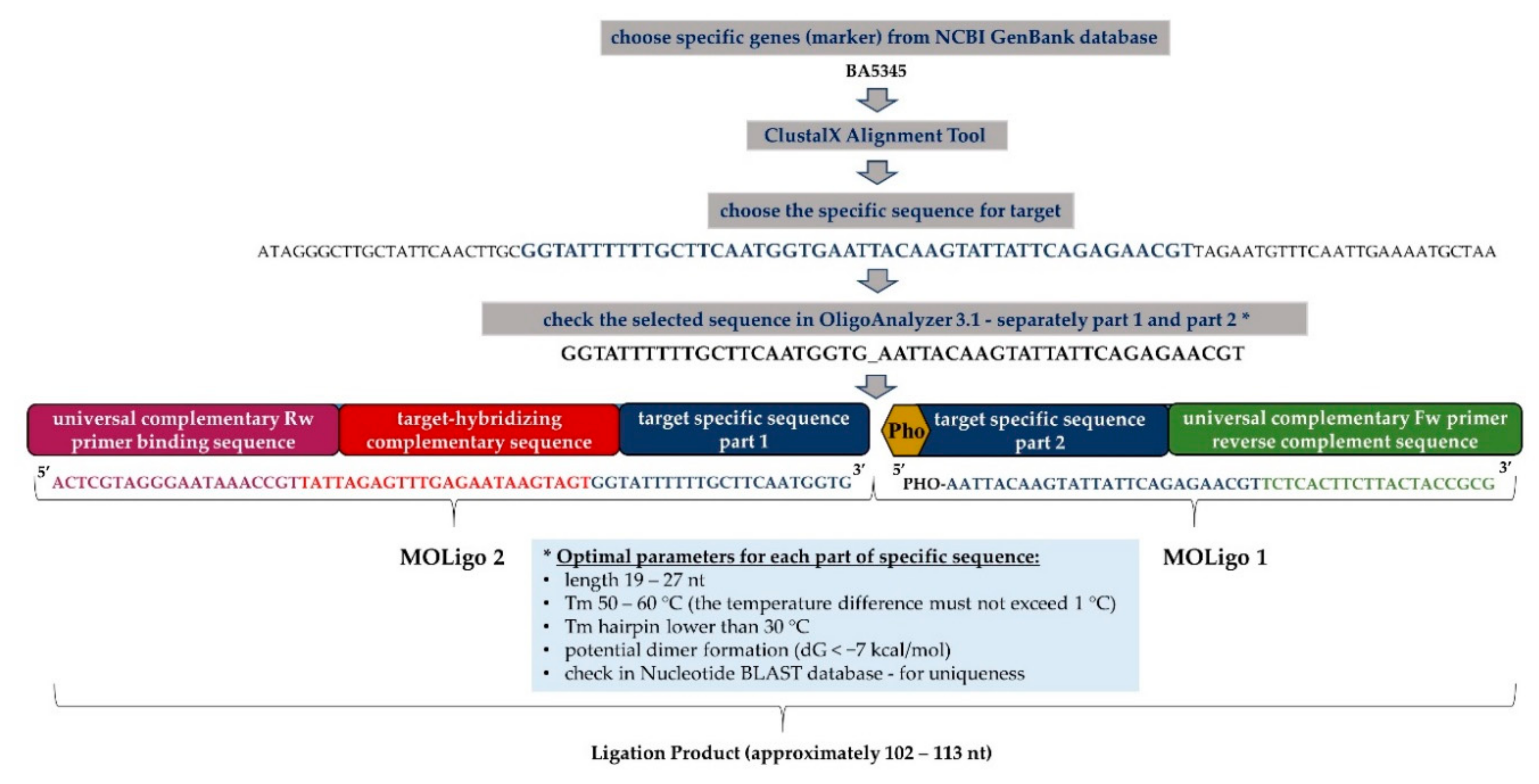
Figure 1. The structure of MOLigo probes demonstrated on a specific marker BA5345 for Bacillus anthracis: ClustalX Alignment Tool—part of a software program BioEdit, designed to perform mulTable 3. 1—analytical tool for oligonucleotides identifying their properties; MOLigo1 consists of a specific sequence for selected target (part 2) and a universal complementary forward primer (Fw)-binding sequence, MOLigo1-specific sequence is phosphorylated at its 5′end; MOLigo2 includes a specific sequence for selected target (part 1), target-hybridizing complementary sequence, which is specific for each microsphere and universal complementary reverse primer (Rw)-binding sequence. The final size of the ligation product ranges from 102 to 113 nucleotides (nt). * Optimal parameters for target specific sequences defined according to Deshpande et al. [26].
Microorganisms 09 00038 g001
Figure 2. Biothreat detection panel. MOL-PCR biothreat assay for detection of (A) B. anthracis, (B) Y. pestis, (C) F. tularensis, and (D) Brucella spp. The biothreat panel was verified using DNA gra-dients from the collection of pathogenic microorganisms (CAPM) and evaluated by MFI after sub-tracting the NTC (H2O only) to the appropriate target. IC confirms that the individual reaction steps have taken place correctly. Exces (DNA from S. aureus) was used for confirmation as another negative control.
Microorganisms 09 00038 g002
Figure 3. Exclusivity of biothreat detection panel based on MOL-PCR method. The exclusivity test was performed on various bacterial strains (Table 1) and evaluated as MFI. The specificity of the probes used in the biothreat panel was confirmed by the values obtained being below 100 MFI when using unrelated DNA. An IC confirmed the correct course of the MOL-PCR reaction.
Microorganisms 09 00038 g003
Figure 4. Sensitivity test.—The detection limit of the MOL-PCR system was performed in the form of a concentration gradient of isolated DNA of all bacteria and for two targets, namely: (A) B. anthracis, (B) Y. pestis, (C) F. tularensis, (D) B. melitensis.
Microorganisms 09 00038 g004
Figure 5. Long-term storage effect of ligation mix.—Experiment based on evaluating the stability of different types of ligation premix storage over time (MOLigo probes separately LP1 + LP2 or all MOLigo probes together—MIX). Better protocol performance was recorded when the two probes premix were stored separately. Abbreviations: LP = ligation premix; N = Negative control; MIX = mix of all probes in the ligation buffer.
Microorganisms 09 00038 g005
Table 1. List of bacteria used in this study.
SpeciesAccession No.
Bacillus anthracisCAPM 5001
Yersinia pestisNCTC 5923 T
Francisella tularensisCAPM 5600
Brucella abortusCAPM 5520
Brucella suisCAPM 6073 T
Brucella melitensisCAPM 5659 T
Staphylococcus aureusCCM 885 T
CAPM 5736
CAPM 5756
CAPM 5755
Methicillin-resistant Staphylococcus aureusCAPM 6565
Listeria monocytogenesCAPM 5580
CAPM 5879
Bacillus cereusCAPM 5631
Yersinia enterocoliticaDSM 9499
Yersinia enterocoliticaDSM 13030 T
Campylobacter jejuniCAPM 6316
CAPM 6341
Salmonella entericaCAPM 5439
Salmonella entericaCAPM 5445
Salmonella entericaCAPM 5456
Salmonella entericaCAPM 6324 T
Escherichia coli O26CAPM 5358
Escherichia coli O157CAPM 6557
Enterococcus faecalisCAPM 6575
CAPM 6577
Enterococcus faeciumCAPM 6563
CAPM 6590
Vibrio parahaemoliticusCAPM 5939
Yersinia pseudotuberculosisCAPM 6495
Clostridium tetaniNCTC 5409
Clostridium botulinumNCTC 3815
Abbreviations: CAPM = Collection of Animal Pathogenic Microorganisms; CCM = Czech Collection of Microorganisms; NCTC = National Collection of Type Cultures; T = type strain, DSM = DSMZ-German Collection of Microorganisms and Cell Cultures.
Table 2. Specific MOLigo pair sequence used for detection with microspheres code and xTAG assignation (bead and xTAG numbers of unique sequences are available in the Luminex catalog).
PathogenMarkerReferenceSequence 5′-3′Size of LPxTAGBead
Bacillus anthracisBA5345[35]PHO-AATTACAAGTATTATTCAGAGAACGTTCTCACTTCTTACTACCGCG
ACTCGTAGGGAATAAACCGTtattagagtttgagaataagtagtGGTATTTTTTGCTTCAATGGTG
112A033033
pagA[36]PHO-CTGTATCAGCGGTATTTAAACTCTCACTTCTTACTACCGCG
ACTCGTAGGGAATAAACCGTtgagtaagtttgtatgtttaagtaATCTAATATCGGCATTTAATCTTG
109A065034
Yersinia pestispla[37]PHO-TTCTGTTGTTTTGCCTTGACATTCTCCTCTCACTTCTTACTACCGCG
ACTCGTAGGGAATAAACCGTgtgttatagaagttaaatgttaagCATAATGACGGGGCGCTCA
110A030030
caf1[38]PHO-AGGAACCACTAGCACATCTTCTCACTTCTTACTACCGCG
ACTCGTAGGGAATAAACCGTgtaagattagaagttaatgaagaaCTTACTCTTGGCGGCTATAAAAC
106A051051
Francisella tularensis23kDA[39]PHO-TGAGATGATAACAAGACAACAGTCTCACTTCTTACTACCGCG
ACTCGTAGGGAATAAACCGTgtaagagtattgaaattagtaagaAACTAAAAAAAGGAGAATGATTATGAG
113A066020
fopA[40]PHO-ACTATCTAGAAATGTTCAAGCAAGTGTTCTCACTTCTTACTACCGCG
ACTCGTAGGGAATAAACCGTagtaagtgttagatagtattgaatGGGTGGTGGTCTTAAGTTTGA
112A038038
Brucella spp.omp2a[41]PHO-CAGGCTACGAATCCAGAAATCTCACTTCTTACTACCGCG
ACTCGTAGGGAATAAACCGTatttgttatgataaatgtgtagtgCGCACTGAATCTCTGTTTTTC
104A042012
bcsp31[42]PHO-TATGCCATTCGCCGCCTGATCTCACTTCTTACTACCGCG
ACTCGTAGGGAATAAACCGTgtgattgaatagtagattgtttaaCATTCTTCACATCCAGGAAACCCGAC
109A046046
Internal controlaDNA[34]Pho-ATTAGCACAATGAATAATCATCGTCTCACTTCTTACTACCGCG
ACTCGTAGGGAATAAACCGTattgtgaaagaaagagaagaaattTATACACACGCAATCACCAC
107A014036
Forward primer [43]CGCGGTAGTAAGAAGTGAGA
Reverse primer BODIPY-TMRX-ACTCGTAGGGAATAAACCGT
Abbreviations: Pho = phosphorylation; underlined = specific sequence for target gene; bold = universal forward and reverse primer; lowercase = xTAG sequence; LP = ligation product.
Table 3. Measured limit of detection (LoD) values for the MOL-PCR method using established detection panel for the biothreat bacteria.
PathogenGene (Target)LoD (ng/µL)
B. anthracis5345>0.5
pagA0.25–0.05
Y. pestispla<0.005
caf0.5–0.05
F. tularensisfopA0.44–0.044
23kDA2.2–0.44
B. melitensisbcsp310.24–0.024
omp2a0.24–0.024
Publisher’s Note: MDPI stays neutral with regard to jurisdictional claims in published maps and institutional affiliations.