Communication

Escherichia coli HS and Enterotoxigenic Escherichia coli Hinder Stress Granule Assembly

by
Felipe Velásquez
1,2,
Josefina Marín-Rojas
1,2,
Ricardo Soto-Rifo
1,2,
Alexia Torres
3,
Felipe Del Canto
3 and
Fernando Valiente-Echeverría
1,2,*
1
Molecular and Cellular Virology Laboratory, Virology Program, Faculty of Medicine, Institute of Biomedical Sciences, Universidad de Chile, 8380453 Santiago, Chile
2
HIV/AIDS Workgroup, Faculty of Medicine, Universidad de Chile, 8380453 Santiago, Chile
3
Microbiology and Mycology Program, Faculty of Medicine, Institute of Biomedical Sciences, Universidad de Chile, 8380453 Santiago, Chile
*
Author to whom correspondence should be addressed.
Microorganisms 2021, 9(1), 17; https://doi.org/10.3390/microorganisms9010017
Submission received: 20 November 2020 / Revised: 15 December 2020 / Accepted: 17 December 2020 / Published: 23 December 2020

Abstract

:
Escherichia coli, one of the most abundant bacterial species in the human gut microbiota, has developed a mutualistic relationship with its host, regulating immunological responses. In contrast, enterotoxigenic E. coli (ETEC), one of the main etiologic agents of diarrheal morbidity and mortality in children under the age of five in developing countries, has developed mechanisms to reduce the immune-activator effect to carry out a successful infection. Following infection, the host cell initiates the shutting-off of protein synthesis and stress granule (SG) assembly. This is mostly mediated by the phosphorylation of translation initiator factor 2α (eIF2α). We therefore evaluated the ability of a non-pathogenic E. coli strain (E. coli HS) and an ETEC strain (ETEC 1766a) to induce stress granule assembly, even in response to exogenous stresses. In this work, we found that infection with E. coli HS or ETEC 1766a prevents SG assembly in Caco-2 cells treated with sodium arsenite (Ars) after infection. We also show that this effect occurs through an eIF2α phosphorylation (eIF2α-P)-dependent mechanism. Understanding how bacteria counters host stress responses will lay the groundwork for new therapeutic strategies to bolster host cell immune defenses against these pathogens.

1. Introduction

In eukaryotic cells, stress granule (SG) assembly can be induced by several environmental stresses including heat shock, oxidative stress, viral infection and UV irradiation, amongst others [1]. SGs are non-membranous cytoplasmic foci, which are formed by mRNA, 40S ribosomal subunits, translation initiator factors (eIF4E, eIF4G, eIF4A, eIF4B, eIF3, eIF2) and RNA-binding proteins such as TIAR, G3BP1 and HuR [2].
The classical mechanism for SG induction is mediated by phosphorylation of the α subunit of the translation initiator factor 2 (eIF2α) by one of the four stress-responsive kinases: HRI, PERK, GCN2 or PKR [3]. Phosphorylation of eIF2α inhibits protein translation by reducing the exchange of the eIF2-GDP to the eIF2-GTP ternary complex necessary for translation initiation (reviewed in [3]). SGs participate in mRNA stabilization and transport, cell survival and cell death [4] and are associated with the pathogenesis of diseases such as cancer, neurodegenerative diseases, inflammatory disorders and viral infections [5]. Although the relationship between viruses and SGs has been widely studied, not much is known about SGs during bacterial infections.
Previous studies have indicated that Shigella flexneri affects the ability of infected cells to form canonical SGs and large SG aggregates [6,7]. On the other hand, it has been shown that shiga-toxin-producing Escherichia coli is capable of inducing SGs on Caco-2 cells through the subtilase cytotoxin (SubAB) [8]. Here, we compared the ability to induce SGs of two E. coli strains, the commensal E. coli HS and the enterotoxigenic E. coli (ETEC) 1766a, producing the human heat-stable toxin (ST1b), the heat-labile toxin (LT), the coli surface antigen 23 (CS23) and the TleA serine-protease autotransporter [9,10,11]. E. coli HS was chosen because it is the prototypic commensal (non-pathogenic) E. coli strain, whereas ETEC 1766a was chosen because it is a non-invasive pathogen with a high cell-adherence capacity at short times [10]. Our results indicate that infection of Caco-2 cells with both E. coli strains did not induce SG assembly. Furthermore, E. coli-infected Caco-2 cells subjected to treatment with sodium arsenite partially inhibit SG assembly through the impairment of eIF2α phosphorylation.

2. Materials and Methods

2.1. Cell Line and Bacteria Strains

Caco-2 cells (human Caucasian colon adenocarcinoma, obtained from European Collection of Authenticated Cell Cultures, Salisbury, UK) were grown in Dulbecco’s modified Eagle’s medium (DMEM, Gibco Thermo Fisher Scientific, Waltham, Massachusetts, United States) containing 10% fetal bovine serum (FBS, Sigma-Aldrich by Merck KGaA, Darmstadt, Germany) and 1% penicillin–streptomycin (Invitrogen by Thermo Fisher Scientific, Waltham, MA, USA). Cells were grown at 37 °C in a 5% CO2 atmosphere. ETEC 1766a and Escherichia coli HS was originally isolated from a healthy donor in the US [9,12] and was obtained from the University of Maryland School of Medicine (Baltimore, MD, USA). ETEC 1766a was isolated from a child with watery diarrhea in Santiago, Chile [9,10,11,13].

2.2. Bacterial Infection and Stress Treatment

Bacteria were grown in Luria–Bertani (LB) broth (10 g/L tryptone, 5 g/L yeast extract, 5 g/L NaCl) at 37 °C overnight. Caco-2 cells were infected at a multiplicity of infection (MOI) of 100 bacteria/cell at 37 °C in a 5% CO2 atmosphere for 1 h. To induce cellular oxidative stress, cells were treated with 0.5 mM sodium arsenite (arsenite, NaAsO2; Sigma-Aldrich by Merck KGaA, Darmstadt, Germany) for 1 h or 30 min.

2.3. Immunofluorescence and Imaging Analyses

For immunofluorescence (IF) analyses, cells were washed with PBS and fixed with 4% paraformaldehyde for 20 min. Cells were then washed with PBS, incubated in 0.1 M glycine for 10 min, washed with PBS, incubated in 0.2% Triton X-100 for 5 min and washed in PBS. Next, cells were incubated with primary antibodies: anti-G3BP1 mouse (Santa Cruz Biotechnology, Santa Cruz, CA, USA), anti-TIAR rabbit (kindly provided by Dr. Jorge Vera-Otarola, PUC, Chile) for 1 h, and incubated with secondary antibodies anti-mouse 488 or anti-rabbit 594 (Life Technologies) and for the nuclei, cells were stained with DAPI (Sigma-Aldrich by Merck KGaA, Darmstadt, Germany). Confocal microscopy was performed with a Carl Zeiss LSM 700 microscope, and image acquisition was done with a 40× objective lens. All imaging experiments were performed at least 2 times. Imaging analyses were performed using ImageJ software (NIH).

2.4. Western Blotting

Cell extracts were prepared by lysis with mild lysis buffer [NaCl 100 mM, Tris-HCl 10 mM pH 7.5, EDTA 1mM, NP40 0.5%, Protease inhibitor cocktail (Roche, 124 CH-4070 Basel Switzerland) and 30 μg of total protein were subjected to 10% SDS-PAGE and transferred to a nitrocellulose blotting membrane (GE Healthcare Boston, MA, USA). Membranes were incubated with an anti-eIF2α (Cell Signaling 32 Tozer Road Beverly, MA, USA), anti-p-eIF2α ser51 (Abcam Cambridge, CB2 0AX, UK) and anti-Actin (Santa Cruz Biotechnology, Santa Cruz, CA, USA). Upon incubation with the corresponding HRP-conjugated secondary antibody (Santa Cruz Biotechnologies, Santa Cruz, CA, USA), membranes were revealed with a C-Digit digital scanner (Li-Cor Lincoln, NE, USA) using the PIERCE(TM) ECL Western blotting substrate (Thermo, Waltham, MA, USA). Signal intensity was quantified by ImageJ (NIH).

2.5. Statistical Analysis

The statistical data analysis and graphics described in the text were done using the GraphPad v8.4.3 program (La Jolla, CA, USA). Differences were tested by analysis of variance (Dunnet’s or Bonferroni’s post hoc tests) where p < 0.05 was considered significant.

3. Results

3.1. Non-Pathogenic E. coli HS Strain and ETEC 1766a Pathogenic Strain Do Not Induce Stress Granule Assembly

Our first approach was to determine the capacity of non-pathogenic and pathogenic strains of Escherichia coli to induce stress granule assembly in the Caco-2 cells derived from human colorectal adenocarcinoma frequently used to study E. coli [14]. We used the non-pathogenic commensal E. coli HS strain and the pathogenic enterotoxigenic E. coli (ETEC) 1766a strain. To assay the E. coli-mediated stress granule assembly, Caco-2 cells were exposed to 0.5 mM of sodium arsenite (Ars) for 30 min or 1 h (as a control for SG assembly) and compared to 1 h of E. coli strain infection. Untreated Caco-2 cells, Ars-treated and infected cells were incubated for the indicated times, then fixed and immunostained for the SG marker proteins G3BP1 and TIAR and were stained with DAPI to visualize the nuclei. As shown in Figure 1, untreated cells do not show G3BP1 or TIAR aggregation into dense cytoplasmic foci, consistent with no SG assembly. Ars-treated cells at different times were positive to aggregation and colocalization of G3BP1 and TIAR in cytoplasmic foci. Nevertheless, neither E. coli HS nor ETEC strain showed any signs of SG assembly (Figure 1). The presence of E. coli strains were proven using DAPI staining and, as expected, bacteria were only observed in the infected cell cultures (Figure 1, lateral panels). These results suggest that E. coli infection does not induce SG assembly in Caco-2 cells.

3.2. E. coli HS and ETEC 1766a Strains Prevent Arsenite-Induced Stress Granule Assembly in Caco-2 Cells

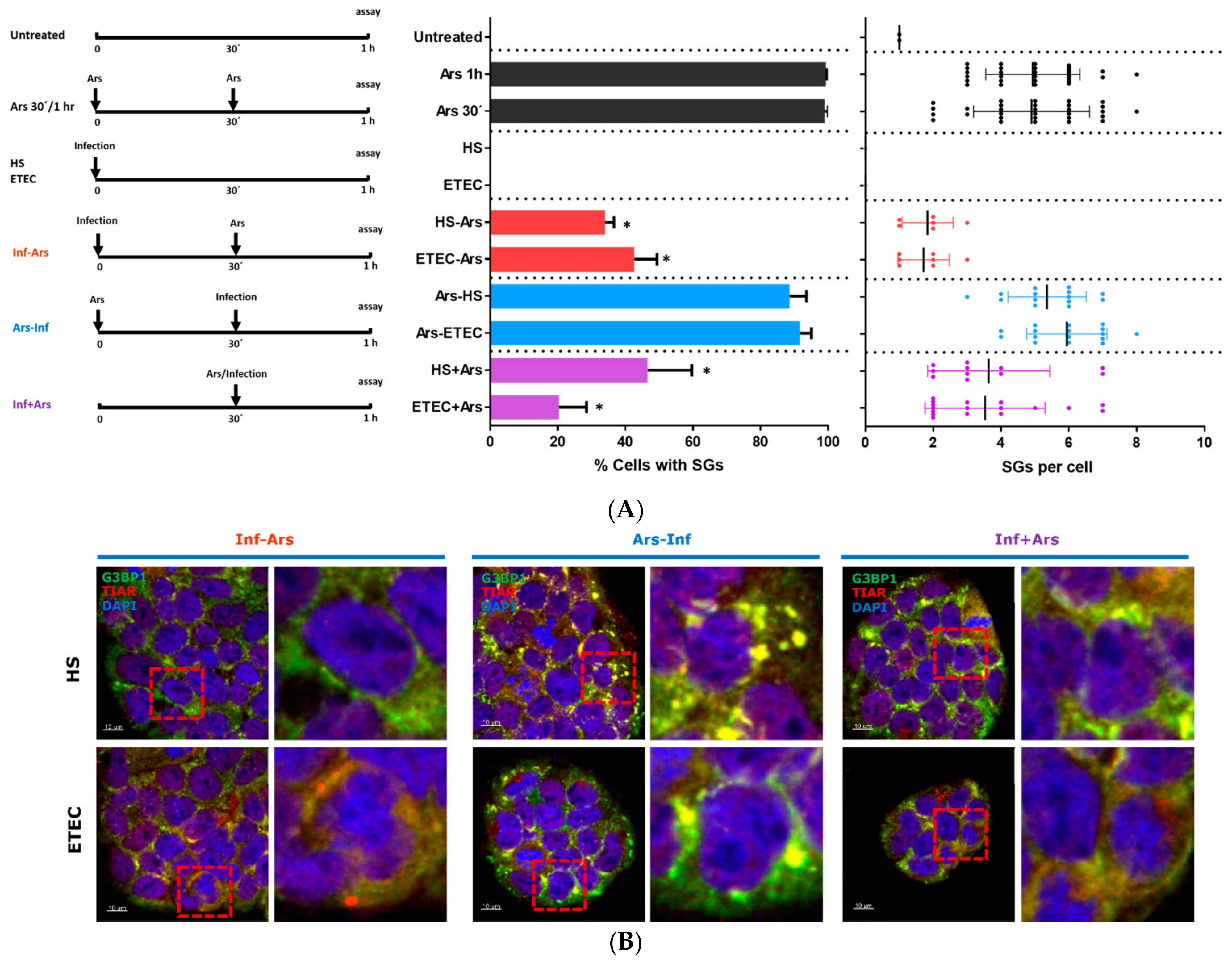
Several pathogens, including viruses, have developed strategies to subvert the SG response, owing to the fact that SGs constitute an antiviral host response (reviewed in [3,15]). To test if E. coli HS and/or ETEC 1766a strains could block SG assembly, we infected Caco-2 cells with E. coli HS or ETEC 1766a prior to and after Ars treatment (Figure 2A, left panel). Our results showed that E. coli HS- or ETEC 1766a-infected cells treated with Ars were able to prevent the assembly of arsenite-induced SGs in 66% and 58% of cases, respectively (Figure 2A, middle panel in red). Otherwise, when Caco-2 cells were treated with Ars for 30 min and then exposed to E. coli HS or ETEC 1766a strains for 30 min, SG assembly was evident. (Figure 2A, middle panel in blue). Finally, we tested the SG assembly upon simultaneous incubation with Ars and E. coli HS or ETEC 1766a. Our results showed a reduced SG assembly, 53% and 80%, respectively (Figure 2A middle panel in purple). Although in both conditions (Inf-Ars and Inf + Ars) there is still a low percentage of SG assembly, we observed fewer number of SGs per cell (Figure 2A right panel in red/purple). These results suggest that the presence of E. coli strains was sufficient to attenuate the SG assembly mediated by sodium arsenite as a stress inductor (Figure 2B).

3.3. E. coli HS and ETEC 1766a Do Not Induce eIF2α Phosphorylation

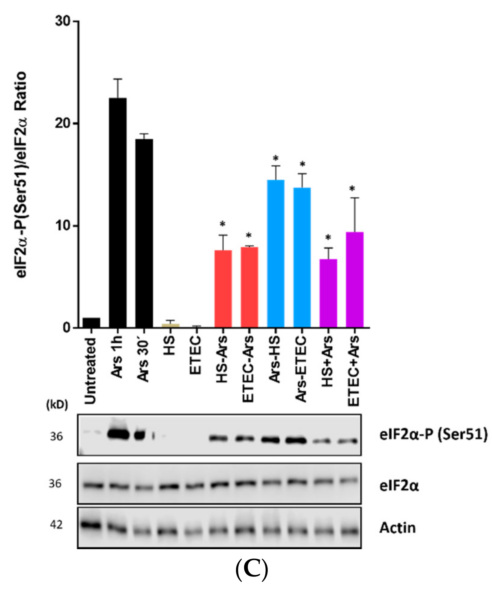
The canonical mechanism to induce SG assembly is mediated by phosphorylation of eIF2α (eIF2α-P) (reviewed in [3]). Therefore, we evaluated whether the decrease in SG assembly reflected an impairment in eIF2α phosphorylation. Interestingly, the detection of eIF2α-P by Western blot under our different experimental conditions showed that Caco-2 cells subjected to arsenite treatment either post-infection (Inf-Ars) or simultaneous to the infection (Inf + Ars) had a lower signal of eIF2α-P (Figure 2C, in red and purple), which was concomitant with a decrease in SG assembly (Figure 2B). These results indicate that E. coli infection prevents SG assembly in response to oxidative stress.

4. Discussion

Although some bacterial pathogens can induce SG assembly during infection, it is not clear in each case whether this response benefits the host or the bacteria [16]. Here, we showed that two different E. coli strains led to an impairment of stress granule assembly in the epithelial cell line Caco-2, despite the use of sodium arsenite as a stress inductor. Infection times were chosen based on the available literature on S. flexneri-induced SG assembly in Caco-2 cells [6,7]. It is worth noting that longer infection times could also bear significant results, like those reported by He et al. [17]. Regardless, our results consistently show that granule assembly impairment was observable at both 30 and 60 min post-infection even in the presence of an exogenous stressor.
Published research showed that the heat-labile enterotoxin from E. coli ETEC causes endoplasmic reticulum stress and can induce the phosphorylation of eIF2α in epithelial cells [18]. Given these results, we were surprised to observe that ETEC-infected cells presented impairment in the assembly of SGs and did not show phosphorylation of eIF2α. In contrast, arsenite-induced eIF2α phosphorylation is lower in the presence of bacterial infection. However, it warrants mention that the previous finding was reported on cells treated with the purified toxin, whereas our results were obtained upon directly infecting the cells with the ETEC strain. This implies that the potential impairment of eIF2α phosphorylation could be carried out through another bacterial factor. Similarly, the E. coli HS strain had the same reducing effect over oxidative stress-induced eIF2α phosphorylation and SG assembly.
The integrated stress response, including SG assembly, can be heavily modulated during infection by bacterial and cellular factors. The infection’s outcomes remains unclear, as so many variables are involved [16]. In this work, we report a regulatory effect in the cellular stress response, taking place upstream of eIF2α phosphorylation. Given that sodium arsenite induces eIF2α phosphorylation through the HRI kinase, our results suggest that this route is likely targeted [19]. This can be aligned with a recent study that demonstrates that HRI leads the integrated stress response (ISR) and concomitant SG assembly in response to bacterial peptidoglycan detection during infection by intracellular bacteria [20]. Strikingly, the modulation exerted by pathogenic and non-pathogenic E. coli strains appears to be similar. This observation could imply that pathogenic bacteria can exploit strategies that allow for commensal bacteria. This could be in part explained by the high average nucleotide identity (ANI) shared by the genomes of the two strains evaluated in this work (98.77%, E. coli HS GenBank assembly accession GCA_000017765.1, ETEC 1766a assembly accession GCA_009868995.1). We propose that a lowered stress induction by the commensal E. coli HS would favor colonization without upsetting the host. Simultaneously, the ETEC strain could avoid SG induction as an immune-evasion strategy to establish itself on the epithelium, inflicting damage through its repertoire of virulence factors, which are not produced by commensal strains.

Author Contributions

F.V., F.D.C. and F.V.-E. conceived and planned the experiments. A.T. performed the bacterial cultures. J.M.-R. performed the statistical analysis. F.V., J.M.-R., F.D.C., R.S.-R. and F.V.-E. wrote the manuscript. F.V.-E., F.D.C. and R.S.-R. were responsible for funding acquisition. All authors listed discussed the results and have made a substantial, direct and intellectual contribution to the work, and approved it for publication. All authors have read and agreed to the published version of the manuscript.

Funding

This work was supported by ANID/Conicyt Chile through the Fondecyt No. 1180798 and 11140502 (FV-E); 1190156 (RS-R), 11150966 and 1200979 (FDC), and ANID/Conicyt grant No. 21190857 (JM-R) and USACH grant to FV.

Data Availability Statement

Data sharing not applicable: No new data were created or analyzed in this study. Data sharing is not applicable to this article.

Acknowledgments

We thank Carolina Hernández (microscopy facilities at Universidad de Chile) for her support in imaging acquisition. We also thank Jorge Vera-Otarola (PUC, Chile) for providing the anti-TIAR antibody. We are immensely grateful to lab members from Roberto Vidal (Universidad de Chile) and Daniela Gutierrez for their technical support on bacterial cultures. We thank Aracelly Gaete-Argel for providing comments on this manuscript.

Conflicts of Interest

The authors declare no conflict of interest.

References

  1. Anderson, P.; Kedersha, N. Stressful initiations. J. Cell Sci. 2002, 115 Pt 16, 3227–3234. [Google Scholar] [PubMed]
  2. Anderson, P.; Kedersha, N. RNA granules. J. Cell Biol. 2006, 172, 803–808. [Google Scholar] [CrossRef] [PubMed]
  3. Gaete-Argel, A.; Marquez, C.L.; Barriga, G.P.; Soto-Rifo, R.; Valiente-Echeverria, F. Strategies for Success. Viral Infections and Membraneless Organelles. Front. Cell. Infect. Microbiol. 2019, 9, 336. [Google Scholar] [CrossRef] [PubMed]
  4. Protter, D.S.W.; Parker, R. Principles and Properties of Stress Granules. Trends Cell Biol. 2016, 26, 668–679. [Google Scholar] [CrossRef] [PubMed] [Green Version]
  5. Buchan, J.R. mRNP granules. Assembly, function, and connections with disease. RNA Biol. 2014, 11, 1019–1030. [Google Scholar] [CrossRef] [PubMed] [Green Version]
  6. Vonaesch, P.; Campbell-Valois, F.X.; Dufour, A.; Sansonetti, P.J.; Schnupf, P. Shigella flexneri modulates stress granule composition and inhibits stress granule aggregation. Cell. Microbiol. 2016, 18, 982–997. [Google Scholar] [CrossRef] [PubMed]
  7. Vonaesch, P.; Sansonetti, P.J.; Schnupf, P. Immunofluorescence Analysis of Stress Granule Formation after Bacterial Challenge of Mammalian Cells. J. Vis. Exp. Jove 2017, 125, e55536. [Google Scholar] [CrossRef] [PubMed]
  8. Tsutsuki, H.; Yahiro, K.; Ogura, K.; Ichimura, K.; Iyoda, S.; Ohnishi, M.; Nagasawa, S.; Seto, K.; Moss, J.; Noda, M. Subtilase cytotoxin produced by locus of enterocyte effacement-negative Shiga-toxigenic Escherichia coli induces stress granule formation. Cell. Microbiol. 2016, 18, 1024–1040. [Google Scholar] [CrossRef] [PubMed]
  9. Rasko, D.A.; Rosovitz, M.J.; Myers, G.S.; Mongodin, E.F.; Fricke, W.F.; Gajer, P.; Crabtree, J.; Sebaihia, M.; Thomson, N.R.; Chaudhuri, R.; et al. The pangenome structure of Escherichia coli: Comparative genomic analysis of E. coli commensal and pathogenic isolates. J. Bacteriol. 2008, 190, 6881–6893. [Google Scholar] [CrossRef] [PubMed] [Green Version]
  10. Del Canto, F.; Botkin, D.J.; Valenzuela, P.; Popov, V.; Ruiz-Perez, F.; Nataro, J.P.; Levine, M.M.; Stine, O.C.; Pop, M.; Torres, A.G.; et al. Identification of Coli Surface Antigen 23, a novel adhesin of enterotoxigenic Escherichia coli. Infect. Immun. 2012, 80, 2791–2801. [Google Scholar] [CrossRef] [PubMed] [Green Version]
  11. Gutierrez, D.; Pardo, M.; Montero, D.; Onate, A.; Farfan, M.J.; Ruiz-Perez, F.; Del Canto, F.; Vidal, R. TleA, a Tsh-like autotransporter identified in a human enterotoxigenic Escherichia coli strain. Infect. Immun. 2015, 83, 1893–1903. [Google Scholar] [CrossRef] [PubMed] [Green Version]
  12. DuPont, H.L.; Formal, S.B.; Hornick, R.B.; Snyder, M.J.; Libonati, J.P.; Sheahan, D.G.; LaBrec, E.H.; Kalas, J.P. Pathogenesis of Escherichia coli diarrhea. N. Engl. J. Med. 1971, 285, 1–9. [Google Scholar] [CrossRef] [PubMed]
  13. Levine, M.M.; Ferreccio, C.; Prado, V.; Cayazzo, M.; Abrego, P.; Martinez, J.; Maggi, L.; Baldini, M.M.; Martin, W.; Maneval, D.; et al. Epidemiologic studies of Escherichia coli diarrheal infections in a low socioeconomic level peri-urban community in Santiago, Chile. Am. J. Epidemiol. 1993, 138, 849–869. [Google Scholar] [CrossRef] [PubMed]
  14. Darfeuille-Michaud, A.; Aubel, D.; Chauviere, G.; Rich, C.; Bourges, M.; Servin, A.; Joly, B. Adhesion of enterotoxigenic Escherichia coli to the human colon carcinoma cell line Caco-2 in culture. Infect. Immun. 1990, 58, 893–902. [Google Scholar] [CrossRef] [PubMed] [Green Version]
  15. Poblete-Duran, N.; Prades-Perez, Y.; Vera-Otarola, J.; Soto-Rifo, R.; Valiente-Echeverria, F. Who Regulates Whom? An Overview of RNA Granules and Viral Infections. Viruses 2016, 8, 180. [Google Scholar] [CrossRef] [PubMed]
  16. Rodrigues, L.; Graca, R.S.F.; Carneiro, L.A.M. Integrated Stress Responses to Bacterial Pathogenesis Patterns. Front. Immunol. 2018, 9, 1306. [Google Scholar] [CrossRef] [PubMed]
  17. He, X.; Mishchuk, D.O.; Shah, J.; Weimer, B.C.; Slupsky, C.M. Cross-talk between E. coli strains and a human colorectal adenocarcinoma-derived cell line. Sci. Rep. 2013, 3, 3416. [Google Scholar] [CrossRef] [PubMed] [Green Version]
  18. Lu, X.; Li, C.; Li, C.; Li, P.; Fu, E.; Xie, Y.; Jin, F. Heat-Labile Enterotoxin-Induced PERK-CHOP Pathway Activation Causes Intestinal Epithelial Cell Apoptosis. Front. Cell. Infect. Microbiol. 2017, 7, 244. [Google Scholar] [CrossRef] [PubMed] [Green Version]
  19. McEwen, E.; Kedersha, N.; Song, B.; Scheuner, D.; Gilks, N.; Han, A.; Chen, J.J.; Anderson, P.; Kaufman, R.J. Heme-regulated inhibitor kinase-mediated phosphorylation of eukaryotic translation initiation factor 2 inhibits translation, induces stress granule formation, and mediates survival upon arsenite exposure. J. Biol. Chem. 2005, 280, 16925–16933. [Google Scholar] [CrossRef] [PubMed] [Green Version]
  20. Abdel-Nour, M.; Carneiro, L.A.M.; Downey, J.; Tsalikis, J.; Outlioua, A.; Prescott, D.; Da Costa, L.S.; Hovingh, E.S.; Farahvash, A.; Gaudet, R.G.; et al. The heme-regulated inhibitor is a cytosolic sensor of protein misfolding that controls innate immune signaling. Science 2019, 365, eaaw4144. [Google Scholar] [CrossRef] [PubMed]
Figure 1. Immunofluorescence (IF) of stress granule marker proteins G3BP1 and TIAR in Caco-2 cells treated with 0.5 mM of sodium arsenite (Ars) at 30 min or 1 h and infected with E. coli HS or ETEC 1766a strains. DAPI-stained nuclei and bacteria (right panel). Scale bars are 10 µm.
Microorganisms 09 00017 g001
Figure 2. (A) Left panel: Schema of the experimental protocol used in this work. The arrows indicate treatment or bacterial infection. Middle panel: Quantification of stress granules (SGs) in Caco-2 cells shown in (B). p value is indicated by *(p < 0.05 compared with Ars). Right panel: Quantification of SGs per cell from (B). Data are presented as mean ± s.d. from three independent experiments with at least 30 cells analyzed in each condition. (B) Immunofluorescence (IF) of stress granule marker proteins G3BP1 and TIAR and nucleus marker DAPI in Caco-2 cells under different experimental conditions. Left panel: Caco-2 cells infected with E. coli strains followed by treatment with Ars (Inf-Ars). Middle panel: Cells treated with Ars followed by infection with E. coli strains (Ars-Inf). Right panel: Simultaneous E. coli infection and Ars treatment (Inf + Ars). Scale bars are 10 µm (C) Caco-2 cell lysates were analyzed for eIF2α-p (Ser51), eIF2α and ACTIN, for untreated or infected cells from (B). Densitometry analysis from three independent experiments and representative Western blot are shown. Values were normalized against untreated cells. p-value is indicated by * (p < 0.05 compared with Ars).
Microorganisms 09 00017 g002aMicroorganisms 09 00017 g002b
Publisher’s Note: MDPI stays neutral with regard to jurisdictional claims in published maps and institutional affiliations.