Article

Microbiome of the Queensland Fruit Fly through Metamorphosis

1
Applied BioSciences, Macquarie University, North Ryde, NSW 2109, Australia
2
Department of Environmental Sciences, Macquarie University, North Ryde, NSW 2109, Australia
3
Biosecurity and Food Safety, NSW Department of Primary Industries, Elizabeth Macarthur Agricultural Institute (EMAI), Menangle, NSW 2568, Australia
*
Author to whom correspondence should be addressed.
Microorganisms 2020, 8(6), 795; https://doi.org/10.3390/microorganisms8060795
Submission received: 19 April 2020 / Revised: 12 May 2020 / Accepted: 20 May 2020 / Published: 26 May 2020
(This article belongs to the Section Systems Microbiology)

Abstract

:
Bactrocera tryoni (Froggatt) (Queensland fruit fly, or “Qfly”) is a highly polyphagous tephritid fruit fly and a serious economic pest in Australia. Qfly biology is intimately linked to the bacteria and fungi of its microbiome. While there are numerous studies of the microbiome in larvae and adults, the transition of the microbiome through the pupal stage remains unknown. To address this knowledge gap, we used high-throughput Next-Generation Sequencing (NGS) to examine microbial communities at each developmental stage in the Qfly life cycle, targeting the bacterial 16S rRNA and fungal ITS regions. We found that microbial communities were similar at the larval and pupal stage and were also similar between adult males and females, yet there were marked differences between the larval and adult stages. Specific bacterial and fungal taxa are present in the larvae and adults (fed hydrolyzed yeast with sugar) which is likely related to differences in nutritional biology of these life stages. We observed a significant abundance of the Acetobacteraceae at the family level, both in the larval and pupal stages. Conversely, Enterobacteriaceae was highly abundant (>80%) only in the adults. The majority of fungal taxa present in Qfly were yeasts or yeast-like fungi. In addition to elucidating changes in the microbiome through developmental stages, this study characterizes the Qfly microbiome present at the establishment of laboratory colonies as they enter the domestication process.

1. Introduction

From humans to insects, the microbiome plays an important role in host health and metabolism [1,2]. The symbiotic relationship between insects and their gut microflora is very complex, but it is essential to insect health [3]. Shin et al. [4] identified that the gut microbiome influences host gene expression in Drosophila, affecting host fitness through body development, nutritional metabolism, and stem cell activity. The interactions between hosts and gut microbial communities have been investigated in diverse insects, including beetles [5], mosquitoes [6], butterflies [7], silkworms [8], house flies [9], and red palm weevil [10]. A vast diversity of insect metabolic and behavioral functions have now been linked to the microbiome. Examples include the role of insect gut microbiota in the breakdown of ingested plant polysaccharides, the synthesis of secondary metabolites from sugar-based diets, the recycling of nutrients, inhibition of pathogen colonization, and the breakdown of xenobiotics or toxic materials (allelochemicals) used in plant defense [7,11,12,13,14]. A comprehensive understanding of an insect’s microbiome is an essential step toward understanding the insect’s nutritional biology and physiology and may also be a first step toward developing novel pest-management strategies for some pest insects [12,15].
Although insect microbiomes are often analyzed as a conglomerate, it is important to recognize that they comprise archaea, bacteria, and fungi, and so have substantial taxonomic breadth. Additionally, different microbial taxa have different relationships with host insects. For example, the bacteria Providencia spp. and the fungi Metarhizium spp. are known insect pathogens [16,17]. On the other hand, bacteria from the family Acetobacteraceae (includes Acetic acid bacteria) have a mutualistic relationship with the honey bee Apis mellifera (Hymenoptera: Apidae) [18,19] and pink sugar cane mealybug Saccharococcus sacchari (Cockerell) (Homoptera: Pseudococcidae) [20], both of which have a sugar-based diet. Additionally, gut-associated fungi (mostly yeast, e.g., Saccharomycetes) are an essential source of amino acids, vitamins, and enzymes, with these playing a role in detoxification, metabolism, and pheromone-production pathways [21,22,23,24,25]. While holistic studies provide a useful framework for understanding the overall role of insect microbiomes, consideration of the specific taxonomy of microbiome members is important for a detailed understanding of specific relationships and their functions.
In addition to this taxonomic and functional complexity in microbiome communities, biologically relevant temporal changes also occur in these communities. Perhaps the most dramatic example of this is the change in microbial communities through metamorphosis. Metamorphosis is a conspicuous and abrupt transformation process in which an insect undergoes a complex remodeling of its external and internal morphology [26,27,28]. All holometabolous insects undergo a metamorphosis process, starting with a larval stage, followed by a pupal stage, and finally an adult stage. Numerous holometabolous insects are economically important, due to their roles as a source of food production (e.g., honey bee), as agricultural pests (e.g., many tephritid flies) [29,30,31], as vectors of infectious diseases (e.g., sandfly and mosquito), and as important experimental models (e.g., Drosophila melanogaster) [26,28]. Changes in the gut microbiota during metamorphosis (e.g., butterflies [7], silkworms [8], ground-dwelling beetles [5], and longhorn beetles [32]) are most likely due to the anatomical transformations of hosts during this period [27]. In tephritid fruit flies, changes in the gut microbiota through the developmental stages have been investigated in various species of Bactrocera, Ceratitis, Anastrepha, and Zeugodacus, including B. carambolae [31], B. dorsalis [33], B. latifrons [34], B. minax (Andongma et al., 2019), C. capitata (aka, Medfly) [35], A. ludens, A. obliqua, A. serpentina, A. striata [29], and Z. tau [36], but to date there is only fragmented knowledge of how the microbiome changes through the developmental stages in Queensland fruit fly, B. tryoni (Froggatt) (“Qfly”).
The Qfly is highly polyphagous and is the most economically damaging insect pest of Australian horticulture [37,38,39,40]. Due to its economic importance, numerous studies have been conducted on Qfly ecology [37,41], domestication [42,43,44], production quality traits [45,46], behavior (e.g., mating performance) [47,48,49,50], larval and adult nutritional requirement [51,52,53], and microbiome [54,55,56,57,58]. Bacteria associated with wild and domesticated larvae [54,55], pupae [59], and adult Qfly [56,57,58] have been described in separate studies. Furthermore, yeasts associated with domesticated larvae [60] and adults [61] have been described. Some of these studies have used culture-dependent approaches to profile the microbiota [54,57,59], while others have used high-throughput sequencing technologies to circumvent the well-documented biases of culture-based methods [54,55,56,58]. However, there has been no comprehensive study of changes in the wild-type Qfly microbiome through metamorphosis from larvae, to pupae, to adults. Such a detailed study is important to overcome the highly constrained interpretation of comparisons across studies of different life stages, undertaken in different laboratories, and applying different techniques.
In the present study, we applied high-throughput NGS technology to profile the bacterial 16S ribosomal rRNA gene and fungal internal transcribed spacer region (ITS) obtained from larvae, pupae, and the gut of the adult Qfly. Here we addressed the microbial communities (both bacteria and fungi) present in Qflies sourced as larvae in infested fruit (generation 0/G0) that provide the starting point for laboratory rearing of Qfly from wild populations. To test the hypothesis that microbiota diversity and community structure changes through metamorphosis, we identified the dominant bacteria and fungi present at each Qfly life stage. We predicted that there would be some common dominant bacterial and fungal taxa that represented the core microbiome of Qfly across the three developmental stages, while other taxa would be specific to a developmental stage.

2. Results

2.1. Identification of Qfly

We confirmed that all larval, pupal and adult fly samples included in this study were Qfly by both morphological and molecular testing. The genetic testing was performed by analyzing the mitochondrial cytochrome oxidase I (COI) gene using Sanger sequencing and this confirmed all 24 tested samples as Qfly. Approximately 600 additional adult Qfly in the established colony were also confirmed as Qfly. No other fly species were identified from the experimental samples.

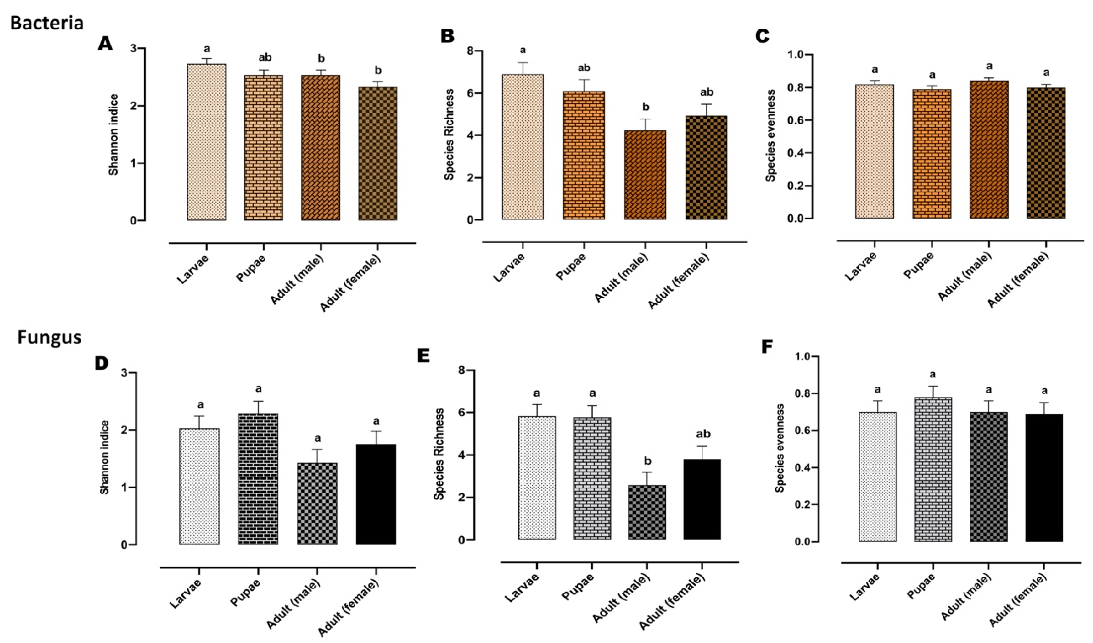
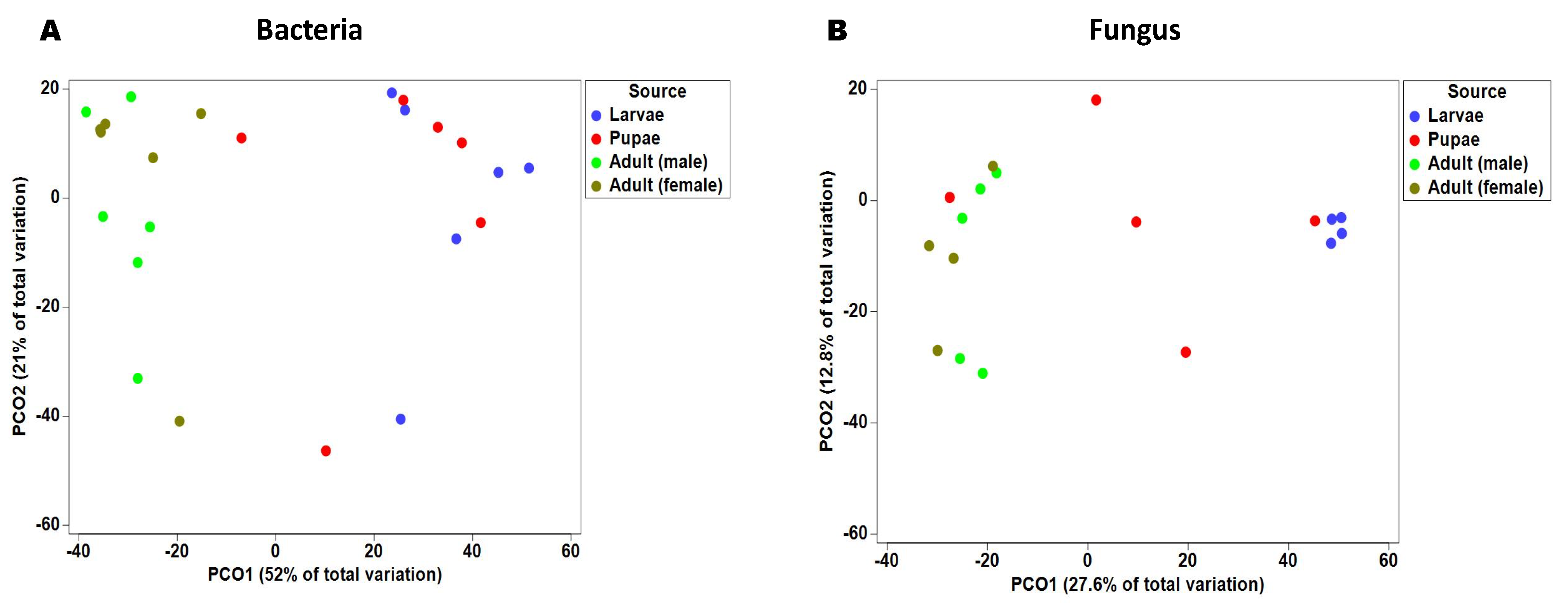
2.2. Gut Bacterial Alpha and Beta Diversity During Metamorphosis

After quality filtering and the removal of chimeric sequences, and then rarefaction to 14,000 reads per sample, 74 bacterial OTUs (operational taxonomic units) were detected (Supplementary Data S1). These taxa spanned 6 phyla, 14 classes, 38 families, and 49 genera; however, despite this broad taxonomic range, only 11 genera were represented by an abundance ≥ 0.1% (Table 1). A Venn diagram analysis revealed that a subset of 14 bacterial taxa were common across all developmental stages of the Qfly (Figure 1A).
Bacterial alpha biodiversity metrics, including Shannon’s biodiversity, species richness, and species evenness indices were compared between the developmental stages (Figure 2A–C). Shannon indices were significantly different between larvae and adults (both male and female flies) (p < 0.05) (Figure 2A). This appears to be driven by a higher species richness in the larvae, when compared with adults, although this was only significant for the male adults (Figure 2B). None of the bacterial alpha diversity metrics showed significant differences between adult males and females, or larvae and pupae.
Beta diversity of the bacterial communities at each Qfly stage was assessed by PERMANOVA analysis (pair-wise test with 999 permutation), based on Bray–Curtis similarities (Supplementary Data S1). Additionally, a principal coordinate analysis (PCoA) of this Bray–Curtis similarity matrix was used to visualize variation among host microbial communities (Figure 3A). Principal coordinate analysis (PCoA) and PERMANOVA both demonstrate a clear structural difference between bacterial communities of Qfly larvae, compared with both male and female adults (PERMANOVA < 0.05, Figure 3A; Supplementary Data S1). We found no differences between the bacterial communities of the larval and pupal microbiome (PERMANOVA test, p = 0.578), or between adult male and female gut microbiomes (PERMANOVA test, adult male and female p = 0.472, Figure 3A).

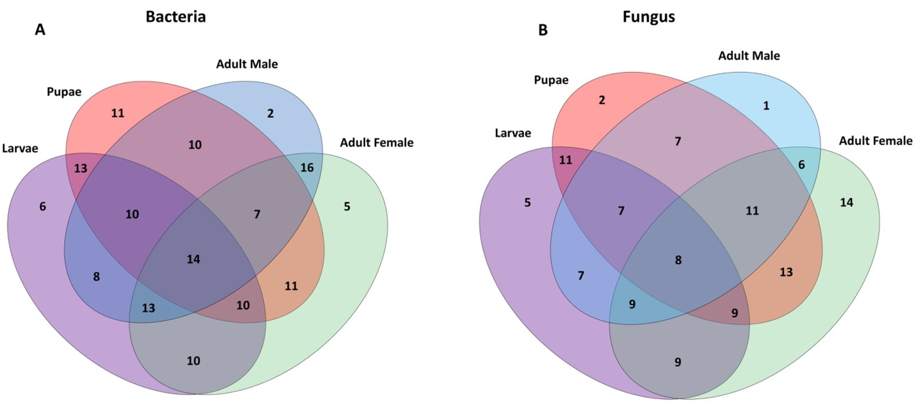
2.3. Bacterial Communities Associated with Metamorphosis

The relative abundance of the bacterial community members in the larval and pupal microbiome, and in adult males and females, was analyzed. At the phylum level, the most abundant taxa in the larval microbiome was Proteobacteria (98.20%), followed by Bacteroidetes (1.70%) and Actinobacteria (0.01%). In adult microbiome, Proteobacteria were observed 99.95% in females and 100% in males. Adult female flies were the only samples to host Firmicutes (0.04%), while the Actinobacteria was only found in the larvae and pupal microbiome. At the family level, the most prevalent taxa were the Enterobacteriaceae, which represented an average relative abundance of 76.1% at all developmental stages. However, they were of greatest relative abundance in adults (males 99.78% and females 98.80%), compared with the larvae (49.59%) and pupae (56.22%). Conversely, the Acetobacteraceae was observed to be highly abundant both in larvae (48.58%) and pupae (42.98%) but were substantially less abundant in adult males (0.18%) and females (1.09%). The Xanthomonadaceae was only observed in the larvae (0.03%) and pupae (0.33%).
At the genus level, bacterial taxa with the greatest relative abundance across the dataset were unassigned Enterobacteriaceae (60.1%), Swaminathania/Asaia (17%), Erwinia (10.5%), Providencia (5.6%), Acetobacter (2.9%), Gluconobacter (2.2%), and unassigned Acetobacteraceae (1.1%) (Figure 4A and Table 1). Highly abundant sequences of unassigned Enterobacteriaceae were reconfirmed with Geneious R10.2.3 [55], a bioinformatic software platform for sequence data analysis, using the NCBI database, and was 99.9% matched with the bacterial genus Enterobacter. The abundant taxa were identified across this dataset; however, these were not equally distributed among the Qfly life stages. For example, Swaminathania/Asaia was highly abundant both in larvae (29.2%) and pupae (37.6%) but had relatively low abundance in adults (male 0.2% and female 1.1%). Furthermore, Erwinia was also abundant in the larvae and pupae (22.6% and 18.1%, respectively), but represented only 1.1% in adult females, and was not detected in adult males (Table 1). In contrast, the average relative abundance of the unassigned Enterobacteriaceae (Enterobacter) was particularly high in adults (males 80.4% and females 94.8%, respectively), compared with larvae 27% and pupae 38.1% (Figure 4A)

2.4. Fungal Alpha and Beta Diversity during Metamorphosis

The fungal microbiome of 24 Qfly samples were sequenced, of which 22 were retained after quality control, and rarefaction at 1000 reads per sample. In total, 96 fungal ITS OTU sequences were identified, with taxa spanning 4 phyla, 11 classes, 35 families, and 40 fungal genera (Supplementary Data S1). Among them, only 14 fungal genera (~10%) were listed as abundant, i.e., comprising ≥ 1% of the fungal microbiome in one or more samples (Table 2). A subset of eight core fungal taxa were commonly found across all developmental stages of the Qfly, using Venn diagram analysis (Figure 1b).
Shannon’s biodiversity, species richness, and species evenness indices were illustrated in the alpha diversity of the fungal communities (Figure 2D–F). When comparing alpha diversity metrics between different developmental stages, fungal community species richness was found to be greater in larvae and pupae than in adults. This was significant for adult males, but not for adult females (p < 0.05) (Figure 2E). Shannon’s biodiversity index also tended to be greater in the larvae and pupae than in adults, although these comparisons were not found to be significant.
Beta diversity of these fungal communities was explored by using a combination of principal coordinate analysis (PCoA) and PERMANOVA (pair-wise test with 999 permutation) (Figure 3B; Supplementary Data S1). Principal coordinate analysis (PCoA) axis PCO1 accounted for 27.6% of total variation seen in fungal communities and correlated with a separation of larvae and pupae samples from adult male and female samples. However, the pupae proved to be extremely disparate, with one sample clustering with the larvae, another sample clustering with the adults, and the remaining samples sitting between the two clusters (Figure 3B). Results from PERMANOVA support this separation, with larvae fungal communities significantly distinct from adult males and females (p < 0.05).

2.5. Fungal Communities Associated with the Qfly Metamorphosis

The most abundant fungal phylum was Ascomycota (94.28%), followed by Basidiomycota (4.94%). The phylum Basidiomycota was observed in the adult male gut but was rarely observed in other developmental stages. Trichocomaceae was the dominant fungal family found with an average relative abundance of 21.40%. Other abundant families included Ascomycota incertae sedis (23.16%), Pichiaceae (9.71%), Nectriaceae (4.92%), Saccharomycetaceae (4.86%), Cladosporiaceae (4.79%), Trichomonascaceae (2.83%), Debaryomycetaceae (1.57%), and Didymosphaeriaceae (1.03%). Trichocomaceae was of greater relative abundance in adult males (47.77%) compared to females and other developmental stages (female adult 10.83%, pupae 26.50%, and larvae 0.48%). The Pichiaceae was highly abundant in larvae (26.47%) and pupae (12.07%), but not in adults. Cladosporiaceae and Debaryomycetaceae were both only abundant in adult females and pupae. The Trichomonascaceae had the lowest relative abundance in larvae (0.48%), compared with other stages (male adult 47.77% female adult 10.83%, pupae 26.50%).
At the genus level, Penicillium, Candida, Pichia, Cyberlindnera, Gibberella, unassigned Tremellomycetes, Cladosporium, Zygosaccharomyces, Zygoascus, Meyerozyma, and Pseudopithomyces were found to be the most abundant fungi across the dataset (Figure 4B; Table 2). Adult males (47.8%) harbored a much higher proportion of the fungal genus Penicillium compared to adult females (10.8%) and larvae (0.4%). Further, unassigned Tremellomycetes (19.4%), Gibberella (19.6%), and Pseudopithomyces (4.1%) were abundant only in the adult male gut microbiome (Table 2). Candida and Pichia were abundant predominantly in the larval and pupal microbiome, but not in adults. Cyberlindnera (32.3%) was abundant only in the female gut and was completely absent in males and other developmental stages. Conversely, Qfly larvae contained Zygosaccharomyces at a relative abundance of 16.1%, but these were not found in other life stages.

3. Discussion

The present study identifies and characterizes the microbial communities present in the different developmental stages of wild-type Qfly (G0) at the point of entry into laboratory rearing. The use of high-throughput sequencing methods to profile both bacterial and fungal elements of the microbiome circumvents the well-known difficulties in isolating microbes through traditional, culture-dependent methods. Indeed, a number of the taxa identified in both bacterial and fungal datasets were novel, having no closely related described culture representatives. This approach enabled us to assess biodiversity independent of culturing, and to examine how the Qfly microbiota changes through development and between the adult sexes, without the biases inherent to culture-based approaches.
For both bacterial and fungal microbiota communities, substantial and significant shifts in beta diversity occurred between larvae and adults. This has been observed in other tephritid fruit fly species of the genera Bactrocera, Zeugodacus, Ceratitis, and Anastrepha [29,30,31,33,36,62], as well as in other insects, including butterflies, beetles, and mosquitoes [5,6,7,32]. Interestingly, the pupae appear to be a transitionary phase for the microbial communities. Minor shifts occurred in the microbial communities of pupae to make them less like larvae but not the same as adults. For bacterial communities, these shifts resulted in a significant decline in overall diversity for adult males and females, compared with the larvae. During the pupal period, the gut microbiome undergoes minimal metabolic activity [15]. Like other holometabolous insects (e.g., bark beetle Dendroctonus rhizophagus), morphological changes during Qfly metamorphosis might impact the bacterial community structure [63]. Previous research on Medfly and butterfly gut bacteria during metamorphosis is consistent with our findings [35,64]. Similarly, Moll et al. [65] found that the gut community structure of the mosquitoes (Diptera: Culicidae) Anopheles punctipennis (Say), Culex pipiens (L.), and Aedes aegypti (L.) changes rapidly during metamorphosis. The environment, diet, and developmental time can all be key factors affecting gut microbial diversity in insects [15,66,67] and might each contribute to the results of the present study. Our results are also consistent with previous findings in microbial analysis across developmental stages of B. dorsalis [68,69], B. carambola [30], B. minax [62], C. capitata [35], four Anastrepha fruit flies—A. ludens, A. obliqua, A. serpentina, and A. striata [29]—and Z. tau [36].
This study revealed that the bacterial community was dominated by proteobacteria comprising 99.52% of identified taxa found across Qfly larvae, pupae, and adults (both male and female). This trend has also been observed in other Bactrocera species, including in studies of bacteria present at various stages of B. carambola metamorphosis [30]. Furthermore, these findings are consistent with the high abundance of Proteobacteria reported in adults of other tephritid species, including B. cacuminata, B. dorsalis, B. jarvisi, B. minax, B. neohumeralis, A. ludens, A. obliqua, A. serpentina, A. striata, Z. tau, and C. capitata [29,31,33,35,36,56,62,70]. Within the Proteobacteria, different families were associated with different life stages. For example, Enterobacteriaceae was found to be the most dominant family in adult Qfly, but Acetobacteraceae were abundant in both larval and pupal stages; Reference [58] also found Enterobacteriaceae to be dominant in the gut microbiome of wild Qfly adults. Enterobacteriaceae are mostly transmitted to Qfly larvae via vertical transmission during oviposition [54,55]. In B. oleae larvae, these bacteria fix nitrogen and perform pectinolysis in the gut [71,72]. Additionally, previous studies of C. capitata larvae showed that supplementation of Enterobacter spp. in the diet decreases developmental time, and increases pupal weight and mating performance during mass rearing [73,74,75,76]. Within the family Enterobacteriaceae, we identified the bacterium Erwinia in the larvae, pupae, and adult female Qfly microbiome. Erwinia plays role in nitrogen fixation and helps insects to sustain themselves in environments with limited oxygen [77]. It is likely that these functions are required at all life stages in the Qfly, potentially explaining the consistency in their relative abundances across the dataset. In comparison, the bacterium Providencia was only found in adult male and female gut microbiota.
We also observed the dominance of three Acetobacteraceae bacterial genera, Swaminathania/Asaia, Acetobacter, and Gluconobacter, in the immature stages (larvae and pupae) of the Qfly and at very low abundance in adults. A similar observation was reported for B. dorsalis [33]. Previously, Swaminathania/Asaia was also detected at low abundance in adults of both Qfly [56,58] and B. oleae [78]. Although the role of the Swaminathania/Asaia is still unknown in tephritid fruit flies [54,58], these bacterial taxa have also been reported in the adult D. melanogaster, Anopheles, and Aedes mosquitoes, as well as the honeybee Apis mellifera [79]. In addition, Chouaia et al. [80] observed that lack of Swaminathania/Asaia spp. delayed larval development in Anopheles stephensi. Acetobacter pomorum and Swaminathania/Asaia supply essential nutrients that improve larval development in Drosophila and Anopheles gambiae mosquitoes [4,81]. Therefore, we hypothesize the reason behind life stage-specific variation in abundance is that Swaminathania/Asaia might be an essential symbiont during the larval stages to maintain larval development but is much less important in adult stages and found in low abundance. Furthermore, it might be that the larval gut bacterial communities (mostly Swaminathania, Acetobacter, and Gluconobacter) are favored by the fruit-based carbohydrate-rich diet, whereas adults consume more protein [68]. Considering this, the bacterial communities in adults (mostly from the bacterial family Enterobacteriaceae) may be more focused on protein metabolism than the larval microbiome.
The fungal microbiome of the Qfly during development includes various types of fungi and yeast. Our study identified many fungi and yeast that are generally difficult to isolate or culture via traditional methods [82,83,84,85]. This study on the fungal microbiome of Qfly across life stages using NGS appears to be the first not only in Qfly but also in any tephritid species.
In the present study, alpha diversity and beta diversity showed similar trends for bacterial and fungal communities and further indicated a simple structure in the fungal community across stages. Fungal species richness was significantly greater in the larval stage than in adult male microbiome. Among all developmental stages of the Qfly, species richness was highest in the larvae, followed by pupae, while adult males contained the lowest number of fungal species. Yeast and yeast-like fungi produce proteins, carbohydrates, and lipids that can provide important nutrition to host insects [25,86,87]. Yeast and yeast-like fungi present in the insect gut also contribute to amino acid and fatty acid metabolic pathways, and, consequently, development through metamorphosis can be compromised if yeasts are not present [25,88]. Although Qflies do not feed during the pupal stage, they maintain a microbiome that might support metabolic activities during this stage. Based on PCoA ordination plots, the fungal microbial communities varied across the Qfly developmental stages, with larval and pupal stages being clearly separated from the adults, but retaining a similarity to each other. It might be that the morphological transformation of the Qfly from the larval stage to pupal stage causes a reduction of metabolic activities, which impacts on the fungal communities that transmit to the adult stage [63,89]. The structure of the gut microbial communities of insects can be modified through enzyme production according to host morphology during metamorphosis and diet [90]. Our findings are somewhat similar to those of Hu et al. [91] for fungal community structure of the Chinese white pine beetle Dendroctonus armandi across different developmental stages. Overall, both bacterial and fungal diversity followed a trend of decreasing from the larval stage to the adult stage. Additionally, the PCoA plots also showed similar results with the bacterial and fungal communities of the larvae and pupae clustering together and separating from the adults, while the adult male and female gut microbial communities clustered closely together.
At the phylum level, Ascomycota was present at the highest abundance in every developmental stage of the Qfly microbiome. The greatest number of ascomycetes were associated genera of Penicillium, Candida, Pichia, Cyberlindnera, Gibberella, Cladosporium, Zygosaccharomyces, Zygoascus, Meyerozyma, Aspergillus, and Saccharomyces (Figure 4B). These fungi are mostly identified as single-cell fungi commonly known as the budding yeasts [7]. Different types of yeast were found at different development stages of Qfly. Therefore, it might be that the Qfly ingests yeast as a food source during larval and adult stages and few strains are able to transmit across all developmental stages. The fungal taxa Candida and Pichia were highly abundant in the larval and pupal stage but were comparatively rare in adults. Similarly, a comprehensive fungal analysis of adult wild B. oleae did not identify any fungi associated with Candida and Pichia [84]. In contrast, these fungi have been reported in other insects including spotted wing drosophila, Drosophila suzukii (Matsumura) (Diptera: Drosophilidae) [92], and Agrilus mali (Coleoptera: Buprestidae) [89]. This result indicated that certain fungi present in the larval and pupal stages did not transmit to the adults. Similarly, the yeast genus Saccharomyces was only found in gut of adult female Qfly. Furthermore, Saccharomyces have been detected in the adult stage of B. oleae (Diptera: Tephritidae) [93]. Vega and Blackwell [25] demonstrated that Saccharomyces and Candida produce digestive enzymes, including β-glucosidases, xylases, and cellulases, to help digestion in the host insect. These yeasts can play a vital role on detoxification of toxic compounds from plants [67].
The percentage of the relative abundance of Penicillium was observed to be significantly lower in the larval stage and increased from the pupal microbiome to adults. Previously, Penicillium has been detected in other fruit flies, including C. capitata [94] and B. oleae pupae and adults (both male and females) [84]. Deutscher et al. [60] isolated Penicillium from the midgut of Qfly larvae, using culture-based methods. Various toxigenic species are included under the fungal genus Penicillium, and most produce mycotoxin. Penicillium chrysogenum and P. notatum are used to produce the commercial antibiotic Penicillin [95,96]. However, Konstantopoulou and Mazomenos [94] demonstrated that toxin from Penicillium was not toxic to insects. A possible explanation is that Penicillium might be capable of outcompeting other fungi in certain conditions or environments, for example, in the low pH levels of the Qfly gut.
Furthermore, we observed fungal genera including Cladosporium, Zygoascus, and Meyerozyma present in adults, mostly in female gut fungal communities. The genus Cladosporium has also been found to be abundant in the gut of both male and female B. oleae [84]. Cladosporium associated with sooty mold communities are mainly abundant in plant phylloplane and carpoplane [97,98]. Additionally, Bensch et al. [99] demonstrated that the fungal genus Cladosporium can cause plant diseases. Cladosporium was found at low levels in the pupal stage of Qfly but became highly abundant in adult females. It might be possible that Cladosporium presents at very low abundance in later larval stages and then only becomes detectable at the pupal stage of the Qfly [100]. The opposite was found in the genus Zygoascus. This yeast was highly abundant in the Qfly pupal stage but was much less abundant in adults. Moreover, the yeast genus Zygoascus has been reported in beetles [101]. The fungal genera Meyerozyma and Aspergillus were abundant in Qfly pupae. Additionally, Meyerozyma have been found in burying beetles Nicrophorus vespilloides [102] and in the hindgut of carrion beetles (Coleoptera, Silphidae) [103]. Aspergillus has also been detected in B. oleae [84]. We found that some plant pathogens were associated with some Qfly developmental stages. We hypothesized that the Qfly might act as a host carrier of these fungal pathogens and ingest them with food or from the environment. For example, fungal species of Colletotrichum are sourced by B. oleae from olive fruits and are present for at least a part of their life cycle with this pest fruit fly. Colletotrichum causes olive anthracnose that greatly effects the quality of both fruits and oil [104,105]. Bactrocera oleae might be an important disease carrier that spreads Colletotrichum among olive fruits [84,106]. A group of unidentified fungi (mostly yeasts) were found in every developmental stage of Qfly but were highly abundant in the larvae. These fungi are likely transmitted horizontally to the larvae from infested fruits during ingestion of fruit flesh, and many of these may be transient.

4. Materials and Methods

4.1. Qfly Sample Collection

Infested pomegranates Punica granatum, green apples Malus pumila, and quinces Cydonia oblonga were collected from different geographic locations in New South Wales (NSW) and Victoria (VIC), Australia (Table 3). The infested fruits were collected from under trees, and most were over-ripe. After collection, all fruits were stored in 60 L plastic bins (Award, Bunnings Warehouse, Greenacre, NSW, Australia) that contained a layer of fine vermiculite (1.0 cm depth) (Grade 1, Sage Horticultural, Hallam, VIC, Australia). All containers with infested fruits were placed in a controlled-environment laboratory (25 ± 0.20 °C, 65 ± 3% RH and 11 h : 1 h : 11 h : 1 h light : dusk : dark : dawn photoperiod). Collected pupae were transferred in mesh cages (Megaview Bugdorm 44545, 47.5 × 47.5 × 47.5 cm, MegaView Science Co., Ltd., Talchung, Taiwan). After emergence, the emerged flies were also placed in the same mesh cages. The adult flies were supplied with hydrolyzed yeast (MP Biomedicals, Irvine, CA, USA, Cat. No. 02103304) and commercial sucrose (CSR® White Sugar, Maribyrnong VIC, Australia). This Qfly colony of Generation 0 (G0) was considered as wild-type, whereas larvae and pupae were collected from the natural host fruits. The adults, however, were fed hydrolyzed yeast with sugar (2:1) and water for 15 days. Each developmental stage of the Qfly from G0, 3rd instar larvae (n = 6), 8 days old pupae (n = 6), and 15 days old sexually mature adults, both male (n = 6) and female flies (n = 6), were collected for high-throughput NGS.

4.2. Sample Preparation

For sample processing, Qfly larvae, pupae, and adult flies (male and female separately) were surface sterilized with 0.5% Tween 80 (Sigma-Aldrich, St. Louis, MO, USA, Cat. No. 9005656), 0.5% Bleach (Sodium hypochlorite) (Sigma-Aldrich, St. Louis, MO, USA, Cat. No. 7681529), and 80% Ethanol (Sigma-Aldrich, St. Louis, MO, USA, Cat. No. 65175) for 30 s, and rinsed 3 times in 1M sterile phosphate-buffered saline (1× PBS) for 30 s [55]. The PBS from the 2nd and 3rd washes were kept and 100 µL spread-plated onto five types of microbial growth media (de Man, Rogosa and Sharpe Agar, Tryptone Soya Agar, Macconkey Agar, Potato Dextrose Agar and Yeast-dextrose Agar medium) (Sigma-Aldrich, St. Louis, MO, USA), to confirm the surface sterilization of the insects. All plates were incubated at 35 °C for 24 to 48 h post-sterilization, and the guts of adult flies were dissected under a stereomicroscope (Leica MZ6, Leica®, Wetzlar, Germany) [55]. Surface sterilization of the larvae, pupae, and adult flies (before dissection) was found to be effective as there was no microbial growth detected in any growth medium after 24 to 48 h incubation. Using sterile pestles, larvae, pupae, and dissected guts from the adults were homogenized separately in a sterile solution of Brain Heart Infusion (BHI) broth (Oxoid Ltd, Basingstoke, UK, Lot # 1656503) and 20% Glycerol (Sigma-Aldrich, St. Louis, MO, USA, Lot # SHBG2711V). Each sample was split into two separate cryovial tubes (Simport Scientific, Saint-Mathieu-de-Beloeil, QC, Canada) for both cytochrome oxidase I (COI) gene identification and high-throughput NGS analysis. All samples were preserved at −80 °C for future use. All procedures were completed in a sterile environment (Biological Air Clean Bench, safe 2020 1.2, Thermo Scientific, Dreieich, Germany).

4.3. Qfly Identification Using Mitochondrial Cytochrome Oxidase I (COI) Gene

The mitochondrial cytochrome oxidase I (COI) gene sequencing of all samples for Qfly identification was performed according to the method described in Reference [55]. In brief, Isolate II genomic DNA (Bioline, Memphis, TN, USA, Cat. no. BIO-52065) was used to extract DNA from Qfly samples, following the manufacturer’s protocol. The Invitrogen™ Qubit® dsDNA High Sensitivity (HS) Assay Kit (Life Technologies, Eugene, OR, USA) was used to determine the DNA extract concentrations. Standard LCO1490 ⁄ HCO2198 primers were used to amplify a 700 bp segment of the CO1 gene [55,107].
All PCR amplifications were performed in an Eppendorf thermocycler (Eppendorf, Hamburg, Germany), using a MyTaqTM HS Mix (Bioline, Memphis, TN, USA), 400 nM of the primers LCO1490 and HC02198 [55], and thermocycling conditions of 95 °C for 2 min, followed by 35 cycles of 95 °C for 15 s, 50 °C for 30 s, and 72 °C for 45 s, and 5 min at 72 °C. Amplicons were visualized, using electrophoresis on a 1% agarose gel (110v for 45 min). Purified amplicons were sequenced, using the LCO1490 primer by the Australian Genome Research Facility (AGRF) (The Westmead Institute, 176 Hawkesbury Rd, Westmead, NSW 2145, Australia). The sequences were trimmed, using Geneious v. 9.1.14 (https://www.geneious.com/) [108] and compared against the National Center for Biotechnology Information (NCBI) nonredundant nucleotide collection, using the Mega-BLAST algorithm [109] available on NCBI’s BLAST web interface [110]. In addition to this molecular confirmation, microscopic examination of larval morphological features was carried out prior to DNA extraction [111]. Additional confirmation was gained through morphological observation of emerged adult flies under a stereomicroscope (Leica MZ6, Wetzlar, Germany) [55].

4.4. Qfly Microbiome Profiling

DNA was extracted with a DNeasy Power Lyzer Power Soil Kit-100 (Qiagen, Hilden, Germany) (Cat. no. 12888-100) for each sample, following the manufacturer’s procedure. DNA extracts were then quantified by an Invitrogen™ Qubit® dsDNA High Sensitivity (HS) Assay Kit (Life Technologies, Eugene, OR, USA). the Australian Genome Research Facility (University of Adelaide, Plant Genomics Centre, Hartley Grove, Urrbrae, SA 5064, Australia) performed the PCR amplification and sequencing. For bacterial identification, the V1-V3 16S rRNA region was amplified by using primers 27F (5′AGAGTTTGATCMTGGCTCAG-3′) and 519R (3′ GWATTACCGCGGCKGCTG-5′) [55]. For the fungi, the ITS region of RNA operon was amplified by using the fungal-specific forward primer ITS1F (5’-CTTGGTCATTTAGAGGAAGTAA-3’) and the reverse primer ITS2 (5’-GCTGCGTTCTTCATCGATGC-3’) [112,113,114]. Reactions contained 1X AmpliTaq Gold 360 mastermix (Life Technologies, Eugene, OR, USA) and 0.20 µM of each forward and reverse primer with 25 µL of DNA. The cycling conditions of the PCR consisted of denaturation at 95 °C for 7 min, 35 cycles of 94 °C for 45 s, 50 °C for 60 s, and 72 °C for 60 s, as well as a final extension of 72 °C for 7 min. A secondary PCR was used to adhere sequencing adaptors and indexes to the amplicons. Primerstar max DNA Polymerase was used for secondary PCR amplicon generation from Takara Bio Inc., Shiga, Japan (Cat. No. #R045Q). The resulting amplicons were measured by fluorimeter (Invitrogen Picogreen, Thermo Fisher Scientific, NSW, Australia) and normalized [115]. The equimolar amounts of each sample were pooled and quantified by qPCR prior to sequencing (Kapa qPCR Library Quantification kit, Roche, Basel, Switzerland). The resulting amplicon library was then sequenced on the Illumina MiSeq platform (San Diego, CA, USA), with 2 × 300 base pairs paired-end chemistry [116].

4.5. Sequence Data Processing

Both bacterial 16s rRNA and fungal ITS amplicons were processed by first aligning forward and reverse reads, using PEAR (v.0.9.5) [117]. For the 16S rRNA data, quality filtering, clustering, and taxonomic assignments were achieved by using the USEARCH tools [55,118,119] and rdp_gold database as a reference (Ribosomal database project, https://rdp.cme.msu.edu) [55,120]. Any 16S rRNA OTUs with taxonomic assignments to eukaryotic organelles (e.g., chloroplast) were removed from the dataset. The ITS sequence data processing was completed, and sequences were quality filtered, using USEARCH tools. Full-length duplicate sequences were removed and sorted by abundance. Singletons or unique reads in the dataset were discarded. Sequences were clustered followed by chimera filtering, using the Unite database as reference. Reads were mapped back to OTUs with a minimum identity of 97%, to obtain the number of reads in each OTU. QIIME taxonomy was assigned by using the Unite database [121] (Unite v.7.1 Dated: 22.08.2016). An in-house python script was applied for rarefaction. To maintain equal sequence depth among all samples, we then rarefied to 14,000 reads per sample for bacteria and 1000 reads for fungi, repeating these 50 times and averaging the counts, to obtain a representative rarefaction. Samples with < 14,000 reads and < 1000 reads for bacteria and fungi, respectively, were removed from analysis. The data were normalized as the percentage of relative abundance, which is henceforth referred to as the OTU table (Supplementary Data S1) [55]. All the figures of bacterial relative abundance at genus levels were plotted in Prism 8, v.8.0.1(145), GraphPad software, Inc., San Diego, CA, USA).

4.6. Statistical Analysis

The bacterial and fungal OTU tables were imported into Primer-E v7 for analysis [55,114,122,123]. The OTU table contained the number of read counts for each OTU detected for each sample. In brief, all statistical testing was performed on fixed factors associated with each developmental stage of the Qfly, both for bacterial and fungal analysis (larvae, pupae, and adult male and female) from which 6 replicates were collected. The DIVERSE function was used to generate univariate biodiversity metrics, species richness, Pielou’s evenness, and Shannon’s biodiversity indices. Statistical differences between these metrics were assessed in JMP Statistical Software Version 10.0.0 (SAS Institute, Cary, NC, USA), using one-way analysis of variance (ANOVA) and Tukey–Kramer’s post hoc analysis [55]. To observe the taxonomic compositional changes in the bacterial and fungal communities, the OTU table was first log transformed, using Primer-E v7. A Bray–Curtis similarity matrix was derived from these transformed data, and a permutation analysis of variance (PERMANOVA) pairwise comparison was conducted to compare all community samples. A p-value of < 0.05 was considered statistically significant. Furthermore, ordination plots of these communities were visualized by using principal coordinates analysis (PCoA) in Primer-E [55].

4.7. Data Availability Statement

The Illumina sequences were deposited and publicly available in the NCBI GenBank, under Bio-project PRJNA556787.

5. Conclusions

The present study demonstrates that microbial communities both of bacteria and fungi differ between the larvae and adults of the Qfly. Our findings contribute to increased understanding of the bacteria and fungi present in the Qfly through development and their role as commensals and pathogens. In addition to illuminating the intimate life-stage-specific ecological relationships between the Qfly and its microbiota, this knowledge may also have more applied value for efforts to manage this challenging pest species. For example, this knowledge may facilitate manipulation of the microbiome to improve the artificial diet both for the larvae and adults, thereby improving the quality of artificially reared Qfly, and it may also provide a useful starting point for the development of novel pest-management solutions.

Supplementary Materials

The following are available online at https://www.mdpi.com/2076-2607/8/6/795/s1.

Author Contributions

R.M. and T.A.C. designed the experiment; R.M. collected the data; R.M. and B.S. analyzed the data; T.A.C. and P.W.T. supervised the project; R.M. wrote the manuscript. All authors have read and agreed to the published version of the manuscript.

Funding

This research was conducted as part of the SITplus collaborative fruit fly program. Project Raising Q-fly Sterile Insect Technique to World Standard (HG14033) is funded by the Hort Frontiers Fruit Fly Fund, part of the Hort Frontiers strategic partnership initiative developed by Hort Innovation, with co-investment from Macquarie University and contributions from the Australian Government. RM is supported by an International Macquarie University Research Excellence Scholarship (IMQRES) from Macquarie University (NSW, Australia).

Acknowledgments

We thank Ania Deutscher for her support regarding fruit samples’ collection and larval sample preparation. Bernie Dominiak and John Webster reviewed and improved an earlier version of this manuscript.

Conflicts of Interest

The authors have no conflicts of interest to declare.

References

  1. Oh, J.; Byrd, A.L.; Deming, C.; Conlan, S.; Barnabas, B.; Blakesley, R.; Bouffard, G.; Brooks, S.; Coleman, H.; Dekhtyar, M. Biogeography and individuality shape function in the human skin metagenome. Nature 2014, 514, 59. [Google Scholar] [CrossRef] [PubMed] [Green Version]
  2. Ottman, N.; Smidt, H.; De Vos, W.M.; Belzer, C. The function of our microbiota: Who is out there and what do they do? Front. Cell. Infect. Microbiol. 2012, 2, 104. [Google Scholar] [CrossRef] [Green Version]
  3. Janson, E.M.; Stireman III, J.O.; Singer, M.S.; Abbot, P. Phytophagous insect–microbe mutualisms and adaptive evolutionary diversification. Evol. Int. J. Org. Evol. 2008, 62, 997–1012. [Google Scholar] [CrossRef] [PubMed] [Green Version]
  4. Shin, S.C.; Kim, S.H.; You, H.; Kim, B.; Kim, A.C.; Lee, K.A.; Yoon, J.H.; Ryu, J.H.; Lee, W.J. Drosophila microbiome modulates host developmental and metabolic homeostasis via insulin signaling. Science 2011, 334, 670–674. [Google Scholar] [CrossRef] [PubMed] [Green Version]
  5. Kudo, R.; Masuya, H.; Endoh, R.; Kikuchi, T.; Ikeda, H. Gut bacterial and fungal communities in ground-dwelling beetles are associated with host food habit and habitat. ISME J. 2019, 13, 676. [Google Scholar] [CrossRef]
  6. Coon, K.L.; Vogel, K.J.; Brown, M.R.; Strand, M.R. Mosquitoes rely on their gut microbiota for development. Mol. Ecol. 2014, 23, 2727–2739. [Google Scholar] [CrossRef] [PubMed] [Green Version]
  7. Ravenscraft, A.; Berry, M.; Hammer, T.; Peay, K.; Boggs, C. Structure and function of the bacterial and fungal gut microbiota of Neotropical butterflies. Ecol. Monogr. 2019, 89, e01346. [Google Scholar] [CrossRef] [Green Version]
  8. Chen, B.; Du, K.; Sun, C.; Vimalanathan, A.; Liang, X.; Li, Y.; Wang, B.; Lu, X.; Li, L.; Shao, Y. Gut bacterial and fungal communities of the domesticated silkworm (Bombyx mori) and wild mulberry-feeding relatives. ISME J. 2018, 12, 2252. [Google Scholar] [CrossRef] [Green Version]
  9. Zhao, Y.; Wang, W.; Zhu, F.; Wang, X.; Wang, X.; Lei, C. The gut microbiota in larvae of the housefly Musca domestica and their horizontal transfer through feeding. AMB Express 2017, 7, 147. [Google Scholar] [CrossRef] [Green Version]
  10. Tagliavia, M.; Messina, E.; Manachini, B.; Cappello, S.; Quatrini, P. The gut microbiota of larvae of Rhynchophorus ferrugineus Oliver (Coleoptera: Curculionidae). BMC Microbiol. 2014, 14, 136. [Google Scholar] [CrossRef] [Green Version]
  11. Calderón-Cortés, N.; Quesada, M.; Watanabe, H.; Cano-Camacho, H.; Oyama, K. Endogenous plant cell wall digestion: A key mechanism in insect evolution. Annu. Rev. Ecol. Evol. Syst. 2012, 43, 45–71. [Google Scholar] [CrossRef]
  12. Dillon, R.; Dillon, V. The gut bacteria of insects: Nonpathogenic interactions. Annu. Rev. Entomol. 2004, 49, 71–92. [Google Scholar] [CrossRef] [PubMed]
  13. Douglas, A.E. The microbial dimension in insect nutritional ecology. Funct. Ecol. 2009, 23, 38–47. [Google Scholar] [CrossRef]
  14. Hammer, T.J.; Bowers, M.D. Gut microbes may facilitate insect herbivory of chemically defended plants. Oecologia 2015, 179, 1–14. [Google Scholar] [CrossRef]
  15. Chen, B.; Teh, B.S.; Sun, C.; Hu, S.; Lu, X.; Boland, W.; Shao, Y. Biodiversity and activity of the gut microbiota across the life history of the insect herbivore Spodoptera littoralis. Sci. Rep. 2016, 6, 29505. [Google Scholar] [CrossRef]
  16. Galac, M.R.; Lazzaro, B.P. Comparative pathology of bacteria in the genus Providencia to a natural host, Drosophila melanogaster. Microbes Infect. 2011, 13, 673–683. [Google Scholar] [CrossRef] [Green Version]
  17. Lu, H.L.; Wang, J.B.; Brown, M.A.; Euerle, C.; Leger, R.J.S. Identification of Drosophila mutants affecting defense to an entomopathogenic fungus. Sci. Rep. 2015, 5, 12350. [Google Scholar] [CrossRef] [Green Version]
  18. Babendreier, D.; Joller, D.; Romeis, J.; Bigler, F.; Widmer, F. Bacterial community structures in honeybee intestines and their response to two insecticidal proteins. FEMS Microbiol. Ecol. 2007, 59, 600–610. [Google Scholar] [CrossRef]
  19. Mohr, K.I.; Tebbe, C.C. Diversity and phylotype consistency of bacteria in the guts of three bee species (Apoidea) at an oilseed rape field. Environ. Microbiol. 2006, 8, 258–272. [Google Scholar] [CrossRef]
  20. Ashbolt, N.J.; Inkerman, P.A. Acetic acid bacterial biota of the pink sugar cane mealybug, Saccharococcus sacchari, and its environs. Appl. Environ. Microbiol. 1990, 56, 707–712. [Google Scholar] [CrossRef] [Green Version]
  21. Dowd, P. Symbiont-mediated detoxification in insect herbivores. Microb. Mediat. Plant Herbiv. Interact. 1991, 411, 440. [Google Scholar]
  22. Dowd, P.F. In situ production of hydrolytic detoxifying enzymes by symbiotic yeasts in the cigarette beetle (Coleoptera: Anobiidae). J. Econ. Entomol. 1989, 82, 396–400. [Google Scholar] [CrossRef]
  23. Nguyen, N.H.; Suh, S.O.; Blackwell, M. Five novel Candida species in insect-associated yeast clades isolated from Neuroptera and other insects. Mycologia 2007, 99, 842–858. [Google Scholar] [CrossRef] [PubMed]
  24. Stefanini, I. Yeast-insect associations: It takes guts. Yeast 2018, 35, 315–330. [Google Scholar] [CrossRef] [PubMed] [Green Version]
  25. Vega, F.E.; Blackwell, M. Insect-Fungal Associations: Ecology and Evolution; Oxford University Press: Oxford, UK, 2005. [Google Scholar]
  26. Grimaldi, D.; Engel, M.S.; Engel, M.S. Evolution of the Insects; Cambridge University Press: Cambridge, UK, 2005. [Google Scholar]
  27. Johnston, P.R.; Rolff, J. Host and symbiont jointly control gut microbiota during complete metamorphosis. PLoS Pathog. 2015, 11, e1005246. [Google Scholar] [CrossRef] [PubMed] [Green Version]
  28. Truman, J.W.; Riddiford, L.M. The origins of insect metamorphosis. Nature 1999, 401, 447. [Google Scholar] [CrossRef]
  29. Ventura, C.; Briones-Roblero, C.I.; Hernández, E.; Rivera-Orduña, F.N.; Zúñiga, G. Comparative analysis of the gut bacterial community of four Anastrepha fruit flies (Diptera: Tephritidae) based on pyrosequencing. Curr. Microbiol. 2018, 75, 966–976. [Google Scholar] [CrossRef]
  30. Yong, H.S.; Song, S.L.; Chua, K.O.; Lim, P.E. High diversity of bacterial communities in developmental stages of Bactrocera carambolae (Insecta: Tephritidae) revealed by Illumina MiSeq sequencing of 16S rRNA gene. Curr. Microbiol. 2017, 74, 1076–1082. [Google Scholar] [CrossRef]
  31. Yong, H.S.; Song, S.L.; Chua, K.O.; Lim, P.E. Microbiota associated with Bactrocera carambolae and B. dorsalis (Insecta: Tephritidae) revealed by next-generation sequencing of 16S rRNA gene. Meta Gene 2017, 11, 189–196. [Google Scholar] [CrossRef]
  32. Mohammed, W.S.; Ziganshina, E.E.; Shagimardanova, E.I.; Gogoleva, N.E.; Ziganshin, A.M. Comparison of intestinal bacterial and fungal communities across various xylophagous beetle larvae (Coleoptera: Cerambycidae). Sci. Rep. 2018, 8, 10073. [Google Scholar] [CrossRef]
  33. Andongma, A.A.; Wan, L.; Dong, Y.C.; Desneux, N.; White, J.A.; Niu, C.Y. Pyrosequencing reveals a shift in symbiotic bacteria populations across life stages of Bactrocera dorsalis. Sci. Rep. 2015, 5, 9470. [Google Scholar] [CrossRef] [PubMed]
  34. Yong, H.S.; Song, S.L.; Chua, K.O.; Lim, P.E. Predominance of Wolbachia endosymbiont in the microbiota across life stages of Bactrocera latifrons (Insecta: Tephritidae). Meta Gene 2017, 14, 6–11. [Google Scholar] [CrossRef]
  35. Malacrinò, A.; Campolo, O.; Medina, R.F.; Palmeri, V. Instar-and host-associated differentiation of bacterial communities in the Mediterranean fruit fly Ceratitis capitata. PLoS ONE 2018, 13, e0194131. [Google Scholar] [CrossRef] [PubMed]
  36. Noman, M.S.; Shi, G.; Liu, L.J.; Li, Z.H. The diversity of bacteria in different life stages and their impact on the development and reproduction of Zeugodacus tau (Diptera: Tephritidae). Insect Sci. 2020, 00, 1–14. [Google Scholar] [CrossRef] [PubMed]
  37. Clarke, A.R.; Powell, K.S.; Weldon, C.W.; Taylor, P.W. The ecology of Bactrocera tryoni (Diptera: Tephritidae): What do we know to assist pest management? Ann. Appl. Biol. 2011, 158, 26–54. [Google Scholar] [CrossRef] [Green Version]
  38. Dominiak, B.C.; Mapson, R. Revised distribution of Bactrocera tryoni in eastern Australia and effect on possible incursions of Mediterranean fruit fly: Development of Australia’s eastern trading block. J. Econ. Entomol. 2017, 110, 2459–2465. [Google Scholar] [CrossRef]
  39. Hancock, D.; Hamacek, E.; Lloyd, A.; Elson-Harris, M. The Distribution and Host Plants of Fruit Flies (Diptera: Tephritidae) in Australia; Queensland Department of Primary Industries: Brisbane, Queensland, Australia, 2000. [Google Scholar]
  40. Stringer, L.D.; Kean, J.M.; Beggs, J.R.; Suckling, D.M. Management and eradication options for Queensland fruit fly. Popul. Ecol. 2017, 59, 259–273. [Google Scholar] [CrossRef]
  41. Fletcher, B. The ecology of a natural population of the Queensland fruit fly, Dacus tryoni. V. The dispersal of adults. Aust. J. Zool. 1974, 22, 189–202. [Google Scholar] [CrossRef]
  42. Gilchrist, A.; Cameron, E.; Sved, J.; Meats, A. Genetic consequences of domestication and mass rearing of pest fruit fly Bactrocera tryoni (Diptera: Tephritidae). J. Econ. Entomol. 2012, 105, 1051–1056. [Google Scholar] [CrossRef]
  43. Meats, A.; Holmes, H.; Kelly, G. Laboratory adaptation of Bactrocera tryoni (Diptera: Tephritidae) decreases mating age and increases protein consumption and number of eggs produced per milligram of protein. Bull. Entomol. Res. 2004, 94, 517–524. [Google Scholar] [CrossRef]
  44. Pérez, J.; Park, S.J.; Taylor, P.W. Domestication modifies the volatile emissions produced by male Queensland fruit flies during sexual advertisement. Sci. Rep. 2018, 8, 16503. [Google Scholar] [CrossRef] [PubMed]
  45. Collins, S.R.; Taylor, P.W. Fecundity, fertility and reproductive recovery of irradiated Queensland fruit fly Bactrocera tryoni. Physiol. Entomol. 2011, 36, 247–252. [Google Scholar] [CrossRef]
  46. Dominiak, B.; Sundaralingam, S.; Jiang, L.; Jessup, A.; Barchia, I. Quality parameters of mass production adult Queensland fruit fly Bactrocera tryoni (Froggatt) (Diptera: Tephritidae) in 1998/1999. Plant Prot. Q. 2007, 22, 59. [Google Scholar]
  47. Pérez-Staples, D.; Prabhu, V.; Taylor, P.W. Post-teneral protein feeding enhances sexual performance of Queensland fruit flies. Physiol. Entomol. 2007, 32, 225–232. [Google Scholar] [CrossRef]
  48. Radhakrishnan, P.; Pérez-Staples, D.; Weldon, C.W.; Taylor, P.W. Multiple mating and sperm depletion in male Queensland fruit flies: Effects on female remating behaviour. Anim. Behav. 2009, 78, 839–846. [Google Scholar] [CrossRef]
  49. Tychsen, P. Mating behaviour of the Queensland fruit fly, Dacus tryoni (Diptera: Tephritidae) in field cages. J. Aust. Entomol. Soc. 1977, 16, 459–465. [Google Scholar] [CrossRef]
  50. Vijaysegaran, S.; Walter, G.; Drew, R. Mouthpart structure, feeding mechanisms, and natural food sources of adult Bactrocera (Diptera: Tephritidae). Ann. Entomol. Soc. Am. 1997, 90, 184–201. [Google Scholar] [CrossRef]
  51. Fanson, B.G.; Taylor, P.W. Protein: Carbohydrate ratios explain life span patterns found in Queensland fruit fly on diets varying in yeast: Sugar ratios. Age 2012, 34, 1361–1368. [Google Scholar] [CrossRef] [Green Version]
  52. Fanson, B.G.; Weldon, C.W.; Pérez-Staples, D.; Simpson, S.J.; Taylor, P.W. Nutrients, not caloric restriction, extend lifespan in Queensland fruit flies (Bactrocera tryoni). Aging Cell 2009, 8, 514–523. [Google Scholar] [CrossRef]
  53. Moadeli, T.; Mainali, B.; Ponton, F.; Taylor, P. Evaluation of yeasts in gel larval diet for Queensland fruit fly, Bactrocera tryoni. J. Appl. Entomol. 2018, 142, 679–688. [Google Scholar] [CrossRef]
  54. Deutscher, A.T.; Burke, C.M.; Darling, A.E.; Riegler, M.; Reynolds, O.L.; Chapman, T.A. Near full-length 16S rRNA gene next-generation sequencing revealed Asaia as a common midgut bacterium of wild and domesticated Queensland fruit fly larvae. Microbiome 2018, 6, 85. [Google Scholar] [CrossRef] [PubMed] [Green Version]
  55. Majumder, R.; Sutcliffe, B.; Taylor, P.W.; Chapman, T.A. Next-Generation Sequencing reveals relationship between the larval microbiome and food substrate in the polyphagous Queensland fruit fly. Sci. Rep. 2019, 9, 14292. [Google Scholar] [CrossRef] [Green Version]
  56. Morrow, J.L.; Frommer, M.; Shearman, D.C.; Riegler, M. The microbiome of field-caught and laboratory-adapted Australian tephritid fruit fly species with different host plant use and specialisation. Microb. Ecol. 2015, 70, 498–508. [Google Scholar] [CrossRef] [PubMed]
  57. Thaochan, N.; Drew, R.; Hughes, J.; Vijaysegaran, S.; Chinajariyawong, A. Alimentary tract bacteria isolated and identified with API-20E and molecular cloning techniques from Australian tropical fruit flies, Bactrocera cacuminata and B. tryoni. J. Insect Sci. 2010, 10, 131. [Google Scholar] [CrossRef] [Green Version]
  58. Woruba, D.N.; Morrow, J.L.; Reynolds, O.L.; Chapman, T.A.; Collins, D.P.; Riegler, M. Diet and irradiation effects on the bacterial community composition and structure in the gut of domesticated teneral and mature Queensland fruit fly, Bactrocera tryoni (Diptera: Tephritidae). BMC Microbiol. 2019, 19, 281. [Google Scholar] [CrossRef] [Green Version]
  59. Fitt, G.P.; O’Brien, R. Bacteria associated with four species of Dacus (Diptera: Tephritidae) and their role in the nutrition of the larvae. Oecologia 1985, 67, 447–454. [Google Scholar] [CrossRef]
  60. Deutscher, A.T.; Reynolds, O.L.; Chapman, T.A. Yeast: An overlooked component of Bactrocera tryoni (Diptera: Tephritidae) larval gut microbiota. J. Econ. Entomol. 2016, 110, 298–300. [Google Scholar] [CrossRef]
  61. Piper, A.M.; Farnier, K.; Linder, T.; Speight, R.; Cunningham, J.P. Two gut-associated yeasts in a Tephritid fruit fly have contrasting effects on adult attraction and larval survival. J. Chem. Ecol. 2017, 43, 891–901. [Google Scholar] [CrossRef]
  62. Andongma, A.A.; Wan, L.; Dong, Y.C.; Wang, Y.L.; He, J.; Niu, C.Y. Assessment of the Bacteria community structure across life stages of the Chinese Citrus Fly, Bactrocera minax (Diptera: Tephritidae). BMC Microbiol. 2019, 19, 285. [Google Scholar] [CrossRef] [Green Version]
  63. Morales-Jiménez, J.; Zúñiga, G.; Ramírez-Saad, H.C.; Hernández-Rodríguez, C. Gut-associated bacteria throughout the life cycle of the bark beetle Dendroctonus rhizophagus Thomas and Bright (Curculionidae: Scolytinae) and their cellulolytic activities. Microb. Ecol. 2012, 64, 268–278. [Google Scholar] [CrossRef]
  64. Hammer, T.J.; McMillan, W.O.; Fierer, N. Metamorphosis of a butterfly-associated bacterial community. PLoS ONE 2014, 9, e86995. [Google Scholar] [CrossRef]
  65. Moll, R.M.; Romoser, W.S.; Modrakowski, M.C.; Moncayo, A.C.; Lerdthusnee, K. Meconial peritrophic membranes and the fate of midgut bacteria during mosquito (Diptera: Culicidae) metamorphosis. J. Med. Entomol. 2001, 38, 29–32. [Google Scholar] [CrossRef]
  66. Martinson, V.G.; Douglas, A.E.; Jaenike, J. Community structure of the gut microbiota in sympatric species of wild Drosophila. Ecol. Lett. 2017, 20, 629–639. [Google Scholar] [CrossRef]
  67. Yun, J.H.; Roh, S.W.; Whon, T.W.; Jung, M.J.; Kim, M.S.; Park, D.S.; Yoon, C.; Nam, Y.D.; Kim, Y.J.; Choi, J.H. Insect gut bacterial diversity determined by environmental habitat, diet, developmental stage, and phylogeny of host. Appl. Environ. Microbiol. 2014, 80, 5254–5264. [Google Scholar] [CrossRef] [Green Version]
  68. Stathopoulou, P.; Asimakis, E.D.; Khan, M.; Caceres, C.; Bourtzis, K.; Tsiamis, G. Irradiation effect on the structure of bacterial communities associated with the oriental fruit fly, Bactrocera dorsalis. Entomol. Exp. Appl. 2019, 167, 209–219. [Google Scholar] [CrossRef] [Green Version]
  69. Zhao, X.; Zhang, X.; Chen, Z.; Wang, Z.; Lu, Y.; Cheng, D. The divergence in bacterial components associated with Bactrocera dorsalis across developmental stages. Front. Microbiol. 2018, 9, 114. [Google Scholar] [CrossRef] [Green Version]
  70. Wang, A.; Yao, Z.; Zheng, W.; Zhang, H. Bacterial communities in the gut and reproductive organs of Bactrocera minax (Diptera: Tephritidae) based on 454 pyrosequencing. PLoS ONE 2014, 9, e106988. [Google Scholar] [CrossRef]
  71. Behar, A.; Jurkevitch, E.; Yuval, B. Bringing back the fruit into fruit fly–bacteria interactions. Mol. Ecol. 2008, 17, 1375–1386. [Google Scholar] [CrossRef]
  72. Ben-Yosef, M.; Pasternak, Z.; Jurkevitch, E.; Yuval, B. Symbiotic bacteria enable olive flies (Bactrocera oleae) to exploit intractable sources of nitrogen. J. Evol. Biol. 2014, 27, 2695–2705. [Google Scholar] [CrossRef]
  73. Augustinos, A.A.; Kyritsis, G.A.; Papadopoulos, N.T.; Abd-Alla, A.M.; Cáceres, C.; Bourtzis, K. Exploitation of the medfly gut microbiota for the enhancement of sterile insect technique: Use of Enterobacter sp. in larval diet-based probiotic applications. PLoS ONE 2015, 10, e0136459. [Google Scholar] [CrossRef] [Green Version]
  74. Ben Ami, E.; Yuval, B.; Jurkevitch, E. Manipulation of the microbiota of mass-reared Mediterranean fruit flies Ceratitis capitata (Diptera: Tephritidae) improves sterile male sexual performance. ISME J. 2010, 4, 28–37. [Google Scholar]
  75. Gavriel, S.; Jurkevitch, E.; Gazit, Y.; Yuval, B. Bacterially enriched diet improves sexual performance of sterile male Mediterranean fruit flies. J. Appl. Entomol. 2011, 135, 564–573. [Google Scholar] [CrossRef]
  76. Hamden, H.; Guerfali, M.M.S.; Fadhl, S.; Saidi, M.; Chevrier, C. Fitness improvement of mass-reared sterile males of Ceratitis capitata (Vienna 8 strain) (Diptera: Tephritidae) after gut enrichment with probiotics. J. Econ. Entomol. 2013, 106, 641–647. [Google Scholar] [CrossRef]
  77. Martínez-Falcón, A.P.; Durbán, A.; Latorre, A.; Antón, J.; de los Ángeles Marcos-García, M. Bacteria associated with Copestylum (Diptera, Syrphidae) larvae and their cactus host Isolatocereus dumortieri. PLoS ONE 2011, 6, e27443. [Google Scholar] [CrossRef] [Green Version]
  78. Sacchetti, P.; Granchietti, A.; Landini, S.; Viti, C.; Giovannetti, L.; Belcari, A. Relationships between the olive fly and bacteria. J. Appl. Entomol. 2008, 132, 682–689. [Google Scholar] [CrossRef]
  79. Crotti, E.; Rizzi, A.; Chouaia, B.; Ricci, I.; Favia, G.; Alma, A.; Sacchi, L.; Bourtzis, K.; Mandrioli, M.; Cherif, A. Acetic acid bacteria, newly emerging symbionts of insects. Appl. Environ. Microbiol. 2010, 76, 6963–6970. [Google Scholar] [CrossRef] [Green Version]
  80. Chouaia, B.; Rossi, P.; Montagna, M.; Ricci, I.; Crotti, E.; Damiani, C.; Epis, S.; Faye, I.; Sagnon, N.F.; Alma, A. Molecular evidence for multiple infections as revealed by typing of Asaia bacterial symbionts of four mosquito species. Appl. Environ. Microbiol. 2010, 76, 7444–7450. [Google Scholar] [CrossRef] [Green Version]
  81. Mitraka, E.; Stathopoulos, S.; Siden-Kiamos, I.; Christophides, G.K.; Louis, C. Asaia accelerates larval development of Anopheles gambiae. Pathog. Glob. Health 2013, 107, 305–311. [Google Scholar] [CrossRef] [Green Version]
  82. Bellemain, E.; Carlsen, T.; Brochmann, C.; Coissac, E.; Taberlet, P.; Kauserud, H. ITS as an environmental DNA barcode for fungi: An in silico approach reveals potential PCR biases. BMC Microbiol. 2010, 10, 189. [Google Scholar] [CrossRef] [Green Version]
  83. Kiss, L. Limits of nuclear ribosomal DNA internal transcribed spacer (ITS) sequences as species barcodes for Fungi. Proc. Natl. Acad. Sci. USA 2012, 109, E1811. [Google Scholar] [CrossRef] [Green Version]
  84. Malacrinò, A.; Schena, L.; Campolo, O.; Laudani, F.; Palmeri, V. Molecular analysis of the fungal microbiome associated with the olive fruit fly Bactrocera oleae. Fungal Ecol. 2015, 18, 67–74. [Google Scholar] [CrossRef]
  85. Schoch, C.L.; Seifert, K.A.; Huhndorf, S.; Robert, V.; Spouge, J.L.; Levesque, C.A.; Chen, W.; Consortium, F.B. Nuclear ribosomal internal transcribed spacer (ITS) region as a universal DNA barcode marker for Fungi. Proc. Natl. Acad. Sci. USA 2012, 109, 6241–6246. [Google Scholar] [CrossRef] [Green Version]
  86. Martin, M.M. Invertebrate-Microbial Interactions. Ingested Fungal Enzymes in Arthropod Biology; Cornell University Press: Ithaca, NY, USA, 1987. [Google Scholar]
  87. Nardon, P.; Grenier, A. Endocytobiosis in Coleoptera: Biological, biochemical, and genetic aspects. Insect Endocytobiosis 1989, 11, 175–215. [Google Scholar]
  88. Carvalho, M.; Schwudke, D.; Sampaio, J.L.; Palm, W.; Riezman, I.; Dey, G.; Gupta, G.D.; Mayor, S.; Riezman, H.; Shevchenko, A. Survival strategies of a sterol auxotroph. Development 2010, 137, 3675–3685. [Google Scholar] [CrossRef] [Green Version]
  89. Zhang, Z.; Jiao, S.; Li, X.; Li, M. Bacterial and fungal gut communities of Agrilus mali at different developmental stages and fed different diets. Sci. Rep. 2018, 8, 15634. [Google Scholar] [CrossRef] [Green Version]
  90. Franzini, P.Z.; Ramond, J.-B.; Scholtz, C.H.; Sole, C.L.; Ronca, S.; Cowan, D.A. The gut microbiomes of two Pachysoma MacLeay desert dung beetle species (Coleoptera: Scarabaeidae: Scarabaeinae) feeding on different diets. PLoS ONE 2016, 11, e0161118. [Google Scholar]
  91. Hu, X.; Li, M.; Chen, H. Community structure of gut fungi during different developmental stages of the Chinese white pine beetle (Dendroctonus armandi). Sci. Rep. 2015, 5, 8411. [Google Scholar] [CrossRef]
  92. Hamby, K.A.; Hernández, A.; Boundy-Mills, K.; Zalom, F.G. Associations of yeasts with spotted-wing Drosophila (Drosophila suzukii; Diptera: Drosophilidae) in cherries and raspberries. Appl. Environ. Microbiol. 2012, 78, 4869–4873. [Google Scholar] [CrossRef] [Green Version]
  93. El Haidani, A.; Chakri, M.; Mostakim, M.; El Mzibri, M.; Boudouma, J.; El Hassouni, M.; Haggoud, A.; Iraqui, M.; Houari, A.; Ibnsouda, S.K. Isolation and characterisation of yeast strains for the olive fly Bactrocera oleae biological control. Moroc. J. Biol. 2008, 2, 19–29. [Google Scholar]
  94. Konstantopoulou, M.; Mazomenos, B. Evaluation of Beauveria bassiana and B. brongniartii strains and four wild-type fungal species against adults of Bactrocera oleae and Ceratitis capitata. BioControl 2005, 50, 293–305. [Google Scholar] [CrossRef]
  95. Demain, A.L.; Fang, A. The natural functions of secondary metabolites. In History of Modern Biotechnology I; Springer: Berlin/Heidelberg, Germany, 2000; pp. 1–39. [Google Scholar]
  96. Sweeney, M.J.; Dobson, A.D. Mycotoxin production by Aspergillus, Fusarium and Penicillium species. Int. J. Food Microbiol. 1998, 43, 141–158. [Google Scholar] [CrossRef]
  97. Flessa, F.; Peršoh, D.; Rambold, G. Annuality of Central European deciduous tree leaves delimits community development of epifoliar pigmented fungi. Fungal Ecol. 2012, 5, 554–561. [Google Scholar] [CrossRef]
  98. Frisullo, S.; Carlucci, A. Minor fungal diseases of olive. In Olive Diseases and Disorders; Schena, L., Agosteo, G., Cacciola, S.O., Eds.; Transworld Research Network: Kerala, India, 2011; pp. 209–304. [Google Scholar]
  99. Bensch, K.; Braun, U.; Groenewald, J.Z.; Crous, P.W. The genus cladosporium. Stud. Mycol. 2012, 72, 1–401. [Google Scholar] [CrossRef] [Green Version]
  100. Swett, C.L.; Hamby, K.A.; Hellman, E.M.; Carignan, C.; Bourret, T.B.; Koivunen, E.E. Characterizing members of the Cladosporium cladosporioides species complex as fruit rot pathogens of red raspberries in the mid-Atlantic and co-occurrence with Drosophila suzukii (spotted wing drosophila). Phytoparasitica 2019, 47, 415–428. [Google Scholar] [CrossRef]
  101. Sung-Oui, S.; Mchugh, J.V.; Pollock, D.D.; Blackwell, M. The beetle gut: A hyperdiverse source of novel yeasts. Mycol. Res. 2005, 109, 261–265. [Google Scholar]
  102. Vogel, H.; Shukla, S.P.; Engl, T.; Weiss, B.; Fischer, R.; Steiger, S.; Heckel, D.G.; Kaltenpoth, M.; Vilcinskas, A. The digestive and defensive basis of carcass utilization by the burying beetle and its microbiota. Nat. Commun. 2017, 8, 15186. [Google Scholar] [CrossRef] [Green Version]
  103. Kaltenpoth, M.; Steiger, S. Unearthing carrion beetles’ microbiome: Characterization of bacterial and fungal hindgut communities across the S ilphidae. Mol. Ecol. 2014, 23, 1251–1267. [Google Scholar] [CrossRef]
  104. Cacciola, S.; Faedda, R.; Sinatra, F.; Agosteo, G.; Schena, L.; Frisullo, S.; di San Lio, G.M. Olive anthracnose. J. Plant Pathol. 2012, 94, 29–44. [Google Scholar]
  105. Schena, L.; Mosca, S.; Cacciola, S.O.; Faedda, R.; Sanzani, S.M.; Agosteo, G.E.; Sergeeva, V.; Magnano di San Lio, G. Species of the Colletotrichum gloeosporioides and C. boninense complexes associated with olive anthracnose. Plant Pathol. 2014, 63, 437–446. [Google Scholar] [CrossRef]
  106. Moral, J.; de Oliveira, R.; Trapero, A. Elucidation of the disease cycle of olive anthracnose caused by Colletotrichum acutatum. Phytopathology 2009, 99, 548–556. [Google Scholar] [CrossRef] [Green Version]
  107. Folmer, o.; Black, M.; Hoeh, W.; Lutz, R.; Vrijenhoek, R. DNA primers for amplification of mitochondrial cytochrome c oxidase subunit I from diverse metazoan invertebrates. Mol. Mar. Biol. Biotechnol. 1994, 3, 294–299. [Google Scholar] [PubMed]
  108. Kearse, M.; Moir, R.; Wilson, A.; Stones-Havas, S.; Cheung, M.; Sturrock, S.; Buxton, S.; Cooper, A.; Markowitz, S.; Duran, C. Geneious Basic: An integrated and extendable desktop software platform for the organization and analysis of sequence data. Bioinformatics 2012, 28, 1647–1649. [Google Scholar] [CrossRef]
  109. Morgulis, A.; Coulouris, G.; Raytselis, Y.; Madden, T.L.; Agarwala, R.; Schäffer, A.A. Database indexing for production MegaBLAST searches. Bioinformatics 2008, 24, 1757–1764. [Google Scholar] [CrossRef] [PubMed]
  110. Johnson, M.; Zaretskaya, I.; Raytselis, Y.; Merezhuk, Y.; McGinnis, S.; Madden, T.L. NCBI BLAST: A better web interface. Nucleic Acids Res. 2008, 36, W5–W9. [Google Scholar] [CrossRef] [PubMed]
  111. White, I.M.; Elson-Harris, M.M. Fruit Flies of Economic Significance: Their Identification and Bionomics; CAB International: Wallingford, UK, 1992. [Google Scholar]
  112. Hoggard, M.; Vesty, A.; Wong, G.; Montgomery, J.M.; Fourie, C.; Douglas, R.G.; Biswas, K.; Taylor, M.W. Characterizing the human mycobiota: A comparison of small subunit rRNA, ITS1, ITS2, and large subunit rRNA genomic targets. Front. Microbiol. 2018, 9, 2208. [Google Scholar] [CrossRef] [PubMed] [Green Version]
  113. Schmidt, P.A.; Bálint, M.; Greshake, B.; Bandow, C.; Römbke, J.; Schmitt, I. Illumina metabarcoding of a soil fungal community. Soil Biol. Biochem. 2013, 65, 128–132. [Google Scholar] [CrossRef]
  114. Sutcliffe, B.; Chariton, A.A.; Harford, A.J.; Hose, G.C.; Greenfield, P.; Midgley, D.J.; Paulsen, I.T. Diverse fungal lineages in subtropical ponds are altered by sediment-bound copper. Fungal Ecol. 2018, 34, 28–42. [Google Scholar] [CrossRef]
  115. Fouts, D.E.; Szpakowski, S.; Purushe, J.; Torralba, M.; Waterman, R.C.; MacNeil, M.D.; Alexander, L.J.; Nelson, K.E. Next generation sequencing to define prokaryotic and fungal diversity in the bovine rumen. PLoS ONE 2012, 7, e48289. [Google Scholar] [CrossRef]
  116. Caporaso, J.G.; Kuczynski, J.; Stombaugh, J.; Bittinger, K.; Bushman, F.D.; Costello, E.K.; Fierer, N.; Pena, A.G.; Goodrich, J.K.; Gordon, J.I. QIIME allows analysis of high-throughput community sequencing data. Nat. Methods 2010, 7, 335. [Google Scholar] [CrossRef] [Green Version]
  117. Zhang, J.; Kobert, K.; Flouri, T.; Stamatakis, A. PEAR: A fast and accurate Illumina Paired-End reAd mergeR. Bioinformatics 2013, 30, 614–620. [Google Scholar] [CrossRef] [Green Version]
  118. Edgar, R.C. Search and clustering orders of magnitude faster than BLAST. Bioinformatics 2010, 26, 2460–2461. [Google Scholar] [CrossRef] [PubMed] [Green Version]
  119. Edgar, R.C.; Haas, B.J.; Clemente, J.C.; Quince, C.; Knight, R. UCHIME improves sensitivity and speed of chimera detection. Bioinformatics 2011, 27, 2194–2200. [Google Scholar] [CrossRef] [PubMed] [Green Version]
  120. Cole, J.R.; Wang, Q.; Fish, J.A.; Chai, B.; McGarrell, D.M.; Sun, Y.; Brown, C.T.; Porras-Alfaro, A.; Kuske, C.R.; Tiedje, J.M. Ribosomal Database Project: Data and tools for high throughput rRNA analysis. Nucleic Acids Res. 2013, 42, D633–D642. [Google Scholar] [CrossRef] [PubMed] [Green Version]
  121. Kõljalg, U.; Larsson, K.H.; Abarenkov, K.; Nilsson, R.H.; Alexander, I.J.; Eberhardt, U.; Erland, S.; Høiland, K.; Kjøller, R.; Larsson, E. UNITE: A database providing web-based methods for the molecular identification of ectomycorrhizal fungi. New Phytol. 2005, 166, 1063–1068. [Google Scholar] [CrossRef] [PubMed]
  122. Clarke, K.; Ainsworth, M. A method of linking multivariate community structure to environmental variables. Mar. Ecol. Prog. Ser. 1993, 92, 205. [Google Scholar] [CrossRef]
  123. Sutcliffe, B.; Chariton, A.A.; Harford, A.J.; Hose, G.C.; Greenfield, P.; Elbourne, L.D.; Oytam, Y.; Stephenson, S.; Midgley, D.J.; Paulsen, I.T. Effects of uranium concentration on microbial community structure and functional potential. Environ. Microbiol. 2017, 19, 3323–3341. [Google Scholar] [CrossRef] [PubMed]
Figure 1. Venn diagram of the (A) bacterial and (B) fungal genera present in different developmental stages of the Qfly.
Microorganisms 08 00795 g001
Figure 2. Alpha diversity of the bacterial and fungal microbiome of the Qfly developmental stages includes (A) Shannon indices, (B) species richness, and (C) species evenness of the bacterial microbiome. (D) Shannon indices, (E) species richness, and (F) species evenness of the fungal microbiome. Different letters indicate significant Tukey’s post hoc comparisons (p < 0.05).
Microorganisms 08 00795 g002
Figure 3. Principal co-ordinate analysis of the Qfly in the different developmental stages: (A) bacterial communities; (B) fungal communities. Different colors indicate the microbial communities in the different life stages of the Qfly.
Microorganisms 08 00795 g003
Figure 4. Relative abundance of the (A) bacterial and (B) fungal genus present in the different developmental stages of the Qfly.
Microorganisms 08 00795 g004
Table 1. Taxonomic identification of the of the 11 most abundant bacterial taxa in the Qfly across all developmental stages (% based on genus level).
DomainPhylumClassOrderFamilyGenusLarvaePupaeAdult MaleAdult Female
BacteriaProteobacteriaGammaproteobacteriaEnterobacterialesEnterobacteriaceae 27.0%38.1%80.4%94.8%
BacteriaProteobacteriaAlphaproteobacteriaRhodospirillalesAcetobacteraceaeSwaminathania/Asaia29.2%37.6%0.2%1.1%
BacteriaProteobacteriaGammaproteobacteriaEnterobacterialesEnterobacteriaceaeErwinia22.6%18.1%0.0%1.1%
BacteriaProteobacteriaGammaproteobacteriaEnterobacterialesEnterobacteriaceaeProvidencia0.00%0.00%19.4%2.8%
BacteriaProteobacteriaAlphaproteobacteriaRhodospirillalesAcetobacteraceaeAcetobacter10.2%1.4%0.0%0.0%
BacteriaProteobacteriaAlphaproteobacteriaRhodospirillalesAcetobacteraceaeGluconobacter5.3%3.4%0.0%0.0%
BacteriaProteobacteriaAlphaproteobacteriaRhodospirillalesAcetobacteraceae 3.8%0.6%0.0%0.0%
BacteriaBacteroidetesFlavobacteriiaFlavobacterialesWeeksellaceaeWautersiella1.7%0.0%0.0%0.0%
BacteriaProteobacteriaBetaproteobacteriaBurkholderialesComamonadaceae 0.0%0.4%0.0%0.0%
BacteriaProteobacteriaGammaproteobacteriaXanthomonadalesXanthomonadaceae 0.0%0.3%0.0%0.0%
Bacteria 0.1%0.1%0.0%0.0%
Table 2. Taxonomic identification of the 14 most abundant fungal taxa in the Qfly across all developmental stages (% based on genus level).
DomainPhylumClassOrderFamilyGenusLarvaePupaeAdult MaleAdult Female
FungiAscomycotaEurotiomycetesEurotialesTrichocomaceaePenicillium0.4%23.2%47.8%10.8%
FungiAscomycotaSaccharomycetesSaccharomycetalesIncertae-sedisCandida38.3%21.5%0.0%0.4%
FungiAscomycotaSaccharomycetesSaccharomycetalesPichiaceaePichia26.5%12.1%0.1%0.1%
FungiAscomycotaSaccharomycetesSaccharomycetalesIncertae-sedisCyberlindnera0.0%0.0%0.0%32.3%
FungiAscomycotaSordariomycetesHypocrealesNectriaceaeGibberella0.0%0.0%19.6%0.1%
FungiBasidiomycotaTremellomycetes 0.0%0.0%19.4%0.0%
FungiAscomycotaDothideomycetesCapnodialesCladosporiaceaeCladosporium0.0%2.9%0.0%16.3%
FungiAscomycotaSaccharomycetesSaccharomycetalesSaccharomycetaceaeZygosaccharomyces16.1%0.0%0.0%0.0%
FungiAscomycotaSaccharomycetesSaccharomycetalesTrichomonascaceaeZygoascus0.0%9.2%0.3%1.8%
FungiAscomycotaSaccharomycetesSaccharomycetalesDebaryomycetaceaeMeyerozyma0.0%6.2%0.0%0.1%
FungiAscomycotaDothideomycetesPleosporalesDidymosphaeriaceaePseudopithomyces0.0%0.0%4.1%0.0%
FungiAscomycotaEurotiomycetesEurotialesTrichocomaceaeAspergillus0.0%3.3%0.0%0.0%
FungiAscomycotaSaccharomycetesSaccharomycetalesSaccharomycetaceaeSaccharomyces0.0%0.0%0.0%3.3%
Fungi 18.2%20.1%8.6%33.3%
Table 3. Fruit types and origin used for the wild-type Bactrocera tryoni colony (G0).
Geographic Location of CollectionFruit Source and Number of Fruits CollectedCollection Date
Coomealla, NSW
GPS: Lat 34° 5¡ä50.97", Long 142° 3¡ä7.21"
Pomegranate
37 pieces
5/05/17
St. Germains, Between Tatura and Echuca in Victoria
GPS: Lat 36°10’48.86", Long 145° 8’50.74"
Green Apple
41 pieces
05/05/17
Downer road between Tatura and Toolamba in Victoria
GPS: Lat 26°38’34.92", Long 152°56’22.99"
Quince
52 pieces
05/05/17

