Article

Taxonomic Characterization and Secondary Metabolite Analysis of NEAU-wh3-1: An Embleya Strain with Antitumor and Antibacterial Activity

1
Key Laboratory of Agricultural Microbiology of Heilongjiang Province, Northeast Agricultural University, No. 600 Changjiang Road, Xiangfang District, Harbin 150030, China
2
College of Science, Northeast Agricultural University, No. 600 Changjiang Road, Xiangfang District, Harbin 150030, China
*
Authors to whom correspondence should be addressed.
These authors contributed equally to this work.
Microorganisms 2020, 8(3), 441; https://doi.org/10.3390/microorganisms8030441
Submission received: 28 February 2020 / Revised: 17 March 2020 / Accepted: 18 March 2020 / Published: 20 March 2020

Abstract

:
Cancer is a serious threat to human health. With the increasing resistance to known drugs, it is still urgent to find new drugs or pro-drugs with anti-tumor effects. Natural products produced by microorganisms have played an important role in the history of drug discovery, particularly in the anticancer and anti-infective areas. The plant rhizosphere ecosystem is a rich resource for the discovery of actinomycetes with potential applications in pharmaceutical science, especially Streptomyces. We screened Streptomyces-like strains from the rhizosphere soil of wheat (Triticum aestivum L.) in Hebei province, China, and thirty-nine strains were obtained. Among them, the extracts of 14 isolates inhibited the growth of colon tumor cell line HCT-116. Strain NEAU-wh-3-1 exhibited better inhibitory activity, and its active ingredients were further studied. Then, 16S rRNA gene sequence similarity studies showed that strain NEAU-wh3-1 with high sequence similarities to Embleya scabrispora DSM 41855T (99.65%), Embleya hyalina MB891-A1T (99.45%), and Streptomyces lasii 5H-CA11T (98.62%). Moreover, multilocus sequence analysis based on the five other house-keeping genes (atpD, gyrB, rpoB, recA, and trpB) and polyphasic taxonomic approach comprising chemotaxonomic, phylogenetic, morphological, and physiological characterization indicated that the isolate should be assigned to the genus Embleya and was different from its closely related strains, therefore, it is proposed that strain NEAU-wh3-1 may be classified as representatives of a novel species of the genus Embleya. Furthermore, active substances in the fermentation broth of strain NEAU-wh-3-1 were isolated by bioassay-guided analysis and identified by nuclear magnetic resonance (NMR) and mass spectrometry (MS) analyses. Consequently, one new Zincophorin analogue together with seven known compounds was detected. The new compound showed highest antitumor activity against three human cell lines with the 50% inhibition (IC50) values of 8.8–11.6 μg/mL and good antibacterial activity against four pathogenic bacteria, the other known compounds also exhibit certain biological activity.

1. Introduction

Tumor, especially malignant tumor, has become one of the major diseases, which is a serious threat to the health of people around the world [1,2]. According to records of the World Health Organization (WHO) in 2018, more than 9 million people died of cancer, which was the second leading cause of death worldwide [3]. This figure will further rise because of aging, intensification of industrialization and urbanization, lifestyle modifications, etc. [2]. Thus, the burden of cancer cannot be ignored and the search for effective anticancer drugs is urgent [4]. On the other hand, the severe cancer incidence is also an invisible spur to the development of anti-tumor drugs throughout the world. So far, chemotherapy is still an important method for cancer treatment. Among the chemotherapeutics used, antitumor antibiotics derived from natural products account for a large proportion [5,6,7]. Natural product antibiotics were derived from various source materials including terrestrial plants, terrestrial microorganisms, marine organisms, and some invertebrates [8].
Microbial natural products have, in fact, been an excellent resource for drug discovery, particularly in the anticancer and anti-infective areas [9,10,11]. The phylum Actinobacteria accounts for a high proportion of soil microbial biomass and contains the most economically significant prokaryotes, producing more than half of the bioactive compounds in a literature survey, including antibiotics, antitumor agents, and enzymes [8,12,13,14]. Many famous antibiotics, such as bleomycin (BLM), mitomycin, anthracyclines [15], actinomycin D (ActD), polyether ionophore antibiotics, tetracyclines, quinolones, and so on, are derived from actinomycetes, which played an important role in the drug market [16]. Zincophorin, also referred to as M144255 or griseochellin, is a polyoxygenated ionophoric antibiotic [17], and has been reported to possess strong activity against Gram-positive bacteria and have strong cytotoxicity against human lung carcinoma cells A549 and Madin-Darby canine kidney cells MDCK [17,18], which was also isolated from actinomycetes. As the main genus of Actinobacteria, Streptomyces is the largest antibiotic producer. More than 70% of nearly 10,000 microbial origin compounds are produced by Streptomyces while some rare actinobacterial genera only accounted for less than 30% [19,20,21,22]. As abundant resources of larger number and wider variety of new antibiotics, Streptomyces strains have been continuously noted rather than any other actinomycete genera [19,23]. Streptomyces are widely distributed in terrestrial ecosystems, especially in the soil [24,25]. However, as time goes on, the possibility of finding novel compounds from Streptomyces in conventional soil has decreased and the rediscovery rate is high [22,26]. In recent years, studies on actinomycetes from diverse habitats have suggested new chemical structures and bioactive compounds [27,28]. Rhizosphere soil, the thin layer of soil around the roots of plants, has been a potential region for the discovery of functional microbes due to its special ecological environment. As early as the beginning of the last century, Hiltner proposed that there are more microorganisms in rhizosphere soil than surrounding soil [29,30,31]. There is a close relationship between rhizosphere microorganisms and plants. Plants can release organic compounds and signal molecules through root secretions to recruit microbial flora that are beneficial to their own growth. Microbes can control plant pathogens and pests by synthesizing multiple antibiotics, thereby indirectly promoting plant growth [32,33,34]. In recent years, many biologically active microorganisms and active substances produced by their secondary metabolism have been isolated from plant rhizosphere soil [35,36,37,38].
The genus Embleya, was very recently transferred from genus Streptomyces and established by Nouioui et al [39] and is a new member of the family Streptomycetaceae in the order Streptomycetales [39,40]. Embleya forms well-branched substrate mycelia with long aerial hyphae in open spirals and contains LL-diaminopimelic acid in the cell wall peptidoglycan, MK-9(H4) or MK-9(H6) as the major isoprenoid quinone and phosphatidylethanolamine (PE) as the predominant phospholipid [41], which is very similar to that of Streptomyces [42]. At present, the genus comprises only two species: Embleya scabrispora and Embleya hyaline. Embleya scabrispora was originally proposed as Streptomyces scabrisporus sp. nov. [43], and it has been reclassified to the genus Embleya as the type species [39,40], it could produce hitachimycin with antitumor, antibacterial, and antiprotozoal activities [44,45,46]; and Embleya hyaline was first described as Streptomyces hyalinum [41,47], and it has been reported to produce nybomycin which is an effective agent against antibiotic-resistant Staphylococcus aureus and it was called a reverse antibiotic [48].
In this study, an Embleya strain, NEAU-wh-3-1, with better antitumor activity was isolated from the wheat rhizosphere soil. The taxonomic identity of strain NEAU-wh3-1 was determined by a combination of 16S rRNA gene sequence and five other house-keeping genes (atpD, gyrB, rpoB, recA, and trpB) analysis with morphological and physiological characteristics. The active substances of strain NEAU-wh-3-1 were also isolated, identified, and determined. Furthermore, the cytotoxicity and antimicrobial activity of the isolated compounds were tested.

2. Materials and Methods

2.1. Isolation of Streptomyces-Like Strains

Rhizosphere soil of wheat (Triticum aestivum L.) was collected from Langfang, Hebei Province, Central China (39°32′ N, 116°40′ E). The soil sample should be protected from light and air-dried at room temperature for 14 days before isolation for Streptomyces-like strains. After drying, the soil sample was ground into powder and then suspended in sterile distilled water followed by a standard serial dilution technique. The diluted soil suspension was spread on humic acid-vitamin agar (HV) [49] supplemented with cycloheximide (50 mg L−1) and nalidixic acid (20 mg L−1). After 28 days of aerobic incubation at 28 °C, colonies were transferred and purified on the International Streptomyces Project (ISP) medium 3 [50], and maintained as glycerol suspensions (20%, v/v) at −80 °C for long-period preservation.

2.2. Screening of Strains with Antitumor Activity

All the isolated were cultured on ISP medium 2 (yeast extract-malt extract agar) and incubated at 28 °C for 7 days. The spores of the strains were transferred into 250 mL Erlenmeyer flasks containing 30 mL of the production broth containing maltodextrin 4%, lactose 4%, yeast extract 0.5%, and MOPS 2%, at pH 7.2–7.4. on a rotary shaker at 250 r.p.m at 28 °C. After seven days, the production broth was extracted with an equal volume of methanol for approximately 24 h. After filtration, the filtrate substances were evaporated under reduced pressure at 50 °C to yield the crude extract and then dissolved in DMSO (dimethyl sulfoxide) at concentrations of 20 μg/mL and 100 μg/mL. The HCT-116 (human colorectal carcinoma) cell lines were maintained in Dulbecco’s modified Eagle’s medium supplemented with 10% (w/v) fetal bovine serum in a humidified incubator at 37 °C of 5% CO2 incubator. The antitumor activities of extracts with two concentrations were investigated by the SRB (Sulforhodamine B) colorimetric method. Briefly, treated cells were harvested and seeded at a density of 5 × 10⁴ cells/well into a sterile flat bottom 96-well plate for 24 h, the cells were treated with different concentrations of the extracts for 48 h and growth inhibition was measured by determining the optical density at 510 nm, and the assay was performed basing on an established method [51].

2.3. Morphological and Physiological and Biochemical Characteristics of NEAU-wh3-1

Gram staining was carried out by using the standard method and morphological characteristics were observed by light microscopy (Nikon ECLIPSE E200, Nikon Corporation, Tokyo, Japan) and scanning electron microscopy (Hitachi SU8010, Hitachi Co., Tokyo, Japan) using cultures grown on ISP 3 agar at 28 °C for 2 weeks. Samples for scanning electron microscopy were prepared as described by Jin et al. [52]. Growth at different temperatures (4, 10, 15, 20, 25, 28, 32, 37, 40, and 45 °C) was determined on ISP 3 medium after incubation for 14 days. Growth tests for pH range (pH 4.0–12.0, at intervals of 1.0 pH unit) using the buffer system described by Zhao et al. [53] and tolerance of various NaCl concentrations (0–10%, with an interval of 1%, w/v) were tested in GY (Glucose-yeast extract powder) medium (glucose 1%, yeast extract 1%, K2HPO4 3H2O 0.05%, MgSO4 7H2O 0.05%, w/v, pH 7.2) at 28 °C for 14 days on a rotary shaker. Hydrolysis of Tweens (20, 40, and 80) and production of urease were tested as described by Smibert and Krieg [54]. The utilization of sole carbon and nitrogen sources, decomposition of cellulose, hydrolysis of starch and aesculin, reduction of nitrate, coagulation and peptonization of milk, liquefaction of gelatin, and production of H2S were examined as described previously [55,56].

2.4. Chemotaxonomic Analysis of NEAU-wh3-1

Biomass for chemotaxonomic characterization was prepared by growing strain NEAU-wh3-1 in ISP 2 broth in shake flasks at 28 °C for 7 days. Cells were harvested by centrifugation, washed twice with distilled water, and freeze-dried. The whole-cell sugars were analyzed according to the procedures developed by Lechevalier and Lechevalier [57]. The polar lipids were examined by two-dimensional TLC (thin layer chromatography) and identified using the method of Minnikin et al. [58]. Menaquinones were extracted from freeze-dried biomass and purified according to Collins [59]. Streptomyces lutosisoli DSM 42165T [60] was used as the reference strain for identification of menaquinones. Extracts were analyzed by a HPLC-UV method [61] using an Agilent Extend-C18 Column (150 × 4.6 mm, i.d. 5 μm) (Agilent Corp., Santa Clara, CA, USA) at 270 nm.

2.5. Phylogenetic Analysis of NEAU-wh3-1

Extraction of genomic DNA, PCR amplification of the 16S rRNA gene sequence and sequencing of PCR products were carried out using a standard procedure [62]. The PCR product was purified and cloned into the vector pMD19-T (Takara Bio Inc., Dalian, China) and sequenced using an Applied Biosystems DNA sequencer (model 3730XL, Applied Biosystems Inc., Foster City, California, USA). The almost complete 16S rRNA gene sequence of strain NEAU-wh3-1, comprising 1487 bp, was obtained and compared with type strains available in the EzBioCloud server [63] and retrieved using NCBI BLAST (https://blast.ncbi.nlm.nih.gov/Blast.cgi;), and then submitted to the GenBank database. The phylogenetic tree was constructed based on the 16S rRNA gene sequences of strain NEAU-wh3-1 and related reference species. Sequences were multiply aligned in Molecular Evolutionary Genetics Analysis (MEGA) using the Clustal W algorithm and trimmed manually where necessary. Phylogenetic trees were generated with the neighbor-joining [64] and maximum-likelihood [65] algorithms using MEGA software version MEGA 7.0 [66]. The stability of the topology of the phylogenetic tree was assessed using the bootstrap method with 1000 replicates [67]. A distance matrix was generated using Kimura’s two-parameter model [68]. All positions containing gaps and missing data were eliminated from the dataset (complete deletion option). The gyrB gene was amplified with primers PF-1 and PR-2 [69] under the PCR program for 16S rRNA gene. PCR of the atpD, recA, rpoB, and trpB genes were performed using primers and amplification conditions described by Guo et al. [70]. The sequence data were exported as single gene alignments or a concatenated five-gene alignment for subsequent analysis as described above. Trimmed sequences of the five housekeeping genes were concatenated head-to-tail in-frame in the order atpD (430 bp)-gyrB (354 bp)-recA (431 bp)-rpoB (208 bp)-trpB (556 bp). Phylogenetic analysis was performed as described above.

2.6. Production

The strain Embleya sp. NEAU-wh3-1 was grown on the ISP medium 2 (yeast extract-malt extract agar) and incubated for 6–7 days at 28 °C. The spores of the strain were transferred into two 1.0 L Erlenmeyer flasks containing 250 mL of the seed medium and incubated at 28 °C for 48 h on a rotary shaker at 250 r.p.m. All of the media were sterilized at 121 °C for 30 min. The seed culture (8%) was transferred into 60 flasks (1.0 L) containing 250 mL of production broth. The production broth was composed of maltodextrin 4%, lactose 4%, yeast extract 0.5%, MOPS 2%, at pH 7.2–7.4. The flasks were incubated at 28 °C for 7 days, shaken at 250 r.p.m.

2.7. Extraction and Isolation

The final 15.0 L production broth was filtered to separate supernatant and mycelial cake. The supernatant was subjected to a Diaion HP-20 resin column and eluted with 95% EtOH. The mycelial cake was washed with water (3 L) and subsequently extracted with MeOH (3 L) to obtain soluble material. The MeOH extract and the EtOH eluents were evaporated under reduced pressure at 50 °C to yield the crude extract (24 g). The crude extract was chromatographed on a silica gel column and eluted with a stepwise gradient of CH2Cl2/MeOH (95:5/90:10/85:15/80:20/70:30/65:35, v/v) and giving three fractions (Fr.1–Fr.3) based on the TLC profiles, which was performed on silica-gel plates with solvent system of CHCl3/MeOH (9:1, v/v). The Fr.1 was subjected to a Sephadex LH-20 column eluted with CH2Cl2/MeOH (1:1, v/v) and detected by TLC to give two subfractions (Fr.1-1-Fr.1-2). The Fr.1-1 was further isolated by semi-preparative HPLC (Agilent 1100, Zorbax SB-C18, 5 μm, 250 × 9.4 mm inner diameter; 1.5 mL min−1; 254 nm; Agilent, PaloAlto, CA, USA) eluting with CH3CN/H2O (90:10, v/v) to give compound 1 (tR 25.06 min, 10.5 mg), the Fr.1-2 was further isolated by preparative HPLC (Shimadzu LC-8 A, Shimadzu-C18, 5 μm, 250 × 20 mm inner diameter; 20 mL min−1; 220 /254 nm; Shimadzu, Kyoto, Japan) eluting with a stepwise gradient MeOH/H2O (30–80%, v/v 30 min), and giving compound 2 (tR 12.7 min, 7.5 mg), compound 3 (tR 17.5 min, 12.7 mg) and compound 4 (tR 22.6 min, 16.3 mg). The Fr.2 was subjected to another silica gel column eluted with n-hexane/acetone (95:5-60:40, v/v) and further purified by semi-preparative HPLC (Agilent 1100, Zorbax SB-C18, 5 μm, 250 × 9.4 mm inner diameter; 1.5 mLmin−1; 254 nm; Agilent, PaloAlto, CA, USA) eluting with CH3CN/H2O (75:25, v/v) to give compound 5 (tR 15.1 min, 13.5 mg) and compound 6 (tR 24.3 min, 18.5 mg). Fr.3 was treated by an another silica gel column and eluted with a stepwise gradient of n-hexane/acetone (100:0-40:60, v/v) to give three fractions Fr.3-1–Fr.3-3 according to their TLC profiles, which was observed on silica-gel plates with solvent system of n-hexane/acetone (1:3, v/v). The Fr.3-3 was further purified by semi-preparative HPLC (Agilent 1260, Zorbax SB-C18, 5 μm, 250 × 9.4 mm inner diameter; 1.5 mL min−1; 220nm; 254 nm; Agilent, PaloAlto, CA, USA) eluting with CH3CN/H2O (45:55, v/v) to obtain compounds 7 (tR 25.1 min, 13.0 mg) and 8 (tR 30.1 min, 7.5 mg).

2.8. General Experimental Procedures

IR spectra were recorded on a Thermo Nicolet Avatar FT-IR-750 spectrophotometer (Thermo, Tokyo, Japan) using KBr disks. Optical rotations were measured on a Perkin-Elmer 341 polarimeter (PerkinElmer, Inc. Suzhou, China). UV spectra were recorded on a Varian CARY 300 BIO spectrophotometer (Varian, Cary, NC, USA). The HR-ESI-MS and ESI-MS were taken on a Q-TOF Micro LC-MS-MS mass spectrometer (Waters Co, Milford, MA, U.S.A.). Nuclear magnetic resonance (NMR) spectra (400 MHz for 1H and 100 MHz for 13C) were measured with a Bruker DRX-400 spectrometer (Bruker, Rheinstetten, Germany). HPLC analysis was performed on a preparative HPLC (Shimadzu LC-8 A, Shimadzu-C18, 5 μm, 250 × 20 mm inner diameter; 20 mL min−1; 220/254 nm; Shimadzu, Kyoto, Japan) as well as a semipreparative HPLC (Agilent 1100, Zorbax SB-C18, 5 μm, 250 × 9.4 mm inner diameter; 1.5 mL/min; 220/254 nm; Agilent, Palo Alto, CA, USA). Column chromatography were consisted of silica gel (100–200 mesh, Qingdao Haiyang Chemical Group Co., Qingdao, China) as well as Sephadex LH-20 gel (GE Healthcare, Glies, UK), which were analyzed by thin-layer chromatography (TLC). TLC was performed on silica-gel plates (HSGF254, Yantai Chemical Industry Research Institute, Yantai, China) and the developed plates were observed under a UV lamp at 254 nm or by heating after spraying with sulfuric acid-ethanol, 5:95 (v/v).

2.9. Biological Assays

The cytotoxicity of the eight compounds was assayed by cell counting kit-8 (CCK-8) colorimetric method [71] in vitro against the human leukemia cells K562, hepatocellular liver carcinoma cell line HepG2, and the human colon tumor cell line HCT-116. The cell lines were routinely in Dulbecco’s Modified Eagle’s Medium (DMEM) containing 10% calf serum at 37 °C for 4 h in a humidified atmosphere of 5% CO2 incubator. The adherent cells at logarithmic phase were digested by pancreatic enzymes and inoculated onto 96-well culture plate at a density of 1.0 × 10⁴ cells per/well. Test samples and control were dissolved in DMSO (dimethyl sulfoxide) and then added to the medium, incubated for 72 h. Then, the cell counting kit-8 (CCK-8, Dojindo, Kumamoto, Japan) reagent was added to the medium followed by further incubation for 3 h. Absorbance at 450 nm with a 600 nm reference was measured thereafter using a SpectraMax M5 microplate reader (Molecular Devices Inc., Sunnyvale, CA, USA). The inhibitory rate of cell proliferation was expressed as IC50 values and calculated by the following formula:
Growth inhibition (%) = [ODcontrol-ODtreated]/ODcontrol×100
Doxorubicin was tested as a positive control, and cell solutions containing 0.5% DMSO were tested as a negative control.
The antibacterial activities of the isolated compounds were tested against Gram-positive bacteria Staphylococcus aureus, Bacillus subtilis, and Sarcina lutea and Gram-negative bacteria Klebsiella pneumoniae and Escherichia coli with the minimum inhibitory concentration (MIC) method recommended by the Clinical and Laboratory Standards Institute [72].

3. Results

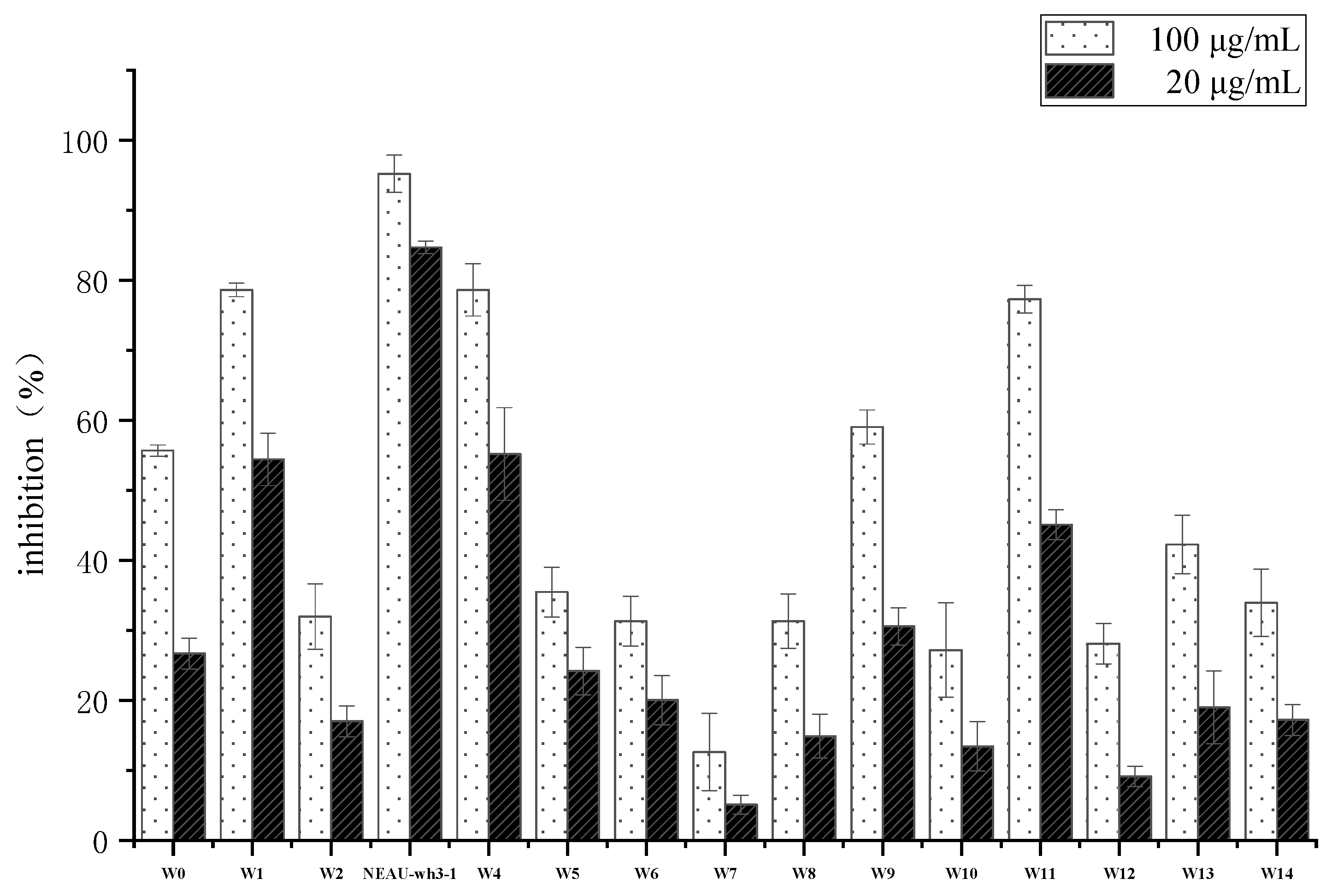
3.1. Isolation and Screening of an Antitumor Compound Producing Strains

Thirty-nine strains belonging to actinomycetes were isolated from the soil samples. The crude extracts of these isolates were examined for their cytotoxic activity at dilution concentrations of 100 μg/mL and 20 μg/mL. As a result of primary screening, fourteen strains showed cytotoxic activity to human colon tumor cell line HCT-116 (Figure 1). Due to the superior cytotoxic activity of strain NEAU-wh3-1, which inhibition rate was greater than 80% at both concentrations, further chemical investigations were performed on this strain.

3.2. Polyphasic Taxonomic Characterization of NEAU-wh3-1

Morphological observation of 2-week-old cultures of strain NEAU-wh3-1 grown on ISP 3 medium revealed that the strain has the typical characteristics of genus Embleya and formed well-developed, branched substrate hyphae and aerial mycelium that differentiated into spiral spore chains consisted of cylindrical spores (0.6–0.8μm × 0.9–1.3μm), the spores were rough-surfaced and non-motile (Figure 2). Strain NEAU-wh3-1 was found to grow at a temperature range of 4 to 37 °C (optimum temperature 28 °C), pH 5 to 12 (optimum pH 7), and NaCl tolerance of 0% to 3% (optimum NaCl of 1%). The physiological and biochemical properties of strain NEAU-wh3-1, Embleya scabrispora DSM 41855T, Embleya hyalina MB891-A1T, and Streptomyces lasii 5H-CA11T are given in Table 1.
Chemotaxonomic analyses revealed that strain NEAU-wh3-1 contained LL-diaminopimelic acid as cell wall diamino acid. The whole-cell sugar was found to contain arabinose, glucose, and ribose. The phospholipid profile consisted of diphosphatidylglycerol (DPG), phosphatidylethanolamine (PE), phosphatidylinositol (PI), and two unidentified lipids (ULs) (Supplementary Figure S1). The menaquinones detected were MK-9(H4) (46.5%), MK-9(H6) (45.8%), and MK-9(H8) (7.7%).
The almost complete 16S rRNA gene sequence of strain NEAU-wh3-1 (1487 bp) was determined and deposited with the accession number MN928616 in the GenBank/EMBL (European Molecular Biology Laboratory)/DDBJ (DNA Data Bank of Japan) databases. EzBioCloud analysis suggests that strain NEAU-wh3-1 shared the highest 16S rRNA gene sequence similarities with Embleya scabrispora DSM 41855T (99.65%), Embleya hyalina MB891-A1T (99.45%), and Streptomyces lasii 5H-CA11T (98.62%). Phylogenetic analysis based on the 16S rRNA gene sequences indicated that the strain formed a stable cluster with E. scabrispora DSM 41855T, E. hyalina MB891-A1T, and S. lasii 5H-CA11T based on neighbor-joining algorithm (Figure 3) and also supported by the maximum-likelihood algorithm (Supplementary Figure S2). To further clarify the affiliation of strain NEAU-wh3-1 to its closely related strains, partial sequences of housekeeping genes including atpD, gyrB, recA, rpoB, and trpB were obtained. GenBank accession numbers of the sequences are displayed in Table S1. The phylogenetic tree based on the neighbor-joining tree constructed from the concatenated sequence alignment (1979 bp) of five housekeeping genes (Figure 4) suggested that the isolate clustered with E. scabrispora DSM 41855T and E. hyalina MB891-A1T, and also supported by the maximum-likelihood algorithm (Streptomyces lasii 5H-CA11T lacks housekeeping genes; Supplementary Figure S3). Moreover, pairwise distances calculated for NEAU-wh3-1 and the related species using concatenated sequences of atpD-gyrB-recA-rpoB-trpB were well above 0.007 (Table S2) for the related species, which was considered to be the threshold for species determination [74].

3.3. Structural Elucidation

The strain NEAU-wh3-1 was grown preparative scale in 15.0 L of production broth for 7 days. Bioassay-guided isolation of the active components of the strain yielded eight main bioactive compounds. Compounds 28 are known compounds, which structures were elucidated as conglobatin (2) [75], piericidin C1 (3) [76], piericidin C5 (4) [77], piericidin A1 (5) [78], piericidin A3 (6) [76], Mer-A 2026 A (7) [79], and BE-52211 D (8) [80] by analysis of their spectroscopic data and comparison with literature values (Figure 5, Figures S12–S27). Compound 1 is a new zincophorin analogue (Figure 6, Figures S4–S11) [17].
Analysis of 1H NMR spectrum of 1 revealed the presence of three olefinic protons at δH 5.52 (1H, m), 5.36 (1H, m), 5.20 (1H, d, J = 8.9 Hz), seven aliphatic methine protons at δH 4.07 (1H, m), 4.06 (1H, m), 3.77 (1H, d, J = 8.9 Hz), 3.72 (1H, dd, J = 9.6, 2.1 Hz), 3.58 (1H, d, J = 9.2 Hz), 3.49 (1H, m), 3.28 (1H, m), seven methylene protons at δH 2.18 (1H, m), 2.13 (1H, m), 1.77 (1H, m), 1.67 (2H, m), 1.40 (1H, m), 1.35 (1H, m), 1.28 (1H, m), one singlet methyl at δH 1.63 s, in addition to eight doublet methyl protons at δH 1.18 (3H, d, J = 7.1 Hz), 1.15 (3H, d, J = 7.2 Hz), 1.11 (3H, d, J = 7.0 Hz), 0.98 (3H, d, J = 6.5 Hz), 0.97 (3H, d, J = 6.6 Hz), 0.86 (3H, d, J = 6.7 Hz), 0.81 (3H, d, J = 6.5 Hz), 0.70 (3H, d, J = 6.7 Hz). The 13C NMR and DEPT135 spectra (Table 2) of 1 showed 31 resonances attributable to a carbonyl carbon at δC 175.6, one sp2 quaternary carbon at δC 132.3, three sp2 methines at δC 136.6, 134.5, 132.4. In the sp3-carbon region, the spectrum showed six oxygenated methines at δC 84.3, 83.3, 81.7, 76.1, 74.0, 69.4, six methines at δC 42.2, 37.1, 36.5, 36.0, 33.0, 26.1, four methylenes at δC 34.2, 28.6, 27.1, 24.9 and nine methyl carbons at δC 22.9, 22.9, 17.1, 16.7, 15.4, 12.3, 11.3, 10.8, 10.1. The 1H–1H COSY correlations (Figure 6) of H-2/H-3/H2-4/H2-5/H-6/H-7/H-8/H-9/H-10/H-11/H-12/H-13/H2-14/H2-15/H-16/H-17/H-18/H-19 established connectivity from H-2 atom along the chain through to C-19 atom. The correlations between H-21/H-22/H3-23/H3-24, H-12/H3-27, H-10/H3-28, H-18/H3-26, H-8/H3-29 protons in the 1H–1H COSY spectrum (Figure 6) indicated the five structural units of C-21–C-24, C-18–C-26, C-12–C-27, C-10-C-28, C-8-C-29. The observed HMBC (heteronuclear multiple bond correlation) correlations (Figure 6) from H3-23, H3-24 to C-21, C-22, from H3-25 to C-19, C-21, from H3-26 to C-17, C-18, and C-19, from H3-27 to C-11, C-12, and C-13, from H3-28 to C-9, C-10, C-11, from H3-29 to C-7, C-8, C-9, from H3-30 to C-5, C-6, C-7, from H3-31 to C-2 and C-3, from H-21 to C-19, H-20 to C-18, from H-19 to C-17, from H3-23 to C-21 established the linkage of C-2–C-22. The carbonyl group was connected with C-2 by the HMBC corrections from H-2 and H3-31 to C-1 (δC 175.6). The correlations from H-7 (δH 3.77 d, J = 8.9 Hz) to C-3 (δC 74.0) indicated the linkage of C-3 and C-7 through an oxygen atom to form a tetrahydropyran ring. Taking the molecular formula of C31H56O7 into account, four hydroxyl groups were situated at C-9, C-11, C-13, C-19, respectively, and a carboxyl group was situated at C-1. Comparison the NMR data of 1 with Zincophorin [17], a mocarboxylic acid ionophore contains one single tetrahydropyran ring, which was isolated from a strain of Streptomyces griseus, implied that 1 was identified to be an analogue of Zincophorin, the difference between two compounds was that the terminal ethyl group in Zincophorin was replaced by a H proton in compound 1. On the basis of the above spectroscopic data, a gross structure of 1 was established and named Zincophorin B, and the 1H and 13C resonances in 1 were assigned (Table 2).

3.4. Biological Activity

The cytotoxic activities of compounds 1–8 against K562, HCT-116, and HepG2 cancer cell lines are showed in Table 3. Eight compounds restrained proliferation of the tested cells and compound 1 showed the highest cytotoxic activity, and the average IC50 values were lower than 10.0 μg/mL.
The result of minimum inhibitory concentrations (MICs) showed that compound 1 showed good activities against Gram-positive bacteria Staphylococcus aureus, Sarcina lutea, and Bacillus subtilis, and the Gram-negative bacteria Klebsiella pneumoniae in vitro (Table 4). Compound 8 showed weak antibacterial activity against two Gram-positive bacterium and the minimum inhibitory concentrations (MICs) of compounds 27 were determined to be >10 mg/mL, so they had no activity against these tested pathogens.

4. Discussion

In this research, the results of morphological, physiological, and biochemical tests showed that strain NEAU-wh3-1 has typical characteristics of the genus Embleya [41]. Such as containing LL-diaminopimelic acid as the cell wall peptidoglycan, MK-9(H4), and MK-9(H6) as the major menaquinones, phosphatidylethanolamine (PE) as the predominant phospholipid and arabinose in the whole sugars. Moreover, strain NEAU-wh3-1 formed spiral spore chains and the spore surface was rough, which are consistent with E. scabrispora DSM 41855T and E. hyalina MB891-A1T [39,41]. In addition, the phylogenetic trees constructed from the 16S rRNA gene sequences and the concatenated sequences alignment (1979 bp) of five housekeeping genes all suggested that the isolate should be assigned to the genus Embleya.
However, some obvious differences could also be found between strain NEAU-wh3-1 and its closely related strains regarding several phenotypic and chemotaxonomic characteristics (Table 1). The isolate was able to grow at 4 °C, in contrast to its closely related strains, which were not. The composition of phospholipids and menaquinones of strain NEAU-wh3-1 was also different from its related species, E. scabrispora DSM 41855T, E. hyalina MB891-A1T and S. lasii 5H-CA11T. Most notably, the whole-cell sugars of strain NEAU-wh3-1 was found to contain arabinose, glucose, and ribose, while E. scabrispora DSM 41855T only contains arabinose, E. hyalina MB891-A1T contains arabinose and glucose and S. lasii 5H-CA11T contains glucose and ribose, which also could distinguish the strain from its closely related strains. Other phenotypic differences include the temperature and pH range of growth, patterns of carbon and nitrogen utilization, hydrolysis of cellulose, starch, and Tweens (20, 40, and 80), liquefaction of gelatin, peptonization of milk, production of H2S, and urease and reduction of nitrate. Therefore, it is evident from the phenotypic, genotypic, and chemotaxonomic data that strain NEAU-wh3-1 may represent a novel species of the genus Embleya.
The genus Embleya, recently transferred from genus Streptomyces, is a new member of the family Streptomycetaceae [39,40]. At present, it contains only two species: Embleya scabrispora, could produce hitachimycin with antitumor, antibacterial, and antiprotozoal activities [44,45,46]; and Embleya hyaline, could produce nybomycin which is an effective agent against antibiotic-resistant Staphylococcus aureus and is called a reverse antibiotic [48]. During the study of the chemical properties of the active ingredients of strain NEAU-wh3-1, eight active compounds were obtained, including one macrolide dilactone, five piericidins, one β-hydroxy acetamides, an analogue of monocarboxylic acid ionophore, which were observed to fit into at least three types based on their molecular skeletons. This shows to some extent that this strain has the ability to produce metabolites with a wide variety of different skeletal structures. Out of these compounds, Piericidins (3-7) were a class of polyene alpha-pyridone heterocyclic antibiotics, among them, Piericidin A1(5) was first reported [81], which was isolated from Streptomyces mobaraensis. Piericidins exhibit interesting biological activities, in particular antitrypanosomal [82]. In our research, compounds 3-7 exhibited different degrees of cytotoxicity on three types of tumor cells, but they did not show any antibacterial activity, which was consistent with previous reports [78,81]. As a Piericidins-producing strain, NEAU-wh3-1 has certain application potential in pest control. Conglobatin (2) is an unusual 16-membered macrocyclic diolide originally isolated from a polyether-producing strain of Streptomyces conglobatus ATCC 31005T and was reported to be essentially devoid of antifungal, antibacterial, antitumor, and antiprotozoal activity at that time [75]. However, in recent research, FW-04-806 is identical in structure to conglobatin, and it has been reported to inhibit the growth of a human chronic myelocytic leukemia K562 cell line with an IC50 of 6.66 μg/mL, further study also investigated the effects of FW-04-806 on SKBR3 and MCF-7, respectively [83]. Its mechanism of action appears to be novel, via direct binding to the N-terminal domain of Hsp90 and disruption of its interaction with co-chaperone Cdc37 [84]. In our antitumor activity test, Conglobatin (2) showed good bioactivity against two tumor cell lines, supporting it at least partially accounted for the cytotoxic activity of the strain NEAU-wh3-1 extract. BE-52211 D (8) was a cytotoxic metabolite from a strain of Streptomyces and had been reported to have moderate cytotoxicity against human hepatocellular liver carcinoma cells HepG2, human leukemia cells K562, and human colon carcinoma cells HCT-116 with the IC50 values of >10 μg/mL [80], which is consistent with the result in the present study. Compound 1 structurally related to zincophorin, which was also referred to as M144255 or griseochellin and is a polyoxygenated ionophoric antibiotic isolating from Streptomyces griseus in 1984 [17]. It has been reported to possess strong in vivo activity against Gram-positive bacteria and have strong cytotoxicity against human lung carcinoma cells A549 and Madin-Darby canine kidney cells MDCK [18]. No biological activity has been reported against Gram-negative bacteria, yeasts, and fungi [85]. The second member in the zincophorin family named CP-78545, was found in the culture broth of Streptomyces sp. N731-45. The structural difference between them is that CP-78545 has an extra terminal double bond; but they have similar spectrum and potency on biological properties except for the antitumor activity (no reports) [86]. In our antitumor and antimicrobial assays, compound 1 showed the highest antitumor activity against three human cell lines and good antibacterial activity against Gram-negative bacteria. To our knowledge, this is the first report of this kind of compound with antibacterial activity against Gram-negative bacteria. This study has enriched the activity spectrum of Zincophorins.

5. Conclusions

Strain producing a new compound with strong antitumor activity, isolated from the rhizosphere soil of wheat (Triticum aestivum L.) in HeBei province, China. Morphological and chemotaxonomic features together with phylogenetic analysis suggested that strain NEAU-wh3-1 belonged to the genus Embleya. Cultural and biochemical characteristics combined with multilocus sequence analysis clearly revealed that strain NEAU-wh3-1 may represent a novel species of the genus Embleya. Moreover, eight compounds, including one new compound with higher antitumor activities against three human cell lines, were isolated from the strain.

Supplementary Materials

The following are available online at https://www.mdpi.com/2076-2607/8/3/441/s1, Table S1: GenBank accession numbers of the sequences used in MLSA; Table S2: MLSA distance values for selected strains in this study; Figure S1: The polar lipids of strain NEAU-wh3-1; Figure S2: Maximum-likelihood tree based on 16S rRNA gene sequences showing relationship between strain NEAU-wh3-1 and related taxa; Figure S3: Maximum-likelihood tree based on multilocus sequence analysis (MLSA)analysis of the concatenated partial sequences (1979 bp) from five housekeeping genes (atpD, gyrB, recA, rpoB, and trpB) of strain NEAU-wh3-1 (in bold) with related taxa; Figure S4: 1H NMR (400 MHz) spectrum of compound 1 in CDCl3; Figure S5: 13C NMR (150 MHz) spectrum of compound 1 (in CDCl3); Figure S: 1H-1H COSY spectrum (400 MHz) of compound 1 (in CDCl3); Figure S7: HSQC spectrum (400 MHz) of compound 1 (in CDCl3); Figure S8: HMBC spectrum (400 MHz) of compound 1 (in CDCl3); Figure S9: IR spectrum of compound 1(in EtOH); Figure S10: UV spectrum of compound 1 (in EtOH); Figure S11: The HRESIMS spectrum of compound 1; Figure S12: 1H NMR (400 MHz) spectrum of compound 2 in CDCl3; Figure S13: 13C NMR (150 MHz) spectrum of compound 2 in CDCl3; Figure S14: The ESI-MS spectrum of compound 2; Figure S15: 1H NMR (400 MHz) spectrum of compound 3 in CDCl3; Figure S16: The ESI-MS spectrum of compound 3; Figure S17: 1H NMR (400 MHz) spectrum of compound 4 in CDCl3; Figure S18: The ESI-MS spectrum of compound 4; Figure S19: 1H NMR (400 MHz) spectrum of compound 5 in CDCl3; Figure S20: The ESI-MS spectrum of compound 5; Figure S21: 1H NMR (400 MHz) spectrum of compound 6 in CDCl3; Figure S22: The ESI-MS spectrum of compound 6; Figure S23: 1H NMR (400 MHz) spectrum of compound 7 in CDCl3; Figure S24: ESI-MS spectrum of compound of compound 7; Figure S25: 1H NMR (400 MHz) spectrum of compound 8 in CDCl3; Figure S26: 13C NMR (150 MHz) spectrum of compound 8 in CDCl3; Figure S27: The HRESIMS spectrum of compound 8.

Author Contributions

H.W., T.S., and W.S. performed the experiments. X.G. prepared the figures and tables. P.C. and X.X. analyzed the data. Y.S. and J.Z. designed the experiments and reviewed the manuscript. All authors have read and agreed to the published version of the manuscript.

Funding

This work was supported in part by grants from the Heilongjiang Postdoctoral Fund (LBH-Z17015) and the Scientific Research Foundation for Settled Postdoctoral of Heilongjiang Province (LBH-Q19082).

Conflicts of Interest

The authors declare that there are no conflict of interest.

References

  1. Torre, L.A.; Bray, F.; Siegel, R.L.; Ferlay, J.; Lortet-Tieulent, J.; Jemal, A. Global cancer statistics, 2012. CA. Cancer J. Clin. 2015, 65, 87–108. [Google Scholar] [CrossRef] [Green Version]
  2. Commander, H.; Whiteside, G.; Perry, C. Vandetanib: First global approval. Drugs 2011, 71, 1355–1365. [Google Scholar] [CrossRef]
  3. Zhu, F.; Zhao, X.; Li, J.; Guo, L.; Bai, L.; Qi, X. A new compound Trichomicin exerts antitumor activity through STAT3 signaling inhibition. Biomed. Pharmacother. 2020, 121, 109608. [Google Scholar] [CrossRef]
  4. Zhang, Z.; Yu, X.; Wang, Z.; Wu, P.; Huang, J. Anthracyclines potentiate anti-tumor immunity: A new opportunity for chemoimmunotherapy. Cancer Lett. 2015, 369, 331–335. [Google Scholar] [CrossRef]
  5. Sznarkowska, A.; Kostecka, A.; Meller, K.; Bielawski, K.P. Inhibition of cancer antioxidant defense by natural compounds. Oncotarget 2017, 8, 15996–16016. [Google Scholar] [CrossRef] [Green Version]
  6. Newman, D.J.; Cragg, G.M. Natural products as sources of new drugs over the last 25 years. J. Nat. Prod. 2007, 70, 461–477. [Google Scholar] [CrossRef] [Green Version]
  7. Cragg, G.M.; Newman, D.J.; Snader, K.M. Natural products in drug discovery and development. J. Nat. Prod. 1997, 60, 52–60. [Google Scholar] [CrossRef]
  8. Chin, Y.W.; Balunas, M.J.; Chai, H.B.; Kinghorn, A.D. Drug discovery from natural sources. Drug Addict. From Basic Res. Ther. 2008, 8, 17–39. [Google Scholar]
  9. Pettit, R.K. Culturability and Secondary Metabolite Diversity of Extreme Microbes: Expanding Contribution of Deep Sea and Deep-Sea Vent Microbes to Natural Product Discovery. Mar. Biotechnol. 2011, 13, 1–11. [Google Scholar] [CrossRef]
  10. Qin, S.; Li, J.; Chen, H.H.; Zhao, G.Z.; Zhu, W.Y.; Jiang, C.L.; Xu, L.H.; Li, W.J. Isolation, diversity, and antimicrobial activity of rare actinobacteria from medicinal plants of tropical rain forests in Xishuangbanna China. Appl. Environ. Microbiol. 2009, 75, 6176–6186. [Google Scholar] [CrossRef] [Green Version]
  11. Parkinson, D.R.; Arbuck, S.G.; Moore, T.; Pluda, J.M.; Christian, M.C. Clinical development of anticancer agents from natural products. Stem Cells 1994, 12, 30–43. [Google Scholar] [CrossRef]
  12. Berdy, J. Bioactive microbial metabolites. J Antibiot. 2005, 58, 1–26. [Google Scholar] [CrossRef] [Green Version]
  13. Ventura, M.; Canchaya, C.; Tauch, A.; Chandra, G.; Fitzgerald, G.F.; Chater, K.F.; van Sinderen, D. Genomics of Actinobacteria: Tracing the Evolutionary History of an Ancient Phylum. Microbiol. Mol. Biol. Rev. 2007, 71, 495–548. [Google Scholar] [CrossRef] [Green Version]
  14. Goodfellow, M.; Williams, S.T. Ecology of actinomycetes. Annu. Rev. Microbiol. 1983, 37, 189–216. [Google Scholar] [CrossRef]
  15. Rabbani, A.; Finn, R.M.; Ausió, J. The anthracycline antibiotics: Antitumor drugs that alter chromatin structure. BioEssays 2005, 27, 50–56. [Google Scholar] [CrossRef]
  16. Demain, A.L.; Sanchez, S. Microbial drug discovery: 80 Years of progress. J. Antibiot. 2009, 62, 5–16. [Google Scholar] [CrossRef] [Green Version]
  17. Brooks, H.A.; Gardner, D.; Poyser, J.P.; King, T.J. The structure and absolute stereochemistry of zincophorin (antibiotic m144255): a monobasic carboxylic acid ionophore having a remarkable specificity for divalent cations. J. Antibiot. 1984, 37(11), 1501–1504. [Google Scholar] [CrossRef] [Green Version]
  18. Walther, E.; Boldt, S.; Kage, H.; Lauterbach, T.; Martin, K.; Roth, M.; Hertweck, C.; Sauerbrei, A.; Schmidtke, M.; Nett, M. Zincophorin-biosynthesis in Streptomyces griseus and antibiotic properties. GMS Infect. Dis. 2016, 4, Doc08. [Google Scholar]
  19. Yang, S.X.; Gao, J.M.; Zhang, A.L.; Laatsch, H.S. A novel toxic macrolactam polyketide glycoside produced by actinomycete Streptomyces sannanensis. Bioorg. Med. Chem. Lett. 2011, 21, 3905–3908. [Google Scholar] [CrossRef]
  20. Subramani, R.; Aalbersberg, W. Culturable rare Actinomycetes: Diversity, isolation and marine natural product discovery. Appl. Microbiol. Biotechnol. 2013, 97, 9291–9321. [Google Scholar] [CrossRef]
  21. Heidari, B.; Mohammadipanah, F. Isolation and identification of two alkaloid structures with radical scavenging activity from Actinokineospora sp. UTMC 968, a new promising source of alkaloid compounds. Mol. Biol. Rep. 2018, 45, 2325–2332. [Google Scholar] [CrossRef] [PubMed]
  22. Gao, M.Y.; Qi, H.; Li, J.S.; Zhang, H.; Zhang, J.; Wang, J.D.; Xiang, W.S. A new polysubstituted cyclopentene derivative from Streptomyces sp. HS-NF-1046. J. Antibiot. 2017, 70, 216–218. [Google Scholar] [CrossRef] [PubMed]
  23. Peláez, F. The historical delivery of antibiotics from microbial natural products - Can history repeat? Biochem. Pharmacol. 2006, 71, 981–990. [Google Scholar] [CrossRef]
  24. Kekuda, P.; Onkarappa, R.; Jayanna, N. Characterization and Antibacterial Activity of a Glycoside Antibiotic from Streptomyces variabilis PO-178. Sci. Technol. Arts. Res. J. 2015, 3, 116. [Google Scholar] [CrossRef]
  25. Miao, V.; Davies, J. Actinobacteria: The good, the bad, and the ugly. Antonie van Leeuwenhoek 2010, 98, 143–150. [Google Scholar] [CrossRef]
  26. Subramani, R.; Aalbersberg, W. Marine actinomycetes: An ongoing source of novel bioactive metabolites. Microbiol. Res. 2012, 167, 571–580. [Google Scholar] [CrossRef]
  27. Clardy, J.; Fischbach, M.A.; Walsh, C.T. New antibiotics from bacterial natural products. Nat. Biotechnol. 2006, 24, 1541–1550. [Google Scholar] [CrossRef]
  28. Ayed, A.; Slama, N.; Mankai, H.; Bachkouel, S.; ElKahoui, S.; Tabbene, O.; Limam, F. Streptomyces tunisialbus sp. nov., a novel Streptomyces species with antimicrobial activity. Antonie van Leeuwenhoek 2018, 111, 1571–1581. [Google Scholar] [CrossRef]
  29. Mccully, M.; Harper, J.D.I.; An, M.; Kent, J.H. The rhizosphere: the key functional unit in plant soil microbial interactions in the field implications for the understanding of allelopathic effects. Pol. J. Vet. Sci. 2005, 15, 493–498. [Google Scholar]
  30. Hiltner, L. Uber neuer erfahrungen und probleme auf dem gebiet der berücksichtigung unter besonderer berücksichtigung der gründüngung und brache. Arbeiten der Deustchen Landwirtschaftsgesellesschaft 1904, 32, 1405–1417. [Google Scholar]
  31. Berendsen, R.L.; Pieterse, C.M.J.; Bakker, P.A.H.M. The rhizosphere microbiome and plant health. Trends. Plant Sci. 2012, 17, 478–486. [Google Scholar] [CrossRef]
  32. Loper, J.E.; Gross, H. Genomic analysis of antifungal metabolite production by Pseudomonas fluorescens Pf-5. Eur. J. Plant Pathol. 2007, 119, 265–278. [Google Scholar] [CrossRef]
  33. Reiter, B.; Sessitsch, A.; Nowak, J.; Cle, C. Endophytic colonization of Vitis vinifera L. by plant growth-promoting bacterium Burkholderia sp. strain PsJN. Society 2005, 71, 1685–1693. [Google Scholar]
  34. Lanteigne, C.; Gadkar, V.J.; Wallon, T.; Novinscak, A.; Filion, M. Production of DAPG and HCN by Pseudomonas sp. LBUM300 contributes to the biological control of bacterial canker of tomato. Phytopathology 2012, 102, 967–973. [Google Scholar] [CrossRef] [Green Version]
  35. Osei, E.; Kwain, S.; Mawuli, G.T.; Anang, A.K.; Owusu, K.B.A.; Camas, M.; Camas, A.S.; Ohashi, M.; Alexandru-Crivac, C.N.; Deng, H. Paenidigyamycin A, potent antiparasitic imidazole alkaloid from the ghanaian Paenibacillus sp. De2Sh. Mar. Drugs 2019, 17, 9. [Google Scholar] [CrossRef] [Green Version]
  36. Mukhtar, S.; Mehnaz, S.; Mirza, M.S.; Malik, K.A. Isolation and characterization of bacteria associated with the rhizosphere of halophytes (Salsola stocksii and Atriplex amnicola) for production of hydrolytic enzymes. Brazilian J. Microbiol. 2019, 50, 85–97. [Google Scholar] [CrossRef]
  37. Orfali, R.; Perveen, S. Secondary metabolites from the Aspergillus sp. in the rhizosphere soil of Phoenix dactylifera (Palm tree). BMC Chem. 2019, 13, 1–6. [Google Scholar] [CrossRef]
  38. Wang, R.J.; Zhang, S.Y.; Ye, Y.H.; Yu, Z.; Qi, H.; Zhang, H.; Xue, Z.L.; Wang, J.D.; Wu, M. Three new isoflavonoid glycosides from the mangrove-derived actinomycete micromonospora aurantiaca 110B. Mar. Drugs 2019, 17, 294. [Google Scholar] [CrossRef] [Green Version]
  39. Nouioui, I.; Carro, L.; García-López, M.; Meier-Kolthoff, J.P.; Woyke, T.; Kyrpides, N.C.; Pukall, R.; Klenk, H.-P.; Goodfellow, M.; Göker, M. Genome-Based Taxonomic Classification of the Phylum Actinobacteria. Front. Microbiol. 2018, 9, 2007. [Google Scholar] [CrossRef] [Green Version]
  40. Oren, A.; Garrity, G.M. List of new names and new combinations previously effectively, but not validly, published. Int. J. Syst. Evol. Microbiol. 2018, 68, 2707–2709. [Google Scholar] [CrossRef]
  41. Komaki, H.; Hosoyama, A.; Kimura, A.; Ichikawa, N.; Igarashi, Y.; Tamura, T. Classification of ‘Streptomyces hyalinum’ Hamada and Yokoyama as Embleya hyalina sp. nov., the second species in the genus Embleya, and emendation of the genus Embleya. Int. J. Syst. Evol. Microbiol. 2020, 1–5. [Google Scholar] [CrossRef]
  42. Kämpfer, P.; Genus, I. Streptomyces Waksman and Henrici 1943, 339 AL. In Bergey’s Manual of Systematic Bacteriology, 2nd ed.; Springer: New York, NY, USA, 2012; pp. 1679–1680. [Google Scholar]
  43. Ping, X.; Takahashi, Y.; Seino, A.; Iwai, Y.; Omura, S. Streptomyces scarbrisporus sp. nov. Int. J. Syst. Evol. Microbiol. 2004, 54, 577–581. [Google Scholar] [CrossRef]
  44. Omura, S.; Nakagawa, A.; Shibata, K.; Sano, H. The structure of hitachimycin, a novel macrocyclic lactam involving β-phenylalanine. Tetrahedron Lett. 1982, 23, 4713–4716. [Google Scholar] [CrossRef]
  45. Komiyama, K.; Edanami, K.I.; Yamamoto, H.; Umezawa, I. Antitumor activity of a new antitumor antibiotic, stubomycin. J. Antibiot. 1982, 35, 703–706. [Google Scholar] [CrossRef] [Green Version]
  46. Shibata, K.; Satsumabayashi, S.; Sano, H.; Komiyama, K.; Nakagawa, A.; Omura, S. Chemical modification of hitachimycin. Synthesis, antibacterial, cytocidal and in vivo antitumor activities of hitachimycin derivatives. J. Antibiot. 1988, 41, 614–623. [Google Scholar] [CrossRef] [Green Version]
  47. Naganawa, H.; Wakashiro, T.; Yagi, A.; Kondo, S.; Takita, T.; Hamada, M.; Maeda, K.; Umezawa, H. Deoxynybomycin from a streptomyces. J. Antibiot. 1970, 23, 365–368. [Google Scholar] [CrossRef] [Green Version]
  48. Hiramatsu, K.; Igarashi, M.; Morimoto, Y.; Baba, T.; Umekita, M.; Akamatsu, Y. Curing bacteria of antibiotic resistance: Reverse antibiotics, a novel class of antibiotics in nature. Int. J. Antimicrob. Agents 2012, 39, 478–485. [Google Scholar] [CrossRef] [Green Version]
  49. Hayakawa, M.; Nonomura, H. Humic acid-vitamin agar, a new medium for the selective isolation of soil actinomycetes. J. Ferment. Technol. 1987, 65, 501–509. [Google Scholar] [CrossRef]
  50. Shirling, E.B.; Gottlieb, D. Methods for characterization of Streptomyces species. Int. J. Syst. Bacteriol. 1966, 16, 313–340. [Google Scholar] [CrossRef] [Green Version]
  51. Vichai, V.; Kirtikara, K. Sulforhodamine B colorimetric assay for cytotoxicity screening. Nat. Protoc. 2006, 1, 1112–1116. [Google Scholar] [CrossRef]
  52. Jin, L.; Zhao, Y.; Song, W.; Duan, L.; Jiang, S.; Wang, X.; Zhao, J.; Xiang, W. Streptomyces inhibens sp. Nov., a novel actinomycete isolated from rhizosphere soil of wheat (Triticum aestivum L.). Int. J. Syst. Evol. Microbiol. 2019, 69, 688–695. [Google Scholar] [CrossRef] [PubMed]
  53. Zhao, J.W.; Han, L.Y.; Yu, M.Y.; Cao, P.; Li, D.M.; Guo, X.W.; Liu, Y.Q.; Wang, X.J.; Xiang, W.S. Characterization of Streptomyces sporangiiformans sp. nov., a Novel Soil Actinomycete with Antibacterial Activity against Ralstonia solanacearum. Microorganisms 2019, 7, 360. [Google Scholar] [CrossRef] [PubMed] [Green Version]
  54. Smibert, R.M.; Krieg, N.R. Phenotypic characterization. In Methods for General and Molecular Bacteriology, 2nd ed.; Gerhardt, P., Murray, R.G.E., Wood, W.A., Krieg, N.R., Eds.; American Society for Microbiology: Washington, DC, USA, 1994; pp. 607–654. [Google Scholar]
  55. Gordon, R.E.; Barnett, D.A.; Handerhan, J.E.; Pang, C. Nocardia coeliaca, Nocardia autotrophica, and the nocardin strain. Int. J. Syst. Bacteriol. 1974, 24, 54–63. [Google Scholar] [CrossRef] [Green Version]
  56. Yokota, A.; Tamura, T.; Hasegawa; Huang, L.H. Catenuloplanes japonicas gen. nov., sp. nov., nom. rev., a new genus of the order Actinomycetales. Int. J. Syst. Bacteriol. 1993, 43, 805–812. [Google Scholar] [CrossRef] [Green Version]
  57. Lechevalier, M.P.; Lechevalier, H.A. The chemotaxonomy of actinomycetes. In Actinomycete taxonomy, 2nd ed.; Dietz, A., Thayer, D.W., Eds.; special publication for Society of Industrial Microbiology: Arlington, TX, USA, 1980; pp. 227–291. [Google Scholar]
  58. Minnikin, D.E.; O’Donnell, A.G.; Goodfellow, M.; Alderson, G.; Athalye, M.; Schaal, A.; Parlett, J.H. An integrated procedure for the extraction of bacterial isoprenoid quinones and polar lipids. J. Microbiol. Methods 1984, 2, 233–241. [Google Scholar] [CrossRef]
  59. Collins, M.D. Isoprenoid quinone analyses in bacterial classification and identification. In Chemical methods in bacterial systematics; Goodfellow, M., Minnikin, D.E., Eds.; Academic Press: London, UK, 1985; pp. 267–284. [Google Scholar]
  60. Shen, Y.; Sun, T.; Jiang, S.; Mu, S.; Li, D.; Guo, X.; Zhang, J.; Zhao, J.; Xiang, W. Streptomyces lutosisoli sp. nov., a novel actinomycete isolated from muddy soil. Antonie Van Leeuwenhoek, Int. J. Gen. Mol. Microbiol. 2018, 111, 2403–2412. [Google Scholar] [CrossRef]
  61. Wu, C.; Lu, X.; Qin, M.; Wang, Y.; Ruan, J. Analysis of menaquinone compound in microbial cells by HPLC. Microbiology 1989, 16, 176–178. [Google Scholar]
  62. Kim, S.B.; Brown, R.; Oldfield, C.; Gilbert, S.C.; Iliarionov, S.; Goodfellow, M. Gordonia amicalis sp. nov., a novel dibenzothiophene-desulphurizing actinomycete. Int. J. Syst. Evol. Microbiol. 2000, 50, 2031–2036. [Google Scholar] [CrossRef] [Green Version]
  63. Yoon, S.H.; Ha, S.M.; Kwon, S.; Lim, J.; Kim, Y.; Seo, H.; Chun, J. Introducing EzBioCloud: A taxonomically united database of 16S rRNA gene sequences and whole-genome assemblies. Int. J. Syst. Evol. Microbiol. 2017, 67, 1613–1617. [Google Scholar] [CrossRef]
  64. Saitou, N.; Nei, M. The neighbor-joining method: a new method for reconstructing phylogenetic trees. Mol. Biol. Evol. 1987, 4, 406–425. [Google Scholar]
  65. Felsenstein, J. Evolutionary trees from DNA sequences: A maximum likelihood approach. J. Mol. Evol. 1981, 17, 368–376. [Google Scholar] [CrossRef] [PubMed]
  66. Kumar, S.; Stecher, G.; Tamura, K. Mega7: molecular evolutionary genetics analysis version 7.0 for bigger datasets. Mol. Biol. Evol. 2016, 33, 1870–1874. [Google Scholar] [CrossRef] [PubMed] [Green Version]
  67. Felsenstein, J. Confidence Limits on Phylogenies: an Approach Using the Bootstrap. Evolution (N. Y). 1985, 39, 783–791. [Google Scholar]
  68. Kimura, M. A simple method for estimating evolutionary rates of base substitutions through comparative studies of nucleotide sequences. J. Mol. Evol. 1980, 16, 111–120. [Google Scholar] [CrossRef]
  69. Hatano, K.; Nishii, T.; Kasai, H. Taxonomic re-evaluation of whorl-forming Streptomyces (formerly Streptoverticillium) species by using phenotypes, DNA-DNA hybridization and sequences of gyrB, and proposal of Streptomyces luteireticuli (ex Katoh and Arai 1957) corrig., sp. nov., nom. rev. Int. J. Syst. Evol. Microbiol. 2003, 53, 1519–1529. [Google Scholar] [CrossRef]
  70. Guo, Y.P.; Zheng, W.; Rong, X.Y.; Huang, Y. A multilocus phylogeny of the Streptomyces griseus 16S rRNA gene clade: Use of multilocus sequence analysis for streptomycete systematics. Int. J. Syst. Evol. Microbiol. 2008, 58, 149–159. [Google Scholar] [CrossRef]
  71. Wang, J.; Zhang, H.; Ying, L.; Wang, C.; Jiang, N.; Zhou, Y.; Wang, H.; Bai, H. Five new epothilone metabolites from Sorangium cellulosum strain So0157-2. J. Antibiot. 2009, 62, 483–487. [Google Scholar] [CrossRef] [Green Version]
  72. Wayne, P.A. Clinical and Laboratory Standards Institute. Methods for dilution antimicrobial susceptibility tests for bacteria that grow aerobically; approved standard—ninth edition. Clin. Lab. Standards Institute 2012, 32, 2. [Google Scholar]
  73. Liu, C.; Han, C.; Jiang, S.; Zhao, X.; Tian, Y.; Yan, K.; Wang, X.; Xiang, W. Streptomyces lasii sp. nov., a Novel Actinomycete with Antifungal Activity Isolated from the Head of an Ant (Lasius flavus). Curr. Microbiol. 2018, 75, 353–358. [Google Scholar] [CrossRef]
  74. Rong, X.; Huang, Y. Taxonomic evaluation of the Streptomyces hygroscopicus clade using multilocus sequence analysis and DNA-DNA hybridization, validating the MLSA scheme for systematics of the whole genus. Syst. Appl. Microbiol. 2012, 35, 7–18. [Google Scholar] [CrossRef]
  75. Westley, J.W.; Liu, C.M.; Evans, R.H.; Blount, J.F. Conglobatin, a novel macrolide dilactone from Streptomyces conglobatus ATCC 31005. J. Antibiot. 1979, 32, 874–877. [Google Scholar] [CrossRef] [Green Version]
  76. Yoshida, S.; Yoneyama, K.; Shiraishi, S.; Watanabe, A.; Takahashi, N. Chemical structures of new piericidins produced by Streptomyces pactum. Agric. Biol. Chem. 1977, 41, 855–862. [Google Scholar] [CrossRef]
  77. Kubota, N.K.; Ohta, E.; Ohta, S.; Koizumi, F.; Suzuki, M.; Ichimura, M.; Ikegami, S. Piericidins C5 and C6: New 4-pyridinol compounds produced by Streptomyces sp. and Nocardioides sp. Bioorganic Med. Chem. 2003, 11, 4569–4575. [Google Scholar] [CrossRef]
  78. Tamura, S.; Takahashi, N.; Miyamoto, S.; Mori, R.; Suzuki, S.; Nagatsu, J. Isolation and physiological activities of piericidin A, a natural insecticide produced by Streptomyces. Agr. Biol. Chem. 1963, 27, 576–582. [Google Scholar] [CrossRef]
  79. Kominato, K.; Watanabe, Y.; Hirano, S.I.; Kioka, T.; Tone, H. Mer-a2026a and b, novel piericidins with vasodilating effect. ii. physico-chemical properties and chemical structures. J. Antibiot. 1994, 48, 103–105. [Google Scholar] [CrossRef] [Green Version]
  80. Wang, H.; Zhao, X.L.; Gao, Y.H.; Qi, H.; Zhang, H.; Xiang, W.S.; Wang, J.D.; Wang, X.J. Two new cytotoxic metabolites from Streptomyces sp. HS-NF-813. J. Asian. Nat. Prod. Res. 2018, 22, 249–256. [Google Scholar] [CrossRef]
  81. Hall, C.; Wu, M.; Crane, F.L.; Takahashi, H.; Tamura, S.; Folkers, K. Piericidin A: A new inhibitor of mitochondrial electron transport. Biochem. Biophys. Res. Commun. 1966, 25, 373–377. [Google Scholar] [CrossRef]
  82. Shaaban, K.A.; Helmke, E.; Kelter, G.; Fiebig, H.H.; Laatsch, H. Glucopiericidin C: A cytotoxic piericidin glucoside antibiotic produced by a marine Streptomyces isolate. J. Antibiot. 2011, 64, 205–209. [Google Scholar] [CrossRef] [Green Version]
  83. Huang, W.; Ye, M.; Zhang, L.; Wu, Q.; Zhang, M.; Xu, J.; Zheng, W. FW-04-806 inhibits proliferation and induces apoptosis in human breast cancer cells by binding to N-terminus of Hsp90 and disrupting Hsp90-Cdc37 complex formation. Mol. Cancer 2014, 13, 1–13. [Google Scholar] [CrossRef] [Green Version]
  84. Huang, W.; Wu, Q.D.; Zhang, M.; Kong, Y.L.; Cao, P.R.; Zheng, W.; Xu, J.H.; Ye, M. Novel Hsp90 inhibitor FW-04-806 displays potent antitumor effects in HER2-positive breast cancer cells as a single agent or in combination with lapatinib. Cancer Lett. 2015, 356, 862–871. [Google Scholar] [CrossRef]
  85. Song, Z.; Lohse, A.G.; Hsung, R.P. Challenges in the synthesis of a unique mono-carboxylic acid antibiotic, (+)-zincophorin. Cheminform 2009, 26, 560–571. [Google Scholar] [CrossRef] [PubMed] [Green Version]
  86. Dirlam, J.P.; Belton, A.M.; Chang, S.P.; Cullen, W.P.; Huang, L.H.; Kojima, Y.; Maeda, H.; Nishiyama, S.; Oscarson, J.R.; Sakakibara, T.; et al. Cp-78,545, a new monocarboxylic acid ionophore antibiotic related to zincophorin and produced by a streptomyces. J. Antibiot. 1989, 42, 1213–1220. [Google Scholar] [CrossRef] [PubMed]
Figure 1. Antitumor activities of extracts obtained from fourteen isolates against human colon tumor cell line HCT-116.
Microorganisms 08 00441 g001
Figure 2. Scanning electron micrograph of spore chains of strain NEAU-wh3-1 grown on ISP 3 agar for 2 weeks at 28 °C.
Microorganisms 08 00441 g002
Figure 3. Neighbor-joining tree showing the phylogenetic position of strain NEAU-wh3-1 (1487 bp) and related taxa based on 16S rRNA gene sequences. Bootstrap values > 50% (based on 1000 replications) are shown at branch points. Arthrobacter globiformis DSM 20124T (M23411) was used as an outgroup. Asterisks indicate branches also recovered in the maximum-likelihood tree; Bar, 0.01 substitutions per nucleotide position.
Microorganisms 08 00441 g003
Figure 4. Neighbor-joining tree based on multilocus sequence analysis (MLSA) analysis of the concatenated partial sequences (1979 bp) from five housekeeping genes (atpD, gyrB, recA, rpoB, and trpB) of strain NEAU-wh3-1 (in bold) with related taxa. Only bootstrap values above 50% (percentages of 1000 replications) are indicated. Asterisks indicate branches also recovered in the maximum-likelihood tree; Bar, 0.05 substitutions per nucleotide position.
Microorganisms 08 00441 g004
Figure 5. The structures of compounds 18. Compound 1 was isolated as white solid with [ α ] D 25 + 15 (c 0.043, EtOH) and UV (EtOH) λmax nm (log ε): 202 (4.53). Its molecular formula was established as C31H56O7 by HR-ESI-MS at m/z 539.3942 [M-H]- (calcd 539.3953 as C31H55O7). The IR spectrum revealed hydroxyl absorption at 3320 cm−1 and carbonyl absorption at 1735 cm−1, as well as methyl and methylene absorptions at 2953 cm−1 and 2924 cm−1.
Microorganisms 08 00441 g005
Figure 6. 2D nuclear magnetic resonance (NMR) correlations of compound 1.
Microorganisms 08 00441 g006
Table 1. The physiological and biochemical properties of strain NEAU-wh3-1, Embleya scabrispora DSM 41855T, Embleya hyalina MB891-A1T, Streptomyces lasii 5H-CA11T.
Characteristic12a,c3b4c
Decomposition of
CelluloseND+
Tween 20+ND+
Tween 40++ND+
Tween 80+ND+
Liquefaction of gelatin++
Growth temperature (°C)4–3718–3610–2815–38
pH range for growth5–124–106–115–11
NaCl tolerance range (w/v, %)0–30–30–10–2.5
Milk coagulationw+
Nitrate reduction+
Starch hydrolysisw
Carbon source utilization
l-arabinose+±
Dulcitol+wW
d-Fructose++
d-Galactose++
d-Glucose++++
Inositol++
Lactose+++
d-Maltose+±
d-Mannitol+
d-Mannose++
d-Raffinose++
d-Ribose+ND
d-Sorbitol+
d-Sucrose+±W+
d-Xylose++
l-Rhamnose++
Nitrogen source utilization
l-Alanine+WND+
l-Arginine+WND+
l-Asparagine+WND
l-Aspartic acid++ND+
Creatine+wND
l-Glutamic acid+wND+
l-Glutamine+wND+
Glycine+wND+
l-Proline+ND+
l-Serine+ND
l-Threonine+wND+
l-Tyrosine++ND+
PhospholipidsDPG, PE,
PI, UL
PE, PGLDPG, PE, PIDPG, PME,
PI, PIM, GL
MenaquinonesMK-9(H4),
MK-9(H6),
MK-9(H8)
MK-9(H2),
MK-9(H4),
MK-9(H6)
MK-9(H4),
MK-9(H6),
MK-9(H8)
MK-9(H4),
MK-9(H6),
MK-9(H8)
Whole cell-wall sugarsArabinose, glucose, riboseArabinoseArabinose, glucoseGlucose, ribose
Strains: 1, NEAU-wh3-1; 2, Embleya scabrispora DSM 41855T; 3, Embleya hyalina MB891-A1T; 4, Streptomyces lasii 5H-CA11T. Abbreviation: +, positive; –, negative; ±, ambiguous; ND, not determined; w, weak; DPG, diphosphatidylglycerol; PME, phosphatidylmonomethylethanolamine; PE, phosphatidylethanolamine; PI, phosphatidylinositol; PIM, phosphatidylinositolmannoside; UL, unidentified lipid; GL, glucosamine-containing lipid; PGL, phospholipid containing glucosamine. All data are from this study except where marked. a Data from Ping et al. [43]; b Data from Komaki et al. [41]; c Data from Liu et al. [73].
Table 2. 1H and 13C NMR data of compound 1 in CDCl3.
No.δH (J in Hz)δC (p.p.m)No.δH (J in Hz)δC (p.p.m)
1 175.6165.52 m132.4
23.28 m37.1175.36 m134.5
34.07 m74.0
41.67 m24.9182.27 m42.2
193.58 d (9.2)81.7
5a1.28 m27.120 132.3
5b1.40 m 215.20 d (8.9)136.6
61.52 m26.1222.59 m26.1
73.77 d (8.9)76.1230.97 d (6.6)22.9
82.04 m33.0240.98 d (6.5)22.9
93.72 dd84.3251.63 s10.1
(9.6, 2.1)
102.01 m36.5260.86 d (6.7)16.7
113.49 m83.3271.15 d (7.2)10.8
121.75 m36.0280.70 d (6.7)12.3
291.11 d (7.0)11.3
134.06 m69.4300.81 d (6.5)17.1
14a1.35 m34.2311.18 d (7.1)15.4
14b1.77 m
15a2.13 m28.6
15b2.18 m
Table 3. The cytotoxicity of compounds 1–8.
CompoundIC50 (μg/mL)
K562HCT-116HepG2
18.8 ± 1.59.5 ± 0.89.6 ± 5.6
2
3
4
5
6
7
57.1 ± 7.3


28.3 ± 1.1
36.6 ± 2.4
75.42 ± 2.1
68.39 ± 3.3
36.8 ± 5.6
14.3 ± 1.6
21.6 ± 4.1
112.3 ± 5.7

53.78 ± 6.7
17.5 ± 1.9
27.3 ± 5.8
79.7 ± 5.9
811.42 ± 3.0515.13 ± 1.7610.83 ± 3.47
Doxorubicin1.1 ± 0.10.9 ± 0.32.1 ± 0.2
Table 4. The antibacterial activity of compounds 1-8.
CompoundsMIC (μg/mL)
Gram-Positive BacteriaGram-Negative Bacteria
Staphylococcus aureusSarcina luteaBacillus subtilisKlebsiella pneumonieEscherichia coli
131.0 ± 2.544.0 ± 5.83.5 ± 0.525.0 ± 1.5
2–7
8210.0 ± 20.0190.0 ± 15.0

