Article

Indoor Microbiome: Quantification of Exposure and Association with Geographical Location, Meteorological Factors, and Land Use in France

1
Department of Parasitology and Mycology, University Hospital, 25030 Besançon CEDEX, France
2
Chrono-Environnement Research Team UMR/CNRS-6249, Bourgogne-Franche-Comté University, 25000 Besançon, France
3
INED French Institute for Demographic Studies, ELFE Joint Unit Campus Condorcet 9, 93322 Aubervilliers CEDEX, France
4
INSERM Bordeaux Population Health Research Center U1219, Bordeaux University, 33076 Bordeaux, France
5
Inserm U1168, VIMA Aging and Chronic Disease, 94809 Villejuif, France
6
UMR S 1168, Versailles Saint Quentin University, 78180 Montigny le Bretonneux, France
7
Department of Pneumology, University Hospital, 33000 Bordeaux, France
*
Author to whom correspondence should be addressed.
Microorganisms 2020, 8(3), 341; https://doi.org/10.3390/microorganisms8030341
Submission received: 12 February 2020 / Accepted: 25 February 2020 / Published: 28 February 2020
(This article belongs to the Section Environmental Microbiology)

Abstract

:
The indoor microbial community is a mixture of microorganisms resulting from outdoor ecosystems that seed the built environment. However, the biogeography of the indoor microbial community is still inadequately studied. Dust from more than 3000 dwellings across France was analyzed by qPCR using 17 targets: 10 molds, 3 bacteria groups, and 4 mites. Thus, the first spatial description of the main indoor microbial allergens on the French territory, in relation with biogeographical factors influencing the distribution of microorganisms, was realized in this study. Ten microorganisms out of 17 exhibited increasing abundance profiles across the country: Five microorganisms (Dermatophagoïdes pteronyssinus, Dermatophagoïdes spp., Streptomyces spp., Cladosporium sphaerospermum, Epicoccum nigrum) from northeast to southwest, two (Cryptococcus spp., Alternaria alternata) from northwest to southeast, Mycobacteria from east to west, Aspergillus fumigatus from south to north, and Penicillium chrysogenum from south to northeast. These geographical patterns were partly linked to climate and land cover. Multivariate analysis showed that composition of communities seemed to depend on landscapes, with species related to closed and rather cold and humid landscapes (forests, located in the northeast) and others to more open, hot, and dry landscapes (herbaceous and coastal regions, located in the west). This study highlights the importance of geographical location and outdoor factors that shape communities. In order to study the effect of microorganisms on human health (allergic diseases in particular), it is important to identify biogeographic factors that structure microbial communities on large spatial scales and to quantify the exposure with quantitative tools, such as the multi-qPCR approach.

Graphical Abstract

1. Introduction

Individual allergic sensitivity is the main risk factor for asthma development and the early childhood environment is important in the development of allergic diseases [1]. Both protective [2] and harmful effects [3,4,5] of environmental indoor microorganisms have been suggested, but the exact role of bacteria, molds and mites remains unknown, and there is no at-risk or protective threshold available for any species [6]. One explanation is the lack of objective quantification of indoor microbial exposure, which is a major limit in most studies. Moreover, the impact of simultaneous exposure to multiple contaminants (molds, bacteria, and mites) has been poorly studied up to now.
The consideration of the indoor microbiome is indeed new [7] and the relative implication of factors that structure the composition of microbial communities in the built environment remains unknown [8].
The indoor microbial community is a mixture of microorganisms from various outdoor ecosystems that seed the built environment [9]. Regarding the outside environment, the atmosphere has an incredible richness of microbial diversity, with some microorganisms able to have metabolic activities in clouds [10]. Fungi and bacteria are capable of dispersing over great distances through the effect of wind [11], moving up to thousands of kilometres [12,13]. However, the idea that there can be homogeneity in the distribution of spores in the atmosphere is false. Instead, there are varieties of regional and local atmospheres and, above them, certain atmospheric corridors between remote regions defining a biogeographic distribution of microorganisms [10]. Thus, it is expected that the geographical location of a dwelling (and therefore the associated climatic parameters and type of land use) may influence its microbial contamination [14,15,16,17]. It has been shown, for example, that the geographical position of an outdoor dust sample can be identified, within a median error of 230 km, based on the fungal biome identified [18]. However, geographical location as a factor influencing indoor microbial communities remains largely unstudied, with only three studies that evaluated fungal and/or bacterial contamination across the USA [19,20,21].
In surface ecosystems, biotic and/or abiotic properties of the soil and land use, including type of vegetation cover, modify the diversity of communities to varying degrees [14,22,23,24,25,26]. Meteorological factors, such as temperature, relative humidity, and precipitation, have also been said to influence sporulation and the dispersion of fungi [27]. Snow episodes or low temperatures in sub-Arctic regions have been described as being able to reduce fungal concentrations in outdoor air [28,29]. In addition to these spatial patterns, there is also a temporal and seasonal influence (including season or plant phenological period) on the distribution of microorganisms [17,30]. In temperate climates, peaks of fungal counts are observed in the summer, as in the case of Cladosporium spp. [31], or spore concentrations can also differ between days or years, as previously described for Alternaria spp. [32].
Given the complexity of the indoor exposure, with microorganism biotopes probably defined by a combination of bioclimatic area and ecosystem properties with temporal variation, it is important to analyze parameters that shape this exposure, to better understand the microbiological environment as we should.
In our first study in 2015, we quantified 10 microorganisms (6 fungal species, 1 family and 2 genera of bacteria, 1 house dust mite) by real-time quantitative PCR (qPCR) in dust samples, collected with electrostatic dust collectors (EDCs) in the dwellings of 3193 children of the ELFE cohort (French nationwide birth cohort “Etude Longitudinale Française depuis l’Enfance”) throughout France [33]. These qPCR targets were chosen for their allergic, infectious, or toxic effect and, at that time, we showed that exposure could be defined by 6 “microorganism cocktail” profiles that had geographical disparities. Thus, in an attempt to more fully analyze the biogeography of indoor microbial life, a spatial analysis was done at a finer resolution than in the first study (21 regions vs. 93 departments). To achieve this goal, statistical methods utilized in ecology to analyze nonlinear data with spatial and/or temporal correlations were used. The qPCR panel was also expanded from 10 to 17 microorganisms (including others mites [34] and yeasts [35] compared to the first study) to increase the potential number of species that may have an effect on children’s respiratory health. First, we described the geographical distribution of selected organisms throughout the country and then we selected those having a significant pattern and focused on outdoor factors (climate and biophysical land use) that could influence this geographical distribution. Thus, by analysing the geographical position of the dwellings, considered to be an essential factor [14,19,36,37,38], we provided the first spatial description of the main indoor microbial allergens on the French territory in relation to outdoor factors influencing the distribution of microorganisms.

2. Material and Methods

2.1. Samples and qPCR Method

Sampling was performed using EDC in the dwellings of 3193 children (ELFE subsampling, called EBRA-ELFE (Environnement Biologique et Risque Allergique) at the time of their birth (2011) [33]. EDC were analyzed by qPCR using a panel of 17 targets: 9 molds (Alternaria alternata, Aspergillus fumigatus, Aspergillus versicolor, Cladosporium sphaerospermum, Penicillium chrysogenum, Stachybotrys chartarum, Trichoderma viride, Chaetomium globosum, Epicoccum nigrum), 3 bacteria groups (Enterobacteria, Mycobacteria, and Streptomyces spp.), 4 dust mites (Dermatophagoides spp. (house dust mites, HDM), D. pteronyssinus (HDM), D. farinae (HDM), Acarus siro (storage mites, SM)), and one yeast (Cryptococcus spp.). All primers and probes had already been used in our previous studies [33,34,39,40] except the Cryptococcus spp. system that was designed for this study. Primers and probes sequences are provided in Table 1. They are available on the Environmental Protection Agency website [41] or were designed by our team or taken from other studies [42,43,44].

2.2. qPCR Data

To avoid quantification differences between the different targets (due to the fact that qPCRs are designed on repeated regions in the genome), the results were transformed into copy number equivalents. Plasmids containing each targeted sequence were produced using the Zero Blunt® PCR Cloning Kit (Invitrogen, Carlsbad, CA, USA). The copy number was then calculated for each DNA standard dilution as a function of the amount of DNA, Avogadro’s number (6.022.1023 molecules/moles), and the length of the plasmid and its insert, according to the formula: number of copies (in ng) = (amount of DNA × 6.022.1023)/(length (in bp) × 1.109 × 650). For each DNA standard dilution, DNA concentrations (in fg/µL) were expressed in copy numbers. The coefficients of the lines thus obtained were used as a correction factor to express the qPCR results in copy number.

2.3. Climate Data and Land Use

For climate data, we chose those described by Joly et al. who defined the French climates (https://journals.openedition.org/cybergeo/23155). The authors proposed a method for analyzing and processing available climate data (Météo France, the official French meteorological institute) to produce maps that best reflect spatial variations in climate. They described 8 types of climates, defined according to 14 variables (6 temperature and 8 precipitation variables) with monthly data from 1971 to 2000. Thus, we used their constructed variables: annual average temperature, number of days with a temperature below −5 °C, number of days with a temperature above 30 °C, annual temperature range, annual precipitation totals, number of precipitation days in January, number of precipitation days in July, and the ratio between autumn downpour (September + October) and July downpour.
We also used Corine Land Cover data, a European database of biophysical land use. Fifteen different possible cover types were used: urban fabric; industrial, commercial and transport units; mine, dump and construction sites; artificial, non-agricultural vegetated areas; arable land; permanent crops; pastures; heterogeneous agricultural areas; forests, scrub and/or herbaceous vegetation associations; open spaces with little or no vegetation; inland wetlands; coastal wetlands; inland waters; and marine waters.

2.4. Statistical Analyses

Statistical analyses were carried out in two stages: we observed the distribution of each target independently of the others and examined the entire community through multivariate analyses. All statistical analyses and graphical displays were performed using R [45] and libraries nlme [46], mgcv [47], and mapdata [48].
Spatial distributions of dwelling qPCR targets were described using the spatial coordinates of each French department centroids, according to the 1984 World Geodetic System. These coordinates were denoted by X and Y for longitude and latitude, respectively. A finer spatial grain for dwelling location could not be achieved given the ELFE cohort’s confidentiality rules.
A first single redundancy analysis (RDA) was performed to assess the structuring effects of geographical position of the dwellings (latitude and longitude coordinates) and month of sampling on microorganism composition. RDA is an extension of principal component analysis (PCA) that models response variables as a function of explanatory variables (in this first RDA: months and X and Y coordinates). Partitioning of variance due to each of the variables could thus be calculated.
Generalized additive mixed models (GAMMs) were used to model the spatial patterns for the different qPCR targets [49]. GAMMs allow nonparametric forms of regression in which smooth functions of the explanatory variables replace the sum of regression coefficients x explanatory variables. Two reasons motivated the use of such models: first, nonlinear relationships between taxa abundances and spatial predictors (geographic coordinates) that were expected could be taken into account using GAMMs through cubic regression splines as a smoothing function. The optimal amount of smoothing was defined by cross-validation and referred to in the model outputs as estimated degrees of freedom (edf), where edf = 1 corresponds to linear relationships and an edf higher than 1 indicates nonlinear relationships. In order to prevent overfitting, maximal edf was set at 4 in the models. Then, the non-independence of samples that were collected at different months could be handled with GAMMs by considering month as a random effect and spatial coordinates as fixed effects in the model. Akaike information criterion (AIC) was used to assess the need to consider month as a random part to improve model performance.
The amount of each target with geographical distribution was modeled with climate and biophysical land use variables. Linear mixed effect models were applied using the nlme function, incorporating a random effect for the month of sampling. The backwards selection method (based on AIC) was used to assess model improvement and to define the final model. Then, the influence of climate and biophysical land use on community composition was assessed through partial redundancy analysis (pRDA), with month of sampling and departments’ centroids (X and Y) considered as covariables to remove their influence. A forward selection was performed to select only significant covariates using the ordiR2step function, with model choice adjusted R2 and p-value.

3. Results

3.1. qPCR Analysis

The analysis of the contamination of the 3143 dwellings was carried out after excluding housing from the Corsica department and that for which parents did not complete the questionnaires. C. globosum was not included in the analyses because there were more than 95% of EDCs with a negative result for this target. The number of positive EDCs and the median and maximum copy number for each target are presented in Table 2.

3.2. Spatio-Temporal Distribution of Microorganisms

According to the first RDA (structuring effects of geographical position (X: longitude and Y: latitude) and month of sampling), the microorganism distribution was structured by X coordinates (p < 0.001), Y coordinates (p < 0.001), and months of sampling (p < 0.001) (Figure 1). These three variables explained 9% of microorganism variability and partitioning of variance showed that X and Y coordinates each accounted for 1% of variability and month of sampling for 7%.
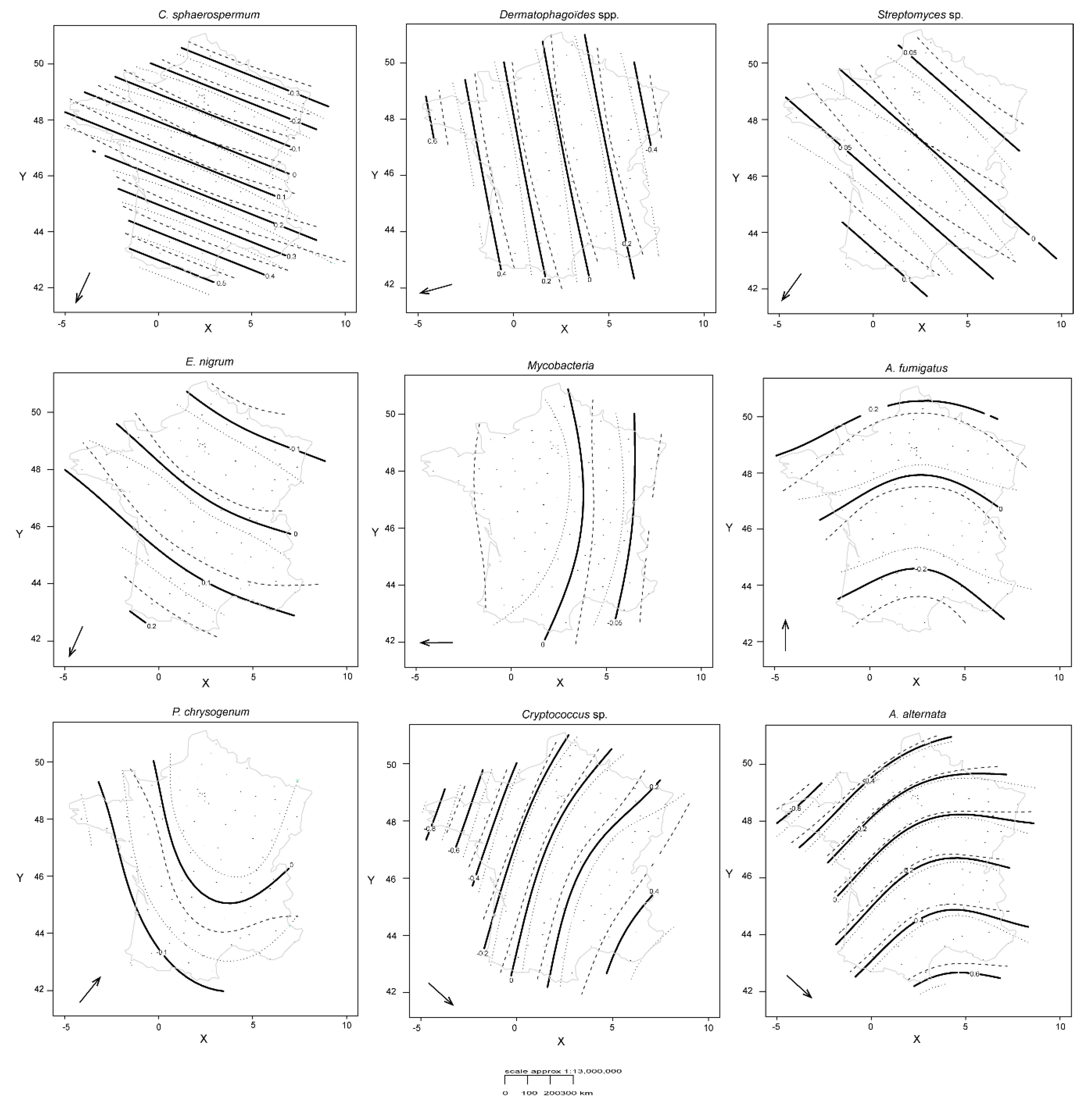
In GAMMs, the consideration of sampling month as a random effect significantly improved model predictions (reduction in the AIC value). Among the 17 qPCR targets, 10 exhibited significant spatial variability, although the amount of variability explained by the spatial structure remained rather restricted (Table 3). Non-linear patterns were found for some targets (Figure 2), especially for A. alternata (with greater effective degrees of freedom, edf, in Table 3). The higher the edf, the more non-linear the smoothing spline is.
Although some microorganisms showed distribution gradients in the same geographical direction, the correlation analysis (Pearson coefficient) was not significant. Five microorganisms had northeast to southwest increasing abundance profiles (on a line from Reims to Bordeaux): D. pteronyssinus and whole Dermatophagoïdes spp. (260° SW), Streptomyces spp. (220° SW), C. sphaerospermum (200° SW), and E. nigrum (200° SW). The mean of the azimuth of this group was 230° SW. Mycobacteria had an east to west increasing profile (270° W), A. fumigatus showed an increasing gradient from south to north (0° N), and a south to northeast gradient was found for P. chrysogenum (40° NE). Cryptococcus spp. and A. alternata exhibited an increasing gradient from northwest to southeast (130° SE) (a line from Brest to Nice). The seven other microorganisms (S. chartarum, A. versicolor, T. viride, D. farinea, A. siro, Enterobacteriaceae) appeared to be evenly distributed throughout the country.
For microorganisms that have the same gradient direction (C. sphaerospermum and E. nigrum), spatial patterns of microorganisms showed nuances that were more or less fine (smoother contours and confidence intervals quite narrow in Figure 2).

3.3. Climate and Biophysical Land Use for Each Individual Target

Twenty-two variables (8/8 for climate and 14/15 for land use) were considered linked to the qPCR targets that showed spatial variability in the previous section. Table 4 presents the results of the backward selection and the direction of the relationship (positive or negative) for each individual target. The coastal wetland factor was not selected in any model.
Some qPCR targets were associated with temperature parameters: some were linked to the number of warm days, such as C. sphaerospermum, with a positive relation to the number of days >30 °C and a negative relation to the number of days < 5 °C. For others, such as A. alternata, distribution was linked to the average annual temperature and annual temperature range (difference between average temperature in July and January). In contrast, Dermatophagoïdes spp. were also linked to average annual temperature, but not to annual temperature range. Concerning precipitation, the number of rainfalls in January was the variable that was most frequently related to the distribution of microorganisms, with 5 positive (C. sphaerospermum, Streptomyces spp., Mycobacteria, A. fumigatus, P. chrysogenum) and 2 negative (Cryptococcus spp., A. alternata) relationships.
For biophysical land use, the prairie variable was the most frequently related to the distribution of microorganisms, with two positive (C. sphaerospermum, A. fumigatus) and seven negative (Dermatophagoïdes spp., Streptomyces spp., E. nigrum, Mycobacteria, P. chrysogenum, Cryptococcus spp., A. alternata) relationships. In contrast, mines, landfills, and construction sites were only positively related to A. fumigatus.

3.4. Microorganism Community

Partial redundancy analysis (pRDA) triplot, depicting the structuring effect of climate and biophysical land use on microorganism communities, is presented in Figure 3. Considered together, the month of samplings and departmental coordinates (X and Y) accounted for 16% of the dataset variance.
Among the 23 variables of climate and biophysical land use, 9 were selected by a forward selection model and were significant (p < 0.05), thus explaining a part of the microorganism distribution in dwellings. However, these variables could only account for a small fraction of the variance (1.6%). In Figure 3, explanatory variables constrain the first ordination axes in order to interpret the species matrix. Thus, from the observations that emerged from this analysis, E. nigrum was mainly associated with areas covered by forests, whereas C. sphaerospermum was linked to annual temperature range and scrub/herbaceous vegetation areas. Dermatophagoïdes spp. were related to the number of days of precipitation in January. These links were not the same in the linear mixed effects models when microorganisms were analyzed individually and not included in a community. More generally, it was as if communities were constrained with a gradient ranging from closed and rather cold and humid landscapes (forests) (on the left side of the graph in Figure 3) to more open, hot, and dry landscapes (herbaceous and coastal).

4. Discussion

The present study addressed the biogeography of indoor microbial exposure, providing an assessment of the microbial composition of French dwellings in terms of molds, bacteria, and dust mites using a single standardized quantification method (qPCR). Given the importance of seasonality on the distribution of microorganisms, the purpose of this paper was to be able to assess the respective weight of geographical location by taking into account the month effect by means of statistical processing (GAMMs and pRDA) and then to evaluate the influence of, probably, the two most important parameters (climate and land cover). Analysis showed that the geographical location of dwellings induces variations in the quantification of different microbial communities (10 out of 17 microorganisms), which is partly linked to patterns of biogeography and climate.
While linear regression is an important tool for statistical analysis, it may be interesting to look at other methods that may be more adapted to biology/ecology, which are areas that do not necessarily have to follow linear relationships. To assess the distribution of each microorganism individually, we used GAMMs, which allowed us to model nested data and spatio-temporal correlation structures in count data and is a popular approach to modeling complex structures in ecological data or pollution studies [49].
We then performed a redundancy analysis, commonly used to simultaneously analyze the effects of multiple environmental factors on multiple species. This made it possible to represent the most important and interpretable environmental gradients available and, more importantly, to provide an overall descriptive analysis, rather than multiple univariate analyses. In our pRDA, month of sampling and X and Y coordinates were considered as condition variables so that their influence was removed prior to assessing the effect of climate and land cover variables. This procedure examined the determinants of community variability once the temporal (month of sampling) and spatial (X and Y) variability were accounted for.
With the statistical analyses conducted, we demonstrated that, if we consider the individual microorganisms (linear models) or community of microorganisms (pRDA), the climatic and occupancy variables that are selected in the models and are linked to the distribution of the microorganism(s) are different.
Outdoor geographical clusters of microbial communities have already been documented on local, regional, and continental scales [14] and geography is also a major factor that influences indoor fungal communities [36]. Thus, we expected that some qPCR targets would be dependent on the geographic coordinates of the dwellings, such as Alternaria, Cladosporium, Penicillium, and Aspergillus, which are among the most common genera found indoors and were previously described as strongly influenced by meteorology. A recent publication, conducted on a European scale reported that outdoor concentrations of Alternaria and Cladosporium spores differed between sites and years of sampling [27]. With our work, we have eliminated the month of sampling effect by taking it into account in the models and have shown that, at the scale of France, we also observe differences in concentrations of Alternaria and Cladosporium according to geographic locations and this is the case within dwellings as well. In contrast, geographical gradients were less predictable for other targets, but this is probably due to our “mistaken” vision of reality that is linked to cultivation methods. For example, the Epicoccum genus is rarely found in studies that use culture as a means of studying fungal exposure, particularly due to difficulties with culture and sporulation on classical media. However, this genus is increasingly reported in studies, including targeted metabarcoding approaches, that place it in first place among fungi [35,40,50,51,52]. Another advantage of using biomolecular methods, and particularly qPCR, is the ability to quantify different types of microorganisms with the same technique without competition between microorganisms (as in the case of culture investigations). Moreover, qPCR takes into account live and dead cells, which represent a significant part of fungal exposure, also influencing immunological responses [53,54]. We were thus able to show that there were different concentration gradients in a given geographical area for yeasts (Cryptococcus spp.), bacteria (Mycobacteria and Streptomyces spp.), and HDM (Dermatophagoïdes spp. and D. pteronyssinus).
Some climate parameters, land use, and/or vegetation cover have been linked to the geographical pattern of microorganisms in univariate analysis, but, above all, the pRDA allows us to place some targeted species on a scale of landscapes and climates (closed and rather cold and humid landscapes to more open, hot and dry landscapes). We have been working on land use and vegetation cover data collected for 2011. For climate, we selected 14 climate variables (6 temperature and 8 precipitation variables), defining 8 French climates.
Some microorganisms not highlighted by our statistical analysis (S. chartarum, A. versicolor, T. viride, D. farinea, A. siro, Enterobacteriaceae) are probably not or are less driven by the outdoor parameters we studied. They can be driven by other outdoor factors (wind or vapor pressure [27]) or maybe by indoor characteristics, such as thermo-isolation and ventilation, infiltration, and water damage, but also occupants (pets, humans, plants), the type of use of the indoor location, architecture, and materials [1,55,56,57]. For example, it would not be surprising that some microorganisms, such as S. chartarum, described as linked to water damage [58] or A. siro, found in the foodstuffs in our kitchens [59], are not related to the landscape cover type.
Given the information mentioned here, the complexity of exposure and the influence of the various factors, which are probably interconnected, it seems difficult to reason in terms of exposure to a single microorganism when we want to address the issue of allergic diseases. Above all, nothing is single-factorial. On the other hand, as long as we use qualitative methods to measure the presence of microorganisms, we can certainly believe that “everything is everywhere”. However, quantification methods show us that we are dealing with various concentrations in different environments. Thus, to assess an allergic risk related to microbial exposure, it would undoubtedly be useful to build “composite” indices, mixing exposure measurements, environmental factors (geography, climate, vegetation), and housing factors (humidity, temperature, ventilation).

Author Contributions

L.M., G.R., S.R., E.S., B.L. and C.R. participated in the conceptualization of the study and funding acquisition. C.Z. and M.-N.D. did the logistics (distribution of the electrostatics dust collectors, EDCs) and allowed access to the data. S.R., A.R., A.L. and B.V. make EDCs analysis. S.R. and B.V. made statistical analysis and the figures. L.M., G.R., S.K., E.S., B.L. and C.R. validated analysis. S.R., G.R. and L.M. wrote the manuscript. S.R., G.R., L.M., E.S., B.V., B.L. and C.R. revised the manuscript. All authors have read and agreed to the published version of the manuscript.

Funding

This work was support by the French National Research Agency (ANR) (EMBRASE ANR17-CE34-0002-01).

Acknowledgments

We are grateful to Victor Frossard (Savoie Mont-Blanc University, CARTEL Laboratory) for his help with generalized additive mixed models. We also thank Pamela Albert for her editorial assistance.

Conflicts of Interest

The authors declare no conflict of interest.

References

  1. Kirjavainen, P.V.; Hyytiäinen, H.; Täubel, M. The environmental microbiota and asthma. In The Lung Microbiome (ERS Monograph); Cox, M.J., Ege, M.J., von Mutius, E., Eds.; European Respiratory Society: Lausanne, Switzerland, 2019; pp. 216–239. [Google Scholar] [CrossRef]
  2. von Mutius, E. Allergies, infections and the hygiene hypothesis—The epidemiological evidence. Immunobiology 2007, 212, 433–439. [Google Scholar] [CrossRef] [PubMed]
  3. Tischer, C.G.; Hohmann, C.; Thiering, E.; Herbarth, O.; Muller, A.; Henderson, J.; Granell, R.; Fantini, M.P.; Luciano, L.; Bergstrom, A.; et al. Meta-analysis of mould and dampness exposure on asthma and allergy in eight European birth cohorts: An ENRIECO initiative. Allergy 2011, 66, 1570–1579. [Google Scholar] [CrossRef] [PubMed]
  4. Douwes, J.; Thorne, P.; Pearce, N.; Heederik, D. Bioaerosol Health Effects and Exposure Assessment: Progress and Prospects. Ann. Occup. Hyg. 2003, 47, 187–200. [Google Scholar] [CrossRef] [PubMed] [Green Version]
  5. Platts-Mills, T.A.; Vervloet, D.; Thomas, W.R.; Aalberse, R.C.; Chapman, M.D. Indoor allergens and asthma: Report of the Third International Workshop. J. Allergy Clin. Immunol. 1997, 100, S2–S24. [Google Scholar] [CrossRef]
  6. Kanchongkittiphon, W.; Mendell, M.J.; Gaffin, J.M.; Wang, G.; Phipatanakul, W. Indoor environmental exposures and exacerbation of asthma: An update to the 2000 review by the Institute of Medicine. Environ. Health Perspect. 2015, 123, 6–20. [Google Scholar] [CrossRef] [Green Version]
  7. Martin, L.J.; Adams, R.I.; Bateman, A.; Bik, H.M.; Hawks, J.; Hird, S.M.; Hughes, D.; Kembel, S.W.; Kinney, K.; Kolokotronis, S.-O.; et al. Evolution of the indoor biome. Trends Ecol. Evol. 2015, 30, 223–232. [Google Scholar] [CrossRef]
  8. Gibbons, S.M. The Built Environment Is a Microbial Wasteland. mSystems 2016, 1, e00033-16. [Google Scholar] [CrossRef] [Green Version]
  9. Horve, P.F.; Lloyd, S.; Mhuireach, G.A.; Dietz, L.; Fretz, M.; MacCrone, G.; Van Den Wymelenberg, K.; Ishaq, S.L. Building upon current knowledge and techniques of indoor microbiology to construct the next era of theory into microorganisms, health, and the built environment. J. Expo. Sci. Environ. Epidemiol. 2019, 30, 219–235. [Google Scholar] [CrossRef] [Green Version]
  10. Amato, P.; Joly, M.; Besaury, L.; Oudart, A.; Taib, N.; Mone, A.I.; Deguillaume, L.; Delort, A.M.; Debroas, D. Active microorganisms thrive among extremely diverse communities in cloud water. PLoS ONE 2017, 12, e0182869. [Google Scholar] [CrossRef] [Green Version]
  11. Golan, J.J.; Pringle, A. Long-Distance Dispersal of Fungi. Microbiol. Spectr. 2017, 5. [Google Scholar] [CrossRef] [Green Version]
  12. Prospero, J.M.; Blades, E.; Mathison, G.; Naidu, R. Interhemispheric transport of viable fungi and bacteria from Africa to the Caribbean with soil dust. Aerobiologia 2005, 21, 1–19. [Google Scholar] [CrossRef] [Green Version]
  13. Brown, J.K.; Hovmoller, M.S. Aerial dispersal of pathogens on the global and continental scales and its impact on plant disease. Science 2002, 297, 537–541. [Google Scholar] [CrossRef] [PubMed] [Green Version]
  14. Leung, M.H.; Lee, P.K. The roles of the outdoors and occupants in contributing to a potential pan-microbiome of the built environment: A review. Microbiome 2016, 4, 21. [Google Scholar] [CrossRef] [PubMed] [Green Version]
  15. Pitkäranta, M.; Meklin, T.; Hyvärinen, A.; Paulin, L.; Auvinen, P.; Nevalainen, A.; Rintala, H. Analysis of fungal flora in indoor dust by ribosomal DNA sequence analysis, quantitative PCR, and culture. Appl. Environ. Microbiol. 2008, 74, 233–244. [Google Scholar] [CrossRef] [PubMed] [Green Version]
  16. Bowers, R.M.; McLetchie, S.; Knight, R.; Fierer, N. Spatial variability in airborne bacterial communities across land-use types and their relationship to the bacterial communities of potential source environments. ISME J. 2010, 5, 601. [Google Scholar] [CrossRef] [PubMed] [Green Version]
  17. Weikl, F.; Tischer, C.; Probst, A.J.; Heinrich, J.; Markevych, I.; Jochner, S.; Pritsch, K. Fungal and bacterial communities in indoor dust follow different environmental determinants. PLoS ONE 2016, 11, e0154131. [Google Scholar] [CrossRef] [PubMed]
  18. Grantham, N.S.; Reich, B.J.; Pacifici, K.; Laber, E.B.; Menninger, H.L.; Henley, J.B.; Barberan, A.; Leff, J.W.; Fierer, N.; Dunn, R.R. Fungi identify the geographic origin of dust samples. PLoS ONE 2015, 10, e0122605. [Google Scholar] [CrossRef]
  19. Barberan, A.; Ladau, J.; Leff, J.W.; Pollard, K.S.; Menninger, H.L.; Dunn, R.R.; Fierer, N. Continental-scale distributions of dust-associated bacteria and fungi. Proc. Natl. Acad. Sci. USA 2015, 112, 5756–5761. [Google Scholar] [CrossRef] [Green Version]
  20. Vesper, S.; Wakefield, J.; Ashley, P.; Cox, D.; Dewalt, G.; Friedman, W. Geographic distribution of Environmental Relative Moldiness Index molds in USA homes. J. Environ. Public Health 2011, 2011, 242457. [Google Scholar] [CrossRef]
  21. Shelton, B.G.; Kirkland, K.H.; Flanders, W.D.; Morris, G.K. Profiles of airborne fungi in buildings and outdoor environments in the United States. Appl. Environ. Microbiol. 2002, 68, 1743–1753. [Google Scholar] [CrossRef] [Green Version]
  22. Pakpour, S.; Li, D.-W.; Klironomos, J. Relationships of fungal spore concentrations in the air and meteorological factors. Fungal Ecol. 2015, 13, 130–134. [Google Scholar] [CrossRef]
  23. Wang, J.; Chen, C.; Ye, Z.; Li, J.; Feng, Y.; Lu, Q. Relationships between fungal and plant communities differ between desert and grassland in a typical dryland region of northwest China. Front. Microbiol. 2018, 9, 2327. [Google Scholar] [CrossRef] [PubMed]
  24. Tedersoo, L.; Bahram, M.; Polme, S.; Koljalg, U.; Yorou, N.S.; Wijesundera, R.; Villarreal Ruiz, L.; Vasco-Palacios, A.M.; Thu, P.Q.; Suija, A.; et al. Fungal biogeography. Global diversity and geography of soil fungi. Science 2014, 346, 1256688. [Google Scholar] [CrossRef] [PubMed] [Green Version]
  25. Knorr, K.; Jorgensen, L.N.; Nicolaisen, M. Fungicides have complex effects on the wheat phyllosphere mycobiome. PLoS ONE 2019, 14, e0213176. [Google Scholar] [CrossRef] [PubMed] [Green Version]
  26. Pellissier, L.; Oppliger, A.; Hirzel, A.H.; Savova-Bianchi, D.; Mbayo, G.; Mascher, F.; Kellenberger, S.; Niculita-Hirzel, H. Airborne and Grain Dust Fungal Community Compositions Are Shaped Regionally by Plant Genotypes and Farming Practices. Appl. Environ. Microbiol. 2016, 82, 2121–2131. [Google Scholar] [CrossRef] [PubMed] [Green Version]
  27. Grinn-Gofron, A.; Nowosad, J.; Bosiacka, B.; Camacho, I.; Pashley, C.; Belmonte, J.; De Linares, C.; Ianovici, N.; Manzano, J.M.M.; Sadys, M.; et al. Airborne Alternaria and Cladosporium fungal spores in Europe: Forecasting possibilities and relationships with meteorological parameters. Sci. Total Environ. 2019, 653, 938–946. [Google Scholar] [CrossRef]
  28. Crawford, J.A.; Rosenbaum, P.F.; Anagnost, S.E.; Hunt, A.; Abraham, J.L. Indicators of airborne fungal concentrations in urban homes: Understanding the conditions that affect indoor fungal exposures. Sci. Total Environ. 2015, 517, 113–124. [Google Scholar] [CrossRef]
  29. Salonen, H.; Duchaine, C.; Mazaheri, M.; Clifford, S.; Lappalainen, S.; Reijula, K.; Morawska, L. Airborne viable fungi in school environments in different climatic regions—A review. Atmos. Environ. 2015, 104, 186–194. [Google Scholar] [CrossRef] [Green Version]
  30. Adams, R.I.; Miletto, M.; Lindow, S.E.; Taylor, J.W.; Bruns, T.D. Airborne bacterial communities in residences: Similarities and differences with fungi. PLoS ONE 2014, 9, e91283. [Google Scholar] [CrossRef]
  31. Caillaud, D.; Cheriaux, M.; Martin, S.; Segala, C.; Dupuy, N.; Evrard, B.; Thibaudon, M. Short-term effect of outdoor mould spore exposure on prescribed allergy medication sales in Central France. Clin. Exp. Allergy 2018, 48, 837–845. [Google Scholar] [CrossRef]
  32. Grewling, Ł.; Nowak, M.; Szymańska, A.; Kostecki, Ł.; Bogawski, P. Temporal variability in the allergenicity of airborne Alternaria spores. Med. Mycol. 2019, 57, 403–411. [Google Scholar] [CrossRef] [PubMed] [Green Version]
  33. Rocchi, S.; Reboux, G.; Frossard, V.; Scherer, E.; Valot, B.; Laboissiere, A.; Zaros, C.; Vacheyrou, M.; Gillet, F.; Roussel, S.; et al. Microbiological characterization of 3193 French dwellings of Elfe cohort children. Sci. Total Environ. 2015, 505, 1026–1035. [Google Scholar] [CrossRef] [PubMed]
  34. Reboux, G.; Valot, B.; Rocchi, S.; Scherer, E.; Roussel, S.; Millon, L. Storage mite concentrations are underestimated compared to house dust mite concentrations. Exp. Appl. Acarol. 2019, 77, 511–525. [Google Scholar] [CrossRef] [PubMed]
  35. Dannemiller, K.C.; Mendell, M.J.; Macher, J.M.; Kumagai, K.; Bradman, A.; Holland, N.; Harley, K.; Eskenazi, B.; Peccia, J. Next-generation DNA sequencing reveals that low fungal diversity in house dust is associated with childhood asthma development. Indoor Air 2014, 24, 236–247. [Google Scholar] [CrossRef] [Green Version]
  36. Nevalainen, A.; Täubel, M.; Hyvärinen, A. Indoor fungi: Companions and contaminants. Indoor Air 2015, 25, 125–156. [Google Scholar] [CrossRef]
  37. Amend, A.S.; Seifert, K.A.; Samson, R.; Bruns, T.D. Indoor fungal composition is geographically patterned and more diverse in temperate zones than in the tropics. Proc. Natl. Acad. Sci. USA 2010, 107, 13748–13753. [Google Scholar] [CrossRef] [Green Version]
  38. Chase, J.; Fouquier, J.; Zare, M.; Sonderegger, D.L.; Knight, R.; Kelley, S.T.; Siegel, J.; Caporaso, J.G. Geography and location are the primary drivers of office microbiome composition. mSystems 2016, 1, e00022-16. [Google Scholar] [CrossRef] [Green Version]
  39. Barrera, C.; Rocchi, S.; Degano, B.; Soumagne, T.; Laurent, L.; Bellanger, A.P.; Laplante, J.J.; Millon, L.; Dalphin, J.C.; Reboux, G. Microbial exposure to dairy farmers’ dwellings and COPD occurrence. Int. J. Environ. Health Res. 2019, 29, 387–399. [Google Scholar] [CrossRef]
  40. Rocchi, S.; Valot, B.; Reboux, G.; Millon, L. DNA metabarcoding to assess indoor fungal communities: Electrostatic dust collectors and Illumina sequencing. J. Microbiol. Methods 2017, 139, 107–112. [Google Scholar] [CrossRef]
  41. EPA. Microbiological and Chemical Exposure Assessment, EPA Technology for Mold Identification and Enumeration. Available online: https://www.google.com/url?sa=t&rct=j&q=&esrc=s&source=web&cd=1&ved=2ahUKEwiCpMX5tvXjAhURfxoKHa4fCJwQFjAAegQIARAC&url=https%3A%2F%2Firp-cdn.multiscreensite.com%2Fc4e267ab%2Ffiles%2Fuploaded%2FgCQnkBNWQuSD96fPIikY_EPA_Technology%2520for%2520Mold%2520Identification%2520and%2520Enumeration.pdf&usg=AOvVaw0hz56KTQnKxxabVWqzeW3t (accessed on 20 August 2019).
  42. Sen, K.; Asher, D.M. Multiplex PCR for detection of Enterobacteriaceae in blood. Transfusion 2001, 41, 1356–1364. [Google Scholar] [CrossRef]
  43. Torvinen, E.; Torkko, P.; Rintala, A.N. Real-time PCR detection of environmental mycobacteria in house dust. J. Microbiol. Methods 2010, 82, 78–84. [Google Scholar] [CrossRef] [PubMed]
  44. Rintala, H.; Nevalainen, A. Quantitative measurement of streptomycetes using real-time PCR. J. Environ/ Monitor. 2006, 8, 745–749. [Google Scholar] [CrossRef] [PubMed]
  45. R Development Core Team. R: A Language and Environment for Statistical Computing; R Foundation for Statistical Computing: Vienna, Austria, 2017. [Google Scholar]
  46. Pinheiro, J.; Bates, D.; DebRoy, S.; Sarkar, D.; Heisterkamp, S.; van Willigen, B.; R-Core Team. Nlme: Linear and Nonlinear Mixed Effects Models; R Package Version 3.1-122. Available online: https://cran.r-project.org/web/packages/nlme/nlme.pdf (accessed on 2 February 2020).
  47. Wood, S. Generalized Additive Models: An Introduction with R; CRC Press: Boca Raton, FL, USA, 2006. [Google Scholar]
  48. Becker, R.A.; Wilks, A.R. Mapdata: Extra Map Databases; R Package Version 2.2-2. Available online: https://cran.r-project.org/web/packages/mapdata/index.html (accessed on 20 August 2018).
  49. Zuur, A.F.; Ieno, E.N.; Walker, N.J.; Saveliev, A.A.; Smith, G.M. Mixed Effects Models and Extensions in Ecology with R; Springer: Berlin/Heidelberg, Germany, 2009. [Google Scholar]
  50. Adams, R.I.; Miletto, M.; Taylor, J.W.; Bruns, T.D. Dispersal in microbes: Fungi in indoor air are dominated by outdoor air and show dispersal limitation at short distances. ISME J. 2013, 7, 1262–1273. [Google Scholar] [CrossRef] [PubMed] [Green Version]
  51. Adams, R.I.; Amend, A.S.; Taylor, J.W.; Bruns, T.D. A unique signal distorts the perception of species richness and composition in high-throughput sequencing surveys of microbial communities: A case study of fungi in indoor dust. Microb. Ecol. 2013, 66, 735–741. [Google Scholar] [CrossRef] [Green Version]
  52. Amend, A.S.; Seifert, K.A.; Bruns, T.D. Quantifying microbial communities with 454 pyrosequencing: Does read abundance count? Mol. Ecol. 2010, 19, 5555–5565. [Google Scholar] [CrossRef]
  53. Adhikari, A.; Reponen, T.; Rylander, R. Airborne fungal cell fragments in homes in relation to total fungal biomass. Indoor Air 2013, 23, 142–147. [Google Scholar] [CrossRef]
  54. Reponen, T.; Seo, S.C.; Grimsley, F.; Lee, T.; Crawford, C.; Grinshpun, S.A. Fungal fragments in moldy houses: A field study in homes in New Orleans and Southern Ohio. Atmos. Environ. 2007, 41, 8140–8149. [Google Scholar] [CrossRef] [Green Version]
  55. Madsen, A.M.; Matthiesen, C.B.; Frederiksen, M.W.; Frederiksen, M.; Frankel, M.; Spilak, M.; Gunnarsen, L.; Timm, M. Sampling, extraction and measurement of bacteria, endotoxin, fungi and inflammatory potential of settling indoor dust. J. Environ. Monitor. 2012, 14, 3230–3239. [Google Scholar] [CrossRef]
  56. Sordillo, J.E.; Alwis, U.K.; Hoffman, E.; Gold, D.R.; Milton, D.K. Home characteristics as predictors of bacterial and fungal microbial biomarkers in house dust. Environ. Health Perspect. 2011, 119, 189–195. [Google Scholar] [CrossRef] [Green Version]
  57. Adams, R.I.; Bhangar, S.; Dannemiller, K.C.; Eisen, J.A.; Fierer, N.; Gilbert, J.A.; Green, J.L.; Marr, L.C.; Miller, S.L.; Siegel, J.A.; et al. Ten questions concerning the microbiomes of buildings. Build. Environ. 2016, 109, 224–234. [Google Scholar] [CrossRef] [Green Version]
  58. Fog Nielsen, K. Mycotoxin production by indoor molds. Fungal Genet. Biol. 2003, 39, 103–117. [Google Scholar] [CrossRef]
  59. Thind, B.B.; Clarke, P.G. The occurrence of mites in cereal-based foods destined for human consumption and possible consequences of infestation. Exp. Appl. Acarol. 2001, 25, 203–215. [Google Scholar] [CrossRef] [PubMed]
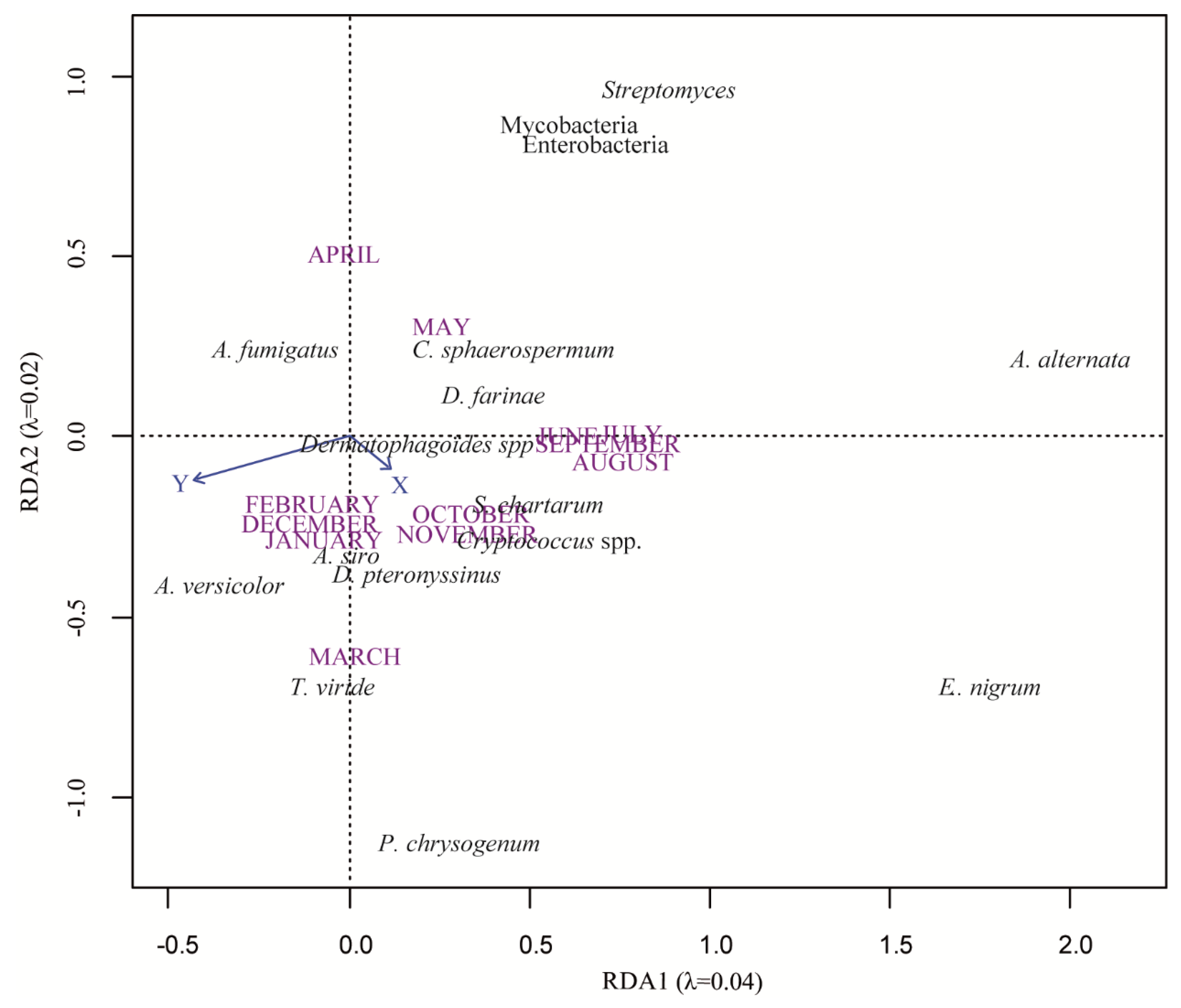
Figure 1. Redundancy analysis (RDA) plot showing target microorganisms and spatio-temporal (X and Y coordinates and months of samplings) distribution. Microorganisms are in black, coordinate variables in blue, and months in purple. Solid arrows indicate quantitative variables (X: longitude and Y: latitude). Months of samplings are qualitative variables and are represented without arrows.
Microorganisms 08 00341 g001
Figure 2. Spatial patterns for log-transformed copy number of qPCR targets for nine microorganisms exhibiting significant spatial variability. As Dermatophagoïdes spp. and D. pteronyssinus targets showed the same distributions, only one of the two is represented. Solid lines represent smoother contours (smoothing component f(Xi,Yi), with X: longitude and Y: latitude) and the dotted lines are 95% confidence bands. Arrows indicate the direction of the abundance gradient from lower to higher abundances.
Microorganisms 08 00341 g002
Figure 3. Partial redundancy analysis (pRDA) triplot showing target microorganisms and model selected climate and landscape variables. Microorganisms are in black, temperature variables in orange, precipitations variables in blue, and land cover in green.
Microorganisms 08 00341 g003
Table 1. Primers and probes sequences (and their references) used in this study.
Targets and Designers [Reference](5’–3’) Sequences
MoldsAlternaria alternata
designed by EPA [41]
Forward primerGGCGGGCTGGAACCTC
Reverse primerGCAATTACAAAAGGTTTATGTTTGTCGTA
ProbeTTACAGCCTTGCTGAATTATTCACCCTTGTCTTT
Aspergillus fumigatus
designed by EPA [41]
Forward primerGCCCGCCGTTTCGAC
Reverse primerCCGTTGTTGAAAGTTTTAACTGATTAC
ProbeCCCGCCGAAGACCCCAACATG
Aspergillus versicolor
designed by EPA [41]
Forward primerCGGCGGGGAGCCCT
Reverse primerCCATTGTTGAAAGTTTTGAcTGATCTTA
ProbeAGACTGCATCACTCTCAGGCATGAAGTTCAG
Cladosporium sphaerospermum
designed by EPA [41]
Forward primerACCGGCTGGGTCTTTCG
Reverse primerGGGGTTGTTTTACGGCGTG
ProbeCCCGCGGCACCCTTTAGCGA
Epicoccum nigrum
designed by EPA [41] #
Forward primerTTGTAGACTTCGGTCTGCTACCTCTT
Reverse primerTGCAACTGCAAAGGGTTTGAAT
ProbeCATGTCTTTTGAGTACCTTCGTTTCCTCGGC
Penicillium chrysogenum
Modified from EPA [40]
Forward primerTGCCTGTCCGAGCGTCATT
Reverse primerCCCCCGGGATCGGAG
ProbeCCAACACACAAGCCGTGCTTGAGG
Stachybotrys chartarum
designed by EPA [41]
Forward primerTCCCAAACCCTTATGTGAACC
Reverse primerGTTTGCCACTCAGAGAATACTGAAA
ProbeCTGCGCCCGGATCCAGGC
Trichoderma viride
designed by EPA [41] #
Forward primerCCCAAACCCAATGTGAACCA
Reverse primerTCCGCGAGGGGACTACAG
ProbeCCAAACTGTTGCCTCGGCGGG
Chaetomium globosum
designed by EPA [41] #
Forward primerCCGCAGGCCCTGAAAAG
Reverse primerCGCGGCGCGACCA
ProbeAGATGTATGCTACTACGCTCGGTGCGACAG
YeastsCryptococcus spp.
designed for this study #
Forward primerCCTGCGGAAGGATCATTAATG
Reverse primerGCACAGGTGTTATGGATATGATGTG
ProbeTTGACCGTCTGTCGAGCTTGCTCACA
MitesAcarus siro
Designed by our team [34] #
Forward primerCGCAAACTGTGGTGCGAGTA
Reverse primerGCTCCTTGGTCCGTGTTTCA
ProbeTCGGTCTCCACCCGACCCGTC
Dermatophagoïdes spp.
Designed by our team [34]
Forward primerTGTTGTGGTTAAAAAGCTCGTAGTTG
Reverse primerATGCGATAATCTGCTCAGTATGACA
ProbeCAGCTCATGTATGGCGGTCCACCTG
Dermatophagoïdes farinea
Designed by our team [34] #
Forward primerCACACATTCAACCAGAGTGGTACTT
Reverse primerGGCTAACACTCCCCCTAGTTTAGA
ProbeCGCTTACGCGATCCTACGAGCCATT
Dermatophagoïdes pteronyssinus
Designed by our team [34] #
Forward primerCATCCAACCAGAGTGGTATTTCC
Reverse primerGCTATTGCGCATACTCCACCTA
ProbeTATGCAATCCTTCGGGCTATCCCATCA
BacteriaEnterobacteriaceae
designed by Sen and Asher [42]
Forward primerGGCGGCAGGCCTAAC
Reverse primerCAGGCAGTTTCCCAGACATTACT
ProbeAGCAAGCTCTCTGTGCTACCGCTCGA
Mycobacteria
designed by Torvinen et al. [43]
Forward primerGATGCAACGCGAAGAACCTT
Reverse primerTGCACCACCTGCACACAGG
ProbeCCTGGGTTTGACATGCACAGGACG
Streptomyces spp.
designed by Rintala and Nevalainen [44]
Forward primerGCCGATTGTGGTGAAGTGGA
Reverse primerGTACGGGCCGCCATGAAA
ProbeATCCTATGCTGTCGAGAAAAGCCTCTAGCG
EPA: Environmental Protection Agency. The seven additional qPCR targets to the 10 used in the first study published in 2015 [33] are notified by “#”.
Table 2. Positivity and quantification of each target by qPCR in the 3143 electrostatic dust collectors (EDC) analyzed.
qPCR TargetsNumber of Positive EDCsMedian Value (copy/µL)Max Value (copy/µL)
Fungi
(molds and yeasts)
E. nigrum27637639106
A. alternata27526632634
P. chrysogenum2660919169
C. sphaerospermum2195812162
A. versicolor230725671
A. fumigatus146805138
T. viride139001536
S. chartarum8480333
Cryptococcus spp.2314<17
BacteriaEnterobacteriaceae308613478163
Mycobacteria305323512511
Streptomyces2879515887
Dust mitesDermatophagoïdes spp.1505<197673
D. pteronyssinus190809657
D. farinae2192<11330
A. siro2300<1348
Table 3. Detailed output on the smoothers and parametric terms in the Generalized additive mixed models (GAMMs).
qPCR TargetspedfFadjR2
MoldsA. alternata<0.001 *2.954162.60.12
A. fumigatus<0.001 *2.710.540.00909
E. nigrum<0.001 *2.34510.850.0069
C. sphaerospermum<0.001 *259.590.0418
P. chrysogenum0.028 *2.7384.4720.00212
S. chartarum0.1222.8531.8080.00119
A. versicolor0.12622.0710.000853
T. viride0.79420.231−0.000452
YeastsCryptococcus spp.<0.001 *2.85242.880.0391
Dust mitesDermatophagoïdes spp.<0.001 *2.12239.510.0286
D. pteronyssinus<0.001 *215.770.00918
D. farinea0.6722.2480.44−0.000307
A. siro0.77920.249−0.000525
BacteriaEnterobacteriaceae0.122.251.8230.00139
Mycobacteria0.009 *2.6943.5440.00456
Streptomyces<0.001 *211.710.00851
p (p-value) is given for each GAMM with “*” when <0.05; edf: estimated degrees of freedom (can be viewed as an estimation of the strength of the smoothness considered in the models); F: F-statistics; adjR2: Adjusted R-squared.
Table 4. Climate and biophysical land use contributing to the distribution of microorganisms (linear mixed-effects models).
C. sphaerospermumDermatophagoïdes spp.StreptomycesE. nigrumMycobacteriaA. fumigatusP. chrysogenumCryptococcus spp.A. alternata
Annual average temperature ++ +
Number of days temperature < −5 °C ++
Number of days temperature >30 °C+ + +
Annual temperature range * + +
Total annual precipitation +
Number of precipitation days in January+ + +++
Number of precipitation days in July
Ratio between autumn ** and July precipitations
Urbanized areas +
Industrial or commercial areas +
Mines, landfills, and construction sites +
Artificial, non-agricultural green spaces+
Arable land+
Permanent crops+
Prairies++
Heterogeneous agricultural areas +
Forests+ +
Scrub and/or herbaceous vegetation associations+ +
Open spaces, with little or no vegetation +
Inland wetlands+
Inland waters+
Maritime waters
Positive relations are indicated by “+” and negative by “−”. Empty cells mean that the variable was removed from the model (ordiR2step function). * Annual temperature range measures the difference between the average temperature in July and January. **Autumn precipitations: September + October.