Brief Report

Evaluation of a kDNA-Based qPCR Assay for the Detection and Quantification of Old World Leishmania Species

1
Department of Biomolecular Sciences, University of Urbino “Carlo Bo”, 61029 Urbino, PU, Italy
2
National Reference Center for Leishmaniasis (C.Re.Na.L.), Istituto Zooprofilattico Sperimentale della Sicilia, 90129 Palermo, PU, Italy
*
Author to whom correspondence should be addressed.
Microorganisms 2020, 8(12), 2006; https://doi.org/10.3390/microorganisms8122006
Submission received: 8 September 2020 / Revised: 9 December 2020 / Accepted: 14 December 2020 / Published: 16 December 2020
(This article belongs to the Special Issue Rapid and Novel Diagnostics for Infectious Diseases)

Abstract

:
The parasite protozoan Leishmania, the causative agent of leishmaniasis, includes two subgenera of medical interest: Leishmania (Leishmania) and Leishmania (Viannia). Parasite species detection and characterization is crucial to choose treatment protocols and to monitor the disease evolution. Molecular approaches can speed up and simplify the diagnostic process. In particular, several molecular assays target the mitochondrial DNA minicircle network (kDNA) that characterizes the Leishmania genus. We previously proposed a qPCR assay targeting kDNA, followed by high resolution melt (HRM) analysis (qPCR-ML) to distinguish L. (L.) infantum and L. (L.) amazonensis from L. Viannia species. Successively, this assay has been integrated with other qPCR assays, to differentiate L. (L.) infantum, L. (L.) amazonensis and L. (L.) mexicana. In this work, we tested the applicability of our qPCR-ML assay on L. (L.) donovani, L. (L.) major, L. (L.) tropica and L. (L.) aethiopica, showing that the qPCR-ML assay can also amplify Old World species, different from L. (L.) infantum, with good quantification limits (1 × 10⁻⁴–1 × 10⁻⁶ ng/pcr tube). Moreover, we evaluated 11 L. (L.) infantum strains/isolates, evidencing the variability of the kDNA minicircle target molecules among the strains/isolates of the same species, and pointing out the possibility of quantification using different strains as reference. Taken together, these data account for the consideration of qPCR-ML as a quantitative pan-Leishmania assay.

1. Introduction

Leishmania spp. is a protozoan parasite widespread in Asia, Africa, Europe and America. The parasite is the etiological agent of leishmaniasis, one of the most important tropical diseases that encompasses a broad spectrum of clinical manifestations. The genus Leishmania ascribes two subgenera of medical interest: Leishmania (Leishmania) and Leishmania (Viannia) [1]. The subgenus Leishmania (Leishmania) includes species responsible for visceral leishmaniasis (VL) [L. (L.) donovani, L. (L.) infantum] and cutaneous leishmaniasis (CL) [L. (L.) infantum, L. (L.) major, L. (L.) tropica, L. (L.) aethiopica, L. (L.) mexicana, L. (L.) amazonensis], while the species belonging to the subgenus Leishmania (Viannia) are the etiological agents of CL and mucocutaneous leishmaniasis (MCL) [2]. However, a few Leishmania (Leishmania) species [i.e., L. (L.) infantum, L. (L.) major and L. (L.) tropica] have also been reported to be the etiological agent of MCL [3]. The diagnosis of leishmaniasis is based on a combination of clinical manifestations, epidemiological data and laboratory test (i.e., serological, parasitological or molecular methods). However, a real gold-standard method for human or veterinary leishmaniasis is still lacking [4]. Among the diagnostic methods, molecular assays allow to obtain fast results and high sensitivity. In particular, a number of PCR-based assays are targeting the mitochondrial DNA minicircle network (kDNA) that characterizes the Leishmania genus, ensuring very high sensitivity [5]. Previously, we proposed a qPCR assay targeting the conserved region of kDNA minicircles using primers MLF and MLR (qPCR-ML), followed by high resolution melt (HRM) analysis to detect and distinguish L. (Leishmania) from L. (Viannia) subgenus [6]. Successively, this assay has been integrated with other qPCR assays to differentiate L. (L.) infantum from L. (L.) amazonensis [7] and from L. (L.) mexicana [8]. These approaches have also been applied to Brazilian clinical samples, allowing to distinguish different species causing infection in the New World [9]. However, considering the Old World Leishmania species, the qPCR-ML assay has been tested only on L. (L.) infantum. In this work, we tested the applicability of our qPCR-ML assay in Old World Leishmania species different from L. (L.) infantum [i.e., L. (L.) donovani, L. (L.) major, L. (L.) tropica and L. (L.) aethiopica], in an attempt to test the pan-Leishmania potential of our approach. Moreover, we quantitatively evaluated the target sequence of qPCR-ML in different L. (L.) infantum strains and isolates to investigate the possibility of quantification in clinical applications.

2. Materials and Methods

2.1. Leishmania Strains, Clinical Isolates and Clinical Samples

Leishmania strains or isolates used in this study are listed in Table 1. The DNA from all strains and isolates was Chelex-purified from cultured promastigotes and was provided by the Istituto Zooprofilattico Sperimentale della Sicilia, OIE Reference Laboratory, National Reference Center for Leishmaniasis (C.Re.Na.L.), (Palermo, Italy), with the following exceptions: L. (L.) donovani MHOM/IN/02/BPK282/0cl4, provided by the Istituto Zooprofilattico Sperimentale della Lombardia e dell’Emilia-Romagna (Modena, Italy); L. (V.) braziliensis MHOM/BR/1987/M11272, provided by Universidade Federal da Grande Dourados (Brazil); L. (L.) amazonensis MHOM/BR/1973/M2269, L. (L.) donovani MHOM/SD/62/1S, L. (L.) major SV39 and L. (S.) tarentolae Parrot Tar II, provided by Instituto de Investigação e Inovação em Saúde Universidade do Porto, Portugal. Moreover, the L. (V.) braziliensis clinical isolate AN1 was obtained from a pharyngo-laryngeal biopsy during routine diagnosis of a human patient with MCL. The human clinical samples Psalb and Daedio were obtained during routine diagnosis of two patients with VL (blood) [10] and CL (cutaneous biopsy), respectively. The DNA samples were quantified using a Qubit fluorometer 3 (Thermofisher, Carlsbad, CA, USA).

2.2. qPCR Assay

The qPCR assays were performed using MLF-MLR primer pair (MLF: 5′-CGTTCTGCGAAAACCGAAA-3′; MLR: 5′-CGGCCCTATTTTACACCAACC-3′) as previously reported [6]. Briefly, the reactions were carried out in 25 μL volume with 1 μL of template DNA and 24 μL TB Green Premix Ex TaqII Mastermix (Takara Bio Europe, France) containing 200 nM MLF and MLR primers. The reactions were performed in a Rotor-Gene 6000 instrument (Corbett Life Science, Mortlake, Australia). The cycle threshold (Cq) values were determined using the quantitation analysis of the Rotor-Gene 6000 software, setting a threshold to 0.15.
Standard curves were established using 1 × 100 to 1 × 10⁻⁷ ng DNA of L. (L.) major MHOM/SU/73/5ASKH, L. (L.) donovani MHOM/IN/80/DD8, L. (L.) aethiopica MHOM/ET/72/L100, L. (L.) tropica MHOM/SU/74/K27, L. (L.) infantum MHOM/FR/78/LEM75 and L. (S.) tarentolae Parrot Tar II. To evaluate the potential interference of host DNA as background in the qPCR assay, 30 ng of human DNA purified from human cell line (CACO-2 or MCF7) was spiked in each qPCR reaction tube (with the exception of L. (S.) tarentolae Parrot Tar II because not pathogenic to humans). The standard curves were obtained from two independent experiments for each strain, processed in duplicate.
To determine the variability of kDNA minicircle target sequences in L. (L.) infantum species, 0.02 ng DNA extracted from 11 L. (L.) infantum strains/isolates were amplified in 6 independent experiments and Cq were compared by Kruskal–Wallis nonparametric test.

2.3. High-Resolution Melt (HRM) Analysis

The qPCR-ML amplicons, obtained by all stains/isolates listed in Table 1, were analyzed by HRM protocol on a Rotor-Gene 6000 instrument as described previously [6] with slight modifications. Briefly, HRM was carried out over the range from 77 °C to 89 °C, rising at 0.1 °C/s and waiting for 2 s at each temperature. Each sample was run in duplicate and the gain was optimized before melting on all tubes. HRM curve analysis was performed with the derivative of the raw data, after smoothing, with the Rotor-Gene 6000 software. Only the samples with Cq values ˂ 30 were considered [11]. The HRM temperature average and the standard deviation derived from 3 independent experiments.

2.4. PCR Product Sequencing

The qPCR-ML products obtained from L. (L.) major MHOM/SU/73/5ASKH, L. (L.) major MRHO/SU/59/P-strain, L. (L.) donovani MHOM/IN/80/DD8, L. (L.) aethiopica MHOM/ET/72/L100, L. (L.) tropica MHOM/SU/74/K27, L. (L.) infantum MHOM/FR/78/LEM75, L. (V.) braziliensis MHOM/BR/75/M2904 were purified using the MinElute PCR purification kit (Qiagen) and directly sequenced, using both MLF and MLR primers, as previously described [6,7]. The DNA sequencing was performed using the BigDye Terminator v. 1.1 Cycle Sequencing Kit on ABI PRISM 310 Genetic Analyzer (Applied Biosystems, Foster City, CA, USA). Sequences were manually edited and nucleotide composition analyses were conducted using MEGA6 [12]. A phylogenetic tree based on the Maximum Likelihood method was constructed with 10,000 bootstrap replications, using the nearest neighbor interchange method in MEGA6 [12].

2.5. Statistical Analysis

To compare the variability on Cq values on L. (L.) infantum strains/isolates, a Kruskal–Wallis nonparametric test followed by Dunn’s multiple-comparison post-test was performed. To evaluate the different HRM temperatures between the Leishmania and Viannia subgenera, a Mann–Whitney test was performed. All statistical tests were performed using GraphPad Prism version 5 (GraphPad Software, Inc., La Jolla, CA, USA). A p value ≤ 0.05 was considered statistically significant.

3. Results

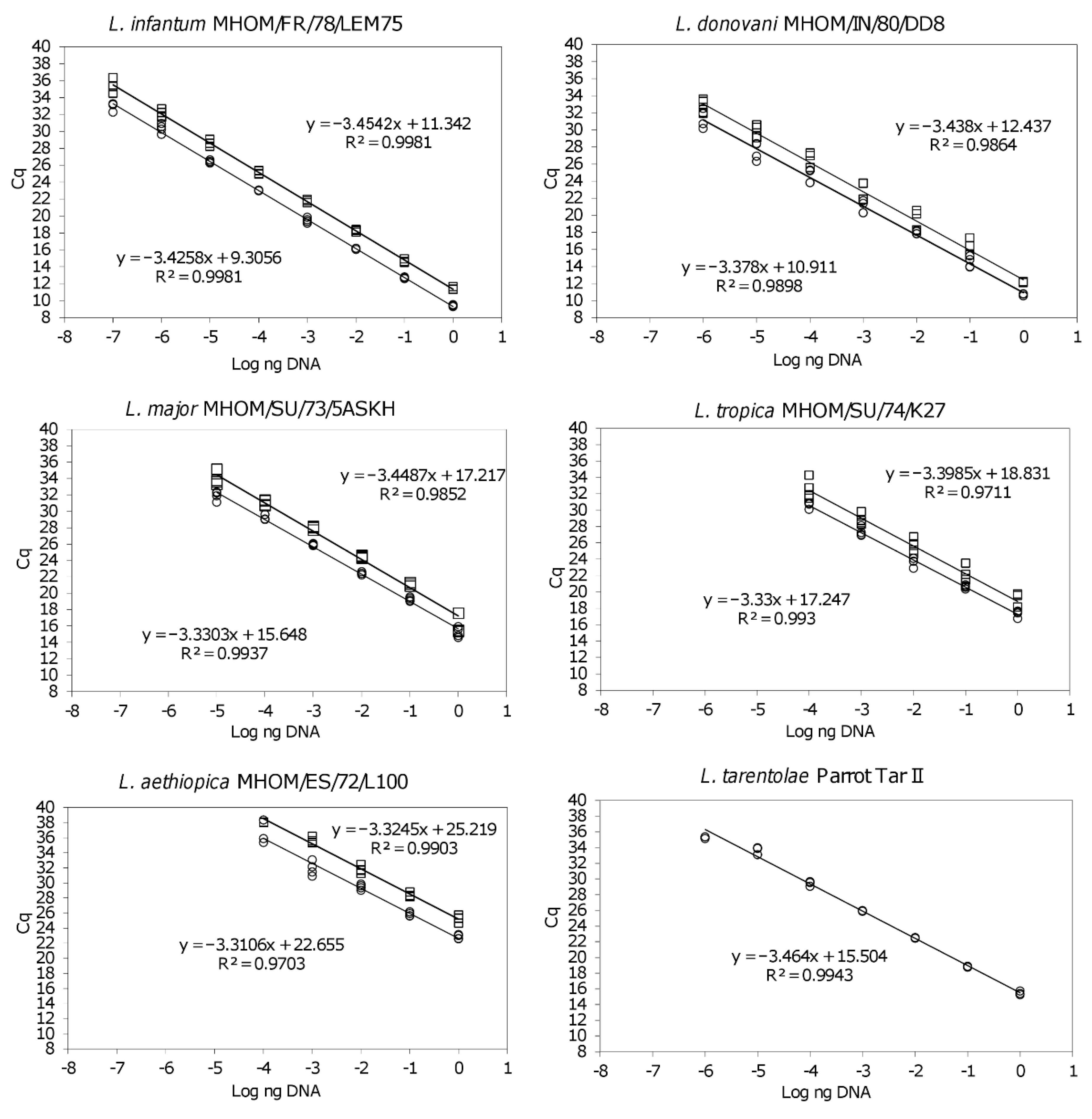
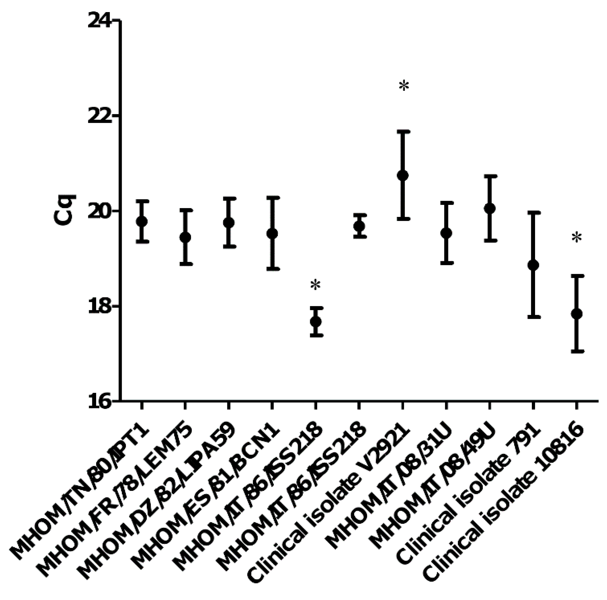
3.1. The qPCR-ML Assay Can Amplify All Medically Relevant Old World Leishmania Species

The qPCR-ML was applied to the strains listed in Table 1 to verify the amplifiability of all Leishmania species with MLF-MLR primers. All Old World species [i.e., L. (L.) infantum, L. (L.) donovani, L. (L.) major, L. (L.) tropica, L. (L.) aethiopica, L. (S.) tarentolae] were successfully amplified. The calibration curves evidenced a limit of quantification of 1 × 10⁻⁴ ng/PCR tube for L. (L.) tropica and L. (L.) aethiopica, 1 × 10⁻⁵ ng/PCR tube for L. (L.) major, 1 × 10⁻⁶ ng/PCR tube for L. (L.) donovani, 1 × 10⁻⁷ ng/PCR tube for L. (L.) infantum and 1 × 10⁻⁶ ng/PCR tube for L. (S.) tarentolae Parrot Tar II, with PCR efficiencies ≥ 94.5% and R2 ˃ 0.97 (Figure 1). Although out of linearity range, the limit of detection for L. (L.) donovani MHOM/IN/80/DD8 and L. (L.) tropica MHOM/SU/74/K27 were 1 × 10⁻⁷ (Cq 32.48 ± 0.46) and 1 × 10⁻⁵ (Cq 33.56 ± 1.14) ng/PCR tube, respectively. In the presence of human DNA as background, the Cq were slightly delayed, but the linearity and limit of quantification of the assay remained unchanged (Figure 1).
The variability of kDNA minicircle target sequence was investigated among 11 L. (L.) infantum strains by amplification of 0.02 ng promastigote DNA. Dunn’s post-test analysis of Cq values revealed significant differences among V2921 clinical isolate, MHOM/IT/86/ISS218 strain and clinical isolate 10816 (Figure 2). The average Cq was 19.36 ± 1.14, very close to the value obtained for the strain MHOM/FR/78/LEM75 (19.44 ± 0.56), suggesting that this strain could be used as reference to obtain approximate quantification values for illness caused by L. (L.) infantum.

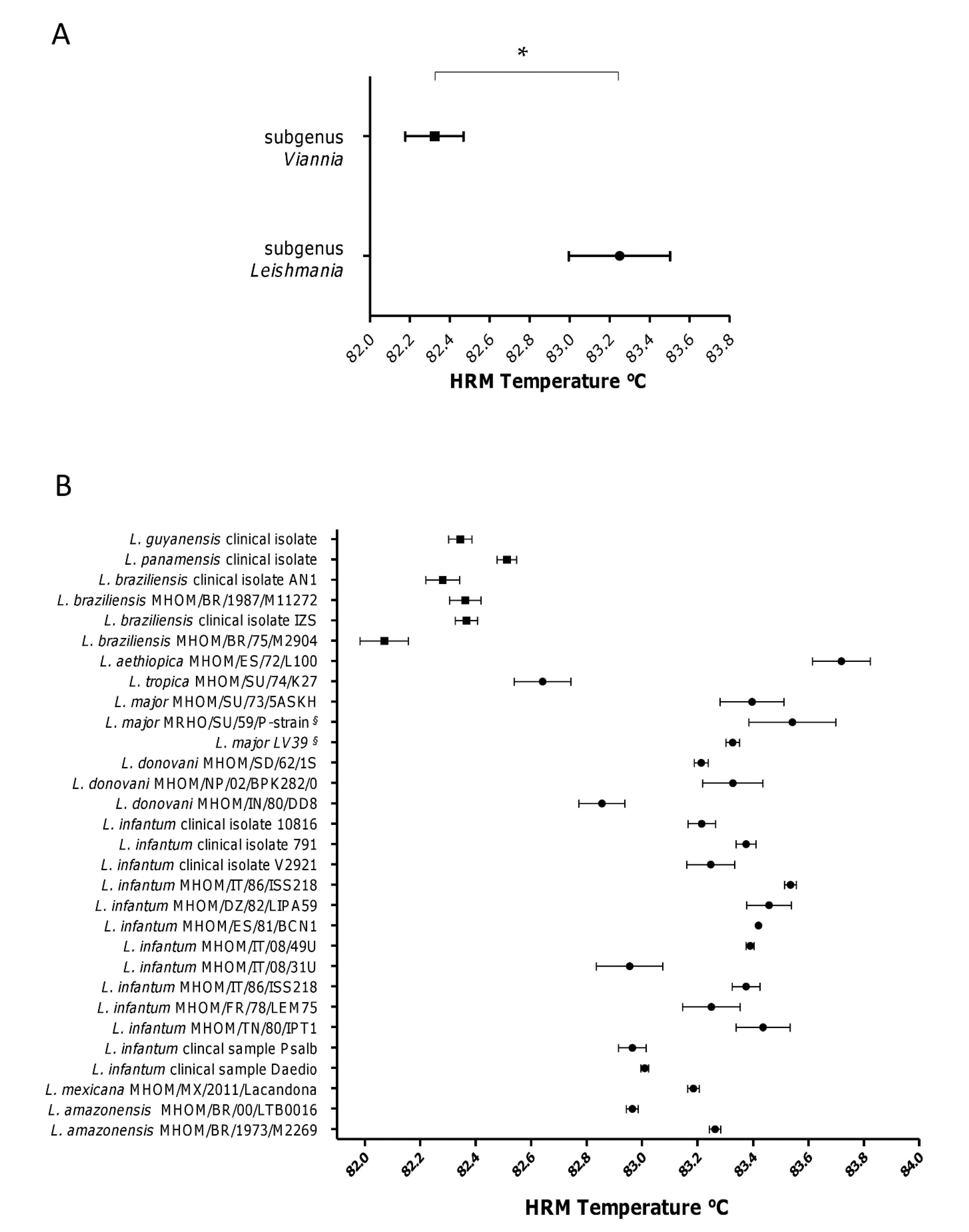
3.2. The subgenera Leishmania and Viannia Can Be Differentiated by HRM Analysis

Species discrimination power of qPCR-ML assay was also evaluated by analysis of HRM profiles on all Leishmania species listed in Table 1. Despite the impossibility to reliably discriminate each species using HRM profiles, the assay evidenced the lower Tm of amplicons belonging to Viannia subgenus, confirming the possibility to discriminate between Leishmania and Viannia subgenus, as previously demonstrated with a limited number of species [6] (Figure 3A). However, the Tm of L. (L.) tropica MHOM/SU/74/K27 amplicon showed a partial overlapping with L. (V.) panamensis, hence the two species were not fully distinguishable (Figure 3B). Accordingly, the specificity of this assay can be considered 96.6%. Nevertheless, this drawback can be overcome by considering the geographical origin of the sample since L. (L.) tropica is an Old World species. It is also noteworthy that L. (S.) tarentolae Parrot Tar II HRM temperature was 80.92 ± 0.08, clearly distinguishable from Viannia and Leishmania subgenera.
The sequence analysis of qPCR-ML products evidenced that the GC content found in L. Viannia strains was lower than that found in L. Leishmania strains (<48.2% and ˃48.6%, respectively), accounting for the lower Tm observed in the Viannia amplicons. In addition, a maximum likelihood phylogenetic tree of qPCR-ML amplicons was performed, confirming the clusterization in two groups of the amplicons obtained from the two subgenera, with the exception of L. (L.) tropica, which clusterizes independently (Figure S1).

4. Discussion

Although leishmaniasis is considered a neglected disease, it affects about 12 million people in Africa, Americas, Asia and Europe, with a heterogeneous clinical spectrum [13]. The diagnosis is complicated, as well as for the different species spread in the world. To improve the diagnostic process and classification, a number of molecular assays have been developed. In particular, qPCR techniques allow obtaining rapid parasitic load estimation and potential species discrimination through melting analysis. Due to their high copy number per cell, the kDNA minicircles are considered molecular targets that allow to design very sensitive assays [14]. However, due to the different composition of minicircle classes in the various species, an assay designed on a particular species can show lower sensitivity in another [7]. For this reason, many works describe minicircle-based qPCR assays targeting only few species or complex. For example, some authors developed qPCR assay for New World species [15,16], while some others tested their primers in Old World species [17,18]. Since different reports describe the infection in travelers [19,20] and in military personnel operating in country endemic for leishmaniasis [21,22,23], the availability of a rapid pan-Leishmania assay, highly sensitive and able to provide a quantification and a partial classification, can certainly be useful. Moreover, a pan-Leishmania assay can also be exploited in eco-epidemiological studies, including identification of vectors and reservoirs [24]. In recent years, only a few attempts to develop pan-Leishmania assays based on nucleic acids amplification (i.e., SL RNA, rDNA or kDNA minicircles) have been made [24,25,26,27]. We previously showed that qPCR-ML was able to detect Old World L. (L.) infantum and New world species [i.e., L. (L.) chagasi/infantum, L. (L.) amazonensis, L. (L.) mexicana, L. (V.) guyanensis, L. (V.) panamensis, L. (V.) braziliensis] and among these species, distinguishing between Leishmania and Viannia subgenera [6,8]. In this work, we show for the first time that the qPCR-ML assay can also amplify the Old World species different from L. (L.) infantum with good quantification limits (1 × 10⁻⁴–1 × 10⁻⁶ ng/pcr tube), including L. (S.) tarentolae, not pathogenic to humans. The differences in the limit of quantification of the different species can be explained by different amount of minicircle class amplified by ML primers, as already shown for other species [7]. Importantly, we confirmed that species related to Viannia subgenus can be distinguished from Old World species of Leishmania subgenus by HRM analysis, with the only exception of L. (L.) tropica that, however, is distinguishable from Viannia subgenus by phylogenetic analysis. Moreover, although caution should be taken due to the few strains tested, it is interesting to note that the related species L. (L.) tropica and L. (L.) aethiopica can be distinguished through HRM analysis (Figure 3). L. (L.) aethiopica is geographically restricted to Kenya and Ethiopia, while L. (L.) tropica is widely distributed from the Eastern Mediterranean to Eastern India and in Africa. These species were distinguished by microsatellite analysis [28] but, to our knowledge, this is the first evidence of their separation using an HRM-based approach. In fact, L. (L.) aethiopica is a species rarely tested using HRM analysis and, when done, it is without discrimination from other species [29]. On the whole, due to its good sensitivity especially in Old World species, the qPCR-ML assay appears to be applicable in human and veterinary medicine in Europe, Africa and Middle East. Similar approaches having ITS1 region as target have been previously published [30,31]. Although these ITS1-based assays have the great potential to discriminate among Leishmania Leishmania species through HRM analysis [30], their contextual application with the Viannia subgenus has not been reported. Moreover, the use of FRET probes for species discrimination [31] could increase the cost compared to approaches based on intercalating dyes. The lack of species specificity of the qPCR-ML assay can be overcome using sequential PCR assays, as already demonstrated [8] or, in part, with the contextualization between the clinical presentation and the geographical origin of the patient.
Finally, we quantitatively evaluated the minicircle subclass amplified by qPCR-ML in 11 different L. (L.) infantum strains/isolates, to verify the possible differences in terms of Cq values. We confirmed the presence of significant variability among some strains/isolates, as previously reported in literature [14,32]. This variation could have an impact on absolute quantification, but the use of a reference strain with average minicircle kDNA amplifiable sequences could minimize the variability of results. Moreover, it could have a negligible consequence on the biological and clinical interpretation of diagnostic results, as already pointed out by Mary et al. [14].

5. Conclusions

In conclusion, the present work represents an update of our assay mentioned above and it demonstrates the applicability of qPCR-ML to detect and to quantify a large spectrum of Leishmania species, including also the Old World species, with a good limit of quantification and detection. We believe that this assay could be now considered as a quantitative pan-Leishmania assay, because it has been tested in the most common Leishmania species of the New and Old World, including L. (S.) tarentolae.

Supplementary Materials

The following are available online at https://www.mdpi.com/2076-2607/8/12/2006/s1, Figure S1: Maximum likelihood phylogenetic tree of qPCR-ML amplicons.

Author Contributions

Conceptualization, M.C. and L.G.; methodology, M.C., G.B., A.D., F.A. and D.B.; validation, M.C., L.G., F.V. and M.M.; formal analysis, L.G., A.D., F.A., D.B. and G.B.; investigation, M.C., A.D., G.B., G.C. and F.B.; resources, F.V. and M.M.; writing—original draft preparation, M.C. and L.G.; writing—review and editing, A.D., G.B., G.C., F.B. and M.M. All authors have read and agreed to the published version of the manuscript.

Funding

This research was partially funded by the Department of Biomolecular Sciences of University of Urbino and by FanoAteneo.

Acknowledgments

We would like to thank Gianluca Rugna and Elena Carra for providing L. donovani MHOM/IN/02/BPK282/0cl4 DNA, and Nuno Santarém and Anabela Cordeiro Da Silva for providing DNA from L. amazonensis MHOM/BR/1973/M2269, L. donovani MHOM/SD/62/1S, L. major SV39 and L tarentolae Parrot Tar II.

Conflicts of Interest

The authors declare no conflict of interest. The funders had no role in the design of the study; in the collection, analyses, or interpretation of data; in the writing of the manuscript, or in the decision to publish the results.

References

  1. Sereno, D.; Akhoundi, M.; Sayehmri, K.; Mirzaei, A.; Holzmuller, P.; Lejon, V.; Waleckx, E. Noninvasive Biological Samples to Detect and Diagnose Infections due to Trypanosomatidae Parasites: A Systematic Review and Meta-Analysis. Int. J. Mol. Sci. 2020, 21, 1684. [Google Scholar] [CrossRef] [Green Version]
  2. World Health Organization Leishmaniasis. Available online: http://www.who.int/mediacentre/factsheets/fs375/en/ (accessed on 16 March 2018).
  3. Shirian, S.; Oryan, A.; Hatam, G.R.; Daneshbod, Y. Three Leishmania/L. species—L. infantum, L. major, L. tropica—As causative agents of mucosal leishmaniasis in Iran. Pathog. Glob. Health 2013, 107, 267–272. [Google Scholar] [CrossRef] [Green Version]
  4. Rodriguez-Cortes, A.; Ojeda, A.; Francino, O.; Lopez-Fuertes, L.; Timon, M.; Alberola, J. Leishmania Infection: Laboratory Diagnosing in the Absence of a “Gold Standard”. Am. J. Trop. Med. Hyg. 2010, 82, 251–256. [Google Scholar] [CrossRef] [Green Version]
  5. Galluzzi, L.; Ceccarelli, M.; Diotallevi, A.; Menotta, M.; Magnani, M. Real-time PCR applications for diagnosis of leishmaniasis. Parasit. Vectors 2018, 11, 273. [Google Scholar] [CrossRef]
  6. Ceccarelli, M.; Galluzzi, L.; Migliazzo, A.; Magnani, M. Detection and Characterization of Leishmania (Leishmania) and Leishmania (Viannia) by SYBR Green-Based Real-Time PCR and High Resolution Melt Analysis Targeting Kinetoplast Minicircle DNA. PLoS ONE 2014, 9, e88845. [Google Scholar] [CrossRef] [Green Version]
  7. Ceccarelli, M.; Galluzzi, L.; Diotallevi, A.; Andreoni, F.; Fowler, H.; Petersen, C.; Vitale, F.; Magnani, M. The use of kDNA minicircle subclass relative abundance to differentiate between Leishmania (L.) infantum and Leishmania (L.) amazonensis. Parasit. Vectors 2017, 10, 239. [Google Scholar] [CrossRef] [PubMed]
  8. Ceccarelli, M.; Diotallevi, A.; Buffi, G.; De Santi, M.; Fernández-Figueroa, E.A.; Rangel-Escareño, C.; Muñoz-Montero, S.A.; Becker, I.; Magnani, M.; Galluzzi, L. Differentiation of Leishmania (L.) infantum, Leishmania (L.) amazonensis and Leishmania (L.) mexicana Using Sequential qPCR Assays and High-Resolution Melt Analysis. Microorganisms 2020, 8, 818. [Google Scholar] [CrossRef] [PubMed]
  9. Diotallevi, A.; Buffi, G.; Ceccarelli, M.; Neitzke-Abreu, H.C.; Gnutzmann, L.V.; da Costa Lima, M.S.; Di Domenico, A.; De Santi, M.; Magnani, M.; Galluzzi, L. Real-time PCR to differentiate among Leishmania (Viannia) subgenus, Leishmania (Leishmania) infantum and Leishmania (Leishmania) amazonensis: Application on Brazilian clinical samples. Acta Trop. 2020, 201, 105178. [Google Scholar] [CrossRef] [PubMed]
  10. Ceccarelli, M.; Diotallevi, A.; Andreoni, F.; Vitale, F.; Galluzzi, L.; Magnani, M. Exploiting genetic polymorphisms in metabolic enzymes for rapid screening of Leishmania infantum genotypes. Parasit. Vectors 2018, 11, 572. [Google Scholar] [CrossRef]
  11. White, H.; Potts, G. Mutation Scanning by High Resolution Melt Analysis. Evaluation of RotorGene 6000 (Corbett Life Science), HR1 and 384 Well LightScanner (Idaho Technology); National Genetics Reference Laboratory: Wessex, UK, 2006. [Google Scholar]
  12. Tamura, K.; Stecher, G.; Peterson, D.; Filipski, A.; Kumar, S. MEGA6: Molecular Evolutionary Genetics Analysis version 6.0. Mol. Biol. Evol. 2013, 30, 2725–2729. [Google Scholar] [CrossRef] [Green Version]
  13. Alvar, J.; Vélez, I.D.; Bern, C.; Herrero, M.; Desjeux, P.; Cano, J.; Jannin, J.; Boer, M. den Leishmaniasis Worldwide and Global Estimates of Its Incidence. PLoS ONE 2012, 7, e35671. [Google Scholar] [CrossRef] [PubMed]
  14. Mary, C.; Faraut, F.; Lascombe, L.; Dumon, H. Quantification of Leishmania infantum DNA by a real-time PCR assay with high sensitivity. J. Clin. Microbiol. 2004, 42, 5249–5255. [Google Scholar] [CrossRef] [PubMed] [Green Version]
  15. de Paiva Cavalcanti, M.; Dantas-Torres, F.; da Cunha Gonçalves de Albuquerque, S.; Silva de Morais, R.C.; de Brito, M.E.F.; Otranto, D.; Brandão-Filho, S.P. Quantitative real time PCR assays for the detection of Leishmania (Viannia) braziliensis in animals and humans. Mol. Cell. Probes 2013, 27, 122–128. [Google Scholar] [CrossRef] [PubMed]
  16. Pita-Pereira, D.; Lins, R.; Oliveira, M.P.; Lima, R.B.; Pereira, B.A.S.; Moreira, O.C.; Brazil, R.P.; Britto, C. SYBR Green-based real-time PCR targeting kinetoplast DNA can be used to discriminate between the main etiologic agents of Brazilian cutaneous and visceral leishmaniases. Parasit. Vectors 2012, 5, 15. [Google Scholar] [CrossRef] [PubMed] [Green Version]
  17. Nicolas, L.; Prina, E.; Lang, T.; Milon, G. Real-Time PCR for Detection and Quantitation of Leishmania in Mouse Tissues. J. Clin. Microbiol. 2002, 40, 1666–1669. [Google Scholar] [CrossRef] [Green Version]
  18. De Monbrison, F.; Mihoubi, I.; Picot, S. Real-time PCR assay for the identification of cutaneous Leishmania parasite species in Constantine region of Algeria. Acta Trop. 2007, 102, 79–83. [Google Scholar] [CrossRef]
  19. Mansueto, P.; Seidita, A.; Vitale, G.; Cascio, A. Leishmaniasis in travelers: A literature review. Travel Med. Infect. Dis. 2014, 12, 563–581. [Google Scholar] [CrossRef]
  20. Pavli, A.; Maltezou, H.C. Leishmaniasis, an emerging infection in travelers. Int. J. Infect. Dis. 2010, 14, e1032–e1039. [Google Scholar] [CrossRef] [Green Version]
  21. Mody, R.M.; Lakhal-Naouar, I.; Sherwood, J.E.; Koles, N.L.; Shaw, D.; Bigley, D.P.; Co, E.M.A.; Copeland, N.K.; Jagodzinski, L.L.; Mukbel, R.M.; et al. Asymptomatic Visceral Leishmania infantum Infection in US Soldiers Deployed to Iraq. Clin. Infect. Dis. 2019, 68, 2036–2044. [Google Scholar] [CrossRef] [Green Version]
  22. Kniha, E.; Walochnik, J.; Poeppl, W.; Mooseder, G.; Obwaller, A.G. Leishmania spp. seropositivity in Austrian soldiers returning from the Kosovo. Wien. Klin. Wochenschr. 2020, 132, 47–49. [Google Scholar] [CrossRef] [Green Version]
  23. Bailey, M.S.; Langman, G. Misdiagnosis of cutaneous leishmaniasis and recurrence after surgical excision. J. R. Army Med. Corps 2013, 160, 314–316. [Google Scholar] [CrossRef] [PubMed]
  24. Pareyn, M.; Hendrickx, R.; Girma, N.; Hendrickx, S.; Bockstal, L.V.; Houtte, N.V.; Shibru, S.; Maes, L.; Leirs, H.; Caljon, G. Evaluation of a pan—Leishmania SL RNA qPCR assay for parasite detection in laboratory-reared and field-collected sand flies and reservoir hosts. Parasit. Vectors 2020, 13, 1–10. [Google Scholar] [CrossRef] [PubMed]
  25. Nateghi Rostami, M.; Darzi, F.; Farahmand, M.; Aghaei, M.; Parvizi, P. Performance of a universal PCR assay to identify different Leishmania species causative of Old World cutaneous leishmaniasis. Parasit. Vectors 2020, 13, 431. [Google Scholar] [CrossRef] [PubMed]
  26. Adams, E.R.; Schoone, G.; Versteeg, I.; Gomez, M.A.; Diro, E.; Mori, Y.; Perlee, D.; Downing, T.; Saravia, N.; Assaye, A.; et al. Development and Evaluation of a Novel Loop-Mediated Isothermal Amplification Assay for Diagnosis of Cutaneous and Visceral Leishmaniasis. J. Clin. Microbiol. 2018, 56, 56. [Google Scholar] [CrossRef] [PubMed] [Green Version]
  27. Gow, I.; Millar, D.; Ellis, J.; Melki, J.; Stark, D. Semi-Quantitative, Duplexed qPCR Assay for the Detection of Leishmania spp. Using Bisulphite Conversion Technology. Trop. Med. Infect. Dis. 2019, 4, 135. [Google Scholar] [CrossRef] [Green Version]
  28. Krayter, L.; Schnur, L.F.; Schönian, G. The Genetic Relationship between Leishmania aethiopica and Leishmania tropica Revealed by Comparing Microsatellite Profiles. PLoS ONE 2015, 10, e0131227. [Google Scholar] [CrossRef] [Green Version]
  29. Schulz, A.; Mellenthin, K.; Schonian, G.; Fleischer, B.; Drosten, C. Detection, Differentiation, and Quantitation of Pathogenic Leishmania Organisms by a Fluorescence Resonance Energy Transfer-Based Real-Time PCR Assay. J. Clin. Microbiol. 2003, 41, 1529–1535. [Google Scholar] [CrossRef] [Green Version]
  30. Talmi-Frank, D.; Nasereddin, A.; Schnur, L.F.; Schönian, G.; Töz, S.Ö.; Jaffe, C.L.; Baneth, G. Detection and Identification of Old World Leishmania by High Resolution Melt Analysis. PLoS Negl. Trop. Dis. 2010, 4, e581. [Google Scholar] [CrossRef] [Green Version]
  31. Toz, S.O.; Culha, G.; Zeyrek, F.Y.; Ertabaklar, H.; Alkan, M.Z.; Vardarlı, A.T.; Gunduz, C.; Ozbel, Y. A Real-Time ITS1-PCR Based Method in the Diagnosis and Species Identification of Leishmania Parasite from Human and Dog Clinical Samples in Turkey. PLoS Negl. Trop. Dis. 2013, 7, e2205. [Google Scholar] [CrossRef] [Green Version]
  32. Weirather, J.L.; Jeronimo, S.M.B.; Gautam, S.; Sundar, S.; Kang, M.; Kurtz, M.A.; Haque, R.; Schriefer, A.; Talhari, S.; Carvalho, E.M.; et al. Serial Quantitative PCR Assay for Detection, Species Discrimination, and Quantification of Leishmania spp. in Human Samples. J. Clin. Microbiol. 2011, 49, 3892–3904. [Google Scholar] [CrossRef] [Green Version]
Figure 1. qPCR-ML standard curves obtained from DNA serial dilutions of L. (L.) infantum MHOM/FR/78/LEM75, L. (L.) donovani MHOM/IN/80/DD8, L. (L.) major MHOM/SU/73/5ASKH, L. (L.) tropica MHOM/SU/74/K27, L. (L.) aethiopica MHOM/ET/72/L100, L. (S.) tarentolae Parrot Tar II. The standard curves were spiked (upper curve, square points) or non-spiked (lower curve, circle points) with 30 ng human DNA. The standard curve of L. (S.) tarentolae Parrot Tar II, not pathogenic to humans, was not spiked. Each point derived from a duplicate of two independent experiments.
Microorganisms 08 02006 g001
Figure 2. qPCR-ML Cq values obtained from 11 L. (L.) infantum strains/isolates. The results were obtained from six independent experiments. * p < 0.01 Dunn’s Multiple Comparison Test.
Microorganisms 08 02006 g002
Figure 3. Leishmania spp. qPCR-ML HRM temperatures analysis. (A) Comparison between HRM temperatures (mean and standard deviation) of all tested samples belonging to subgenus Leishmania (n = 24) and Viannia (n = 6). * p < 0.05 Mann–Whitney test. (B) HRM temperature distribution of all samples tested. Squares and circles represent species belonging to subgenus Viannia and Leishmania, respectively. Mean values and standard deviations of at least three replicates are shown. § synonym strains coming from different laboratories.
Microorganisms 08 02006 g003
Table 1. Leishmania spp. reference strains, clinical isolates and clinical samples used in this study.
SubgenusSpeciesStrain/Isolate
LeishmaniaL. infantumMHOM/TN/80/IPT1
LeishmaniaL. infantumMHOM/FR/78/LEM75
LeishmaniaL. infantumClinical isolate V2921
LeishmaniaL. infantumMHOM/IT/08/31U
LeishmaniaL. infantumMHOM/IT/08/49U
LeishmaniaL. infantumClinical isolate 10816
LeishmaniaL. infantumClinical isolate 791
LeishmaniaL. infantumMHOM/DZ/82/LIPA59
LeishmaniaL. infantumMHOM/ES/81/BCN1
LeishmaniaL. infantumMHOM/IT/86/ISS218
LeishmaniaL. infantumMHOM/IT/93/ISS822
LeishmaniaL. donovaniMHOM/IN/80/DD8
LeishmaniaL. donovaniMHOM/NP/02/BPK282/0cl4
LeishmaniaL. donovaniMHOM/SD/62/1S
LeishmaniaL. majorMHOM/SU/73/5ASKH
LeishmaniaL. majorMRHO/SU/59/P-strain *
LeishmaniaL. majorSV39 *
LeishmaniaL. tropicaMHOM/SU/74/K27
LeishmaniaL. aethiopicaMHOM/ET/72/L100
LeishmaniaL. amazonensisMHOM/BR/00/LTB0016
LeishmaniaL. amazonensisMHOM/BR/1973/M2269
LeishmaniaL. mexicanaMHOM/MX/2011/Lacandona
LeishmaniaL. infantumClinical sample Psalb
LeishmaniaL. infantumClinical sample Daedio
VianniaL. panamensisClinical isolate
VianniaL. guyanensisClinical isolate
VianniaL. braziliensisClinical isolate AN1
VianniaL. braziliensisClinical isolate IZS
VianniaL. braziliensisMHOM/BR/1987/M11272
VianniaL. braziliensisMHOM/BR/75/M2904
SauroleishmaniaL. tarentolaeParrot Tar II
* synonym strains coming from two different laboratories.
Publisher‘s Note: MDPI stays neutral with regard to jurisdictional claims in published maps and institutional affiliations.

