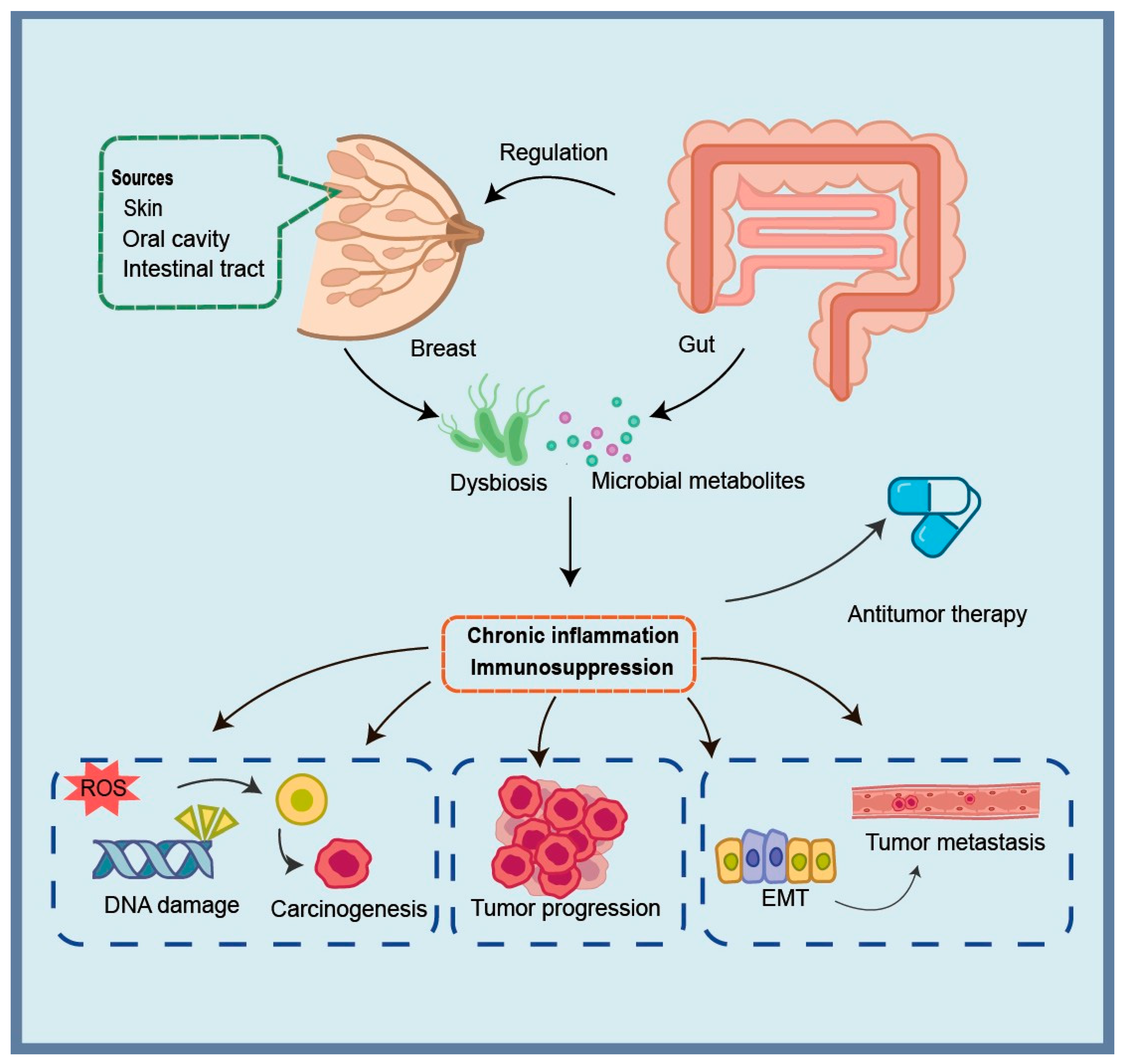
The Impact of Microbiome on Breast Cancer and Regulatory Strategies

Abstract
1. Introduction
2. Literature Search
3. Microbial Dysbiosis in Breast Cancer Patients
3.1. Sources of Breast Microbiome and Dysregulation
3.1.1. Possible Origin of the Breast Microbiome
3.1.2. The Specific Types and Locations of the Microbiome in Breast Tumor Tissues
- Healthy Control Tissue: Breast tissue obtained from individuals without breast cancer (e.g., from reduction mammoplasty), serving as the control baseline.
- Tumor Tissue: Pathologically confirmed breast cancer lesion tissue.
- Tumor Adjacent Normal Tissue: Tissue obtained from breast cancer patients, located outside the visible tumor margin (typically ≥ 5 cm) and pathologically confirmed to be free of cancer cell infiltration, considered “normal” breast tissue from a cancer-bearing host.
3.2. Dysbiosis of Gut Microbiota and Its Mechanisms in Breast Cancer
3.2.1. Digestive Tract Microbiota Dysbiosis
3.2.2. Unique Status of the Gut Microbiome
3.3. Oral Microbiome and Breast Cancer Risk
3.4. Current Landscape and Methodological Challenges in Breast Microbiome Research
4. The Microbiome and the Occurrence, Development, and Metastasis of Breast Cancer
4.1. Initiating Receptors of Microbiome-Related Inflammatory Responses
4.2. Microbiome-Induced Genomic Instability and Tumor Initiation: Roles of Inflammation and Immunosuppression
4.2.1. Direct Genotoxic Bacterial Toxins
4.2.2. Immune-Driven Oxidative DNA Damage
4.3. Microbiome-Driven Mechanisms of Breast Cancer Progression
4.4. Core Mechanisms Linking Inflammation to Tumor Metastasis
4.5. The Influence of Microbial Metabolites on the Onset and Progression of Breast Cancer
5. Therapeutic Applications of Microbiome
5.1. Impacts of Microbiome on Breast Cancer Therapy
5.2. Targeting Microorganisms to Treat or Reduce Complications
6. Conclusions and Perspective
Author Contributions
Funding
Institutional Review Board Statement
Informed Consent Statement
Data Availability Statement
Conflicts of Interest
Abbreviations
| A. muciniphila | Akkermansia muciniphila |
| AP-1 | activator protein 1 |
| BC | breast cancer |
| B. burgdorfer | Borrelia burgdorferi |
| BFT | B. fragilis toxin |
| CTX | cyclophosphamide |
| DCs | dendritic cells |
| EBV | Epstein–Barr virus |
| E. coli | Escherichia coli |
| EMT | epithelial–mesenchymal transition |
| ETBF | enterotoxigenic Bacteroides fragilis |
| FMT | fecal microbiota transplantation |
| F. nucleatum | Fusobacterium nucleatum |
| FPR1 | formyl peptide receptor-1 |
| HCMV | human cytomegalovirus |
| H. hepaticus | Helicobacter hepaticus |
| HMTV | human mammary tumor virus |
| HPV | human papillomavirus |
| ICB | immune checkpoint blockade |
| IHC | immunohistochemical |
| L. acidophilus | Lactobacillus acidophilus |
| LCA | lithocholic acid |
| LPS | lipopolysaccharides |
| MALT | mucosa-associated lymphoid tissue |
| MDSCs | myeloid-derived suppressor cells |
| M. globosa | Malassezia globosa |
| NLRs | NOD-like receptors |
| OS | overall survival |
| OXA | oxaliplatin |
| PAMPs | pathogen-associated molecular patterns |
| PD-L1 | programmed cell death ligand 1 |
| P. gingivalis | Porphyromonas gingivalis |
| PRRs | pattern recognition receptors |
| RNS | reactive nitrogen species |
| ROS | reactive oxygen species |
| SCFAs | short-chain fatty acids |
| S. epidermidis | Staphylococcus epidermidis |
| TLRs | Toll-like receptors |
| TME | tumor microenvironment |
| TNBC | triple-negative breast cancer |
References
- Yoshimura, A.; Imoto, I.; Iwata, H. Functions of Breast Cancer Predisposition Genes: Implications for Clinical Management. Int. J. Mol. Sci. 2022, 23, 7481. [Google Scholar] [CrossRef] [Scilit] [PubMed]
- Filippini, S.E.; Vega, A. Breast Cancer Genes: Beyond BRCA1 and BRCA2. Front. Biosci. 2013, 18, 1358–1372. [Google Scholar] [CrossRef] [Scilit]
- Siegel, R.L.; Kratzer, T.B.; Giaquinto, A.N.; Sung, H.; Jemal, A. Cancer Statistics, 2025. CA Cancer J. Clin. 2025, 75, 10–45. [Google Scholar] [CrossRef] [Scilit] [PubMed]
- Loibl, S.; Poortmans, P.; Morrow, M.; Denkert, C.; Curigliano, G. Breast Cancer. Lancet 2021, 397, 1750–1769. [Google Scholar] [CrossRef] [Scilit] [PubMed]
- Fakhri, N.; Chad, M.A.; Lahkim, M.; Houari, A.; Dehbi, H.; Belmouden, A.; El Kadmiri, N. Risk Factors for Breast Cancer in Women: An Update Review. Med. Oncol. 2022, 39, 197. [Google Scholar] [CrossRef] [Scilit]
- Hill, M.J.; Goddard, P.; Williams, R.E.O. Gut Bacteria and Ætiology of Cancer of the Breast. Lancet 1971, 298, 472–473. [Google Scholar] [CrossRef] [Scilit]
- Papakonstantinou, A.; Nuciforo, P.; Borrell, M.; Zamora, E.; Pimentel, I.; Saura, C.; Oliveira, M. The Conundrum of Breast Cancer and Microbiome—A Comprehensive Review of the Current Evidence. Cancer Treat. Rev. 2022, 111, 102470. [Google Scholar] [CrossRef] [Scilit]
- Fu, A.; Yao, B.; Dong, T.; Chen, Y.; Yao, J.; Liu, Y.; Li, H.; Bai, H.; Liu, X.; Zhang, Y.; et al. Tumor-Resident Intracellular Microbiota Promotes Metastatic Colonization in Breast Cancer. Cell 2022, 185, 1356–1372.e26. [Google Scholar] [CrossRef] [Scilit]
- Sittipo, P.; Shim, J.; Lee, Y.K. Microbial Metabolites Determine Host Health and the Status of Some Diseases. Int. J. Mol. Sci. 2019, 20, 5296. [Google Scholar] [CrossRef] [Scilit]
- Jeffery, I.B.; O’Toole, P.W. Diet-Microbiota Interactions and Their Implications for Healthy Living. Nutrients 2013, 5, 234–252. [Google Scholar] [CrossRef] [Scilit]
- Chapadgaonkar, S.S.; Bajpai, S.S.; Godbole, M.S. Gut Microbiome Influences Incidence and Outcomes of Breast Cancer by Regulating Levels and Activity of Steroid Hormones in Women. Cancer Rep. 2023, 6, e1847. [Google Scholar] [CrossRef] [Scilit] [PubMed]
- Giallourou, N.; Urbaniak, C.; Puebla-Barragan, S.; Vorkas, P.A.; Swann, J.R.; Reid, G. Characterizing the Breast Cancer Lipidome and Its Interaction with the Tissue Microbiota. Commun. Biol. 2021, 4, 1229. [Google Scholar] [CrossRef] [Scilit] [PubMed]
- Banerjee, S.; Wei, Z.; Tian, T.; Bose, D.; Shih, N.N.C.; Feldman, M.D.; Khoury, T.; De Michele, A.; Robertson, E.S. Prognostic Correlations with the Microbiome of Breast Cancer Subtypes. Cell Death Dis. 2021, 12, 831. [Google Scholar] [CrossRef] [Scilit] [PubMed]
- Urbaniak, C.; Cummins, J.; Brackstone, M.; Macklaim, J.M.; Gloor, G.B.; Baban, C.K.; Scott, L.; O’Hanlon, D.M.; Burton, J.P.; Francis, K.P.; et al. Microbiota of Human Breast Tissue. Appl. Environ. Microbiol. 2014, 80, 3007–3014. [Google Scholar] [CrossRef] [Scilit]
- Hieken, T.J.; Chen, J.; Hoskin, T.L.; Walther-Antonio, M.; Johnson, S.; Ramaker, S.; Xiao, J.; Radisky, D.C.; Knutson, K.L.; Kalari, K.R.; et al. The Microbiome of Aseptically Collected Human Breast Tissue in Benign and Malignant Disease. Sci. Rep. 2016, 6, 30751. [Google Scholar] [CrossRef] [Scilit]
- Donnet-Hughes, A.; Perez, P.F.; Doré, J.; Leclerc, M.; Levenez, F.; Benyacoub, J.; Serrant, P.; Segura-Roggero, I.; Schiffrin, E.J. Potential Role of the Intestinal Microbiota of the Mother in Neonatal Immune Education. Proc. Nutr. Soc. 2010, 69, 407–415. [Google Scholar] [CrossRef] [Scilit]
- Zheng, H.-H.; Du, C.-T.; Yu, C.; Tang, X.-Y.; Huang, R.-L.; Zhang, Y.-Z.; Gao, W.; Xie, G.-H. The Relationship of Tumor Microbiome and Oral Bacteria and Intestinal Dysbiosis in Canine Mammary Tumor. Int. J. Mol. Sci. 2022, 23, 10928. [Google Scholar] [CrossRef] [Scilit]
- Urbaniak, C.; Gloor, G.B.; Brackstone, M.; Scott, L.; Tangney, M.; Reid, G. The Microbiota of Breast Tissue and Its Association with Breast Cancer. Appl. Environ. Microbiol. 2016, 82, 5039–5048. [Google Scholar] [CrossRef] [Scilit]
- Hieken, T.J.; Chen, J.; Chen, B.; Johnson, S.; Hoskin, T.L.; Degnim, A.C.; Walther-Antonio, M.R.; Chia, N. The Breast Tissue Microbiome, Stroma, Immune Cells and Breast Cancer. Neoplasia 2022, 27, 100786. [Google Scholar] [CrossRef] [Scilit]
- Tzeng, A.; Sangwan, N.; Jia, M.; Liu, C.-C.; Keslar, K.S.; Downs-Kelly, E.; Fairchild, R.L.; Al-Hilli, Z.; Grobmyer, S.R.; Eng, C. Human Breast Microbiome Correlates with Prognostic Features and Immunological Signatures in Breast Cancer. Genome Med. 2021, 13, 60. [Google Scholar] [CrossRef] [Scilit]
- Rad, S.K.; Yeo, K.K.L.; Wu, F.; Li, R.; Nourmohammadi, S.; Tomita, Y.; Price, T.J.; Ingman, W.V.; Townsend, A.R.; Smith, E. A Systematic Review and Meta-Analysis of 16S rRNA and Cancer Microbiome Atlas Datasets to Characterize Microbiota Signatures in Normal Breast, Mastitis, and Breast Cancer. Microorganisms 2025, 13, 467. [Google Scholar] [CrossRef] [Scilit]
- Chan, A.A.; Bashir, M.; Rivas, M.N.; Duvall, K.; Sieling, P.A.; Pieber, T.R.; Vaishampayan, P.A.; Love, S.M.; Lee, D.J. Characterization of the Microbiome of Nipple Aspirate Fluid of Breast Cancer Survivors. Sci. Rep. 2016, 6, 28061. [Google Scholar] [CrossRef] [Scilit]
- Nejman, D.; Livyatan, I.; Fuks, G.; Gavert, N.; Zwang, Y.; Geller, L.T.; Rotter-Maskowitz, A.; Weiser, R.; Mallel, G.; Gigi, E.; et al. The Human Tumor Microbiome Is Composed of Tumor Type-Specific Intracellular Bacteria. Science 2020, 368, 973–980. [Google Scholar] [CrossRef] [Scilit] [PubMed]
- Banerjee, S.; Tian, T.; Wei, Z.; Shih, N.; Feldman, M.D.; Peck, K.N.; DeMichele, A.M.; Alwine, J.C.; Robertson, E.S. Distinct Microbial Signatures Associated with Different Breast Cancer Types. Front. Microbiol. 2018, 9, 951. [Google Scholar] [CrossRef] [Scilit] [PubMed]
- Li, X.; Sun, X.; Zhang, A.; Pang, J.; Li, Y.; Yan, M.; Xu, Z.; Yu, Y.; Yang, Z.; Chen, X.; et al. Breast Microbiome Associations with Breast Tumor Characteristics and Neoadjuvant Chemotherapy: A Case-Control Study. Front. Oncol. 2022, 12, 926920. [Google Scholar] [CrossRef] [Scilit] [PubMed]
- Kartti, S.; Bendani, H.; Boumajdi, N.; Bouricha, E.M.; Zarrik, O.; EL Agouri, H.; Fokar, M.; Aghlallou, Y.; EL Jaoudi, R.; Belyamani, L.; et al. Metagenomics Analysis of Breast Microbiome Highlights the Abundance of Rothia Genus in Tumor Tissues. J. Pers. Med. 2023, 13, 450. [Google Scholar] [CrossRef] [Scilit]
- Brantley, K.D.; Tamimi, R.M. The Association between Infectious Agents and Breast Cancer: A Review of the Epidemiologic Evidence. Breast Cancer Res. Treat. 2024, 207, 235–252. [Google Scholar] [CrossRef] [Scilit]
- Alibek, K.; Kakpenova, A.; Mussabekova, A.; Sypabekova, M.; Karatayeva, N. Role of Viruses in the Development of Breast Cancer. Infect. Agent. Cancer 2013, 8, 32. [Google Scholar] [CrossRef] [Scilit]
- Gannon, O.M.; Antonsson, A.; Bennett, I.C.; Saunders, N.A. Viral Infections and Breast Cancer—A Current Perspective. Cancer Lett. 2018, 420, 182–189. [Google Scholar] [CrossRef] [Scilit]
- Narunsky-Haziza, L.; Sepich-Poore, G.D.; Livyatan, I.; Asraf, O.; Martino, C.; Nejman, D.; Gavert, N.; Stajich, J.E.; Amit, G.; González, A.; et al. Pan-Cancer Analyses Reveal Cancer-Type-Specific Fungal Ecologies and Bacteriome Interactions. Cell 2022, 185, 3789–3806.e17. [Google Scholar] [CrossRef] [Scilit]
- Hetta, H.F.; Ramadan, Y.N.; Alharbi, A.A.; Alsharef, S.; Alkindy, T.T.; Alkhamali, A.; Albalawi, A.S.; El Amin, H. Gut Microbiome as a Target of Intervention in Inflammatory Bowel Disease Pathogenesis and Therapy. Immuno 2024, 4, 400–425. [Google Scholar] [CrossRef] [Scilit]
- Bobin-Dubigeon, C.; Luu, H.T.; Leuillet, S.; Lavergne, S.N.; Carton, T.; Le Vacon, F.; Michel, C.; Nazih, H.; Bard, J.-M. Faecal Microbiota Composition Varies between Patients with Breast Cancer and Healthy Women: A Comparative Case-Control Study. Nutrients 2021, 13, 2705. [Google Scholar] [CrossRef] [Scilit] [PubMed]
- Luan, B.; Ge, F.; Lu, X.; Li, Z.; Zhang, H.; Wu, J.; Yang, Q.; Chen, L.; Zhang, W.; Chen, W. Changes in the Fecal Microbiota of Breast Cancer Patients Based on 16S rRNA Gene Sequencing: A Systematic Review and Meta-Analysis. Clin. Transl. Oncol. 2024, 26, 1480–1496. [Google Scholar] [CrossRef] [Scilit] [PubMed]
- Zhu, J.; Liao, M.; Yao, Z.; Liang, W.; Li, Q.; Liu, J.; Yang, H.; Ji, Y.; Wei, W.; Tan, A.; et al. Breast Cancer in Postmenopausal Women Is Associated with an Altered Gut Metagenome. Microbiome 2018, 6, 136. [Google Scholar] [CrossRef] [Scilit]
- Issrani, R.; Reddy, R.J.; El-Metwally, T.H.; Prabhu, N. Periodontitis as a Risk Factor for Breast Cancer—What We Know Till Date? Asian Pac. J. Cancer Prev. 2021, 22, 3109–3114. [Google Scholar] [CrossRef] [Scilit]
- Cheng, R.; Billet, S.; Liu, C.; Haldar, S.; Choudhury, D.; Tripathi, M.; Hav, M.; Merchant, A.; Hu, T.; Huang, H.; et al. Periodontal Inflammation Recruits Distant Metastatic Breast Cancer Cells by Increasing Myeloid-Derived Suppressor Cells. Oncogene 2020, 39, 1543–1556. [Google Scholar] [CrossRef] [Scilit]
- Shi, Q.; Wang, J.; Zhou, M.; Zheng, R.; Zhang, X.; Liu, B. Gut Lactobacillus Contribute to the Progression of Breast Cancer by Affecting the Anti-Tumor Activities of Immune Cells in the TME of Tumor-Bearing Mice. Int. Immunopharmacol. 2023, 124, 111039. [Google Scholar] [CrossRef] [Scilit]
- Feng, K.; Ren, F.; Shang, Q.; Wang, X.; Wang, X. Association between Oral Microbiome and Breast Cancer in the East Asian Population: A Mendelian Randomization and Case-Control Study. Thorac. Cancer 2024, 15, 974–986. [Google Scholar] [CrossRef] [Scilit]
- Nearing, J.T.; De Clercq, V.; Langille, M.G.I. Investigating the Oral Microbiome in Retrospective and Prospective Cases of Prostate, Colon, and Breast Cancer. NPJ Biofilms Microbiomes 2023, 9, 23. [Google Scholar] [CrossRef] [Scilit]
- Lu, K.; Mahbub, R.; Fox, J.G. Xenobiotics: Interaction with the Intestinal Microflora. ILAR J. 2015, 56, 218–227. [Google Scholar] [CrossRef] [Scilit]
- Rea, D.; Coppola, G.; Palma, G.; Barbieri, A.; Luciano, A.; Del Prete, P.; Rossetti, S.; Berretta, M.; Facchini, G.; Perdonà, S.; et al. Microbiota Effects on Cancer: From Risks to Therapies. Oncotarget 2018, 9, 17915–17927. [Google Scholar] [CrossRef] [Scilit] [PubMed]
- Bose, C.; Uczkowski, N.G.; Sukla, K.; Raval, N.; Haque, M.M.; Zhang, Y.; Varma, B.; Satagopan, J.M. Breast Cancer and Microbiome: A Systematic Review Highlighting Challenges for Clinical Translation. BMC Womens Health 2025, 25, 416. [Google Scholar] [CrossRef] [Scilit] [PubMed]
- Gihawi, A.; Ge, Y.; Lu, J.; Puiu, D.; Xu, A.; Cooper, C.S.; Brewer, D.S.; Pertea, M.; Salzberg, S.L. Major Data Analysis Errors Invalidate Cancer Microbiome Findings. mBio 2023, 14, e01607-23. [Google Scholar] [CrossRef] [Scilit] [PubMed]
- O’Callaghan, J.L.; Willner, D.; Buttini, M.; Huygens, F.; Pelzer, E.S. Limitations of 16S rRNA Gene Sequencing to Characterize Lactobacillus Species in the Upper Genital Tract. Front. Cell Dev. Biol. 2021, 9, 641921. [Google Scholar] [CrossRef] [Scilit]
- Bhatelia, K.; Singh, K.; Singh, R. TLRs: Linking Inflammation and Breast cancer TLRs. Cell. Signal. 2014, 26, 2350–2357. [Google Scholar] [CrossRef] [Scilit]
- Letran, S.E.; Lee, S.-J.; Shaikh, A.; Uematsu, S.; Akira, S.; McSorley, S.J. TLR5 Functions as an Endocytic Receptor to Enhance Flagellin-Specific Adaptive Immunity. Eur. J. Immunol. 2011, 41, 29–38. [Google Scholar] [CrossRef] [Scilit]
- Zhao, H.; Wu, L.; Yan, G.; Chen, Y.; Zhou, M.; Wu, Y.; Li, Y. Inflammation and Tumor Progression: Signaling Pathways and Targeted Intervention. Signal Transduct. Target. Ther. 2021, 6, 263. [Google Scholar] [CrossRef] [Scilit]
- González-Reyes, S.; Marín, L.; González, L.; González, L.O.; del Casar, J.M.; Lamelas, M.L.; González-Quintana, J.M.; Vizoso, F.J. Study of TLR3, TLR4 and TLR9 in Breast Carcinomas and Their Association with Metastasis. BMC Cancer 2010, 10, 665. [Google Scholar] [CrossRef] [Scilit]
- Ma, W.; Zhang, L.; Chen, W.; Chang, Z.; Tu, J.; Qin, Y.; Yao, Y.; Dong, M.; Ding, J.; Li, S.; et al. Microbiota Enterotoxigenic Bacteroides Fragilis-Secreted BFT-1 Promotes Breast Cancer Cell Stemness and Chemoresistance through Its Functional Receptor NOD1. Protein Cell 2024, 15, 419–440. [Google Scholar] [CrossRef] [Scilit]
- Carty, M.; Guy, C.; Bowie, A.G. Detection of Viral Infections by Innate Immunity. Biochem. Pharmacol. 2021, 183, 114316. [Google Scholar] [CrossRef] [Scilit]
- Kovács, T.; Mikó, E.; Ujlaki, G.; Yousef, H.; Csontos, V.; Uray, K.; Bai, P. The Involvement of Oncobiosis and Bacterial Metabolite Signaling in Metastasis Formation in Breast Cancer. Cancer Metastasis Rev. 2021, 40, 1223–1249. [Google Scholar] [CrossRef] [Scilit] [PubMed]
- Cuevas-Ramos, G.; Petit, C.R.; Marcq, I.; Boury, M.; Oswald, E.; Nougayrède, J.-P. Escherichia Coli Induces DNA Damage in Vivo and Triggers Genomic Instability in Mammalian Cells. Proc. Natl. Acad. Sci. USA 2010, 107, 11537–11542. [Google Scholar] [CrossRef] [Scilit] [PubMed]
- Verma, D.; Nandi, D.; Parida, S.; Saxena, A.; Sharma, D. An Enteropathogenic Microbial Toxin Modulates the Breast Cancer Epigenome Resulting in Concurrent Silencing of Tumor Suppressor Genes. Breast Cancer Res. 2025, 27, 163. [Google Scholar] [CrossRef] [Scilit] [PubMed]
- Parida, S.; Wu, S.; Siddharth, S.; Wang, G.; Muniraj, N.; Nagalingam, A.; Hum, C.; Mistriotis, P.; Hao, H.; Talbot, C.C., Jr.; et al. A Procarcinogenic Colon Microbe Promotes Breast Tumorigenesis and Metastatic Progression and Concomitantly Activates Notch and β-Catenin Axes. Cancer Discov. 2021, 11, 1138–1157. [Google Scholar] [CrossRef] [Scilit]
- Aggarwal, V.; Tuli, H.S.; Varol, A.; Thakral, F.; Yerer, M.B.; Sak, K.; Varol, M.; Jain, A.; Khan, M.A.; Sethi, G. Role of Reactive Oxygen Species in Cancer Progression: Molecular Mechanisms and Recent Advancements. Biomolecules 2019, 9, 735. [Google Scholar] [CrossRef] [Scilit]
- Kong, X.; Zhang, Y.; Xiang, L.; You, Y.; Duan, Y.; Zhao, Y.; Li, S.; Wu, R.; Zhang, J.; Zhou, L.; et al. Fusobacterium Nucleatum-Triggered Neutrophil Extracellular Traps Facilitate Colorectal Carcinoma Progression. J. Exp. Clin. Cancer Res. 2023, 42, 236. [Google Scholar] [CrossRef] [Scilit]
- Wu, S.; Chen, Y.; Chen, Z.; Wei, F.; Zhou, Q.; Li, P.; Gu, Q. Reactive Oxygen Species and Gastric Carcinogenesis: The Complex Interaction between Helicobacter Pylori and Host. Helicobacter 2023, 28, e13024. [Google Scholar] [CrossRef] [Scilit]
- Marquis, R.E. Applied and Ecological Aspects of Oxidative-Stress Damage to Bacterial Spores and to Oral Microbes. Sci. Prog. 2004, 87, 153–177. [Google Scholar] [CrossRef] [Scilit]
- Kay, J.; Thadhani, E.; Samson, L.; Engelward, B. Inflammation-Induced DNA Damage, Mutations and Cancer. DNA Repair 2019, 83, 102673. [Google Scholar] [CrossRef] [Scilit]
- Ko, S.H.; Jeon, J.I.; Woo, H.A.; Kim, J.M. Bacteroides Fragilis Enterotoxin Upregulates Heme Oxygenase-1 in Dendritic Cells via Reactive Oxygen Species-, Mitogen-Activated Protein Kinase-, and Nrf2-Dependent Pathway. World J. Gastroenterol. 2020, 26, 291–306. [Google Scholar] [CrossRef] [Scilit]
- Malla, R.; Surepalli, N.; Farran, B.; Malhotra, S.V.; Nagaraju, G.P. Reactive Oxygen Species (ROS): Critical Roles in Breast Tumor Microenvironment. Crit. Rev. Oncol./Hematol. 2021, 160, 103285. [Google Scholar] [CrossRef] [Scilit]
- Okuyama, A.; Shibata, A.; Nishimoto, H. Critical Points for Interpreting Patients’ Survival Rate Using Cancer Registries: A Literature Review. J. Epidemiol. 2018, 28, 61–66. [Google Scholar] [CrossRef] [Scilit]
- Kennel, K.B.; Greten, F.R. Immune Cell—Produced ROS and Their Impact on Tumor Growth and Metastasis. Redox Biol. 2021, 42, 101891. [Google Scholar] [CrossRef] [Scilit] [PubMed]
- Jabłońska-Trypuć, A. The Role of the Microbiome in Inflammation and Carcinogenesis. Front. Biosci. 2023, 15, 28. [Google Scholar] [CrossRef] [Scilit] [PubMed]
- Deng, H.; Muthupalani, S.; Erdman, S.; Liu, H.; Niu, Z.; Wang, T.C.; Fox, J.G. Translocation of Helicobacter Hepaticus Synergizes with Myeloid-Derived Suppressor Cells and Contributes to Breast Carcinogenesis. Oncoimmunology 2022, 11, 2057399. [Google Scholar] [CrossRef] [Scilit] [PubMed]
- Lakritz, J.R.; Poutahidis, T.; Mirabal, S.; Varian, B.J.; Levkovich, T.; Ibrahim, Y.M.; Ward, J.M.; Teng, E.C.; Fisher, B.; Parry, N.; et al. Gut Bacteria Require Neutrophils to Promote Mammary Tumorigenesis. Oncotarget 2015, 6, 9387–9396. [Google Scholar] [CrossRef] [Scilit]
- Ma, J.-H.; Qin, L.; Li, X. Role of STAT3 Signaling Pathway in Breast Cancer. Cell Commun. Signal 2020, 18, 33. [Google Scholar] [CrossRef] [Scilit]
- Zhang, N.; Ma, Z.P.; Wang, J.; Bai, H.L.; Li, Y.X.; Sun, Q.; Yang, L.; Tao, L.; Zhao, J.; Cao, Y.W.; et al. Human Papillomavirus Infection Correlates with Inflammatory Stat3 Signaling Activity and IL-17 Expression in Patients with Breast Cancer. Am. J. Transl. Res. 2016, 8, 3214–3226. [Google Scholar]
- Khodabandehlou, N.; Mostafaei, S.; Etemadi, A.; Ghasemi, A.; Payandeh, M.; Hadifar, S.; Norooznezhad, A.H.; Kazemnejad, A.; Moghoofei, M. Human Papilloma Virus and Breast Cancer: The Role of Inflammation and Viral Expressed Proteins. BMC Cancer 2019, 19, 61. [Google Scholar] [CrossRef] [Scilit]
- Ma, J.; Sun, L.; Liu, Y.; Ren, H.; Shen, Y.; Bi, F.; Zhang, T.; Wang, X. Alter between Gut Bacteria and Blood Metabolites and the Anti-Tumor Effects of Faecalibacterium prausnitzii in Breast Cancer. BMC Microbiol. 2020, 20, 82. [Google Scholar] [CrossRef] [Scilit]
- Pavitra, E.; Kancharla, J.; Gupta, V.K.; Prasad, K.; Sung, J.Y.; Kim, J.; Tej, M.B.; Choi, R.; Lee, J.-H.; Han, Y.-K.; et al. The Role of NF-κB in Breast Cancer Initiation, Growth, Metastasis, and Resistance to Chemotherapy. Biomed. Pharmacother. 2023, 163, 114822. [Google Scholar] [CrossRef] [Scilit] [PubMed]
- Khatri, V.A.; Paul, S.; Patel, N.J.; Thippani, S.; Sawant, J.Y.; Durkee, K.L.; Murphy, C.L.; Aleman, G.O.; Valentino, J.A.; Jathan, J.; et al. Global Transcriptomic Analysis of Breast Cancer and Normal Mammary Epithelial Cells Infected with Borrelia burgdorferi. Eur. J. Microbiol. Immunol. 2023, 13, 63. [Google Scholar] [CrossRef] [Scilit] [PubMed]
- Benevides, L.; da Fonseca, D.M.; Donate, P.B.; Tiezzi, D.G.; De Carvalho, D.D.; de Andrade, J.M.; Martins, G.A.; Silva, J.S. IL17 Promotes Mammary Tumor Progression by Changing the Behavior of Tumor Cells and Eliciting Tumorigenic Neutrophils Recruitment. Cancer Res. 2015, 75, 3788–3799. [Google Scholar] [CrossRef] [Scilit] [PubMed]
- Liu, M.-M.; Zhu, H.-H.; Bai, J.; Tian, Z.-Y.; Zhao, Y.-J.; Boekhout, T.; Wang, Q.-M. Breast Cancer Colonization by Malassezia globosa Accelerates Tumor Growth. mBio 2024, 15, e01993-24. [Google Scholar] [CrossRef] [Scilit]
- Parhi, L.; Alon-Maimon, T.; Sol, A.; Nejman, D.; Shhadeh, A.; Fainsod-Levi, T.; Yajuk, O.; Isaacson, B.; Abed, J.; Maalouf, N.; et al. Breast Cancer Colonization by Fusobacterium Nucleatum Accelerates Tumor Growth and Metastatic Progression. Nat. Commun. 2020, 11, 3259. [Google Scholar] [CrossRef] [Scilit]
- Van der Merwe, M.; Van Niekerk, G.; Botha, A.; Engelbrecht, A.-M. The Onco-Immunological Implications of Fusobacterium Nucleatum in Breast Cancer. Immunol. Lett. 2021, 232, 60–66. [Google Scholar] [CrossRef] [Scilit]
- D’Angelo, A.; Zenoniani, A.; Masci, M.; Aceto, G.M.; Piattelli, A.; Curia, M.C. Exploring the Microbiome in Breast Cancer: The Role of Fusobacterium Nucleatum as an Onco-Immune Modulator. Microorganisms 2025, 13, 1995. [Google Scholar] [CrossRef] [Scilit]
- Gur, C.; Ibrahim, Y.; Isaacson, B.; Yamin, R.; Abed, J.; Gamliel, M.; Enk, J.; Bar-On, Y.; Stanietsky-Kaynan, N.; Coppenhagen-Glazer, S.; et al. Binding of the Fap2 Protein of Fusobacterium nucleatum to Human Inhibitory Receptor TIGIT Protects Tumors from Immune Cell Attack. Immunity 2015, 42, 344–355. [Google Scholar] [CrossRef] [Scilit]
- Gur, C.; Maalouf, N.; Shhadeh, A.; Berhani, O.; Singer, B.B.; Bachrach, G.; Mandelboim, O. Fusobacterium nucleatum Supresses Anti-Tumor Immunity by Activating CEACAM1. Oncoimmunology 2019, 8, e1581531. [Google Scholar] [CrossRef] [Scilit]
- Zhu, X.; Liang, R.; Lan, T.; Ding, D.; Huang, S.; Shao, J.; Zheng, Z.; Chen, T.; Huang, Y.; Liu, J.; et al. Tumor-Associated Macrophage-Specific CD155 Contributes to M2-Phenotype Transition, Immunosuppression, and Tumor Progression in Colorectal Cancer. J. Immunother. Cancer 2022, 10, e004219. [Google Scholar] [CrossRef] [Scilit]
- Bernardo, G.; Le Noci, V.; Ottaviano, E.; De Cecco, L.; Camisaschi, C.; Guglielmetti, S.; Di Modica, M.; Gargari, G.; Bianchi, F.; Indino, S.; et al. Reduction of Staphylococcus Epidermidis in the Mammary Tumor Microbiota Induces Antitumor Immunity and Decreases Breast Cancer Aggressiveness. Cancer Lett. 2023, 555, 216041. [Google Scholar] [CrossRef] [Scilit] [PubMed]
- Poole, E.; Neves, T.C.; Oliveira, M.T.; Sinclair, J.; da Silva, M.C.C. Human Cytomegalovirus Interleukin 10 Homologs: Facing the Immune System. Front. Cell Infect. Microbiol. 2020, 10, 245. [Google Scholar] [CrossRef] [Scilit]
- Pasquereau, S.; Al Moussawi, F.; Karam, W.; Diab Assaf, M.; Kumar, A.; Herbein, G. Cytomegalovirus, Macrophages and Breast Cancer. Open Virol. J. 2017, 11, 15–27. [Google Scholar] [CrossRef] [Scilit] [PubMed]
- Rosean, C.B.; Bostic, R.R.; Ferey, J.C.M.; Feng, T.-Y.; Azar, F.N.; Tung, K.S.; Dozmorov, M.G.; Smirnova, E.; Bos, P.D.; Rutkowski, M.R. Pre-Existing Commensal Dysbiosis Is a Host-Intrinsic Regulator of Tissue Inflammation and Tumor Cell Dissemination in Hormone Receptor-Positive Breast Cancer. Cancer Res. 2019, 79, 3662. [Google Scholar] [CrossRef] [Scilit] [PubMed]
- Zhang, Q.; Ma, C.; Duan, Y.; Heinrich, B.; Rosato, U.; Diggs, L.P.; Ma, L.; Roy, S.; Fu, Q.; Brown, Z.J.; et al. Gut Microbiome Directs Hepatocytes to Recruit MDSC and Promote Cholangiocarcinoma. Cancer Discov. 2020, 11, 1248. [Google Scholar] [CrossRef] [Scilit]
- Liu, N.-N.; Yi, C.-X.; Wei, L.-Q.; Zhou, J.-A.; Jiang, T.; Hu, C.-C.; Wang, L.; Wang, Y.-Y.; Zou, Y.; Zhao, Y.-K.; et al. The Intratumor Mycobiome Promotes Lung Cancer Progression via Myeloid-Derived Suppressor Cells. Cancer Cell 2023, 41, 1927–1944.e9. [Google Scholar] [CrossRef] [Scilit]
- Gabrilovich, D.I.; Ostrand-Rosenberg, S.; Bronte, V. Coordinated Regulation of Myeloid Cells by Tumours. Nat. Rev. Immunol. 2012, 12, 253–268. [Google Scholar] [CrossRef] [Scilit]
- Rutkowski, M.R.; Stephen, T.L.; Svoronos, N.; Allegrezza, M.J.; Tesone, A.J.; Perales-Puchalt, A.; Brencicova, E.; Escovar-Fadul, X.; Nguyen, J.M.; Cadungog, M.G.; et al. Microbial Driven TLR5-Dependent Signaling Governs Distal Malignant Progression Through Tumor-Promoting Inflammation. Cancer Cell 2014, 27, 27. [Google Scholar] [CrossRef] [Scilit]
- Moaaz, M.; Lotfy, H.; Motawea, M.A.; Fadali, G. The Interplay of Interleukin-17A and Breast Cancer Tumor Microenvironment as a Novel Immunotherapeutic Approach to Increase Tumor Immunogenicity. Immunobiology 2021, 226, 152068. [Google Scholar] [CrossRef] [Scilit]
- Ingman, W.V. The Gut Microbiome: A New Player in Breast Cancer Metastasis. Cancer Res. 2019, 79, 3539–3541. [Google Scholar] [CrossRef] [Scilit]
- Hilmi, M.; Kamal, M.; Vacher, S.; Dupain, C.; Ibadioune, S.; Halladjian, M.; Sablin, M.P.; Marret, G.; Ajgal, Z.C.; Nijnikoff, M.; et al. Intratumoral Microbiome Is Driven by Metastatic Site and Associated with Immune Histopathological Parameters: An Ancillary Study of the SHIVA Clinical Trial. Eur. J. Cancer 2023, 183, 152–161. [Google Scholar] [CrossRef] [Scilit] [PubMed]
- Dominguez, C.; David, J.M.; Palena, C. Epithelial-Mesenchymal Transition and Inflammation at the Site of the Primary Tumor. Semin. Cancer Biol. 2017, 47, 177–184. [Google Scholar] [CrossRef] [Scilit] [PubMed]
- Soria, G.; Ofri-Shahak, M.; Haas, I.; Yaal-Hahoshen, N.; Leider-Trejo, L.; Leibovich-Rivkin, T.; Weitzenfeld, P.; Meshel, T.; Shabtai, E.; Gutman, M.; et al. Inflammatory Mediators in Breast Cancer: Coordinated Expression of TNFα & IL-1β with CCL2 & CCL5 and Effects on Epithelial-to-Mesenchymal Transition. BMC Cancer 2011, 11, 130. [Google Scholar] [CrossRef] [Scilit]
- Wang, Q.; Yu, C.; Yue, C.; Liu, X. Fusobacterium Nucleatum Produces Cancer Stem Cell Characteristics via EMT-Resembling Variations. Int. J. Clin. Exp. Pathol. 2020, 13, 1819–1828. [Google Scholar]
- Parida, S.; Siddharth, S.; Gatla, H.R.; Wu, S.; Wang, G.; Gabrielson, K.; Sears, C.L.; Ladle, B.H.; Sharma, D. Gut Colonization with an Obesity-Associated Enteropathogenic Microbe Modulates the Premetastatic Niches to Promote Breast Cancer Lung and Liver Metastasis. Front. Immunol. 2023, 14, 1194931. [Google Scholar] [CrossRef] [Scilit]
- Wu, J.; Wu, L.; Fang, C.; Nie, R.; Wang, J.; Wang, X.; Liu, W. Mycoplasmal Lipoprotein P37 Binds Human Protein HER2. Microbiol. Res. 2016, 192, 253–259. [Google Scholar] [CrossRef] [Scilit]
- Ivanov, I.I.; Atarashi, K.; Manel, N.; Brodie, E.L.; Shima, T.; Karaoz, U.; Wei, D.; Goldfarb, K.C.; Santee, C.A.; Lynch, S.V.; et al. Induction of Intestinal Th17 Cells by Segmented Filamentous Bacteria. Cell 2009, 139, 485–498. [Google Scholar] [CrossRef] [Scilit]
- Kim, K.; Kim, G.; Kim, J.-Y.; Yun, H.J.; Lim, S.-C.; Choi, H.S. Interleukin-22 Promotes Epithelial Cell Transformation and Breast Tumorigenesis via MAP3K8 Activation. Carcinogenesis 2014, 35, 1352–1361. [Google Scholar] [CrossRef] [Scilit]
- Shibabaw, T.; Teferi, B.; Ayelign, B. The Role of Th-17 Cells and IL-17 in the Metastatic Spread of Breast Cancer: As a Means of Prognosis and Therapeutic Target. Front. Immunol. 2023, 14, 1094823. [Google Scholar] [CrossRef] [Scilit]
- Chia, S.B.; Johnson, B.J.; Hu, J.; Valença-Pereira, F.; Chadeau-Hyam, M.; Guntoro, F.; Montgomery, H.; Boorgula, M.P.; Sreekanth, V.; Goodspeed, A.; et al. Respiratory Viral Infections Awaken Metastatic Breast Cancer Cells in Lungs. Nature 2025, 645, 496–506. [Google Scholar] [CrossRef] [Scilit]
- He, C.; Chen, J.; Tian, R.; Zeng, X.; Han, Q.; Jiang, C.; Zou, J.; Yu, T. Fungal Colonization by Malassezia globosa Promotes Breast Cancer Progression and M2 Macrophage Polarization Through the MBL-C3a–C3aR Signaling Pathway. Microbiologyopen 2025, 14, e70193. [Google Scholar] [CrossRef] [Scilit] [PubMed]
- Mollinedo, F. Neutrophil Degranulation, Plasticity, and Cancer Metastasis. Trends Immunol. 2019, 40, 228–242. [Google Scholar] [CrossRef] [Scilit] [PubMed]
- Dutta, R.K.; Abu, Y.F.; Tao, J.; Chupikova, I.; Oleas, J.; Singh, P.K.; Vitari, N.A.; Qureshi, R.; Ramakrishnan, S.; Roy, S. Altered Gut Microbiome Drives Heightened Pain Sensitivity in a Murine Model of Metastatic Triple-Negative Breast Cancer. Am. J. Cancer Res. 2024, 14, 274–299. [Google Scholar] [CrossRef] [Scilit] [PubMed]
- Yu, L.; Hong, Y.; Maishi, N.; Matsuda, A.Y.; Hida, Y.; Hasebe, A.; Kitagawa, Y.; Hida, K. Oral Bacterium Streptococcus Mutans Promotes Tumor Metastasis through Thrombosis Formation. Cancer Sci. 2024, 115, 648–659. [Google Scholar] [CrossRef] [Scilit]
- Ma, T.; Tang, Y.; Wang, T.; Yang, Y.; Zhang, Y.; Wang, R.; Zhang, Y.; Li, Y.; Wu, M.; Tang, M.; et al. Chronic Pulmonary Bacterial Infection Facilitates Breast Cancer Lung Metastasis by Recruiting Tumor-Promoting MHCIIhi Neutrophils. Signal Transduct. Target. Ther. 2023, 8, 296. [Google Scholar] [CrossRef] [Scilit]
- Yang, Q.; Wang, B.; Zheng, Q.; Li, H.; Meng, X.; Zhou, F.; Zhang, L. A Review of Gut Microbiota-Derived Metabolites in Tumor Progression and Cancer Therapy. Adv. Sci. 2023, 10, 2207366. [Google Scholar] [CrossRef] [Scilit]
- Mikó, E.; Vida, A.; Kovács, T.; Ujlaki, G.; Trencsényi, G.; Márton, J.; Sári, Z.; Kovács, P.; Boratkó, A.; Hujber, Z.; et al. Lithocholic Acid, a Bacterial Metabolite Reduces Breast Cancer Cell Proliferation and Aggressiveness. Biochim. Biophys. Acta Bioenerg. 2018, 1859, 958–974. [Google Scholar] [CrossRef] [Scilit]
- Sári, Z.; Mikó, E.; Kovács, T.; Jankó, L.; Csonka, T.; Lente, G.; Sebő, É.; Tóth, J.; Tóth, D.; Árkosy, P.; et al. Indolepropionic Acid, a Metabolite of the Microbiome, Has Cytostatic Properties in Breast Cancer by Activating AHR and PXR Receptors and Inducing Oxidative Stress. Cancers 2020, 12, 2411. [Google Scholar] [CrossRef] [Scilit]
- Ujlaki, G.; Kovács, T.; Vida, A.; Kókai, E.; Rauch, B.; Schwarcz, S.; Mikó, E.; Janka, E.; Sipos, A.; Hegedűs, C.; et al. Identification of Bacterial Metabolites Modulating Breast Cancer Cell Proliferation and Epithelial-Mesenchymal Transition. Molecules 2023, 28, 5898. [Google Scholar] [CrossRef] [Scilit]
- Kim, M.; Qie, Y.; Park, J.; Kim, C.H. Gut Microbial Metabolites Fuel Host Antibody Responses. Cell Host Microbe 2016, 20, 202–214. [Google Scholar] [CrossRef] [Scilit]
- Ye, L.; Hou, Y.; Hu, W.; Wang, H.; Yang, R.; Zhang, Q.; Feng, Q.; Zheng, X.; Yao, G.; Hao, H. Repressed Blautia-Acetate Immunological Axis Underlies Breast Cancer Progression Promoted by Chronic Stress. Nat. Commun. 2023, 14, 6160. [Google Scholar] [CrossRef] [Scilit] [PubMed]
- Park, H.-S.; Han, J.-H.; Park, J.W.; Lee, D.-H.; Jang, K.-W.; Lee, M.; Heo, K.-S.; Myung, C.-S. Sodium Propionate Exerts Anticancer Effect in Mice Bearing Breast Cancer Cell Xenograft by Regulating JAK2/STAT3/ROS/P38 MAPK Signaling. Acta Pharmacol. Sin. 2021, 42, 1311–1323. [Google Scholar] [CrossRef] [Scilit] [PubMed]
- Zhu, X.; Li, K.; Liu, G.; Wu, R.; Zhang, Y.; Wang, S.; Xu, M.; Lu, L.; Li, P. Microbial Metabolite Butyrate Promotes Anti-PD-1 Antitumor Efficacy by Modulating T Cell Receptor Signaling of Cytotoxic CD8 T Cell. Gut Microbes 2023, 15, 2249143. [Google Scholar] [CrossRef] [Scilit] [PubMed]
- Luu, M.; Riester, Z.; Baldrich, A.; Reichardt, N.; Yuille, S.; Busetti, A.; Klein, M.; Wempe, A.; Leister, H.; Raifer, H.; et al. Microbial Short-Chain Fatty Acids Modulate CD8+ T Cell Responses and Improve Adoptive Immunotherapy for Cancer. Nat. Commun. 2021, 12, 4077. [Google Scholar] [CrossRef] [Scilit]
- Zhang, D.; Tang, Z.; Huang, H.; Zhou, G.; Cui, C.; Weng, Y.; Liu, W.; Kim, S.; Lee, S.; Perez-Neut, M.; et al. Metabolic Regulation of Gene Expression by Histone Lactylation. Nature 2019, 574, 575–580. [Google Scholar] [CrossRef] [Scilit]
- Duthie, S.J. Folate and Cancer: How DNA Damage, Repair and Methylation Impact on Colon Carcinogenesis. J. Inherit. Metab. Dis. 2011, 34, 101–109. [Google Scholar] [CrossRef] [Scilit]
- Wilkie, T.; Verma, A.K.; Zhao, H.; Charan, M.; Ahirwar, D.K.; Kant, S.; Pancholi, V.; Mishra, S.; Ganju, R.K. Lipopolysaccharide from the Commensal Microbiota of the Breast Enhances Cancer Growth: Role of S100A7 and TLR4. Mol. Oncol. 2022, 16, 1508–1522. [Google Scholar] [CrossRef] [Scilit]
- Yang, H.; Wang, B.; Wang, T.; Xu, L.; He, C.; Wen, H.; Yan, J.; Su, H.; Zhu, X. Toll-like Receptor 4 Prompts Human Breast Cancer Cells Invasiveness via Lipopolysaccharide Stimulation and Is Overexpressed in Patients with Lymph Node Metastasis. PLoS ONE 2014, 9, e109980. [Google Scholar] [CrossRef] [Scilit]
- Li, J.; Yin, J.; Shen, W.; Gao, R.; Liu, Y.; Chen, Y.; Li, X.; Liu, C.; Xiang, R.; Luo, N. TLR4 Promotes Breast Cancer Metastasis via Akt/GSK3β/β-Catenin Pathway upon LPS Stimulation. Anat. Rec. 2017, 300, 1219–1229. [Google Scholar] [CrossRef] [Scilit]
- St-Denis-Bissonnette, F.; Khoury, R.; Mediratta, K.; El-Sahli, S.; Wang, L.; Lavoie, J.R. Applications of Extracellular Vesicles in Triple-Negative Breast Cancer. Cancers 2022, 14, 451. [Google Scholar] [CrossRef] [Scilit]
- Murray, W.R.; Blackwood, A.; Calman, K.C.; MacKay, C. Faecal Bile Acids and Clostridia in Patients with Breast Cancer. Br. J. Cancer 1980, 42, 856. [Google Scholar] [CrossRef] [Scilit] [PubMed]
- Morrison, D.J.; Preston, T. Formation of Short Chain Fatty Acids by the Gut Microbiota and Their Impact on Human Metabolism. Gut Microbes 2016, 7, 189. [Google Scholar] [CrossRef] [Scilit] [PubMed]
- Singh, N.; Gurav, A.; Sivaprakasam, S.; Brady, E.; Padia, R.; Shi, H.; Thangaraju, M.; Prasad, P.D.; Manicassamy, S.; Munn, D.H.; et al. Activation of the Receptor (Gpr109a) for Niacin and the Commensal Metabolite Butyrate Suppresses Colonic Inflammation and Carcinogenesis. Immunity 2014, 40, 128–139. [Google Scholar] [CrossRef] [Scilit] [PubMed]
- Bourriaud, C.; Robins, R.J.; Martin, L.; Kozlowski, F.; Tenailleau, E.; Cherbut, C.; Michel, C. Lactate Is Mainly Fermented to Butyrate by Human Intestinal Microfloras but Inter-Individual Variation Is Evident. J. Appl. Microbiol. 2005, 99, 201–212. [Google Scholar] [CrossRef] [Scilit]
- Zhang, Y.-F.; Shi, W.-W.; Gao, H.-F.; Zhou, L.; Hou, A.-J.; Zhou, Y.-H. Folate Intake and the Risk of Breast Cancer: A Dose-Response Meta-Analysis of Prospective Studies. PLoS ONE 2014, 9, e100044. [Google Scholar] [CrossRef] [Scilit]
- Tio, M.; Andrici, J.; Eslick, G.D. Folate Intake and the Risk of Breast Cancer: A Systematic Review and Meta-Analysis. Breast Cancer Res. Treat. 2014, 145, 513–524. [Google Scholar] [CrossRef] [Scilit]
- Zeng, J.; Wang, K.; Ye, F.; Lei, L.; Zhou, Y.; Chen, J.; Zhao, G.; Chang, H. Folate Intake and the Risk of Breast Cancer: An up-to-Date Meta-Analysis of Prospective Studies. Eur. J. Clin. Nutr. 2019, 73, 1657–1660. [Google Scholar] [CrossRef] [Scilit]
- McKee, A.M.; Kirkup, B.M.; Madgwick, M.; Fowler, W.J.; Price, C.A.; Dreger, S.A.; Ansorge, R.; Makin, K.A.; Caim, S.; Le Gall, G.; et al. Antibiotic-Induced Disturbances of the Gut Microbiota Result in Accelerated Breast Tumor Growth. iScience 2021, 24, 103012. [Google Scholar] [CrossRef] [Scilit]
- Seaman, W.E.; Gindhart, T.D. Effect of Estrogen on Natural Killer Cells. Arthritis Rheum. 1979, 22, 1234–1240. [Google Scholar] [CrossRef] [Scilit]
- Nilsson, N.; Carlsten, H. Estrogen Induces Suppression of Natural Killer Cell Cytotoxicity and Augmentation of Polyclonal B Cell Activation. Cell Immunol. 1994, 158, 131–139. [Google Scholar] [CrossRef] [Scilit]
- McGuinness, C.; Britt, K.L. Estrogen Receptor Regulation of the Immune Microenvironment in Breast Cancer. J. Steroid Biochem. Mol. Biol. 2024, 240, 106517. [Google Scholar] [CrossRef] [Scilit]
- Bengtsson, A.K.; Ryan, E.J.; Giordano, D.; Magaletti, D.M.; Clark, E.A. 17beta-Estradiol (E2) Modulates Cytokine and Chemokine Expression in Human Monocyte-Derived Dendritic Cells. Blood 2004, 104, 1404–1410. [Google Scholar] [CrossRef] [Scilit] [PubMed]
- AlMalki, R.H.; Sebaa, R.; Al-Ansari, M.M.; Al-Alwan, M.; Alwehaibi, M.A.; Rahman, A.M.A.E. Coli Secretome Metabolically Modulates MDA-MB-231 Breast Cancer Cells’ Energy Metabolism. Int. J. Mol. Sci. 2023, 24, 4219. [Google Scholar] [CrossRef] [Scilit] [PubMed]
- AlMalki, R.H.; Jaber, M.A.; Al-Ansari, M.M.; Sumaily, K.M.; Al-Alwan, M.; Sabi, E.M.; Malkawi, A.K.; Abdel Rahman, A.M. Metabolic Alteration of MCF-7 Cells upon Indirect Exposure to E. Coli Secretome: A Model of Studying the Microbiota Effect on Human Breast Tissue. Metabolites 2023, 13, 938. [Google Scholar] [CrossRef] [Scilit] [PubMed]
- Wang, H.; Rong, X.; Zhao, G.; Zhou, Y.; Xiao, Y.; Ma, D.; Jin, X.; Wu, Y.; Yan, Y.; Yang, H.; et al. The Microbial Metabolite Trimethylamine N-Oxide Promotes Antitumor Immunity in Triple-Negative Breast Cancer. Cell Metab. 2022, 34, 581–594.e8. [Google Scholar] [CrossRef] [Scilit]
- Luo, B.-P.; Luo, J.; Hu, Y.-B.; Yao, X.-W.; Wu, F.-H. Cyclin D1b Splice Variant Promotes Avβ3-Mediated EMT Induced by LPS in Breast Cancer Cells. Curr. Med. Sci. 2018, 38, 467–472. [Google Scholar] [CrossRef] [Scilit]
- Macia, L.; Nanan, R.; Hosseini-Beheshti, E.; Grau, G.E. Host- and Microbiota-Derived Extracellular Vesicles, Immune Function, and Disease Development. Int. J. Mol. Sci. 2020, 21, 107. [Google Scholar] [CrossRef] [Scilit]
- Vande Voorde, J.; Sabuncuoğlu, S.; Noppen, S.; Hofer, A.; Ranjbarian, F.; Fieuws, S.; Balzarini, J.; Liekens, S. Nucleoside-Catabolizing Enzymes in Mycoplasma-Infected Tumor Cell Cultures Compromise the Cytostatic Activity of the Anticancer Drug Gemcitabine. J. Biol. Chem. 2014, 289, 13054–13065. [Google Scholar] [CrossRef] [Scilit]
- Alexander, J.L.; Wilson, I.D.; Teare, J.; Marchesi, J.R.; Nicholson, J.K.; Kinross, J.M. Gut Microbiota Modulation of Chemotherapy Efficacy and Toxicity. Nat. Rev. Gastroenterol. Hepatol. 2017, 14, 356–365. [Google Scholar] [CrossRef] [Scilit]
- Viaud, S.; Saccheri, F.; Mignot, G.; Yamazaki, T.; Daillère, R.; Hannani, D.; Enot, D.P.; Pfirschke, C.; Engblom, C.; Pittet, M.J.; et al. The Intestinal Microbiota Modulates the Anticancer Immune Effects of Cyclophosphamide. Science 2013, 342, 971–976. [Google Scholar] [CrossRef] [Scilit]
- Iida, N.; Dzutsev, A.; Stewart, C.A.; Smith, L.; Bouladoux, N.; Weingarten, R.A.; Molina, D.A.; Salcedo, R.; Back, T.; Cramer, S.; et al. Commensal Bacteria Control Cancer Response to Therapy by Modulating the Tumor Microenvironment. Science 2013, 342, 967. [Google Scholar] [CrossRef] [Scilit] [PubMed]
- Bawaneh, A.; Wilson, A.S.; Levi, N.; Howard-McNatt, M.M.; Chiba, A.; Soto-Pantoja, D.R.; Cook, K.L. Intestinal Microbiota Influence Doxorubicin Responsiveness in Triple-Negative Breast Cancer. Cancers 2022, 14, 4849. [Google Scholar] [CrossRef] [Scilit] [PubMed]
- Schettini, F.; Fontana, A.; Gattazzo, F.; Strina, C.; Milani, M.; Cappelletti, M.R.; Cervoni, V.; Morelli, L.; Curigliano, G.; Iebba, V.; et al. Faecal Microbiota Composition Is Related to Response to CDK4/6-Inhibitors in Metastatic Breast Cancer: A Prospective Cross-Sectional Exploratory Study. Eur. J. Cancer 2023, 191, 112948. [Google Scholar] [CrossRef] [Scilit] [PubMed]
- Shiao, S.L.; Kershaw, K.M.; Limon, J.J.; You, S.; Yoon, J.; Ko, E.Y.; Guarnerio, J.; Potdar, A.; McGovern, D.P.B.; Bose, S.; et al. Commensal Bacteria and Fungi Differentially Regulate Tumor Responses to Radiation Therapy. Cancer Cell 2021, 39, 1202. [Google Scholar] [CrossRef] [Scilit]
- Herrera-Quintana, L.; Vázquez-Lorente, H.; Silva, R.C.M.C.; Olivares-Arancibia, J.; Reyes-Amigo, T.; Pires, B.R.B.; Plaza-Diaz, J. The Role of the Microbiome and of Radiotherapy-Derived Metabolites in Breast Cancer. Cancers 2024, 16, 3671. [Google Scholar] [CrossRef] [Scilit]
- Karin, M.; Jobin, C.; Balkwill, F. Chemotherapy, Immunity and Microbiota—A New Triumvirate? Nat. Med. 2014, 20, 126–127. [Google Scholar] [CrossRef] [Scilit]
- Yang, J.; Liu, K.; Qu, J.; Wang, X. The Changes Induced by Cyclophosphamide in Intestinal Barrier and Microflora in Mice. Eur. J. Pharmacol. 2013, 714, 120–124. [Google Scholar] [CrossRef] [Scilit]
- Ozben, T. Oxidative Stress and Apoptosis: Impact on Cancer Therapy. J. Pharm. Sci. 2007, 96, 2181–2196. [Google Scholar] [CrossRef] [Scilit]
- Du, P.; Jing, J.; He, X. Microbiota and Their Metabolites Potentiate Cancer Immunotherapy: Therapeutic Target or Resource for Small Molecule Drug Discovery? Front. Pharmacol. 2022, 13, 1091124. [Google Scholar] [CrossRef] [Scilit]
- Semaan, J.; El-Hakim, S.; Ibrahim, J.-N.; Safi, R.; Elnar, A.A.; El Boustany, C. Comparative Effect of Sodium Butyrate and Sodium Propionate on Proliferation, Cell Cycle and Apoptosis in Human Breast Cancer Cells MCF-7. Breast Cancer 2020, 27, 696–705. [Google Scholar] [CrossRef] [Scilit]
- Chen, W.; Wei, F.; Xu, J.; Wang, Y.; Chen, L.; Wang, J.; Guan, X. Trastuzumab Enhances the Anti-Tumor Effects of the Histone Deacetylase Inhibitor Sodium Butyrate on a HER2-Overexpressing Breast Cancer Cell Line. Int. J. Mol. Med. 2011, 28, 985–991. [Google Scholar] [CrossRef] [Scilit]
- Topçul, M.R.; Çetin, İ.; Pulat, E.; Çalişkan, M. Comparison of the Effects of Crizotinib as Monotherapy and as Combination Therapy with Butyric Acid on Different Breast Cancer Cells. Oncol. Lett. 2024, 29, 38. [Google Scholar] [CrossRef] [Scilit]
- Jia, D.; Wang, Q.; Qi, Y.; Jiang, Y.; He, J.; Lin, Y.; Sun, Y.; Xu, J.; Chen, W.; Fan, L.; et al. Microbial Metabolite Enhances Immunotherapy Efficacy by Modulating T Cell Stemness in Pan-Cancer. Cell 2024, 187, 1651–1665.e21. [Google Scholar] [CrossRef] [Scilit] [PubMed]
- Guo, H.; Chou, W.-C.; Lai, Y.; Liang, K.; Tam, J.W.; Brickey, W.J.; Chen, L.; Montgomery, N.D.; Li, X.; Bohannon, L.M.; et al. Multi-Omics Analyses of Radiation Survivors Identify Radioprotective Microbes and Metabolites. Science 2020, 370, eaay9097. [Google Scholar] [CrossRef] [Scilit] [PubMed]
- Filippou, C.; Themistocleous, S.C.; Marangos, G.; Panayiotou, Y.; Fyrilla, M.; Kousparou, C.A.; Pana, Z.-D.; Tsioutis, C.; Johnson, E.O.; Yiallouris, A. Microbial Therapy and Breast Cancer Management: Exploring Mechanisms, Clinical Efficacy, and Integration within the One Health Approach. Int. J. Mol. Sci. 2024, 25, 1110. [Google Scholar] [CrossRef] [Scilit] [PubMed]
- van Pijkeren, J.P.; Morrissey, D.; Monk, I.R.; Cronin, M.; Rajendran, S.; O’Sullivan, G.C.; Gahan, C.G.M.; Tangney, M. A Novel Listeria Monocytogenes-Based DNA Delivery System for Cancer Gene Therapy. Hum. Gene Ther. 2010, 21, 405–416. [Google Scholar] [CrossRef] [Scilit]
- Goto, Y.; Iwata, S.; Miyahara, M.; Miyako, E. Discovery of Intratumoral Oncolytic Bacteria Toward Targeted Anticancer Theranostics. Adv. Sci. 2023, 10, e2301679. [Google Scholar] [CrossRef] [Scilit]
- Thu, M.S.; Ondee, T.; Nopsopon, T.; Farzana, I.A.K.; Fothergill, J.L.; Hirankarn, N.; Campbell, B.J.; Pongpirul, K. Effect of Probiotics in Breast Cancer: A Systematic Review and Meta-Analysis. Biology 2023, 12, 280. [Google Scholar] [CrossRef] [Scilit]
- Zengul, A.G.; Demark-Wahnefried, W.; Barnes, S.; Morrow, C.D.; Bertrand, B.; Berryhill, T.F.; Frugé, A.D. Associations between Dietary Fiber, the Fecal Microbiota and Estrogen Metabolism in Postmenopausal Women with Breast Cancer. Nutr. Cancer 2021, 73, 1108–1117. [Google Scholar] [CrossRef] [Scilit]
- Bastard, Q.L.; Ward, T.; Sidiropoulos, D.; Hillmann, B.M.; Chun, C.L.; Sadowsky, M.J.; Knights, D.; Montassier, E. Fecal Microbiota Transplantation Reverses Antibiotic and Chemotherapy-Induced Gut Dysbiosis in Mice. Sci. Rep. 2018, 8, 6219. [Google Scholar] [CrossRef] [Scilit]
- Drekonja, D.; Reich, J.; Gezahegn, S.; Greer, N.; Shaukat, A.; MacDonald, R.; Rutks, I.; Wilt, T.J. Fecal Microbiota Transplantation for Clostridium Difficile Infection: A Systematic Review. Ann. Intern. Med. 2015, 162, 630–638. [Google Scholar] [CrossRef] [Scilit] [PubMed]
- Nofouzi, K.; Almasi, P.; Fakhri-Dameshgieh, A.A.; Khordadmehr, M.; Baradaran, B.; Asadi, M.; Sarbakhsh, P.; Hamidian, G. Suppression of the Malignancy of Mammary Tumor in Mice Model by Inactivated Preparation of Mycobacterium Obuense. Vet. Res. Forum 2022, 13, 393–401. [Google Scholar] [CrossRef] [Scilit]
- Vincent, R.L.; Gurbatri, C.R.; Li, F.; Vardoshvili, A.; Coker, C.; Im, J.; Ballister, E.R.; Rouanne, M.; Savage, T.; de Los Santos-Alexis, K.; et al. Probiotic-Guided CAR-T Cells for Solid Tumor Targeting. Science 2023, 382, 211–218. [Google Scholar] [CrossRef] [Scilit] [PubMed]
- Suryawanshi, Y.R.; Zhang, T.; Essani, K. Oncolytic Viruses: Emerging Options for the Treatment of Breast Cancer. Med. Oncol. 2017, 34, 43. [Google Scholar] [CrossRef] [Scilit] [PubMed]
- Liu, X.; Sun, M.; Pu, F.; Ren, J.; Qu, X. Transforming Intratumor Bacteria into Immunopotentiators to Reverse Cold Tumors for Enhanced Immuno-Chemodynamic Therapy of Triple-Negative Breast Cancer. J. Am. Chem. Soc. 2023, 145, 26296–26307. [Google Scholar] [CrossRef] [Scilit]
- Otto-Dobos, L.D.; Strehle, L.D.; Loman, B.R.; Seng, M.M.; Sardesai, S.D.; Williams, N.O.; Gatti-Mays, M.E.; Stover, D.G.; Sudheendra, P.K.; Wesolowski, R.; et al. Baseline Gut Microbiome Alpha Diversity Predicts Chemotherapy-Induced Gastrointestinal Symptoms in Patients with Breast Cancer. NPJ Breast Cancer 2024, 10, 99. [Google Scholar] [CrossRef] [Scilit]
- Salva, S.; Marranzino, G.; Villena, J.; Agüero, G.; Alvarez, S. Probiotic Lactobacillus Strains Protect against Myelosuppression and Immunosuppression in Cyclophosphamide-Treated Mice. Int. Immunopharmacol. 2014, 22, 209–221. [Google Scholar] [CrossRef] [Scilit]
- David, L.A.; Maurice, C.F.; Carmody, R.N.; Gootenberg, D.B.; Button, J.E.; Wolfe, B.E.; Ling, A.V.; Devlin, A.S.; Varma, Y.; Fischbach, M.A.; et al. Diet Rapidly and Reproducibly Alters the Human Gut Microbiome. Nature 2014, 505, 559–563. [Google Scholar] [CrossRef] [Scilit]
- Altinok Dindar, D.; Chun, B.; Palma, A.; Cheney, J.; Krieger, M.; Kasschau, K.; Stagaman, K.; Mitri, Z.I.; Goodyear, S.M.; Shannon, J.; et al. Association between Gut Microbiota and Breast Cancer: Diet as a Potential Modulating Factor. Nutrients 2023, 15, 4628. [Google Scholar] [CrossRef] [Scilit]
- Turnbaugh, P.J.; Ridaura, V.K.; Faith, J.J.; Rey, F.E.; Knight, R.; Gordon, J.I. The Effect of Diet on the Human Gut Microbiome: A Metagenomic Analysis in Humanized Gnotobiotic Mice. Sci. Transl. Med. 2009, 1, 6ra14. [Google Scholar] [CrossRef] [Scilit]
- Tsai, H.-H.; Yu, J.-C.; Hsu, H.-M.; Chu, C.-H.; Chang, T.-M.; Hong, Z.-J.; Feng, A.-C.; Fu, C.-Y.; Hsu, K.-F.; Dai, M.-S.; et al. The Risk of Breast Cancer between Western and Mediterranean Dietary Patterns. Nutrients 2023, 15, 2057. [Google Scholar] [CrossRef] [Scilit] [PubMed]
- Nogal, A.; Valdes, A.M.; Menni, C. The Role of Short-Chain Fatty Acids in the Interplay between Gut Microbiota and Diet in Cardio-Metabolic Health. Gut Microbes 2021, 13, 1897212. [Google Scholar] [CrossRef] [Scilit] [PubMed]
- Slavin, J. Fiber and Prebiotics: Mechanisms and Health Benefits. Nutrients 2013, 5, 1417–1435. [Google Scholar] [CrossRef] [Scilit]
- Aragón, F.; Carino, S.; Perdigón, G.; de Moreno de LeBlanc, A. Inhibition of Growth and Metastasis of Breast Cancer in Mice by Milk Fermented with Lactobacillus Casei CRL 431. J. Immunother. 2015, 38, 185–196. [Google Scholar] [CrossRef] [Scilit] [PubMed]
- Zhang, X.; Yu, L.; Shi, J.; Li, S.; Yang, S.; Gao, W.; Yang, S.; Cheng, M.; Wang, H.; Guo, Z.; et al. Antibiotics Modulate Neoadjuvant Therapy Efficiency in Patients with Breast Cancer: A Pilot Analysis. Sci. Rep. 2021, 11, 14024. [Google Scholar] [CrossRef] [Scilit]
- Di Modica, M.; Gargari, G.; Regondi, V.; Bonizzi, A.; Arioli, S.; Belmonte, B.; De Cecco, L.; Fasano, E.; Bianchi, F.; Bertolotti, A.; et al. Gut Microbiota Condition the Therapeutic Efficacy of Trastuzumab in Gut Microbiota Condition the Therapeutic Efficacy of Trastuzumab in HER2-Positive Breast CancerBreast Cancer. Cancer Res. 2021, 81, 2195–2206. [Google Scholar] [CrossRef] [Scilit]
- Kimata, H.; Imai, T.; Kikumori, T.; Teshigahara, O.; Nagasaka, T.; Goshima, F.; Nishiyama, Y.; Nakao, A. Pilot Study of Oncolytic Viral Therapy Using Mutant Herpes Simplex Virus (HF10) against Recurrent Metastatic Breast Cancer. Ann. Surg. Oncol. 2006, 13, 1078–1084. [Google Scholar] [CrossRef] [Scilit]
- Zeh, H.J.; Downs-Canner, S.; McCart, J.A.; Guo, Z.S.; Rao, U.N.M.; Ramalingam, L.; Thorne, S.H.; Jones, H.L.; Kalinski, P.; Wieckowski, E.; et al. First-in-Man Study of Western Reserve Strain Oncolytic Vaccinia Virus: Safety, Systemic Spread, and Antitumor Activity. Mol. Ther. 2015, 23, 202–214. [Google Scholar] [CrossRef] [Scilit]
- Fountzilas, C.; Patel, S.; Mahalingam, D. Review: Oncolytic Virotherapy, Updates and Future Directions. Oncotarget 2017, 8, 102617–102639. [Google Scholar] [CrossRef] [Scilit]
- Ramadan, Y.N.; Rashed, Z.I.; Ahmed, R.; Salem, H.A.; Alshareef, H.; Ahmad-Abakur, E.H.; Alanazi, F.E.; Elfadil, H.; Hetta, H.F. Bacteriophages: Unlocking a New Frontier in Breast Cancer Management by Defeating Oncogenic Bacteria. Futur. J. Pharm. Sci. 2025, 11, 74. [Google Scholar] [CrossRef] [Scilit]
- Shoae-Hassani, A.; Keyhanvar, P.; Seifalian, A.M.; Mortazavi-Tabatabaei, S.A.; Ghaderi, N.; Issazadeh, K.; Amirmozafari, N.; Verdi, J. λ Phage Nanobioparticle Expressing Apoptin Efficiently Suppress Human Breast Carcinoma Tumor Growth in Vivo. PLoS ONE 2013, 8, e79907. [Google Scholar] [CrossRef] [Scilit]
- Nguyen, S.M.; Tran, H.T.T.; Long, J.; Shrubsole, M.J.; Cai, H.; Yang, Y.; Cai, Q.; Tran, T.V.; Zheng, W.; Shu, X.-O. Gut Microbiome in Association with Chemotherapy-Induced Toxicities among Patients with Breast Cancer. Cancer 2024, 130, 2014–2030. [Google Scholar] [CrossRef] [Scilit]
- Alavi, S.E.; Mansouri, H.; Esfahani, M.K.M.; Movahedi, F.; Akbarzadeh, A.; Chiani, M. Archaeosome: As New Drug Carrier for Delivery of Paclitaxel to Breast Cancer. Indian. J. Clin. Biochem. 2014, 29, 150–153. [Google Scholar] [CrossRef] [Scilit]
- Wallace, B.D.; Wang, H.; Lane, K.T.; Scott, J.E.; Orans, J.; Koo, J.S.; Venkatesh, M.; Jobin, C.; Yeh, L.-A.; Mani, S.; et al. Alleviating Cancer Drug Toxicity by Inhibiting a Bacterial Enzyme. Science 2010, 330, 831–835. [Google Scholar] [CrossRef] [Scilit]
- Flórez, A.B.; Sierra, M.; Ruas-Madiedo, P.; Mayo, B. Susceptibility of Lactic Acid Bacteria, Bifidobacteria and Other Bacteria of Intestinal Origin to Chemotherapeutic Agents. Int. J. Antimicrob. Agents 2016, 48, 547–550. [Google Scholar] [CrossRef] [Scilit]



| Tissue Type | Adjacent vs. Healthy Control Tissue | Tumor vs. Healthy Control Tissue | Tumor vs. Adjacent Tissue |
|---|---|---|---|
| ER+ | Increased: Arcanobacterium, Bifidobacterium, Cardiobacterium, Citrobacter, Escherichia [24] Decreased: Vibrio, Pseudoalteromonas, Photobacterium, Marinobacterium, Prevotella_9 [25] | Decreased: Alkanindiges, Micrococcus, Caulobacter, Proteus, Brevibacillus, Kocuria, Parasediminibacterium [20] (more abundant in non-tumor tissues) | |
| PR+ | Increased: Pelomonas, Ralstonia, Oblitimonas, Lactobacillus, Methylophilus, Achromobacter [20] (within tumor tissue) | ||
| TNBC | Increased: Aerococcus, Arcobacter, Geobacillus, Orientia, Rothia [24] | Increased: Azomonas, Alkanindiges, Caulobacter, Proteus, Brevibacillus, Kocuria, Parasediminibacterium [20] (within tumor tissue) Decreased: Cutibacterium, Pseudoalteromonas, Photobacterium [25] | Increased: Sphingomonas, Sphingomonadaceae family [26] Decreased: Sporosarcina [26] |
| HER2+ | Increased: Acinetobacter, Pseudomonas, Stenotrophomonas, Cutibacterium [25] | Increased: Cloacibacterium, PRD01a011B, Alloprevotella, Stakelama, Filibacter, Blastomonas, Anaerostipes [20] (within tumor tissue) Decreased: Glaciecola, Vibrio, Photobacterium | Increased: Sphingomonas [26] |
| TPBC | Increased: Bordetella, Campylobacter, Chlamydia, Chlamydophila, Legionella, Pasteurella [24] | ||
| Luminal A | Increased: Corynebacterium [26] | ||
| Luminal B | Increased: Alloiococcus [26] | ||
| BC | Increased: Staphylococcus, Acinetobacter, Burkholderia–Caballeronia–Paraburkholderia, Escherichia–Shigella, Shewanella, Mycoplasma, Clostridium_sensu_stricto_7, Psychrobacter, Wolbachia, Glaciecola [25] | Increased: Pseudomonadaceae and Enterobacteriaceae, Porphyromonas and Azomonas [20] (significantly higher in tumor tissue); Actinomyces, Bartonella, Brevundimonas, Coxiella, Mobiluncus, Mycobacterium, Rickettsia, Sphingomonas [24] | Increased: Peptostreptococcales_Tissierella family and Finegoldia, Rothia genus; Streptococcus, Rothia, and Staphylococcus [26] Decreased: Pseudomonadaceae family and Pseudomonas genus; Enterobacteriaceae family [26] |
| Tumor Adjacent Normal Tissue | Increased: Bacillus, Staphylococcus, Enterobacteriaceae (unclassified), Comamondaceae (unclassified), and Bacteroidetes (unclassified) [18] | ||
| Healthy Control Tissue | Healthy Tissue Signature: Propionibacterium, Staphylococcus (abundant in non-tumor tissues) Finegoldia, Granulicatella, Streptococcus, Anaerococcus, Ruminococcaceae UCG-002, Corynebacterium 1, Alicyclobacillus, Odoribacter, Lactococcus, Esherichia/Shigella [20] | Increased: Rickettsia, Acinetobacter, Ralstonia, Delftia, Arthrobacter, Stenotrophomonas [25]; Prevotella, Lactococcus, Streptococcus, Corynebacterium, Micrococcus [18] |
| PRR Family | Member(s) | Ligand/Microbial Component | Relevance in Breast Cancer |
|---|---|---|---|
| TLRs | TLR1, TLR2, TLR6 | Lipoproteins (Gram-positive bacteria) | Differentially expressed across subtypes; associated with tumor progression |
| TLR4 | LPS (Gram-negative bacteria) | High expression correlates with poor prognosis; modulates immune microenvironment | |
| TLR5 | Flagellin (bacterial motility protein) | Involved in bacterial sensing; potential link to microbiome-driven inflammation | |
| TLR3 | Double-stranded RNA (viral/bacterial) | Expression negatively correlates with patient survival | |
| TLR9 | Unmethylated CpG DNA (bacterial/viral) | Elevated in some BC tissues; prognostic implications | |
| NLRs | NOD1 | Peptidoglycan fragments | Participate in intracellular bacterial sensing; may influence BC cell signaling |
| RLRs | RIG-I, MDA5 | Viral RNA | Primarily antiviral; potential role in viral-associated BC hypotheses |
| FPR | FPR1 | Bacterial formylated peptides | Loss-of-function variant (Rs867228) linked to infection risk and luminal B BC susceptibility |
| Metabolite | Source | Effects on Breast Cancer | Mechanism | Ref. | |
|---|---|---|---|---|---|
| LCA | Aerobic floras (Mostly Clostridia) | Influence macrophage polarization and T cell function | Act via MAPK pathway and TGR5 receptors; decreased in early-stage breast cancer | [107] | |
| SCFAs | Acetate | Blautia | Augment anti-tumor immunity and inhibit metastasis. | Enhance CD8+ T cell infiltration | [111] |
| Propionate | Gut microbiota | Inhibit JAK2/STAT3, induce apoptosis by promoting ROS levels and MAPK pathway activation | Causes cell-cycle arrest, increases ROS, phosphorylates p38 MAPK | [112] | |
| Butyrate | Gut microbiota | Regulate immune response, lower inflammation, enhance anti-PD-1 therapy effectiveness | Binds to GPR109A, influences T cell differentiation | [113,114] | |
| Lactate | Lactobacilli | Influence macrophage polarization and T cell function. | Modulate immune cell function; dysregulation leads to immunosuppressive TME. | [37,115] | |
| Folate | Gut microbiota | Enhance bone metastasis risk | Impact DNA methylation, repair, and synthesis | [116] | |
| LPS | Gram-negative bacterial cells (breast cancer) | Suppress tumor immunity to enable tumor growth and metastasis | Downregulation of Akt/GSK3β/β-Catenin signaling pathway; recruit MDSCs via LPS/S100A7/TLR4 and impair TLR4-mediated tumor immunity | [117,118,119] | |
| EVs | F. nucleatum (breast microbiota) | promote BC cell proliferation | EVs/TLR4 | [76] | |
| Probiotics | Exert anti-inflammatory properties | / | [120] | ||
| Therapy Category | Microbial Element | Specific Mechanism | Measurable Outcome | Ref. | |
|---|---|---|---|---|---|
| Chemotherapy | Gemcitabine | Mycoplasma | Upregulates cytidine deaminase | Reduction in drug activation | [138] |
| CTX | Specific gut bacteria | Enhances the anti-tumor immune response by inducing Th17 and Th1 cells | Increase in tumor-infiltrating T cells | [139,140] | |
| Platinum agents | Specific gut bacteria | Generates ROS | DNA damage and cell apoptosis | [141] | |
| Docetaxel | A. muciniphila | - | - | [142] | |
| Immunotherapy | CDK4/6 inhibitor | Clostridium innocuum, Oscillibacter ruminantium, and Eubacterium hallii | Negatively correlated | Neutrophil-to-lymphocyte ratio | [143] |
| Roseburia faecis | Positively correlated | ||||
| Anti-PD-1 therapy | Gut microbiota | Production of the metabolite butyrate | - | [113] | |
| Radiotherapy | Opposing effects of commensal fungi and bacteria | Activates Dectin-1 pathway | Promotion of Th2 cell differentiation | [144,145] | |
| Strategy | Advantages | Limitations | Applications | Ref. | |
|---|---|---|---|---|---|
| Dietary Modifications | Non-invasive, low-cost, reversible | Slow effects, inter-individual variability | Mediterranean diet | [156,157] | |
| Probiotics/Prebiotics | Targets beneficial strains | Strain-specific, colonization challenges | Inulin; Lactobacilli and Bifidobacterium | [158,159] | |
| FMT | Rapid restoration of microbial diversity, safety (infection, immune reactions) | Not supported by clear evidence | Post-antibiotic/chemotherapy recovery | [160,161] | |
| Antibiotics | Rapid pathogen clearance | Disrupts commensal microbiota, may reduce efficacy | Selective use for efficacy enhancement or infection control | [81] | |
| Engineered Microbes | Genetically Modified Bacteria | Metabolic adjustment, multifunctional delivery | Technical complexity, regulatory hurdles, safety risks | Targetedtherapy, precisiondrug/genedelivery, multifunctional | [156,162,163] |
| Oncolytic bacteria and viruses | Complementary mechanisms of action, high target specificity | [157,164] | |||
| Bacterial vaccines | Dual targeting (bacteria and tumor) | [165] | |||
| Treatment-related adverse effects | Gastrointestinal Toxicity | Regulation of the intestinal microenvironment | gutmicrobiota (e.g., Faecalibacterium) | [166] | |
| Lymphedema | ProbioticCombination (Lactobacillus, Bifidobacterium, Streptococcus + Fructooligosaccharides) | [158] | |||
| Myelosuppression and immunosuppression | Lactobacillus | [167] | |||
Disclaimer/Publisher’s Note: The statements, opinions and data contained in all publications are solely those of the individual author(s) and contributor(s) and not of MDPI and/or the editor(s). MDPI and/or the editor(s) disclaim responsibility for any injury to people or property resulting from any ideas, methods, instructions or products referred to in the content. |
© 2025 by the authors. Licensee MDPI, Basel, Switzerland. This article is an open access article distributed under the terms and conditions of the Creative Commons Attribution (CC BY) license.
Share and Cite
Wang, J.; Xu, D.; Hu, S.; Zheng, B.; Chen, Y.; Pan, T. The Impact of Microbiome on Breast Cancer and Regulatory Strategies. Microorganisms 2026, 14, 75. https://doi.org/10.3390/microorganisms14010075
Wang J, Xu D, Hu S, Zheng B, Chen Y, Pan T. The Impact of Microbiome on Breast Cancer and Regulatory Strategies. Microorganisms. 2026; 14(1):75. https://doi.org/10.3390/microorganisms14010075
Chicago/Turabian StyleWang, Jiaxin, Dongyan Xu, Shiyao Hu, Beiwen Zheng, Yiding Chen, and Tao Pan. 2026. "The Impact of Microbiome on Breast Cancer and Regulatory Strategies" Microorganisms 14, no. 1: 75. https://doi.org/10.3390/microorganisms14010075
APA StyleWang, J., Xu, D., Hu, S., Zheng, B., Chen, Y., & Pan, T. (2026). The Impact of Microbiome on Breast Cancer and Regulatory Strategies. Microorganisms, 14(1), 75. https://doi.org/10.3390/microorganisms14010075