Article

Water Parameters Predicting the Seasonal and Spatial Dynamics of the Vibrio Harveyi- and Splendidus-Clade Pathogens

by
Karagan Royer
and
Andrei L. Barkovskii
*
Department of Biological and Environmental Sciences, Georgia College and State University, Milledgeville, GA 30161, USA
*
Author to whom correspondence should be addressed.
Microorganisms 2025, 13(9), 2167; https://doi.org/10.3390/microorganisms13092167
Submission received: 15 July 2025 / Revised: 10 September 2025 / Accepted: 11 September 2025 / Published: 17 September 2025
(This article belongs to the Section Environmental Microbiology)

Abstract

Vibrio species of the Harveyi and Splendidus clades are the causative agents of vibriosis, resulting in mortality rates of up to 100% in common aquaculture species. They are primarily responsible for seafood-related illnesses in humans, causing gastroenteritis. Except for V. parahaemolyticus, the ecological behaviour of these pathogens is poorly understood. We investigated the spatial and temporal distribution of V. parahaemolyticus, V. alginolyticus, V. harveyi/V. campbellii, and V. splendidus in three Georgia (USA) grounds for Crassostrea virginica and Mercenaria mercenaria. DNA from oysters, clams, water, and sediment was collected over a year-long study and analyzed using quantitative PCR (qPCR) to assess the prevalence and concentrations of the above Vibrio species. The study targeted the tlh, VA1198230, rpoA, and recA genes using species-specific primers. Species abundance was estimated based on the concentrations of the corresponding genes. The species abundance was profiled for water parameters and concentrations of the clade-specific virulence genes toxR, luxR, srp, vhhA, vhh, and vhp that were previously detected in the study area. V. parahaemolyticus was the most common species, detected year-round in 61% and 44% of the water and sediment samples, respectively, followed by V. splendidus (67% and 17%) and V. harveyi/V. campbellii (19% and 33%). V. alginolyticus was rarely detected in water and never in sediment. In bivalves, the highest frequency was observed for V. parahaemolyticus. This species was detected in 89% of clam and 100% of oyster samples, followed by V. alginolyticus (22% and 17%) and V. splendidus at 17% in both species. No V. harveyi/V. campbellii has been detected in clams and oysters. Seasonal dynamics and concentrations varied between the species. Water temperature (r = 0.58–0.63, p ≤ 0.05), pH (r = −0.46), and dissolved oxygen (r = −0.42 to −0.56, p ≤ 0.05) were reliable predictors for the abundance of the Harveyi and Splendidus clade pathogens in bivalves and the water column, but not in sediments. In water and sediments, the abundances of V. harveyi/V. campbellii and V. parahaemolyticus were highly correlated (r = 0.80–0.99, p ≤ 0.001) to concentrations of most of the virulence genes, with some heterogeneities between the sites. The study revealed the species-specific dynamic of the Harveyi and Splendidus clade pathogens, provided the first evidence for the presence of V. harveyi/V. campbellii in the Atlantic USA waters, and identified environmental predictors for monitoring the Harveyi and Splendidus clade pathogens in mollusks and the water column.

1. Introduction

The Harveyi clade is the second-largest clade of the Vibrio genus and is composed of 12 non-pathogenic and pathogenic species [1]. The latter share a signature array of virulence genes and cause infections and diseases in fish and shellfish [2], as well as human gastroenteritis, diarrhea, nausea, and vomiting due to the consumption of raw and undercooked seafood [3,4].
In a previous study [5], numerous clade-specific virulence genes were detected in the water and sediment of three Georgia (USA) clams and oyster grounds. These genes were detected with Harveyi-clade-specific primers [6], and their patterns revealed the diversification and possible stratification of their carriers into different ecological niches in response to changes in water parameters, such as temperature, pH, salinity, and turbidity [5]. The most common Harveyi-clade species associated with shellfish and human diseases are Vibrio harveyi, Vibrio alginolyticus, Vibrio campbellii, and Vibrio parahaemolyticus [2].
Vibrio parahaemolyticus is one of the best-known species in this clade. This is primarily associated with its role in infecting humans through the consumption of raw or undercooked seafood [7]. Gastroenteritis is a common symptom of V. parahaemolyticus infection, but immunocompromised patients can exhibit more severe symptoms such as septicemia and multi-organ failure [8]. The symptoms vary depending on the route of transmission. V. parahaemolyticus is also associated with wound infections, where individuals encounter contaminated water while having a cut or lesion on their skin [9]. Although uncommon, V. parahaemolyticus can cause more severe conditions, such as soft tissue damage and necrotizing fasciitis [10,11]. Of the 80,000 cases of vibriosis in the USA, 40% are caused by V. parahaemolyticus [12].
Vibrio alginolyticus is the causal agent of shellfish and fish diseases [13]. This pathogen is a major contributor to mass mortality in Crassostrea gigas aquaculture [14,15] and fish species [16]. V. alginolyticus is pathogenic to humans and causes gastrointestinal and other infections [17]. Of the 80,000 cases of vibriosis in the USA annually, 20% are caused by V. alginolyticus [12]. Studies have shown a notable increase in the number of cases of otitis externa during warmer months, with V. alginolyticus being the causative agent [18]. Between 1998 and 2012, 30 percent of soft tissue and bloodstream infections caused by V. alginolyticus in the USA were reported along the Atlantic coast [17]. Surprisingly, there are no published studies on its distribution and abundance in the Atlantic coastal waters of the Southeast United States.
Vibrio harveyi and Vibrio cambellii are known to affect shrimp, fish, and mollusks in Southeast Asia [19,20,21], leading to significant economic losses. Most infected marine species exhibit lethargy, luminescence, and tissue necrosis, with an 80% mortality rate in shrimp [22], and cause rare incidences of vibriosis in humans [23]. These two Vibrio species, with more than 98% 16S rRNA gene similarity [20] form a tight subclade within the Harveyi clade [24], which complicates species-level differentiation. V. harveyi and V. campbellii also share the same virulence factors [19]. Because of its high similarity to V. harveyi, diseases caused by V. campbellii may be severely underreported [24,25]. Both species have not been previously reported in Atlantic USA waters, but their spread to these waters seems possible due to climate change-related expansion.
V. splendidus is part of the Splendidus clade, is commonly found in coastal regions, and mostly infects bivalves, such as oysters and clams [14,26]. Common hosts for this species include the Pacific oyster (Crassostrea gigas) and carpet shell clam (Ruditapes decussatus) [14,27], causing mass mortality in both clams and oysters and substantial economic losses in aquaculture. While known in the North Atlantic [28], there are no reports on its presence and abundance in the Southeastern USA Atlantic.
While V. parahaemolyticus has been repeatedly described in the coastal waters of the Southeastern USA, including the study area [29], the presence and distribution of other Harveyi-clade pathogenic species in this environment have not yet been documented. A previous study revealed the presence of a wide array of Harveyi-clade-specific virulence genes in the study area [5]. This observation justifies the study on the presence and distribution of the Harveyi-clade pathogenic species in Southeastern USA waters. In the current study, we aimed to evaluate the presence and temporal distribution of the major Harveyi-clade pathogens, V. parahaemolyticus, V. alginolyticus, and V. harveyi/V. campbellii in oysters, clams, water, and sediments of the coastal waters of Georgia (USA). V. splendidus was added to the study as a well-known pathogen of oysters and clams [14,26] that has not been previously evaluated in the Southeastern Atlantic waters of the USA. We also aimed to attribute the previously detected Harveyi-clade-specific virulence genes to their Harveyi-clade carriers and to identify water parameters predictive to the presence and abundance of the above pathogens in the water column, oysters, and clams.

2. Materials and Methods

2.1. Field Sites and Sampling Events

Three sampling sites located offshore of the coastal marshes of Townsend, Georgia, USA, were used to collect water, sediment, oyster (Crassostrea virginica), and clam (Mercenaria mercenaria) samples (Figure 1). Site 1 was located north of Fourmile Island along the Julienton River (31°33′34 N, 81°17′16 W). Site 2 was located at the mouth of the Sapelo River (31°32′33 N, 81°16′53 W). Site 3 was the southernmost site, with coordinates of 31°30′21 N, 81°16′44 W, located along the Mud River. All three sampling sites were tidal rivers that feed into Sapelo Sound Bay. Oysters and clams were collected from privately owned beds operated by Sapelo Sea Farms. Six sampling events occurred during low tide between alternating months from June 2022 to April 2023. DNA obtained from these samples has been previously evaluated on the abundance and divergence of the Haveyi-clade-specific virulence genes [5] in the above sites, preserved at −80 °C, and used for the speciation of Vibrio pathogens and their dynamics in the current study.

2.2. Water Collection, Processing, and Storage of Samples

Water samples were collected in triplicate from three segments of each site. Nine water samples were collected at each site in each sampling month. Before collection, sterile bottles were rinsed with environmental water. Approximate water depth at low tide was around 3 m. Water was collected in 1 L sterile glass bottles at a depth of ~1.5 m using a 4000 Subsurface Grab Bottle Sampler (Conbar, Monroeville, NJ, USA). Each triplicate was taken ~25 m apart to ensure an accurate representation of the site [30]. After collection, the samples were immediately placed on ice and, within 12 h, transported to the laboratory, where filtration and DNA extraction took place immediately after arrival. The samples were filtered through a custom water filtration system using 0.22 μm Millipore nitrocellulose filters (Millipore Sigma, Burlington, MA, USA). Using the Qiagen DNeasy® PowerWater kit (Venlo, The Netherlands), total DNA was extracted then quantified using a NanoDrop ND-1000 Spectrophotometer (Thermo Fisher Scientific®, Waltham, MA, USA). The extracted DNA was stored at −20 °C.

2.3. Sediment Collection, Processing, and Storage of Samples

In each sampling event, sediments were collected at low tide in triplicate from three sections of each of the three sites. To account for site variation, each triplicate was taken from a different clam or oyster bed within ~25 m of each other [30]. Sediments were collected directly from the shallow segments of these beds at a depth of 10–20 cm. Sterile gloves and sterile double-ended laboratory spoons (Cole-Parmer, Vernon Hills, IL, USA) were used to retrieve the sediment samples and packed into sterile 1.5 mL microcentrifuge tubes. The tubes were placed on ice in Nasco Whirl-Pak bags (Chicago, IL, USA) for transportation to the laboratory. After reaching the laboratory, 0.25 g of sediment was used to extract DNA using the Qiagen DNEasy® PowerSoil Kit (Venlo, The Netherlands), following the manufacturer’s protocol. Total DNA was quantified using a NanoDrop ND-1000 Spectrophotometer (Thermo Fisher Scientific, Waltham, MA, USA), and the samples were stored at −20 °C.

2.4. Oyster & Clam Collection, Processing, and Storage of Samples

Oysters and clams were collected manually from each site. Triplicates of five mollusks were harvested from random oyster and clam beds at each of three sites to cover the natural variation of the site. The oysters and clams were placed on ice in separate Nasco Whirl-Pak bags (Chicago, IL, USA) and transported to the laboratory. Once in the lab, the mollusks were aseptically shucked and drained. The entire visceral tissue of five mollusks per sample was collected and rinsed with a 0.8% NaCl solution. The visceral tissue was then diluted with an equal volume of sterile phosphate-buffered saline solution (PBS) to make a 1:1 (wt/vol) dilution (pH 7.4, Fisher Scientific). The tissues were homogenized for 1 min using a Tissue Tearor (Biospec Products, Bartlesville, OK, USA) at a mid-range speed setting. The homogenate (0.025 g) was used to extract DNA using the Qiagen DNeasy® Blood & Tissue Kit (Venlo, The Netherlands). Total DNA was quantified using a NanoDrop ND-1000 Spectrophotometer (Thermo Fisher Scientific, Waltham, MA, USA) and stored at −20 °C.

2.5. Environmental Parameters

The environmental parameters for each site and each month (except April) were recorded using a Horiba-52G Multiparameter Water Quality Meter (Horiba Advanced Techno Co., Ltd., Kyoto, Japan) at an approximate depth of 1.5–2 m. Temperature (°C), pH, salinity (ppt), turbidity (NTU), dissolved oxygen (mg/L), total dissolved solids (g/L), conductivity (mS/cm), and potential water density (∂t) data were collected at each site Table S1).

2.6. Primers and PCR Protocols

Published species-specific primers for the Harveyi clade were selected after careful consideration and were retested for their validity in our lab. The reference species of V. harveyi (ATCC 14126), V. campbellii (ATCC BAA-1116/BB120), V. splendidus (ATCC 33125), V. parahaemolyticus (ATCC 17802), and V. alginolyticus (ATCC 17749) were used for retesting several primers against each species under the study. The species-specificity of the primers, the absence of cross-reactions with other species, and detection thresholds were examined. The reference cultures were grown in Difco Marine Broth 2216 (Franklin Lakes, NJ, USA) overnight, at optimal temperatures for each species. The pure cultures were subjected to DNA extraction using the Qiagen DNeasy Microbial Kit (Venlo, The Netherlands), followed by DNA quantification using a NanoDrop ND-1000 Spectrophotometer (Thermo Fisher Scientific®, Waltham, MA, USA). Extracted DNA concentration ranged from 20 to 100 ng/μL. An assay of 5 μL Bio-Rad PCR Master Mix (Hercules, CA, USA), 3 μL of nuclease-free water, 1 μL of each primer (200 nm), and 1 μL of DNA was assembled to perform PCR. DNA from Escherichia coli ATCC 11775™ was used as a negative control in all the test runs.
Several primers failed to differentiate between the reference species or had a low detection threshold. The primers used in this study (Table S2) and the cycling parameters for qPCR (Table S3) [6,31,32,33] are provided in the Supplementary Materials.
After purification with the Qiagen QIAquick PCR purification kit (Venlo, The Netherlands), the PCR products were quantified using a NanoDrop ND-1000 Spectrophotometer (Thermo Fisher Scientific®, Waltham, MA, USA). The formula used to calculate gene copy number was as follows: number of copies = (ng of DNA × (6.022 × 10²³)/(size of amplicon in bp × (1.0 × 109) × 650). The gene copy numbers in purified DNA ranged from 3.20 × 10¹¹ to 1.62 × 10¹² per ng. Each DNA sample was serially diluted and used to generate standard curves.
A qPCR protocol was developed for each target gene, and standard curves were generated. The dilutions were run in triplicate using dye-based qPCR. The assay included 1 μL of the targeted DNA, 5 μL of the Bio-Rad SYBR Green Master Mix (Hercules, CA, USA), 3.75 μL of nuclease-free molecular-grade water, 1 μL of BSA (0.5 mg/μL), 0.25 μL of MgCl2 (25 mM), and 0.2 μL of the corresponding primer set. A melt curve step was added to the cycling parameters to analyze the qPCR products.
The MASTERO Bio-Rad software 2.3 featured a linear regression of the log copy number values and the cycle threshold (ct) values for each assay was applied. The R2 value and y-intercept from the automated equation were used to calculate the gene copy numbers for the collected environmental samples.
The copy number per well was used as the base value to calculate the copy number per 1 mL of water and 1 g of sediment, clams, and oysters. The formula used to calculate copy number per well is as follows: copy number per well = 10((Cq−y-intercept)/(-slope)).

2.7. Statistical Treatment of Data

The graphs were generated using the RStudio software (R version 2024.12.0+). Pearson’s coefficient and p-values were used to evaluate the correlation between average copy numbers and water parameters. Similarly, Pearson’s statistic was used to correlate the abundance of the previously detected virulence genes and species-specific copy numbers in the study area, with p-values applied. The abundance of the Vibrio species was compared to the abundance of previously detected Harveyi-clade-specific virulence genes [5] in the same samples.

3. Results

3.1. Frequency of Detection of the Targeted Vibrio Species

All targeted Vibrio species were detected in all three sites. V. parahaemolyticus was the most common, detected year-round in 61% and 44% of the water and sediment samples, respectively, followed by V. splendidus (67% and 17%), and V. harveyi/V. campbellii (19% and 33%) (Table 1). V. alginolyticus was rarely detected in water and never in sediment. In bivalves, the highest frequency was observed for V. parahaemolyticus. This species was detected in 89% of clam and 100% of oyster samples, followed by V. alginolyticus (22% and 17%) and V. splendidus at 17% in both species (Table 2). No V. harveyi/V. campbellii has been detected in clams and oysters.

3.2. Seasonal and Spatial Dynamics of V. splendidus and the Harveyi-Clade Species

3.2.1. Vibrio splendidus

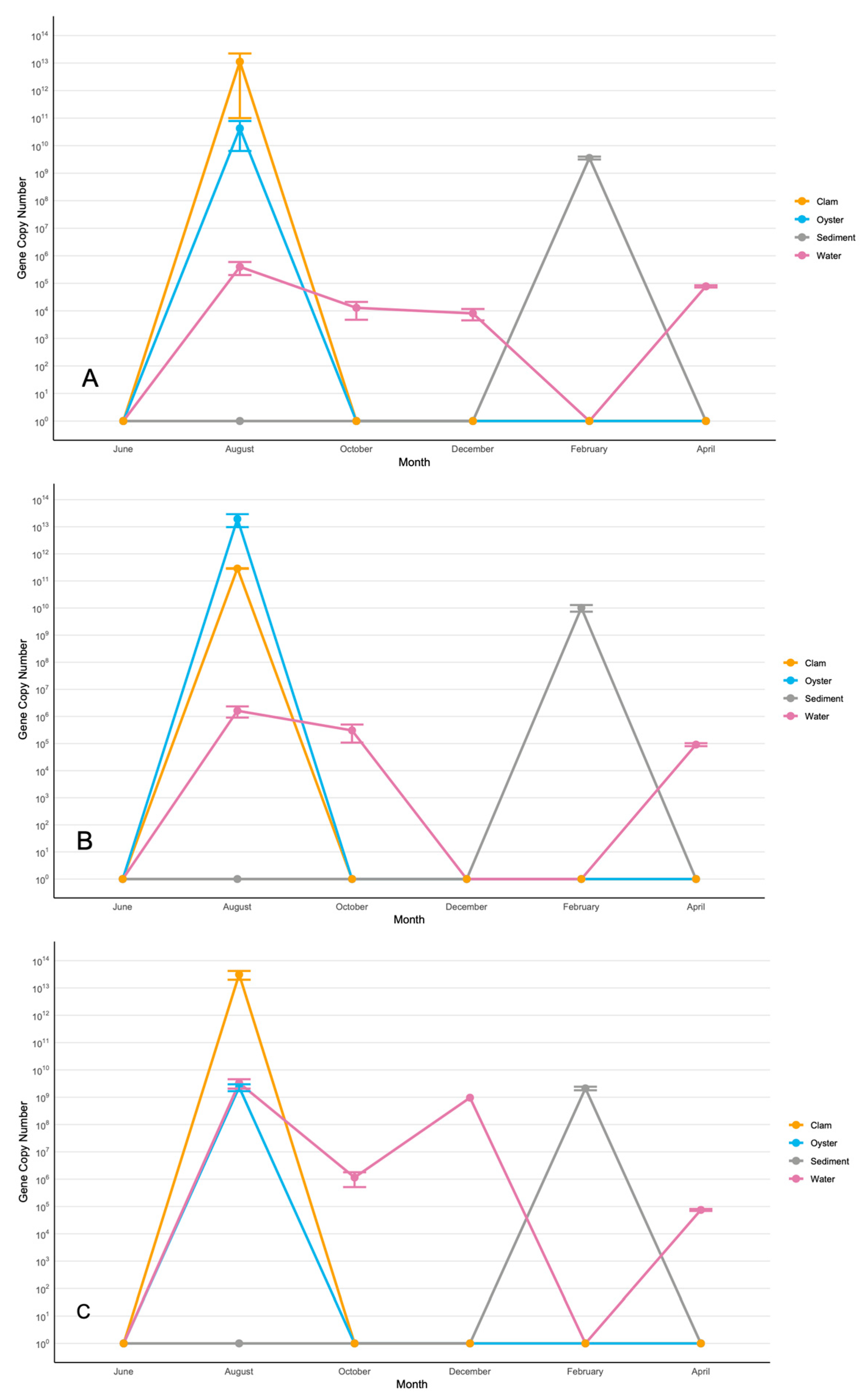
In all three sites, V. splendidus displayed remarkably similar spatial and temporal dynamics (Figure 2). V. splendidus was detected in the water column most months, with peaks in August and April. The August peak of its water concentration at 106–109 gene copies/mL coincided with its peak in bivalves at 1011–1013 gene copies/g. The second peak in April did not correspond to a spike of V. splendidus in the bivalves. In sediments, V. splendidus occurred only in February at 109–1010 gene copies/g. No V. splendidus was detected in the bivalves or the water column in the winter months. These seasonal and spatial dynamics of V. splendidus suggest the water column as a source of this species to oysters and clams in summer months, and sediments as the main ecological niche for V. splendidus in winter.

3.2.2. Vibrio alginolyticus

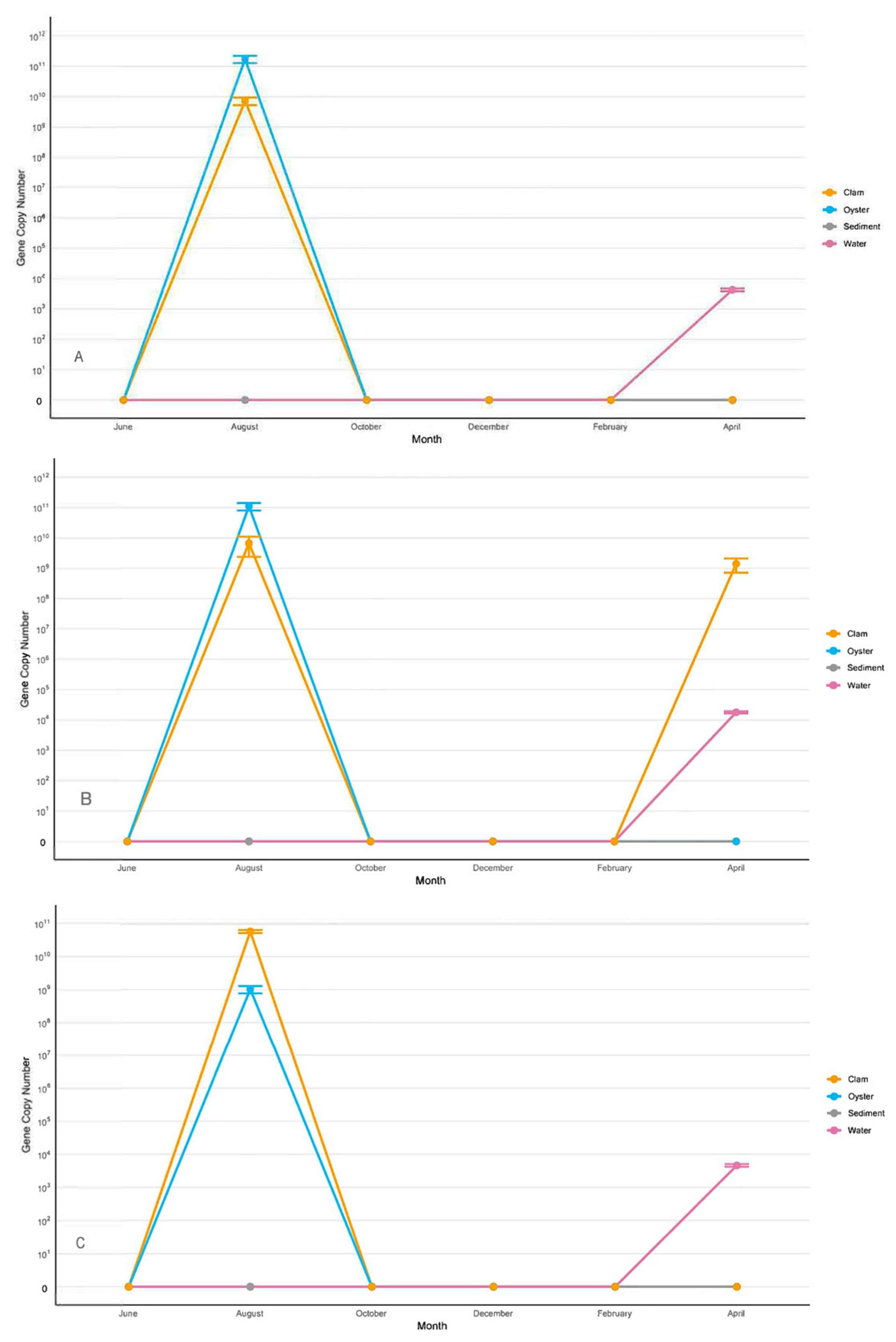
In all three sites, V. alginolyticus displayed remarkably similar spatial and temporal dynamics that were distinctive from that of V. splendidus (Figure 3). As for V. splendidus, this species was detected in oysters in clams in August at the overall highest concentration of 109–1011 gene copies/g. In contrast to V. splendidus, no presence of V. alginolyticus at that time was revealed in the water column. In the water column, V. alginolyticus was detected at ~104 gene copies/mL in April, which coincided with its spike in clams at ~109 gene copies/g, but only in one site (Figure 3B). V. alginolyticus was the only Vibrio species not detected in sediments.

3.2.3. Vibrio harveyi/Vibrio campbellii

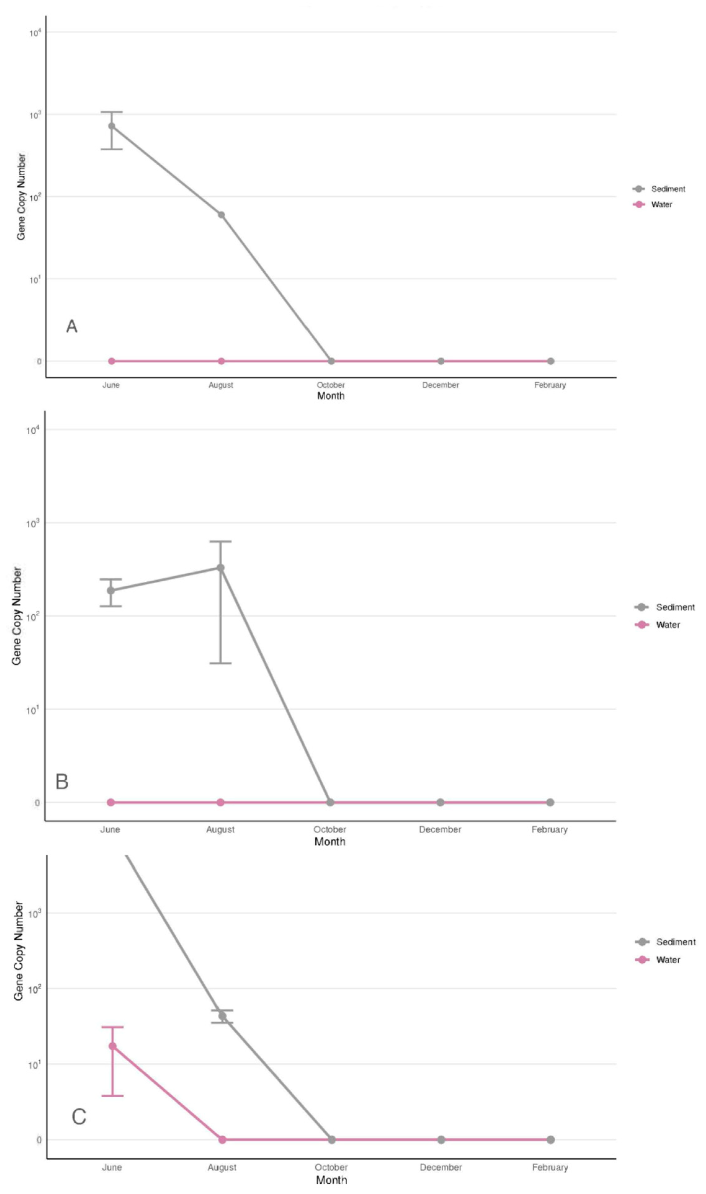
The current study is the first report on the presence of Vibrio harveyi/Vibrio campbellii on the Atlantic coast of the USA. The trend exhibited by V. harveyi/V. campbellii differed from all the other Vibrio species in this study (Figure 4). These species were observed in all three sites but only between June and August. During that time, V. harveyi/V. campbellii resided mostly in sediments with gene copies of 103–104 per gram. June was the only month when these species were recorded in the water column in Site 3 (Figure 4C). Within the site, its concentration was low and varied between the samples in the range of 5–50 gene copies/mL (Figure 4C). No V. harveyi/V. campbellii have been detected in oysters and clams.

3.2.4. Vibrio parahaemolyticus

V. parahaemolyticus was the most prominent Harveyi-clade species in all the sites, with the highest abundance and most predictable spatial and temporal dynamics. This species was present in all environmental niches year-round. The spatial and temporal dynamics were remarkably similar at all the sites. The peaks of V. parahaemolyticus concentration simultaneously occurred in the water column, sediments, and bivalves in August and February (Figure 5). The highest concentration of V. parahaemolyticus in the water column also occurred in August at ~106–107 gene copies/mL (Figure 5).
In contrast to popular belief, V. parahaemolyticus was consistently present in mollusks, even in winter months. Unlike V. splendidus, this pathogen did not move out of the hosts and into sediments during the winter months. The highest concentrations of V. parahaemolyticus in clams and oysters were reached in August, at ~1014 gene copies/g and ~1013 gene copies/g, respectively (Figure 5B,C). The sediment concentration was the second highest, reaching ~1010 gene copies/g in August (Figure 5A,B).

3.3. Impact of Water Parameters on the Distribution and Abundance of Targeted Vibrio Species

In the water column, temperature, water acidity, and oxygen concentration uniformly impacted all the Vibrio species (Table 3), making those reliable predictors for the abundance of Harveyi-clade and V. splendidus pathogens. As expected, all the species positively reacted to the increase in temperature. Observed reverse correlations of all the species to pH and oxygen were not expected. V. harveyi/V. campbellii were the only species with a reversed correlation to salinity, conductivity, TDS, and potential water density, likely indicating the species’ preference for a less saline environment. Due to a single detection of V. alginolyticus in the water column and only at Site 3, no correlation between the environmental parameters and its concentration in the water column could be established.
In the sediments, all the species showed a negative correlation to dissolved oxygen (Table 4). The impacts of other water parameters on the abundance of Vibrio pathogens were species-specific, which separated V. splendidus from the Harveyi-clade species. V. splendidus demonstrated a strong positive correlation to pH and a moderate to strong negative correlation to all the other parameters. This was likely related to its seasonal cycles in which V. splendidus stays in the water table and bivalves for summer and spring, and moves to sediments in winter. Due to a lack of detection of V. alginolyticus in sediments, no correlation for this species could be established.
All the species associated with bivalves uniformly correlated to water parameters. An increase in temperature and salinity, and a decrease in pH and oxygen concentration corresponded to the abundance of Harveyi-clade species and V. splendidus in oysters (Table 5) and clams (Table 6). Therefore, the above parameters predict the abundance of the targeted species in the water column and bivalves, but not in sediments.

3.4. The Harveyi-Clade Carriers of Virulence Genes

The abundance of the Vibrio species was compared to the abundance of previously detected Harveyi-clade-specific virulence genes [5]. Concentrations of six virulent genes, toxR, luxR, srp, vhh, vhhA and vhp, were related to the concentrations of the Harveyi-clade species. The most consistent correlations were observed between the concentrations of V.harveyi/V. campbellii and V. parahaemolyticus to luxR, vhhA, and vhp genes (Table 7). The highest correspondence was between the abundance of these genes in water and sediments and the abundance of V.harveyi/V. campbellii. The second highest was observed between these genes and V. parahaemolyticus. These correlations were high, reliable, and site-specific, possibly indicating uneven distribution of potentially highly virulent strains of each species. In the water column of site 3 and to some extent site 1, but not in any other site or sediments, the abundance of V. splendidus also corresponded to the abundance of the virulent genes. While it was possible that V. splendidus also possessed virulence genes with sequences similar to those carried by the Harveyi clade, it was more likely that this correspondence was due to the abundance of V. harveyi/V. campbellii and V. parahaemolyticus in these two locations, and a coincidental presence of V. splendidus at these locations.

4. Discussion

4.1. Shared Patterns of the Targeted Vibrio Species

This study sought to assess the temporal and spatial distribution and abundance of Harveyi- and Splendidus-clade species in coastal Southeastern USA Atlantic waters. The species abundance was derived from the concentrations of species-specific genes. Each site was sampled at three locations in triplicate with nine samples collected from each site at each sampling event. This approach provides high credibility for the presented data. There was a remarkable similarity in the presence, distribution, and abundance of each species among the three sampling sites.
The highest frequency and abundance of each species targeted in this study was in summer. Several studies have shown a positive correlation between Vibrio concentrations and temperature [34,35,36]. Cantet et al. [37] monitored Vibrio spp. in a coastal region, with the highest detection in July and August, which were also the hottest months. The average copy numbers of species-specific genes were generally higher in the clams and oysters than in the water column and sediment. Oysters and clams are filter feeders; they can selectively or non-selectively accumulate microorganisms in their tissues. In an earlier study, selective bioaccumulation was observed for bacterial carriers of particular antibiotic-resistance genes in Crassostrea virginica oysters [38].
Water parameters predicted the presence and abundance of all the targeted species in the water column, but not in sediments. Temperature, water acidity, and oxygen concentration uniformly (negatively or positively) correlated to the presence of all targeted species in water, which makes those reliable predictors for the abundance of Harveyi-clade pathogens and V. splendidus in the water column. A positive correlation between the abundance of Vibrio pathogens and temperature has been reported in many studies and addressed above. A negative correlation with the pH has been reported in a handful of previous studies. The impact of pH on the growth of V. parahaemolyticus and V. vulnificus was recently described as nonlinear, with maximal growth rates at approximately pH 5.5, 7.0, and 8.2, in planktonic cultures and biofilms [39]. The authors also reported an interplay between pH and temperature. Barkovskii and Brown [5] demonstrated a negative correlation between the abundance of Harveyi-clade-specific virulence genes and pH.
In sediments, the correlation patterns were mixed, less pronounced, and appeared to be species-specific. Sediments were earlier reported as a haven for pathogens under stressful conditions [40]. Sediments may provide more favorable conditions for persistence and regrowth of pathogens via delivering protection from sunlight irradiation and predation, favorable nutrient conditions, and mitigating impacts of cardinal water parameters [41].
A reverse correlation between oxygen concentration and the abundance of Harveyi-clade pathogens and V. splendidus was observed in all the environmental matrices. The strongest negative correlation was observed in mollusks. Numerous studies reported that lower oxygen concentration promotes the synthesis of colonization factors and biofilm formation [42,43], likely causing the movement of Vibrio pathogens into bivalve hosts and their colonization. Overall, temperature, pH, and oxygen concentration consistently revealed positive or negative correlations to the abundance of Harveyi-clade pathogens and V. splendidus in oysters and clams. Therefore, they can serve as reliable indicators for predicting their abundance in bivalves.
Correlations between particular concentrations of virulence genes, V.harveyi/V.campbelli and V. parahaemolyticus were strong and statistically reliable but not in all the sites. A previous study found that infaunal burrows of an estuary in South Carolina are “hot spots” for pathogenic V. parahaemolyticus [44], whose presence was highly correlated with the virulence genes. It is possible that there were faunal burrows created by fiddler crabs in the sampling sites of this study that caused these “hot spots” in sediments for this and other carriers of virulence genes, but this does not explain the existence of “hot spots” in water samples. A random distribution of tdh and trh virulence genes and their Vibrio carriers between individual mollusks has also been previously observed [45].

4.2. V. splendidus

V. splendidus is a member of the Splendidus clade, is commonly found in Pacific coastal regions, and primarily infects bivalves, such as oysters and clams, causing mass mortality [14,26]. The usual hosts for this species are the Pacific oyster (Crassostrea gigas) and carpet shell clam (Ruditapes decussatus) [14,27]. There are no reports of this species in the Southeastern USA Atlantic, and for all the above reasons, it was included in this study.
V. splendidus was observed at a high frequency and concentration in the water column, Crassostrea virginica oysters, Mercenaria mercenaria clams, and sediments with an apparent seasonal trend. Similar correlations between its abundance in the two mollusk species and environmental parameters revealed a low, if any, host dependency of this species. In summer, its concentration in the mollusks was higher than that in the water column either because of selective or non-selective bioaccumulation [38] or because the mollusks provided a more hospitable environment for this pathogen in summer. In winter, no V. splendidus was detected in mollusks or water, but this species was abundant in sediments, suggesting sediments as the preferred habitat in winter.
Previous studies performed in the Adriatic have reported isolation of V. splendidus from water and hosts in winter [31,46]. The temperature might not be the only factor impacting the seasonal distribution of V. splendidus. In particular, the Adriatic and Southeastern USA Atlantic may have experienced different seasonal patterns in salinity, turbidity, pH, and conductivity that have essentially driven the migration of V. splendidus between the water column, mollusks, and sediments in our study. Observed strong positive correlation of V. splendidus to pH and a moderate to strong negative correlation to all the other parameters, likely related to its seasonal cycles in which V. splendidus stays in the water table and bivalves for summer and spring, and moves to sediments in winter. Sediments were earlier reported as a haven for pathogens under stressful conditions [40].
Only at one site and only in the water column did the concentration of V. splendidus correspond to the concentration of the major virulence genes. This suggests a coincidental event rather than inferring this species as a carrier of those genes.

4.3. V. alginolyticus

This species can grow under a broad range of salinity and temperature conditions [47]. This pathogen is a major contributor to mass mortality in Crassostrea gigas aquaculture [14,15] and fish species [16] in the Pacific. It is also known as a human pathogen, causing soft tissue and bloodstream infections [17]. This species was previously reported on the Atlantic USA coast [47], but we did not find reports on its environmental behavior there.
The observed peak of its concentration in oysters and clams in August was expected. V. alginolyticus is closely related to V. parahaemolyticus and mainly occurs in mollusks in the summer months, correlating with the temperature [48]. As V. splendidus, V. alginolyticus was detected at similarly high concentrations in oysters and clams in this study, demonstrating low to no host specificity. This species was detected in the water column at all sites, but only in April. This species was not detected in sediments. Due to these facts, it was impossible to establish a correlation between the virulence genes in these environments and the abundance of this species.
V. alginolyticus left bivalves in October but was absent from the water column until April. Neither did it move to the sediment like V. splendidus did. A possible explanation could be that V. alginolyticus moved to other hosts not researched in this study. Numerous articles show that various fish species are hosts for this pathogen [49,50]. Kataržytė et al. [51] reported red algae wrack as a hospitable environment for V. alginolyticus, and there is always a possibility that V. alginolyticus concentrations in sediments fell below the detection limit.

4.4. Vibrio harveyi/Vibrio campbellii

Among the Harveyi clade, V. harveyi and V. campbellii are the culprits for most of the outbreaks in aquaculture that are notorious for affecting shrimp and fish in Southeast Asia [52], leading to significant economic loss. Most infected species exhibit lethargy, luminescence, and tissue necrosis [22]. This disease has been shown to exhibit high mortality rates, which kills 80% of the infected population [22]. These species have also been found in mollusks, leading to rare incidences of vibriosis in humans [23]. V. harveyi and V. campbellii have been shown to share more than 99% similarity in 16S rRNA genes, indicating a recent common ancestor [53], and making it problematic to detect these species separately in the environment.
V. harveyi and V. campbellii have not been previously reported in coastal waters of the USA Atlantic. Despite its low concentration in sediments and, especially in the water column, the presence of these species revealed the highest correlation to the presence of the clade-specific genes. V. harveyi and V. campbellii share the same virulence factors [19]. Virulence genes such as luxR, chiA, vhhA, toxR, and srp were identified in these pathogens and V. parahaemolyticus [54], and those were the genes whose concentrations corresponded to the presence of these species in sediments and water, making them the most suspicious pathogens in our study. Most consistent correlations were observed between the concentrations of these pathogens and luxR, vhhA, and vhp genes. The luxR encodes for quorum sensing regulator and was reported in V. harveyi, V. campbellii, and V. parahaemolyticus, and related to these Vibrio species [55]. The vhhA encodes for hemolysin in Harveyi-clade species [56], and vhp is the metalloprotease gene of the Harveyi clade [6].
The fact that these species were not detected in oysters and clams may indicate that they have infected other species, e.g., shrimps, that are their preferred hosts [22], which were not included in this study. On the other hand, PCR amplification might have failed due to the presence of inhibitors in DNA extracted from the tissues of mollusks.

4.5. Vibrio parahaemolyticus

V. parahaemolyticus is the most well-known species among the clade members. This is primarily associated with gastroenteritis through the consumption of raw or undercooked seafood [57]. V. parahaemolyticus is the only clade member monitored by most of the States in oysters.
This pathogen was the most persistent in our study, being detected in 94% of all samples, and it revealed a year-round presence in all the environmental matrices. In an earlier study, Prescott and Barkovskii [29] consistently observed V. parahaemolyticus in water, sediments, and oysters collected in the same area. This and other studies [58] reflected similar seasonality in V. parahaemolyticus abundance. In this study, V. parahaemolyticus presented at high concentrations in oysters and clams, making an old saying about their food safety in months having an R in their names obsolete.
V. parahaemolyticus revealed the second strongest correlation, after V. harveyi/V. campbellii, with the concentrations of the virulence genes. This correlation was patchy as in the case of V. harveyi/V. campbellii. As for V. harveyi/V. campbellii, the most consistent positive correlations were observed between the concentrations of V. parahaemolyticus luxR, vvhA, and vhp virulence genes. Earlier, luxR, vvhA, and vhp were reported as shared between those three species [19,54,59]. The uneven correlations of their carriers between the sites and environmental niches suggested an uneven distribution of pathogenic strains within the species. Uneven distribution of pathogenic strains in sediments and bivalves has been previously reported [44,45].
This study revealed the seasonal dynamics of three Harveyi-clade species, V. alginolyticus, V. parahaemolyticus, V. harveyi/V. campbellii, and V. splendidus in the Southeastern USA Atlantic. The species were detected in water, bivalves (except for V. harveyi/V. campbellii), and sediments (except for V. alginolyticus). Although the highest species abundance was observed mostly in summer, the spatial dynamics varied between the species. This is the first report on V. harveyi/V. campbellii in Atlantic USA waters. V. harveyi/V. campbellii and V. parahaemolyticus were recognized as the most likely carriers of the Harveyi-clade-specific virulence genes previously detected in the study area. In the water column and bivalves, temperature, acidity, and oxygen concentration consistently (positively or negatively) correlated with the concentration of the species-specific to Harveyi-clade and V. splendidus genes and were suggested as reliable predictors for the abundance of these species. Except for the reverse correlation to oxygen concentration, the impact of other water parameters on the abundance of Harveyi-clade species and V. splendidus in sediments was species-specific, suggesting a lack of uniform environmental drivers controlling the movement of these species between the water column and sediments.

5. Conclusions

This study prioritizes screening of bivalves for V. parahaemolyticus due to its highest detection rates and demonstrated association with the clade-specific virulence genes. We suggest integrating real-time sensors (T, pH, O2) with periodic qPCR for species markers and virulence genes into the State monitoring program and in farmers’ practice. A solid applied study that operationalizes the relationship between water parameters and Vibrio risk in bivalve production areas, while also investigating a novel biogeographic contribution (V. harveyi/V. campbellii in the US Atlantic), is appropriate. That study could be used as a basis for predictive monitoring systems built on qPCR analysis and environmental sensors. Recommendations for aquaculture and public health practitioners could be developed from the proposed study.

Supplementary Materials

The following supporting information can be downloaded at: https://www.mdpi.com/article/10.3390/microorganisms13092167/s1, Table S1: Water parameters recorded in the study; Table S2: Sequences and amplicons of the species-specific primers utilized in this study; Table S3: PCR and qPCR conditions for species-specific primers utilized in this study.

Author Contributions

A.L.B. designed the project, acquired the funds, developed and designed the methodology, and managed sample analysis. He analyzed the obtained data and wrote the manuscript. K.R. performed experiments and data collection, and participated in drafting the results. All authors have read and agreed to the published version of the manuscript.

Funding

This work was supported by a grant from the U.S. Department of Commerce, NOAA (NA18OAR4170084, sub-award SUB00002797), a Georgia Power Professorship Endowment awarded to Andrei L. Barkovskii, and a Aquatic Science Center grant awarded to Karagan Royer and Andrei L. Barkovskii.

Institutional Review Board Statement

Not applicable.

Informed Consent Statement

Not applicable.

Data Availability Statement

The original contributions presented in this study are included in the article/Supplementary Materials. Further inquiries can be directed to the corresponding author.

Acknowledgments

The authors thank Jodel Nicholas for his help with sample analysis, and GT Moore for his help with R-studio graphics.

Conflicts of Interest

The authors declare no conflict of interest.

References

  1. Culot, A.; Grosset, N.; Bruey, Q.; Auzou, M.; Giard, J.C.; Favard, B.; Wakatsuki, A.; Baron, S.; Frouel, S.; Teacher, C.; et al. Isolation of Harveyi clade Vibrio spp. collected in aquaculture farms: How can the identification be addressed? J. Microbiol. Methods 2021, 180, 106106. [Google Scholar] [CrossRef]
  2. Darshanee Ruwandeepika, H.A.; Sanjeewa Prasad Jayaweera, T.; Paban Bhowmick, P.; Karunasagar, I.; Bossier, P.; Defoirdt, T. Pathogenesis, virulence factors and virulence regulation of vibrios belonging to the Harveyi clade. Rev. Aquac. 2012, 4, 59–74. [Google Scholar] [CrossRef]
  3. Baker-Austin, C.; Oliver, J.D.; Alam, M.; Ali, A.; Waldor, M.K.; Qadri, F.; Martinez-Urtaza, J. Vibrio spp. infections. Nat. Rev. Dis. Prim. 2018, 4, 1–19. [Google Scholar] [CrossRef]
  4. Loo, K. The burden of Vibrio sp. Infections—A scoping review. Prog. Microbes Mol. Biol. 2023, 6. [Google Scholar] [CrossRef]
  5. Barkovskii, A.L.; Brown, C. Environmental Drivers of the Divergence of Harveyi Clade Pathogens with Distinctive Virulence Gene Profiles. Microorganisms 2024, 12, 2234. [Google Scholar] [CrossRef]
  6. Ruwandeepika, H.A.D.; Defoirdt, T.; Bhowmick, P.P.; Karunasagar, I.; Bossier, P. In vitro and in vivo expression of virulence genes in Vibrio isolates belonging to the Harveyi clade in relation to their virulence towards gnotobiotic brine shrimp (Artemia franciscana). Environ. Microbiol. 2011, 12, 506–517. [Google Scholar] [CrossRef] [PubMed]
  7. Letchumanan, V.; Chan, K.G.; Lee, L.H. Vibrio parahaemolyticus: A review on the pathogenesis, prevalence, and advance molecular identification techniques. Front. Microbiol. 2014, 5, 705. [Google Scholar] [CrossRef] [PubMed]
  8. Humphries, R.M.; Linscott, A.J. Practical guidance for clinical microbiology laboratories: Diagnosis of bacterial gastroenteritis. Clin. Microbiol. Rev. 2015, 28, 3–31. [Google Scholar] [CrossRef]
  9. Bryant, T.; Ellenwood, S.; Butters, O.; Saccoccio, F.M. An uncommon cause of soft tissue and knee infection after penetrating injury in a non-immunocompromised adolescent male. SAGE Open Med. Case Rep. 2021, 9, 2050313X2110346. [Google Scholar] [CrossRef]
  10. Daniels, N.A.; Mackinnon, L.; Bishop, R.D.; Altekruse, S.F.; Ray, B.; Hammond, R.; Thompson, S.; Wilson, S.; Bean, N.H.; Griffin, P.M.; et al. Vibrio parahaemolyticus infections in the United States, 1973–1998. J. Infect. Dis. 2000, 181, 1661–1666. [Google Scholar] [CrossRef]
  11. Ahmad, A.; Brumble, L.; Maniaci, M. Vibrio parahaemolyticus induced necrotizing fasciitis: An atypical organism causing an unusual presentation. Case Rep. Infect. Dis. 2013, 2013, 216854. [Google Scholar] [CrossRef] [PubMed]
  12. CDC. Severe Vibrio vulnificus Infections in the United States Associated with Warming Coastal Waters, 1 September 2023. Available online: https://www.cdc.gov/han/2023/han00497.html#:~:text=Vibrio%20are%20bacteria%20that%20cause,About%20150%E2%80%93200%20V.September (accessed on 10 September 2025).
  13. Gopal, S.; Otta, S.K.; Kumar, S.; Karunasagar, I.; Nishibuchi, M.; Karunasagar, I. The occurrence of Vibrio species in tropical shrimp culture environments; implications for food safety. Int. J. Food Microbiol. 2005, 102, 151–159. [Google Scholar] [CrossRef]
  14. Gómez-León, J.; Villamil, L.; Lemos, M.L.; Novoa, B.; Figueras, A. Isolation of Vibrio alginolyticus and Vibrio splendidus from aquacultured carpet shell clam (Ruditapes decussatus) larvae associated with mass mortalities. Appl. Environ. Microbiol. 2005, 71, 98–104. [Google Scholar] [CrossRef]
  15. Yang, B.; Zhai, S.; Li, X.; Tian, J.; Li, Q.; Shan, H.; Liu, S. Identification of Vibrio alginolyticus as a causative pathogen associated with mass summer mortality of the Pacific Oyster (Crassostrea gigas) in China. Aquaculture 2021, 535, 736363. [Google Scholar] [CrossRef]
  16. Manchanayake, T.; Mohamad, A.; Amir-Danial, Z.; Nor, N.A.; Yong-Kit, C.; Nazarudin, M.F.; Nor, R.M.; Hasnan, Q.; Zamri-Saad, M.; Azmai Amal, M.N.; et al. Oral Adjuvanted Vibrio Vaccine Enhances Antibody Production and Lysozyme Activity in the Serum and Mucus of Marine-cultured Red Hybrid Tilapia (Oreochromis sp.) Against V. harveyi and V. alginolyticus. Fish Shellfish. Immunol. 2025, 110503. [Google Scholar] [CrossRef]
  17. Slifka, K.J.; Newton, A.E.; Mahon, B.E. Vibrio alginolyticus infections in the USA, 1988–2012. Epidemiol. Infect. 2017, 145, 1491–1499. [Google Scholar] [CrossRef]
  18. Khan, A.A.; Linkous, B.K.; Lanza, J.T. Vibrio alginolyticus: A rare cause of otitis externa off the coast of Florida. Cureus 2024, 16, e61524. [Google Scholar] [CrossRef] [PubMed]
  19. Haldar, S.; Chatterjee, S.; Sugimoto, N.; Das, S.; Chowdhury, N.; Hinenoya, A.; Asakura, M.; Yamasaki, S. Identification of Vibrio campbellii isolated from diseased farm-shrimps from south India and establishment of its pathogenic potential in an artemia model. Microbiology 2011, 157, 179–188. [Google Scholar] [CrossRef]
  20. Kumar, S.; Kumar, C.B.; Rajendran, V.; Abishaw, N.; Anand, P.S.S.; Kannapan, S.; Nagaleekar, V.K.; Vijayan, K.K.; Alavandi, S.V. Delineating virulence of Vibrio campbellii: A predominant luminescent bacterial pathogen in Indian shrimp hatcheries. Sci. Rep. 2021, 11, 15831. [Google Scholar] [CrossRef]
  21. Zhang, X.H.; He, X.; Austin, B. Vibrio harveyi: A serious pathogen of fish and invertebrates in mariculture. Mar. Life Sci. Technol. 2020, 2, 231–245. [Google Scholar] [CrossRef] [PubMed]
  22. Soto-Rodríguez, S.A.; Gómez-Gil, B.; Lozano, R. ‘Bright-red’ syndrome in pacific white shrimp Litopenaeus vannamei is caused by Vibrio harveyi. Dis. Aquat. Org. 2010, 92, 11–19. [Google Scholar] [CrossRef] [PubMed]
  23. Brehm, T.T.; Berneking, L.; Rohde, H.; Chistner, M.; Schlickewei, C.; Sena Martins, M.; Schmiedel, S. Wound infection with Vibrio harveyi following a traumatic leg amputation after a motorboat propeller injury in Mallorca, Spain: A case report and review of literature. BMC Infect. Dis. 2020, 20, 104. [Google Scholar] [CrossRef] [PubMed]
  24. Cano-Gomez, A.; Høj, L.; Owens, L.; Andreakis, N. Multilocus sequence analysis provides basis for fast and reliable identification of Vibrio harveyi-related species and reveals previous misidentification of important marine pathogens. Syst. Appl. Microbiol. 2011, 34, 561–565. [Google Scholar] [CrossRef]
  25. Srisangthong, I.; Sangseedum, C.; Chaichanit, N.; Surachat, K.; Suanyuk, N.; Mittraparp-arthorn, P. Characterization and genome analysis of Vibrio campbellii lytic bacteriophage OPA17. Microbiol. Spectr. 2023, 11, 01623. [Google Scholar] [CrossRef]
  26. Renault, T.; Pons, A.; Roux, F.L. Two Vibrio splendidus related strains collaborate to kill Crassostrea gigas: Taxonomy and host alterations. Dis. Aquat. Org. 2004, 62, 65–74. [Google Scholar] [CrossRef] [PubMed]
  27. Lacoste, A.; Jalabert, F.; Malham, S.K.; Cueff, A.; Gélébart, F.; Cordevant, C.; Lange, M.; Poulet, S.A. A Vibrio splendidus strain is associated with summer mortality of juvenile oysters Crassostrea gigas in the Bay of Morlaix (North Brittany, France). Dis. Aquat. Org. 2001, 46, 139–145. [Google Scholar] [CrossRef]
  28. Thompson, J.R.; Randa, M.A.; Marcelino, L.A.; Tomita-Mitchell, A.; Lim, E.; Polz, M.F. Diversity and Dynamics of a North Atlantic Coastal Vibrio Community. Appl. Environ. Microbiol. 2004, 70, 4103–4110. [Google Scholar] [CrossRef]
  29. Prescott, J.; Barkovskii, A.L. In situ dynamics of Vibrio parahaemolyticus and Vibrio vulnificus in water, sediment and triploid Crassostrea virginica oysters cultivated in floating gear. J. Appl. Microbiol. 2022, 132, 3343–3354. [Google Scholar] [CrossRef]
  30. MacKnight, S.D. Selection of bottom sediment sampling stations. In Handbook of Techniques for Aquatic Sediments Sampling; CRC Press Inc.: Boca Raton, FL, USA, 1994; pp. 17–28. [Google Scholar]
  31. Torresi, M.; Acciari, V.A.; Piano, A.; Serratore, P.; Prencipe, V.; Migliorati, G. Detection of Vibrio splendidus and related species in Chamelea gallina sampled in the Adriatic along the Abruzzi coastline. Vet. Ital. 2011, 47, 371–378. [Google Scholar]
  32. Nordstrom, J.L.; Vickery, M.C.L.; Blackstone, G.M.; Murray, S.L.; DePaola, A. Development of a multiplex real-time PCR assay with an internal amplification control for the detection of total and pathogenic Vibrio parahaemolyticus bacteria in oysters. Appl. Environ. Microbiol. 2007, 73, 5840–5847. [Google Scholar] [CrossRef]
  33. Kim, H.J.; Ryu, J.O.; Lee, S.Y.; Kim, E.S.; Kim, H.Y. Multiplex PCR for detection of the Vibrio genus and five pathogenic Vibrio species with primer sets designed using comparative genomics. BMC Microbiol. 2015, 15, 239. [Google Scholar] [CrossRef] [PubMed]
  34. Kelly, M.T. Effect of temperature and salinity on Vibrio (beneckea) vulnificus occurrence in a Gulf coast environment. Appl. Environ. Microbiol. 1982, 44, 820–824. [Google Scholar] [CrossRef] [PubMed]
  35. Pfeffer, C.S.; Hite, M.; Oliver, J.D. Ecology of Vibrio vulnificus in estuarine waters of eastern North Carolina. Appl. Environ. Microbiol. 2003, 69, 3526–3531. [Google Scholar] [CrossRef] [PubMed]
  36. Gradoville, M.; Crump, B.; Häse, C.; White, A. Environmental controls of oyster-pathogenic Vibrio spp. in Oregon estuaries and a shellfish hatchery. Appl. Environ. Microbiol. 2018, 84, e02156-17. [Google Scholar] [CrossRef]
  37. Cantet, F.; Hervio-Heath, D.; Caro, A.; Mennec, C.L.; Monteil, C.; Quéméré, C.; Jolivet-Gougeon, A.; Colwell, R.R.; Monfort, P. Quantification of Vibrio parahaemolyticus, Vibrio vulnificus, and Vibrio cholerae in French Mediterranean coastal lagoons. Res. Microbiol. 2013, 164, 867–874. [Google Scholar] [CrossRef]
  38. Barkovskii, A.L.; Thomas, M.; Hurley, D.; Teems, C. Environmental factors responsible for the incidence of antibiotic resistance genes in pristine Crassostrea virginica reefs. Mar. Pollut. Bull. 2012, 64, 2692–2698. [Google Scholar] [CrossRef]
  39. Velez, K.C.; Leighton, R.E.; Decho, A.W.; Pinckney, J.L.; Norman, R.S. Modeling pH and temperature effects as climatic hazards in Vibrio vulnificus and Vibrio parahaemolyticus planktonic growth and biofilm formation. GeoHealth 2023, 7, e2022GH000769. [Google Scholar] [CrossRef]
  40. Stoodley, P.; Cargo, R.; Rupp, C.J.; Wilson, S.; Klapper, I. Biofilm material properties as related to shear-induced deformation and detachment phenomena. J. Ind. Microbiol. Biotechnol. 2002, 29, 361–367. [Google Scholar] [CrossRef]
  41. Yamahara, K.M.; Sassoubre, L.M.; Goodwin, K.D.; Boehm, A.B. Occurrence and persistence of bacterial pathogens and indicator organisms in beach sand along the California coast. Appl. Environ. Microbiol. 2012, 78, 1733–1745. [Google Scholar] [CrossRef]
  42. Marrero, K.; Sánchez, A.; Rodríguez-Ulloa, A.; González, L.J.; Castellanos-Serra, L.; Paz-Lago, D.; Campos, J.; Rodríguez, B.L.; Suzarte, E.; Ledón, T.; et al. Anaerobic growth promotes synthesis of colonization factors encoded at the Vibrio pathogenicity island in Vibrio cholerae El Tor. Res. Microbiol. 2009, 160, 48–56. [Google Scholar] [CrossRef]
  43. Phippen, B.L.; Oliver, J.D. Role of anaerobiosis in capsule production and biofilm formation in Vibrio vulnificus. Infect. Immun. 2015, 83, 551–559. [Google Scholar] [CrossRef]
  44. Gamble, M.D.; Lovell, C.R. Infaunal burrows are enrichment zones for Vibrio parahaemolyticus. Appl. Environ. Microbiol. 2011, 77, 3703–3714. [Google Scholar] [CrossRef]
  45. Klein, S.L.; Lovell, C.R. The hot oyster: Levels of virulent Vibrio parahaemolyticus strains in individual oysters. FEMS Microbiol. Ecol. 2017, 93, fiw232. [Google Scholar] [CrossRef]
  46. Pérez-Cataluña, A.; Lucena, T.; Tarazona, E.; Arahal, D.R.; Macián, M.C.; Pujalte, M.J. An MLSA approach for the taxonomic update of the Splendidus clade, a lineage containing several fish and shellfish pathogenic Vibrio spp. Syst. Appl. Microbiol. 2016, 39, 361–369. [Google Scholar] [CrossRef] [PubMed]
  47. Norfolk, W.A.; Shue, C.; Henderson, W.M.; Glinski, D.A.; Lipp, E.K. Vibrio alginolyticus growth kinetics and the metabolic effects of iron. Microbiol. Spectr. 2023, 11, e02680-23. [Google Scholar] [CrossRef]
  48. Oberbeckmann, S.; Wichels, A.; Wiltshire, K.H.; Gerdts, G. Occurrence of Vibrio parahaemolyticus and Vibrio alginolyticus in the German Bight over a seasonal cycle. Antonie Van Leeuwenhoek 2011, 100, 291–307. [Google Scholar] [CrossRef] [PubMed]
  49. Balebona, M.; Andreu, M.; Bordas, M.; Zorrilla, I.; Moriñigo, M.; Borrego, J. Pathogenicity of Vibrio alginolyticus for cultured gilt-head sea bream (Sparus aurata L.). Appl. Environ. Microbiol. 1998, 64, 4269–4275. [Google Scholar] [CrossRef] [PubMed]
  50. Hao, Y.; Zhao, Y.; Zhang, Y.; Liu, Y.; Wang, G.; He, Z.; Cao, W.; Han, T.; Zhang, X.; Zhang, Z.; et al. Population response of intestinal microbiota to acute Vibrio alginolyticus infection in half-smooth tongue sole (Cynoglossus semilaevis). Front. Microbiol. 2023, 14, 1178575. [Google Scholar] [CrossRef]
  51. Kataržytė, M.; Gyraitė, G.; Kalvaitienė, G.; Vaičiūtė, D.; Budrytė, O.; Bučas, M. Potentially Pathogenic Vibrio spp. in Algal Wrack Accumulations on Baltic Sea Sandy Beaches. Microorganisms 2024, 12, 2101. [Google Scholar] [CrossRef]
  52. King, W.L.; Siboni, N.; Kahlke, T.; Green, T.; Labbate, M.; Seymour, J.R. A new high throughput sequencing assay for characterizing the diversity of natural Vibrio communities and its application to a pacific oyster mortality event. Front. Microbiol. 2019, 10, 2907. [Google Scholar] [CrossRef]
  53. Xiao, J.; Liu, L.; Ke, Y.; Li, X.; Liu, Y.; Pan, Y.; Yan, S.; Wang, Y. Shrimp AHPND-causing plasmids encoding the Pirab toxins as mediated by pirab-tn903 are prevalent in various Vibrio species. Sci. Rep. 2017, 7, 42177. [Google Scholar] [CrossRef] [PubMed]
  54. Mohamad, N.; Amal, M.N.A.; Saad, M.Z.; Yasin, I.S.M.; Zulkiply, N.A.; Mustafa, M.; Nasruddin, N.S. Virulence-associated genes and antibiotic resistance patterns of Vibrio spp. isolated from cultured marine fishes in Malaysia. BMC Vet. Res. 2019, 15, 176. [Google Scholar] [CrossRef]
  55. Ball, A.S.; Chaparian, R.R.; van Kessel, J.C. Quorum sensing gene regulation by LuxR/HapR master regulators in Vibrios. J. Bacteriol. 2017, 199, e00105-17. [Google Scholar] [CrossRef]
  56. Zhong, Y.; Zhang, X.H.; Chen, J.; Chi, Z.; Sun, B.; Li, Y.; Austin, B. Overexpression, purification, characterization, and pathogenicity of Vibrio harveyi hemolysin VHH. Infect. Immun. 2006, 74, 6001–6005. [Google Scholar] [CrossRef]
  57. Raghunath, P. Roles of thermostable direct hemolysin (TDH) and TDH-related hemolysin (TRH) in Vibrio parahaemolyticus. Front. Microbiol. 2015, 5, 805. [Google Scholar] [CrossRef]
  58. Sheikh, H.I.; Najiah, M.; Fadhlina, A.; Laith, A.A.; Nor, M.M.; Jalal, K.C.A.; Kasan, N.A. Temperature upshift mostly but not always enhances the growth of Vibrio species: A systematic review. Front. Mar. Sci. 2022, 9, 959830. [Google Scholar] [CrossRef]
  59. Zhang, X.; Austin, B. Haemolysins in Vibrio species. J. Appl. Microbiol. 2005, 98, 1011–1019. [Google Scholar] [CrossRef] [PubMed]
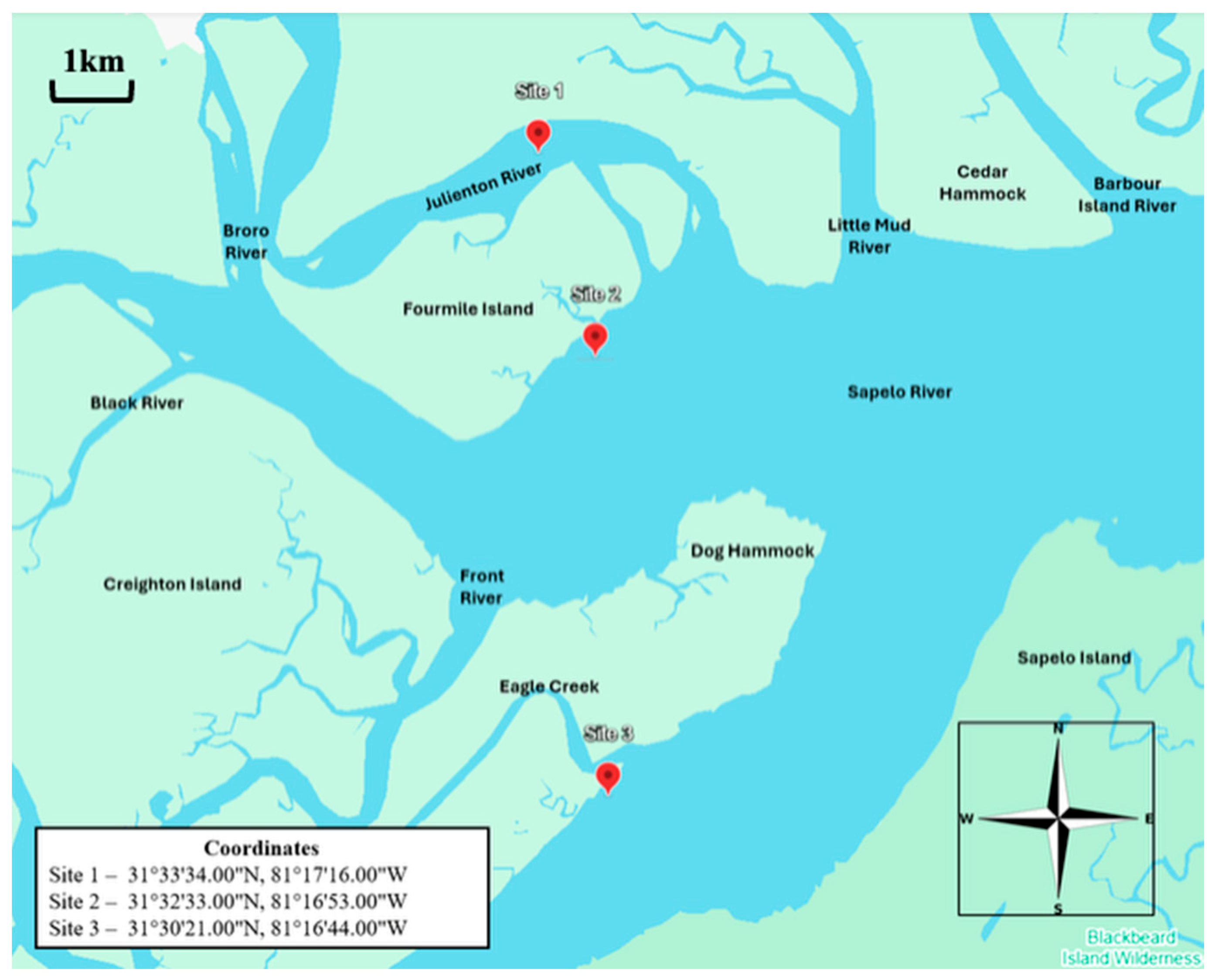
Figure 1. Study sites.
Microorganisms 13 02167 g001
Figure 2. Copy numbers/g/mL of V. splendidus in Sites 1 (A), 2 (B), and 3 (C). The data for the figure were obtained from samples collected in triplicate from three segments of each site. Nine samples were collected at each site in each sampling month. Each triplicate was taken ~25 m apart to ensure an accurate representation of the site. Average copy numbers were displayed per 1 g of sediment, clams, and oysters and 1 mL of water. Error bars represent standard deviations (N = 9).
Microorganisms 13 02167 g002
Figure 3. Copy numbers/g/mL of V. alginolyticus in Sites 1 (A), 2 (B), and 3 (C). The data for the figure were obtained from samples collected in triplicate from three segments of each site. Nine samples were collected at each site in each sampling month. Each triplicate was taken ~25 m apart to ensure an accurate representation of the site. Average copy numbers were displayed per 1 g of sediment, clams, and oysters and 1 mL of water. Error bars represent standard deviations (N = 9).
Microorganisms 13 02167 g003
Figure 4. Copy numbers/g/mL of V. harveyi/V. campbellii in Sites 1 (A), 2 (B), and 3 (C). The data for the figure were obtained from samples collected in triplicate from three segments of each site. Nine samples were collected at each site in each sampling month. Each triplicate was taken ~25 m apart to ensure an accurate representation of the site. Average copy numbers were displayed per 1 g of sediment, clams, and oysters and 1 mL of water. Error bars represent standard deviations (N = 9).
Microorganisms 13 02167 g004
Figure 5. Copy numbers/g/mL of V. parahaemolyticus in Sites 1 (A), 2 (B), and 3 (C). The data for the figure were obtained from samples collected in triplicate from three segments of each site. Nine samples were collected at each site in each sampling month. Each triplicate was taken ~25 m apart to ensure an accurate representation of the site. Average copy numbers were displayed per 1 g of sediment, clams, and oysters and 1 mL of water. Error bars represent standard deviations (N = 9).
Microorganisms 13 02167 g005
Table 1. Occurrence of Vibrio species in sediment and water in Sites 1–3.
Site/Targeted SpeciesVaVsVpVc & Vh
Site 1
June Sediment+
August Sediment++
October Sediment
December Sediment
February Sediment++
April SedimentN/A
June Water
August Water++
October Water++
December Water++
February Water+
April Water++N/A
Site 2
June Sediment+
August Sediment++
October Sediment+
December Sediment
February Sediment++
April SedimentN/A
June Water
August Water++
October Water++
December Water++
February Water+
April Water++N/A
Site 3
June Sediment+
August Sediment++
October Sediment+
December Sediment
February Sediment++
April SedimentN/A
June Water+
August Water++
October Water+
December Water++
February Water+
April Water++N/A
Detection Frequency3/36 (8.33%)15/36 (41.67%)19/36 (52.78%)7/36 (19.44%)
Sediment Total0/18 (0%)3/18 (16.67%)8/18 (44.44%)6/18 (33.33%)
Water Total3/18 (16.67%)12/18 (66.67%)11/18 (61.11%)1/18 (5.55%)
Captions: Va: V. alginolyticus, Vs: V. splendidus, Vp: V. parahaemolyticus, Vc & Vh: V. campbellii and V. harveyi. N/A: not analyzed.
Table 2. Occurrence of Vibrio species in clams and oysters in Sites 1–3.
Site/Targeted SpeciesVaVsVp
Site 1
June Clam+
August Clam+++
October Clam+
December Clam+
February Clam+
April Clam+
June Oyster+
August Oyster+++
October Oyster+
December Oyster+
February Oyster+
April Oyster+
Site 2
June Clam+
August Clam+++
October Clam+
December Clam+
February Clam+
April Clam++
June Oyster+
August Oyster+++
October Oyster+
December Oyster+
February Oyster+
April Oyster+
Site 3
June Clam
August Clam+++
October Clam+
December Clam+
February Clam+
April Clam
June Oyster+
August Oyster+++
October Oyster+
December Oyster+
February Oyster+
April Oyster+
Detection Frequency7/36 (19.44%)6/36 (16.67%)34/36 (94.44%)
Clam Total4/18 (22.22%)3/18 (16.67%)16/18 (88.89%)
Oyster Total3/18 (16.67%)3/18 (16.67%)18/18 (100%)
Captions: Va: V. alginolyticus, Vs: V. splendidus, Vp: V. parahaemolyticus. No V. campbellii and V. harveyi were ever detected.
Table 3. Correlation (Pearson’s Coefficient) between water parameters and abundance of Vibrio species in the water column.
Pearson Coeff.pHTemp. (°C)Salinity (ppt)Turbidity (NTU)dOxygen (mg/L)Conduct (mS/cm)TDS (g/L)Water Density
(g/cm3)
Vp−0.4250.597 *0.331−0.210−0.3750.2560.259−0.127
Va0.0000.0000.0000.0000.0000.0000.0000.000
Vs−0.3510.4080.455−0.070−0.1820.4340.3860.070
Vc & Vh−0.745 **0.462−0.3250.456−0.485−0.372−0.430−0.598 *
Captions: Orange, a moderate positive correlation; Dark Blue: a strong negative correlation; Light Blue: a modest negative correlation; No Color, no correlation. * p-value of 0.05, ** p-value of 0.01, no asterisk indicates no significance of p-values. Va: V. alginolyticus, Vs: V. splendidus, Vp: V. parahaemolyticus, and Vc & Vh: V. campbellii and V. harveyi.
Table 4. Correlation (Pearson’s Coefficient) between water parameters and abundance of Vibrio species in sediment.
Pearson CoefficientpHTemp
(°C)
Salinity (ppt)Turbidity (NTU)Dissolved Oxygen (mg/L)Conduct
(mS/cm)
TDS (g/L)Water Density (g/cm3)
Vp−0.4550.636 *0.315−0.215−0.4090.2270.240−0.166
Va0.0000.0000.0000.0000.0000.0000.0000.000
Vs0.602 *−0.134−0.816 ***−0.867 ***−0.305−0.736 **−0.738 **−0.365
Vc & Vh−0.724 **0.437−0.3340.460−0.467−0.378−0.435−0.588 *
Captions: Va: V. alginolyticus, Vs: V. splendidus, and Vp: V. parahaemolyticus. Orange: a moderate positive correlation; Dark Blue: a strong negative correlation; Light Blue: a modest negative correlation; No Color, no correlation. * p-value of 0.05, ** p-value of 0.01, *** p-value of 0.001, no asterisk indicates no significance of p-values.
Table 5. Correlation (Pearson’s Coefficient) between water parameters and abundance of Vibrio species in oysters.
Pearson CoefficientpHTemp
(°C)
Salinity (ppt)Turbidity (NTU)dOxygen (mg/L)Conduct (mS/cm)TDS (g/L)Water Density
(g/cm3)
Vp−0.1960.579 *−0.064−0.601 *−0.559 *−0.108−0.107−0.339
Va−0.4630.632 *0.303−0.205−0.4150.2240.225−0.171
Vs−0.4640.632 *0.303−0.205−0.4160.2240.225−0.172
Captions: Va: V. alginolyticus, Vs: V. splendidus, and Vp: V. parahaemolyticus. Light Orange: a modest positive correlation; Light Blue: a modest negative correlation; No Color, no correlation. * p-value of 0.05, no asterisk indicates no significance of p-values.
Table 6. Correlation (Pearson’s Coefficient) between water parameters and abundance of Vibrio species in clams.
Pearson CoefficientpHTemp
(°C)
Salinity (ppt)Turbidity (NTU)dOxygen (mg/L)Conduct (mS/cm)TDS (g/L)Water Density (g/cm3)
Vp−0.4640.632 *0.303−0.205−0.4150.2240.225−0.171
Va−0.4640.632 *0.303−0.205−0.4150.2240.225−0.171
Vs−0.4640.632 *0.303−0.205−0.4150.2240.225−0.171
Captions: Va: V. alginolyticus, Vs: V. splendidus, and Vp: V. parahaemolyticuss. Orange: a moderate positive correlation; Dark Blue: a strong negative correlation; Light Blue: a modest negative correlation; No Color, no correlation. * p-value of 0.05, and No asterisk indicates no significance of p-values.
Table 7. Correlations (Pearson’s Coefficient) between copy numbers of the virulence genes and species-specific copy numbers.
Pearson Coefficient WaterSite 1—WaterSite 2—WaterSite 3—WaterSite 1—SedimentSite 2—SedimentSite 3—Sediment
Va ToxRNANANANANANA
Va LuxRNANANANANANA
Va SrpNANANANANANA
Va vhhANANANANANANA
Va VhpNANANANANANA
Va vhhNANANANANANA
Vs toxR−0.266628−0.1783480.614342 *−0.369999−0.308843−0.237613
Vs luxR0.576408 *−0.3092550.956181 ***−0.300033−0.243797−0.379674
Vs srp−0.267089−0.2766350.911818 ***−0.314119−0.029936−0.251624
Vs vhhA0.636287 *0.3364430.956532 ***−0.253705−0.418526−0.255721
Vs vhp0.914971 ***−0.2888700.956312 ***−0.305423−0.359860−0.244952
Vs vhh−0.1823110.0298430.940769 ***−0.237935−0.099112−0.315662
Vp toxR−0.326406−0.1986230.675464 **−0.3764100.993103 ***−0.237613
Vp luxR0.517126 *−0.3297300.994786 ***0.985776 ***0.997616 ***−0.379674
Vp srp−0.326671−0.2984970.961058 ***−0.0312310.962990 ***−0.251624
Vp vhhA0.577854 *0.3193960.994962 ***−0.2457870.821006 ***−0.255721
Vp vhp0.908499 ***−0.3090280.994787 ***−0.2324700.042736−0.244952
Vp vhh−0.2382710.0033210.985684 ***−0.2153600.524304 *−0.315662
Vh/Vc toxR−0.2500000.991771 ***0.564862 *0.864884 ***0.798366 ***−0.252737
Vh/Vc luxR0.590599 *0.999925 ***−0.171827−0.0041370.839814 ***0.863118 ***
Vh/Vc srp−0.2504940.998538 ***−0.1980960.989971 ***0.820817 ***−0.241597
Vh/Vc vhhA0.649705 **0.790431 ***−0.1714630.997152 ***0.998322 ***−0.234020
Vh/Vc vhp0.915609 ***0.999913 ***−0.1729670.983395 ***−0.072447−0.259005
Vh/Vc vhh−0.1657450.565125 *−0.0975610.998677 ***−0.031420−0.336497
Captions: Va: V. alginolyticus, Vs: V. splendidus, Vp: V. parahaemolyticus, and Vc & Vh: V. campbellii and V. harveyi. Dark Orange: a strong positive correlation, Light Orange: a modest positive correlation, Light Blue: a modest negative correlation. * p-value > 0.05, ** p-value ≤ 0.01, and *** p-value ≤ 0.001. NA: not applicable.
Disclaimer/Publisher’s Note: The statements, opinions and data contained in all publications are solely those of the individual author(s) and contributor(s) and not of MDPI and/or the editor(s). MDPI and/or the editor(s) disclaim responsibility for any injury to people or property resulting from any ideas, methods, instructions or products referred to in the content.