Article

Mechanistic Insights into Eimeria tenella-Induced Host Cell Apoptosis Through Modulation of the Mitochondrial Permeability Transition Pore

1
College of Veterinary Medicine, Shanxi Agricultural University, Jinzhong 030801, China
2
College of Animal Science, Shanxi Agricultural University, Jinzhong 030801, China
*
Author to whom correspondence should be addressed.
Microorganisms 2025, 13(9), 2139; https://doi.org/10.3390/microorganisms13092139
Submission received: 23 July 2025 / Revised: 9 September 2025 / Accepted: 11 September 2025 / Published: 12 September 2025
(This article belongs to the Section Molecular Microbiology and Immunology)

Abstract

Coccidiosis due to Eimeria tenella remains a major constraint on the poultry industry. Previous studies have revealed that E. tenella infection triggers apoptosis in host cells. The mitochondrial permeability transition pore (MPTP) plays a pivotal role in the apoptosis and necrosis observed in infected host cells. However, the effect of MPTP opening on mitochondrial apoptotic factors remains unclear. To elucidate the dynamic changes in apoptotic signals during MPTP-mediated apoptosis in host cells infected with E. tenella, we established a chicken embryo caecal epithelial cell infection model. Cyclosporin A (CsA) was used to inhibit the MPTP. The infection rate was assessed by Hematoxylin and eosin (H&E) staining, whereas MPTP opening and the abundances of the mitochondrial apoptotic factors Smac, Endo G, and AIF were determined by flow cytometry and ELISA, respectively. The results revealed that both the degree of MPTP opening was markedly reduced in the E. tenella+CsA group compared to the E. tenella group (p < 0.05). Between 24 and 120 h post-infection (hpi), the cytoplasmic levels of Smac, Endo G, and AIF were significantly elevated in the E. tenella group compared with the control group (p < 0.05), while their mitochondrial levels were markedly decreased (p < 0.05). In contrast, mitochondrial expression of these factors was restored in the E. tenella+CsA group (p < 0.05), accompanied by a reduction in their cytoplasmic abundance (p < 0.05). These findings indicate that E. tenella promotes MPTP-dependent release of mitochondrial pro-apoptotic factors into the cytosol during the mid-to-late stages of infection, whereas pharmacological inhibition of the MPTP limits this redistribution.

1. Introduction

Eimeria tenella (E. tenella) is one of the most virulent coccidian species and predominantly infects the chicken caecum [1]. Infection often leads to reduced weight gain and egg production, and in severe cases, mortality can reach up to 80%, resulting in an estimated global economic loss of approximately $14.5 billion annually [2]. E. tenella infection causes swelling, collapse, and necrotic shedding of mucosal epithelial cells, thereby contributing to severe mucosal damage [3]. This structural damage is accompanied by increased apoptosis in caecal mucosal and intestinal gland epithelial cells [4], which represents a key pathogenic mechanism underlying host tissue injury. During early infection, expression of both anti- and pro-apoptotic genes (e.g., Bax and Bad) declines, yet the anti-/pro-apoptotic Bcl-2 ratio rises, paralleling reduced host–cell apoptosis. In contrast, in mid- to late infection, Bcl-2, Bcl-XL, Bax, Bak, Bid, Bad, and HK-II rise sharply; the anti-/pro-apoptotic ratio falls, and host–cell apoptosis surges [5].
The mitochondrion is a double-membrane-bound organelle with a sac-like structure. The compartment enclosed by the inner mitochondrial membrane is referred to as the inner chamber, while the intermembrane space between the inner and outer membranes constitutes the outer chamber [6,7]. The mitochondrial permeability transition pore (MPTP) is a multiprotein complex at inner–outer membrane contact sites, comprising structural and regulatory subunits [8,9]. Studies have demonstrated that the MPTP-induced increase in mitochondrial membrane permeability is a crucial event in mitochondrial apoptosis signaling pathways [10]. Mitochondrial apoptosis represents a major mechanism of apoptosis during the development of E. tenella [11]. The invasion of E. tenella into chicken caecal epithelial cells triggers the opening of the MPTP, reduces mitochondrial membrane potential, and enhances host cell apoptosis [12]. Cyclosporin A (CsA), a specific inhibitor of the MPTP, binds to cyclophilin D (CypD), a component of the MPTP complex, thereby preventing its opening through inhibition of peptidyl-prolyl cis-trans isomerase (PPIase) activity [13].
Mitochondria contain several key pro-apoptotic proteins, including endonuclease G (Endo G), cytochrome c (cyt c), apoptosis-inducing factor (AIF), and the second mitochondria-derived activator of caspases (Smac). Studies have shown that during the early stage of apoptosis, cyt c is released from the mitochondria into the cytoplasm, thereby triggering the mitochondrial apoptosis signaling pathway and leading to cell death [14,15]. In this pathway, Smac primarily functions by antagonizing the caspase-inhibitory activity of inhibitor of apoptosis proteins (IAPs), thereby facilitating apoptosis [16]. Additionally, upon stimulation by apoptotic signals, Endo G is released together with cyt c and AIF, two other mitochondrial apoptotic proteins. After translocating to the nucleus, Endo G mediates nuclear DNA fragmentation, ultimately leading to apoptosis [17]. Similar mitochondrial pro-apoptotic events have also been observed during infections with other pathogens. For instance, reovirus promotes the release of cyt c and Smac from the mitochondria of human embryonic kidney (HEK) cells [18]. Another study revealed the involvement of AIF and Endo G in the apoptosis of bovine mononuclear macrophages induced by Mycobacterium infection [19].
The intracellular levels of cyt c, Smac, Endo G, and AIF in host cells undergo significant alterations during the development of intracellular parasites, including Toxoplasma gondii, Trypanosoma cruzi, and Leishmania [20,21,22]. Therefore, the mechanism by which these parasites induce host cell apoptosis is believed to be intricately associated with mitochondrial apoptotic signaling pathways [23,24]. We previously demonstrated that cyt c participates in E. tenella-induced host–cell apoptosis via the intrinsic mitochondrial apoptotic pathway [12].
Chicken caecal epithelial cells are the primary target of E. tenella, yet the expression profiles of apoptotic factors in these host cells during infection remain poorly understood. Furthermore, the role of the MPTP in activating apoptotic factors during E. tenella infection has not been fully elucidated. This study aimed to characterize the temporal expression dynamics of mitochondrial apoptotic factors in infected host cells and to elucidate their relationship with MPTP activity. Using a chicken embryo caecal epithelial cell model, we employed hematoxylin and eosin (H&E) staining, flow cytometry, ELISA, and related assays to investigate how MPTP opening modulates the expression of key apoptotic regulators. This study aimed to elucidate the temporal dynamics of mitochondrial apoptotic factors in chicken caecal epithelial cells during E. tenella infection and to determine whether these events are regulated by MPTP opening, thereby providing novel insights into mitochondria-mediated host–pathogen interactions.

2. Materials and Methods

2.1. Experimental Animals

All animal experiments were conducted in accordance with the principles of laboratory animal care and the guidelines established by the Institutional Animal Care and Use Committee of Shanxi Agricultural University. Every effort was made to minimize animal suffering. The study protocol was approved by the Institutional Animal Care and Use Committee of Shanxi Agricultural University. A total of 10 1-day-old chicks and 100 15-day-old specific pathogen-free (SPF) chicken embryos were used and supplied by Beijing Boehringer Ingelheim Vital Biotechnology Co., Ltd. (Beijing, China). The 1-day-old chicks were raised in a strictly pathogen-free environment. Isolator conditions were as follows (temperature and pressure): 1–3 days, 35–36 °C and 25 Pa; 4–7 days, 32–35 °C and 25–35 Pa; 8–14 days, 29–32 °C and 35–45 Pa; and 15–30 days, 21–25 °C and 55–75 Pa.

2.2. Parasites

The E. tenella Shanxi virulent strain (EtSX01) used in this study was kindly provided by the Laboratory of Veterinary Pathology, College of Veterinary Medicine, Shanxi Agricultural University.

2.3. Primary Culture of Chick Embryo Caecal Cells

Chicken embryo caecal epithelial cells were isolated and cultured as previously described [4]. Briefly, 15-day-old SPF chicken embryos were surface-sterilized, and caecal tissues were dissected, then rinsed three times with double-antibiotic PBS (Solarbio, Beijing, China). Tissues were minced into ~1 mm3 fragments and digested with thermophilic protease (50 mg/L; Sigma-Aldrich, St. Louis, MO, USA) at 37 °C for 2 h. The digestion was neutralized with PBS and centrifuged at 344× g for 5 min. The resulting cell pellet was resuspended in DMEM (HyClone, Logan, UT, USA) supplemented with 2.5% FBS (Sijiqing, Hangzhou, China), EGF (0.02 µg/mL; PeproTech, Rocky Hill, NJ, USA), penicillin–streptomycin (100 U/mL), heparin (0.1 mg/mL), sodium pyruvate (1.1 mg/mL), and L-glutamine (0.5 mmol/L) (all from Solarbio). Cells were counted and incubated at 41 °C in 8% CO2. When the adhesion rate reached 85%, they were harvested for subsequent experiments.

2.4. Preparation of Sporozoites of E. tenella

The preparation of E. tenella sporozoites followed the method described in a previous study [25]. Ten SPF chicks were raised in isolators until 20 days of age and orally inoculated with 5 × 10³ sporulated E. tenella oocysts per chick. On days 6–8 post-infection, oocysts were collected from feces, sporulated, and centrifuged at 1490× g for 5 min. The supernatant was discarded, and the pellet was resuspended in 2 mL PBS. The sample was ground using a glass rod until the excystation rate reached approximately 80%. The homogenate containing sporocysts was centrifuged at 955× g for 10 min, and the precipitate was resuspended in an appropriate volume of sporocyst digestion solution (0.75%, v/v, trypsin from Solarbio and 10% bile from SPF chickens). The mixture was shaken and incubated at 41 °C for 70 min until ≥80% sporozoite release was achieved. After filtration, the sample was centrifuged again at 1490× g for 10 min, and the pellet was resuspended in infection medium for counting. The infection medium consisted of low-glucose DMEM (HyClone), MEM199 (HyClone) mixed in a 2:1 (v/v) ratio, 1.5% FBS (Sijiqing), EGF (0.02 μg/mL; PeproTech), glucose (0.3 mg/mL), penicillin (100 U/mL), streptomycin (100 U/mL), heparin (0.1 mg/mL), sodium pyruvate (1.1 mg/mL), insulin (0.05 μg/mL), L-glutamine (0.5 mM), folic acid (0.05 μg/mL), vitamin B6 (0.05 μg/mL), and vitamin B1 (0.05 μg/mL) (all from Solarbio, Beijing, China).

2.5. Experimental Protocol

Chick embryo caecal epithelial cells cultured in six-well plates and chamber slides were harvested at approximately 85% confluence and randomly assigned to three experimental groups: (1) the control group, (2) the E. tenella group (infected with E. tenella sporozoites), and (3) the E. tenella+CsA group (infected with E. tenella sporozoites and treated with CsA; 20 μM, Dalian Mellon Biological Technology Co., Ltd., Dalian, China). In the infected groups, cells were inoculated with 4 × 10⁵ freshly excysted E. tenella sporozoites per well. For the E. tenella+CsA group, cells sampled at 4 h post-infection (hpi) received CsA treatment at the time of infection, whereas those sampled at 24–120 hpi were treated with CsA 24 h prior to sampling. All groups were maintained for up to 120 hpi at 41 °C in a humidified incubator with 8% CO2, and the culture medium was refreshed every 48 h. Samples were collected at 4, 24, 48, 72, 96, and 120 hpi. Five replicates per group were used to assess infection rate, apoptosis rate, and Smac, AIF, and Endo G expression levels in both the mitochondria and the cytoplasm.

2.6. H&E Staining and Quantification of E. tenella Infection Rates

At 4, 24, 48, 72, 96, and 120 hpi, the infection rate was assessed in each group following H&E staining. The procedures were conducted as previously described before the study [12]. In each sample, 200 cells were randomly selected and examined under a light microscope to evaluate E. tenella infection. The infection rate at each time point was calculated using the following formula:
Infection rate (%) = (number of infected cells observed at each time point/200) × 100.

2.7. Dynamic Assessment of MPTP Opening in E. tenella-Infected Cells

The dynamic changes in MPTP opening in E. tenella-infected cells were evaluated by flow cytometry (FCM) using Calcein-AM (Thermo Fisher Scientific, Carlsbad, CA, USA) combined with CoCl2 (Sigma-Aldrich). Briefly, caecal epithelial cells from E. tenella-infected chicken embryos were harvested and suspended in 200 μL of loading buffer. The cells were stained with Calcein-AM and incubated at 37 °C for 15 min, followed by washing with Hank’s balanced salt solution. Next, the cells were treated with CoCl2 at 37 °C for 10 min. After adding 250 μL of binding buffer, the samples were analyzed by flow cytometry. The fluorescence intensity of Calcein-AM reflects the extent of MPTP opening, with higher fluorescence indicating a lower degree of pore opening [8].

2.8. Mitochondrial Isolation and Dynamic Detection of Apoptotic Factor Levels in E. tenella-Infected Host Cells

Mitochondria were isolated from E. tenella-infected chick embryo caecal epithelial cells using a mitochondrial isolation kit (Beyotime, Shanghai, China), following the manufacturer’s instructions. The dynamic changes in the levels of cyt c, Smac, Endo G, and AIF in whole-cell lysates, mitochondrial fractions, and cytoplasmic fractions at various time points post-infection were quantified using ELISA kits specific for Smac, Endo G, and AIF (all from MyBioSource, San Diego, CA, USA), according to the manufacturers’ protocols.

2.9. Statistical Analysis

All quantitative data are expressed as mean ± standard deviation (SD) and were analyzed using SPSS version 22.0 (IBM Corp., Armonk, NY, USA). One-way analysis of variance (ANOVA) followed by Tukey’s post hoc test was employed to determine statistical significance among experimental groups. Data visualization was performed using GraphPad Prism version 5.0 (GraphPad Software, San Diego, CA, USA). Each experiment was repeated at least three times. Differences were considered statistically significant at p < 0.05 and highly significant at p < 0.01.

3. Results

3.1. Inhibition of MPTP Selectively Enhances Intracellular Expansion of E. tenella

H&E staining revealed the presence of intracellular E. tenella in both groups, with a progressive increase in parasite burden over time. At 4 and 24 hpi, there were no statistically significant differences in infection rates between the E. tenella group and the E. tenella+CsA group (p > 0.05). However, starting at 48 hpi, the E. tenella+CsA group exhibited a significantly greater parasite burden in epithelial cells compared to the E. tenella group (p < 0.05, Figure 1A,B). Quantitative analysis of infection rates further confirmed that between 48 and 120 hpi, the infection rate in the E. tenella+CsA group was significantly higher than that in the E. tenella group (p < 0.05; Figure 1B). These results suggest that inhibiting MPTP opening does not affect the early invasion of E. tenella but enhances its intracellular replication and development at later stages of infection.

3.2. CsA Preserves Mitochondrial Integrity by Inhibiting E. tenella-Induced MPTP Opening

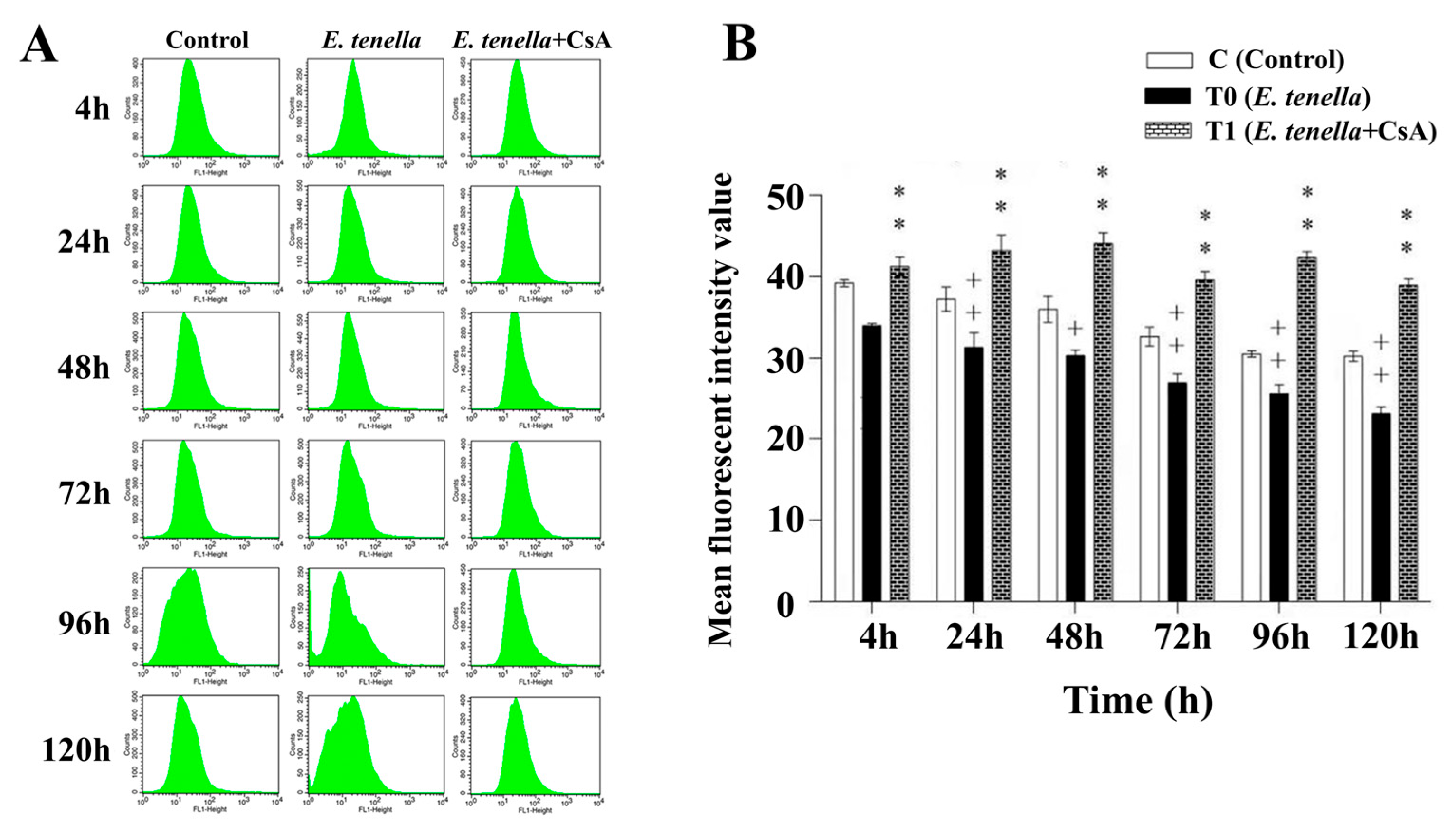
Flow cytometric analysis using the Calcein-AM/CoCl2 fluorescence quenching assay revealed a progressive decline in mitochondrial mean fluorescence intensity (MFI) in E. tenella-infected cells between 4 and 120 hpi, accompanied by a gradual leftward shift in the fluorescence peak (Figure 2A). Quantitative data showed that the mitochondrial MFI in the E. tenella-infected group remained consistently lower than that in the control group at all examined time points, with a statistically significant reduction observed from 24 to 120 hpi (p < 0.01; Figure 2B). In contrast, post-infection CsA treatment helped preserve mitochondrial fluorescence, as indicated by significantly higher calcein-AM fluorescence intensity compared to the E. tenella-infected group (p < 0.01; Figure 2B). These findings suggest that E. tenella induces rapid opening of the MPTP in host intestinal epithelial cells, which progressively intensifies throughout the course of infection, whereas CsA effectively inhibits MPTP opening and preserves mitochondrial integrity.

3.3. Temporal MPTP-Driven Smac Redistribution During E. tenella Infection

The expression of Smac protein in the E. tenella-infected group was significantly decreased at 4 hpi, but markedly increased at 48 hpi compared to the control group (p < 0.05). At both 4 and 48 hpi, Smac levels were markedly lower in the E. tenella+CsA group compared to the E. tenella group (p < 0.05; Figure 3A). In mitochondrial fractions, Smac expression in the E. tenella group was significantly lower than that in the control group throughout 4–120 hpi (p < 0.05), while the E. tenella+CsA group exhibited elevated levels of mitochondrial Smac compared to the E. tenella group from 24 to 120 hpi (p < 0.05; Figure 3B). A significant reduction in cytoplasmic Smac levels was observed in the E. tenella group at 4 hpi (p < 0.01), followed by a marked increase from 24 to 120 hpi compared to the control (p < 0.01). Moreover, cytoplasmic Smac levels in the E. tenella+CsA group were significantly lower than those in the E. tenella group at all examined time points (p < 0.05; Figure 3C). These findings indicate that Smac expression is regulated through MPTP opening during the middle and late stages of E. tenella infection.

3.4. Temporal MPTP-Driven Endo G Redistribution During E. tenella Infection

The total protein expression of Endo G in host cells was significantly reduced in the E. tenella-infected group at 4 hpi (p < 0.05), but significantly increased at 72, 96, and 120 hpi compared to the control group (p < 0.01). The total Endo G expression level in the E. tenella+CsA group was significantly lower than that in the E. tenella group from 24 to 120 hpi (p < 0.05; Figure 4A). In mitochondrial fractions, Endo G expression was significantly lower in the E. tenella group than in the control group from 4 to 120 hpi (p < 0.05), while the E. tenella+CsA group exhibited significantly higher mitochondrial Endo G levels compared to the E. tenella group during the same period (p < 0.05; Figure 4B). In the cytoplasmic fraction, Endo G levels were significantly reduced in the E. tenella group at 4 hpi (p < 0.05), but significantly elevated at 24, 72, 96, and 120 hpi relative to the control (p < 0.05). Moreover, cytoplasmic Endo G expression in the E. tenella+CsA group was consistently lower than that in the E. tenella group from 24 to 120 hpi (p < 0.05; Figure 4C). These results indicate that Endo G expression is regulated by MPTP opening during the middle and late stages of E. tenella infection.

3.5. Temporal MPTP-Driven AIF Redistribution During E. tenella Infection

The expression of AIF in host cells was significantly lower in the E. tenella group than in the control group at 4 hpi (p < 0.05), but significantly higher from 24 to 120 hpi (p < 0.05). AIF expression levels in the E. tenella+CsA group were significantly reduced compared to the E. tenella group throughout the 24–120 hpi period (p < 0.05; Figure 5A). In mitochondrial fractions, AIF expression was significantly elevated in the E. tenella group at 4 hpi (p < 0.01), but significantly decreased from 24 to 120 hpi compared to controls (p < 0.01). Notably, mitochondrial AIF levels were significantly higher in the E. tenella+CsA group than in the E. tenella group during this same period (p < 0.01; Figure 5B). In the cytoplasm, AIF expression in the E. tenella group was significantly lower than that in the control group at 4 hpi (p < 0.01), but significantly higher from 24 to 120 hpi (p < 0.01). Cytoplasmic AIF levels in the E. tenella+CsA group remained significantly lower than those in the E. tenella group at all corresponding time points (p < 0.01; Figure 5C). These results indicate that AIF expression is regulated by MPTP opening during the middle and late stages of E. tenella infection.

4. Discussion

In this study, we investigated the mitochondrial apoptotic signaling pathways in chicken embryonic caecal epithelial cells infected with E. tenella. The results demonstrate that apoptosis is primarily mediated by the opening of the MPTP and the mitochondrial release of pro-apoptotic factors (Smac, Endo G, AIF). This finding is consistent with both our prior evidence of an increase in apoptosis during the mid-to-late stages of infection and the involvement of cyt c in the intrinsic mitochondrial apoptotic pathway [12]. These data provide important insights into the mechanism underlying E. tenella infection in chickens.
Mitochondria serve as central regulators of apoptosis [26,27]. Upon initiation of apoptosis in host cells, the MPTP opens, facilitating the release of cyt c and Smac into the cytoplasm. These molecules subsequently activate caspases, triggering the apoptotic cascade [28,29]. In addition, a caspase-independent apoptotic pathway has also been identified within the mitochondrial network [30]. In this pathway, AIF and Endo G exit mitochondria and enter the cytoplasm, then migrate to the nucleus. They trigger chromatin condensation and DNA fragmentation, culminating in apoptosis [31,32].
At 4 hpi, the MPTP opening in the E. tenella-infected group was greater than that in the control group, whereas the opening in the E. tenella+CsA group was reduced relative to the E. tenella group. Interestingly, this pattern did not correspond to the changes observed in early apoptosis, late apoptosis, and necrosis rates in host cells [12]. This discrepancy may be attributed to the involvement of multiple apoptotic signaling pathways within host cells [33]. Previous studies have shown that early E. tenella infection activates Akt, which lowers Bad, elevates Bcl-2, and restricts cytochrome c release. Collectively, these changes blunt host–cell apoptosis [34]. Moreover, it has been reported that Akt activation suppresses the generation and cytoplasmic release of AIF, Smac, and Endo G, thus interrupting the apoptotic cascade [35,36]. Therefore, the precise molecular mechanisms by which E. tenella modulates early-stage host cell apoptosis remain incompletely understood and merit further investigation.
From 24 to 120 hpi, the opening of the MPTP, the Smac, Endo G, and AIF, as well as the early apoptosis, late apoptosis, and necrosis rates, were all significantly higher in the E. tenella-infected group compared to the control group [12]. In contrast, the mitochondrial levels of Smac, Endo G, and AIF were significantly reduced in the E. tenella group. These findings indicate that E. tenella promotes host cell apoptosis during the middle and late stages of infection by enhancing MPTP opening and facilitating the release of pro-apoptotic factors from the mitochondria. Notably, CsA markedly reduced the cytoplasmic levels of Smac, Endo G, and AIF in infected cells. These results suggest that MPTP opening plays a critical role in regulating the translocation of mitochondrial apoptotic factors into the cytoplasm, thereby promoting apoptosis in host cells during the later stages of E. tenella infection.
In addition, following cadmium treatment, the mitochondrial levels of cyt c in hepatocytes are markedly reduced, while its cytoplasmic expression increases, thereby promoting apoptosis [37]. During apoptosis, Smac is also released into the cytoplasm, where it antagonizes inhibitor of apoptosis proteins (IAPs), facilitating caspase activation [38]. Similarly, pyrroloquinoline quinone translocates Endo G and AIF from mitochondria to the cytoplasm in chondrosarcoma cells, thereby heightening apoptosis [39]. These findings are consistent with our observations and further support the hypothesis that mitochondrial release of pro-apoptotic factors is a key event in the execution of apoptosis [40,41].
In line with our finding that E. tenella modulates host mitochondrial apoptosis through the MPTP, other apicomplexans similarly adjust host cell–death pathways. For example, HSP70 secreted by T. gondii binds AIF and APAF-1, elevates Bcl-2, and blocks AIF translocation and cytochrome c release, thereby curbing host mitochondrial apoptosis [23]. During its hepatic phase, Plasmodium rewires host metabolic and autophagic circuits to sustain cell viability and postpone apoptosis, thereby securing its own development [42]. Cryptosporidium parvum first activates NF-κB and elevates Bcl-2/Survivin to block apoptosis, then later induces pro-apoptotic genes and cell death, representing a stage-dependent biphasic switch [43]. Together, these studies show that apicomplexan parasites establish infection and evade host immunity by finely tuning host apoptotic pathways.
During the middle and late stages of E. tenella infection, host cell apoptosis was significantly elevated [12], characterized by reduced mitochondrial levels and increased cytoplasmic levels of cyt c, Smac, Endo G, and AIF. These results demonstrate that E. tenella promotes apoptosis by triggering MPTP opening and facilitating the translocation of mitochondrial apoptotic factors into the cytoplasm. Chicken embryonic cecal epithelial cells offer a controlled in vitro window on early E. tenella-host interactions, but they cannot recapitulate the immune complexity, microbiota, or mucosal milieu of the living cecum. Accordingly, the timing and function of MPTP opening in parasite growth and tissue injury should be confirmed in chick infection models to gauge the translational promise of MPTP-directed therapies. Despite these limitations, this study elucidates a key mitochondrial pathway involved in E. tenella-induced host cell apoptosis and provides a mechanistic basis for the development of targeted anticoccidial strategies.

5. Conclusions

In summary, we identify the MPTP as a central regulatory node exploited by E. tenella to manipulate host caecal epithelial cell fate. Parasite-induced MPTP opening orchestrates the coordinated release of Smac, Endo G, and AIF, thereby driving apoptosis. Conversely, pharmacological inhibition of MPTP by cyclosporin A preserves mitochondrial integrity but paradoxically supports deeper intracellular parasitism. By reframing host mitochondria as an active battleground rather than passive targets, our findings highlight MPTP modulation as a promising therapeutic strategy to both mitigate host tissue damage and impair parasite fitness.

Author Contributions

Conceptualization, R.B.; data curation, S.Z., H.W., C.L. and R.B.; writing—original draft preparation, W.Z., L.Z., Y.L. and R.B.; writing—review and editing, H.G., X.L. and R.B.; visualization, J.L., X.C. and R.B.; supervision, R.B.; funding acquisition, R.B. All authors have read and agreed to the published version of the manuscript.

Funding

This research was supported by the Shanxi Provincial Key Research and Development Program (Grant No. 2022ZDYF126), the Natural Science Research Program of Shanxi Province (No. 202103021224165), and the Science and Technology Innovation Promotion Project of Shanxi Agricultural University (No. CXGC2025012).

Institutional Review Board Statement

The animal study protocol was approved by Institutional Animal Care and Use Committee of Shanxi Agricultural University (SXAU-EAW-2024C.AW.012025351) approval date: 23 December 2023.

Informed Consent Statement

Not applicable.

Data Availability Statement

The original contributions presented in this study are included in the article. Further inquiries can be directed to the corresponding author.

Conflicts of Interest

The authors declare no conflicts of interest.

References

  1. Xu, L.; Xiang, Q.; Li, M.; Sun, X.; Lu, M.; Yan, R.; Song, X.; Li, X. Pathogenic Effects of Single or Mixed Infections of Eimeria mitis, Eimeria necatrix, and Eimeria tenella in Chickens. Vet. Sci. 2022, 9, 657. [Google Scholar] [CrossRef] [PubMed]
  2. Mustafa, S.; Abbas, R.Z.; Saeed, Z.; Baazaoui, N.; Khan, A.M.A. Use of Metallic Nanoparticles Against Eimeria-the Coccidiosis-Causing Agents: A Comprehensive Review. Biol. Trace Elem. Res. 2025, 203, 3412–3431. [Google Scholar] [CrossRef] [PubMed]
  3. Han, L.; Han, X.; Meng, J.; Yang, J.; Kang, S.; Lv, X.; Cui, X.; Li, J.; Liu, W.; Bai, R. Silymarin Effectively Prevents and Treats Eimeria tenella Infection in Chicks. Poult. Sci. 2024, 103, 103909. [Google Scholar] [CrossRef]
  4. Zhang, X.-S.; Zhao, Y.-J.; Zhang, Y.; Xu, T.; Cui, K.-L.; Duan, B.-T.; Lv, X.-L.; Zhang, L.; Xu, Z.-Y.; Bai, R.; et al. Role of EtMIC4 EGF-like in Regulating the Apoptosis of Eimeria tenella Host Cells via the EGFR Pathway. Poult. Sci. 2022, 101, 102075. [Google Scholar] [CrossRef]
  5. Xu, Z.-Y.; Zheng, M.-X.; Zhang, Y.; Cui, X.-Z.; Yang, S.-S.; Liu, R.-L.; Li, S.; Xi, R.; Gong, X.; Bai, R. Dynamic Changes in the Main Regulatory Genes of Mitochondrial Permeability Transition Pore in Eimeria tenella Host Cells. Exp. Parasitol. 2016, 171, 42–48. [Google Scholar] [CrossRef] [PubMed]
  6. Sunanda, T.; Ray, B.; Mahalakshmi, A.M.; Bhat, A.; Rashan, L.; Rungratanawanich, W.; Song, B.-J.; Essa, M.M.; Sakharkar, M.K.; Chidambaram, S.B. Mitochondria-Endoplasmic Reticulum Crosstalk in Parkinson’s Disease: The Role of Brain Renin Angiotensin System Components. Biomolecules 2021, 11, 1669. [Google Scholar] [CrossRef]
  7. Zong, Y.; Li, H.; Liao, P.; Chen, L.; Pan, Y.; Zheng, Y.; Zhang, C.; Liu, D.; Zheng, M.; Gao, J. Mitochondrial Dysfunction: Mechanisms and Advances in Therapy. Signal Transduct. Target. Ther. 2024, 9, 124. [Google Scholar] [CrossRef]
  8. Morciano, G.; Naumova, N.; Koprowski, P.; Valente, S.; Sardão, V.A.; Potes, Y.; Rimessi, A.; Wieckowski, M.R.; Oliveira, P.J. The Mitochondrial Permeability Transition Pore: An Evolving Concept Critical for Cell Life and Death. Biol. Rev. Camb. Philos. Soc. 2021, 96, 2489–2521. [Google Scholar] [CrossRef]
  9. Song, X.; Zhang, L.; Hui, X.; Sun, X.; Yang, J.; Wang, J.; Wu, H.; Wang, X.; Zheng, Z.; Che, F.; et al. Selenium-Containing Protein from Selenium-Enriched Spirulina Platensis Antagonizes Oxygen Glucose Deprivation-Induced Neurotoxicity by Inhibiting ROS-Mediated Oxidative Damage through Regulating MPTP Opening. Pharm. Biol. 2021, 59, 629–638. [Google Scholar] [CrossRef]
  10. Yao, H.; Xie, Q.; He, Q.; Zeng, L.; Long, J.; Gong, Y.; Li, X.; Li, X.; Liu, W.; Xu, Z.; et al. Pretreatment with Panaxatriol Saponin Attenuates Mitochondrial Apoptosis and Oxidative Stress to Facilitate Treatment of Myocardial Ischemia-Reperfusion Injury via the Regulation of Keap1/Nrf2 Activity. Oxid. Med. Cell. Longev. 2022, 2022, 9626703. [Google Scholar] [CrossRef]
  11. Li, J.-Y.; Huang, H.-B.; Pan, T.-X.; Wang, N.; Shi, C.-W.; Zhang, B.; Wang, C.-F.; Yang, G.-L. Sanguinarine Induces Apoptosis in Eimeria tenella Sporozoites via the Generation of Reactive Oxygen Species. Poult. Sci. 2022, 101, 101771. [Google Scholar] [CrossRef]
  12. Bai, R.; Wang, H.; Yang, T.; Yan, Y.; Zhu, S.; Lv, C.; Pei, Y.; Guo, J.; Li, J.; Cui, X.; et al. Mechanisms of Mitochondria-Mediated Apoptosis During Eimeria tenella Infection. Animals 2025, 15, 577. [Google Scholar] [CrossRef] [PubMed]
  13. Protasoni, M.; López-Polo, V.; Stephan-Otto Attolini, C.; Brandariz, J.; Herranz, N.; Mateo, J.; Ruiz, S.; Fernandez-Capetillo, O.; Kovatcheva, M.; Serrano, M. Cyclophilin D Plays a Critical Role in the Survival of Senescent Cells. EMBO J. 2024, 43, 5972–6000. [Google Scholar] [CrossRef] [PubMed]
  14. Zhou, Z.; Arroum, T.; Luo, X.; Kang, R.; Lee, Y.J.; Tang, D.; Hüttemann, M.; Song, X. Diverse Functions of Cytochrome c in Cell Death and Disease. Cell Death Differ. 2024, 31, 387–404. [Google Scholar] [CrossRef]
  15. Aghaei, M.; Aghaei, S.; Shahmoradi, Z.; Hejazi, S.H. The Role of Metacaspases and Other Proteins Involved in the Apoptosis of Leishmania: Review Article. Iran. J. Parasitol. 2025, 20, 1–12. [Google Scholar] [CrossRef]
  16. Ehrmann, J.F.; Grabarczyk, D.B.; Heinke, M.; Deszcz, L.; Kurzbauer, R.; Hudecz, O.; Shulkina, A.; Gogova, R.; Meinhart, A.; Versteeg, G.A.; et al. Structural Basis for Regulation of Apoptosis and Autophagy by the BIRC6/SMAC Complex. Science 2023, 379, 1117–1123. [Google Scholar] [CrossRef]
  17. Wang, W.; Li, J.; Zhou, Q. The Biological Function of Cytoplasm-Translocated ENDOG (Endonuclease G). Autophagy 2024, 20, 445–447. [Google Scholar] [CrossRef]
  18. Kominsky, D.J.; Bickel, R.J.; Tyler, K.L. Reovirus-Induced Apoptosis Requires Mitochondrial Release of Smac/DIABLO and Involves Reduction of Cellular Inhibitor of Apoptosis Protein Levels. J. Virol. 2002, 76, 11414–11424. [Google Scholar] [CrossRef]
  19. Benítez-Guzmán, A.; Arriaga-Pizano, L.; Morán, J.; Gutiérrez-Pabello, J.A. Endonuclease G Takes Part in AIF-Mediated Caspase-Independent Apoptosis in Mycobacterium bovis-Infected Bovine Macrophages. Vet. Res. 2018, 49, 69. [Google Scholar] [CrossRef] [PubMed]
  20. Azami, M.; Ranjkesh Adermanabadi, V.; Khanahmad, H.; Mohaghegh, M.A.; Zaherinejad, E.; Aghaei, M.; Jalali, A.; Hejazi, S.H. Immunology and Genetic of Leishmania infantum: The Role of Endonuclease G in the Apoptosis. J. Res. Med. Sci. Off. J. Isfahan Univ. Med. Sci. 2018, 23, 36. [Google Scholar] [CrossRef]
  21. Graumann, K.; Schaumburg, F.; Reubold, T.F.; Hippe, D.; Eschenburg, S.; Lüder, C.G.K. Toxoplasma gondii Inhibits Cytochrome C-Induced Caspase Activation in Its Host Cell by Interference with Holo-Apoptosome Assembly. Microb. Cell 2015, 2, 150–162. [Google Scholar] [CrossRef]
  22. Ronco, M.T.; Francés, D.E.; Ingaramo, P.I.; Quiroga, A.D.; Alvarez, M.L.; Pisani, G.B.; Revelli, S.S.; Carnovale, C.E. Tumor Necrosis Factor Alpha Induced by Trypanosoma cruzi Infection Mediates Inflammation and Cell Death in the Liver of Infected Mice. Cytokine 2010, 49, 64–72. [Google Scholar] [CrossRef]
  23. Lian, L.; Sun, Q.; Huang, X.; Li, W.; Cui, Y.; Pan, Y.; Yang, X.; Wang, P. Inhibition of Cell Apoptosis by Apicomplexan Protozoa-Host Interaction in the Early Stage of Infection. Animals 2023, 13, 3817. [Google Scholar] [CrossRef] [PubMed]
  24. Yan, L.; Li, Y.; Yang, X.; Li, R.; Zhu, C.; He, X.; Jin, X.; Zheng, G.; Mehmood, N.; Cho, W.C.; et al. PI3K/AKT Signaling in Parasites and Parasite Diseases: Role and Therapeutic Potential. Virulence 2025, 16, 2532803. [Google Scholar] [CrossRef] [PubMed]
  25. Duan, B.-T.; Zhang, H.-Y.; Song, Z.-H.; Han, X.-Y.; Cui, K.-L.; Xu, T.; Zhang, Y.; Zhao, Y.-J.; Lei, X.; Tan, F.; et al. EtROP38 Suppresses Apoptosis of Host Cells Infected with Eimeria tenella by Inhibition of the P38MAPK Pathway. Vet. Parasitol. 2024, 331, 110296. [Google Scholar] [CrossRef] [PubMed]
  26. Kummer, E.; Ban, N. Mechanisms and Regulation of Protein Synthesis in Mitochondria. Nat. Rev. Mol. Cell Biol. 2021, 22, 307–325. [Google Scholar] [CrossRef]
  27. Zaib, S.; Hayyat, A.; Ali, N.; Gul, A.; Naveed, M.; Khan, I. Role of Mitochondrial Membrane Potential and Lactate Dehydrogenase A in Apoptosis. Anticancer. Agents Med. Chem. 2022, 22, 2048–2062. [Google Scholar] [CrossRef]
  28. Zhou, X.; Chen, H.; Wang, L.; Lenahan, C.; Lian, L.; Ou, Y.; He, Y. Mitochondrial Dynamics: A Potential Therapeutic Target for Ischemic Stroke. Front. Aging Neurosci. 2021, 13, 721428. [Google Scholar] [CrossRef]
  29. Li, B.; Chen, X.; Yang, W.; He, J.; He, K.; Xia, Z.; Zhang, J.; Xiang, G. Single-Walled Carbon Nanohorn Aggregates Promotes Mitochondrial Dysfunction-Induced Apoptosis in Hepatoblastoma Cells by Targeting SIRT3. Int. J. Oncol. 2018, 53, 1129–1137. [Google Scholar] [CrossRef]
  30. Nguyen, T.T.; Wei, S.; Nguyen, T.H.; Jo, Y.; Zhang, Y.; Park, W.; Gariani, K.; Oh, C.-M.; Kim, H.H.; Ha, K.-T.; et al. Mitochondria-Associated Programmed Cell Death as a Therapeutic Target for Age-Related Disease. Exp. Mol. Med. 2023, 55, 1595–1619. [Google Scholar] [CrossRef]
  31. Zong, L.; Liang, Z. Apoptosis-Inducing Factor: A Mitochondrial Protein Associated with Metabolic Diseases-a Narrative Review. Cardiovasc. Diagn. Ther. 2023, 13, 609–622. [Google Scholar] [CrossRef] [PubMed]
  32. Frolova, A.S.; Chepikova, O.E.; Deviataikina, A.S.; Solonkina, A.D.; Zamyatnin, A.A.J. New Perspectives on the Role of Nuclear Proteases in Cell Death Pathways. Biology 2023, 12, 797. [Google Scholar] [CrossRef]
  33. Mammari, N.; Halabi, M.A.; Yaacoub, S.; Chlala, H.; Dardé, M.-L.; Courtioux, B. Toxoplasma gondii Modulates the Host Cell Responses: An Overview of Apoptosis Pathways. Biomed Res. Int. 2019, 2019, 6152489. [Google Scholar] [CrossRef]
  34. Zhang, X.; Li, S.; Zheng, M.; Zhang, L.; Bai, R.; Li, R.; Hao, S.; Bai, B.; Kang, H. Effects of the PI3K/Akt Signaling Pathway on the Apoptosis of Early Host Cells Infected with Eimeria tenella. Parasitol. Res. 2020, 119, 2549–2561. [Google Scholar] [CrossRef]
  35. Hu, W.-L.; Dong, H.-Y.; Li, Y.; Ojcius, D.M.; Li, S.-J.; Yan, J. Bid-Induced Release of AIF/EndoG from Mitochondria Causes Apoptosis of Macrophages during Infection with Leptospira interrogans. Front. Cell. Infect. Microbiol. 2017, 7, 471. [Google Scholar] [CrossRef]
  36. Bai, L.; Xu, S.; Chen, W.; Li, Z.; Wang, X.; Tang, H.; Lin, Y. Blocking NF-ΚB and Akt by Hsp90 Inhibition Sensitizes Smac Mimetic Compound 3-Induced Extrinsic Apoptosis Pathway and Results in Synergistic Cancer Cell Death. Apoptosis 2011, 16, 45–54. [Google Scholar] [CrossRef]
  37. Song, X.-B.; Liu, G.; Wang, Z.-Y.; Wang, L. Puerarin Protects against Cadmium-Induced Proximal Tubular Cell Apoptosis by Restoring Mitochondrial Function. Chem. Biol. Interact. 2016, 260, 219–231. [Google Scholar] [CrossRef]
  38. Saita, S.; Nolte, H.; Fiedler, K.U.; Kashkar, H.; Venne, A.S.; Zahedi, R.P.; Krüger, M.; Langer, T. PARL Mediates Smac Proteolytic Maturation in Mitochondria to Promote Apoptosis. Nat. Cell Biol. 2017, 19, 318–328. [Google Scholar] [CrossRef] [PubMed]
  39. Wu, R.; Pan, J.; Shen, M.; Xing, C. Apoptotic Effect of Pyrroloquinoline Quinone on Chondrosarcoma Cells through Activation of the Mitochondrial Caspase-dependent and Caspase-independent Pathways. Oncol. Rep. 2018, 40, 1614–1620. [Google Scholar] [CrossRef]
  40. Sun, Y.; Ge, X.; Li, X.; He, J.; Wei, X.; Du, J.; Sun, J.; Li, X.; Xun, Z.; Liu, W.; et al. High-Fat Diet Promotes Renal Injury by Inducing Oxidative Stress and Mitochondrial Dysfunction. Cell Death Dis. 2020, 11, 914. [Google Scholar] [CrossRef] [PubMed]
  41. Flores-Romero, H.; Dadsena, S.; García-Sáez, A.J. Mitochondrial Pores at the Crossroad between Cell Death and Inflammatory Signaling. Mol. Cell 2023, 83, 843–856. [Google Scholar] [CrossRef] [PubMed]
  42. Schmuckli-Maurer, J.; Bindschedler, A.F.; Wacker, R.; Würgler, O.M.; Rehmann, R.; Lehmberg, T.; Murphy, L.O.; Nguyen, T.N.; Lazarou, M.; Monfregola, J.; et al. Plasmodium Berghei Liver Stage Parasites Exploit Host GABARAP Proteins for TFEB Activation. Commun. Biol. 2024, 7, 1554. [Google Scholar] [CrossRef] [PubMed]
  43. Chen, X.M.; Levine, S.A.; Splinter, P.L.; Tietz, P.S.; Ganong, A.L.; Jobin, C.; Gores, G.J.; Paya, C.V.; LaRusso, N.F. Cryptosporidium Parvum Activates Nuclear Factor KappaB in Biliary Epithelia Preventing Epithelial Cell Apoptosis. Gastroenterology 2001, 120, 1774–1783. [Google Scholar] [CrossRef] [PubMed]
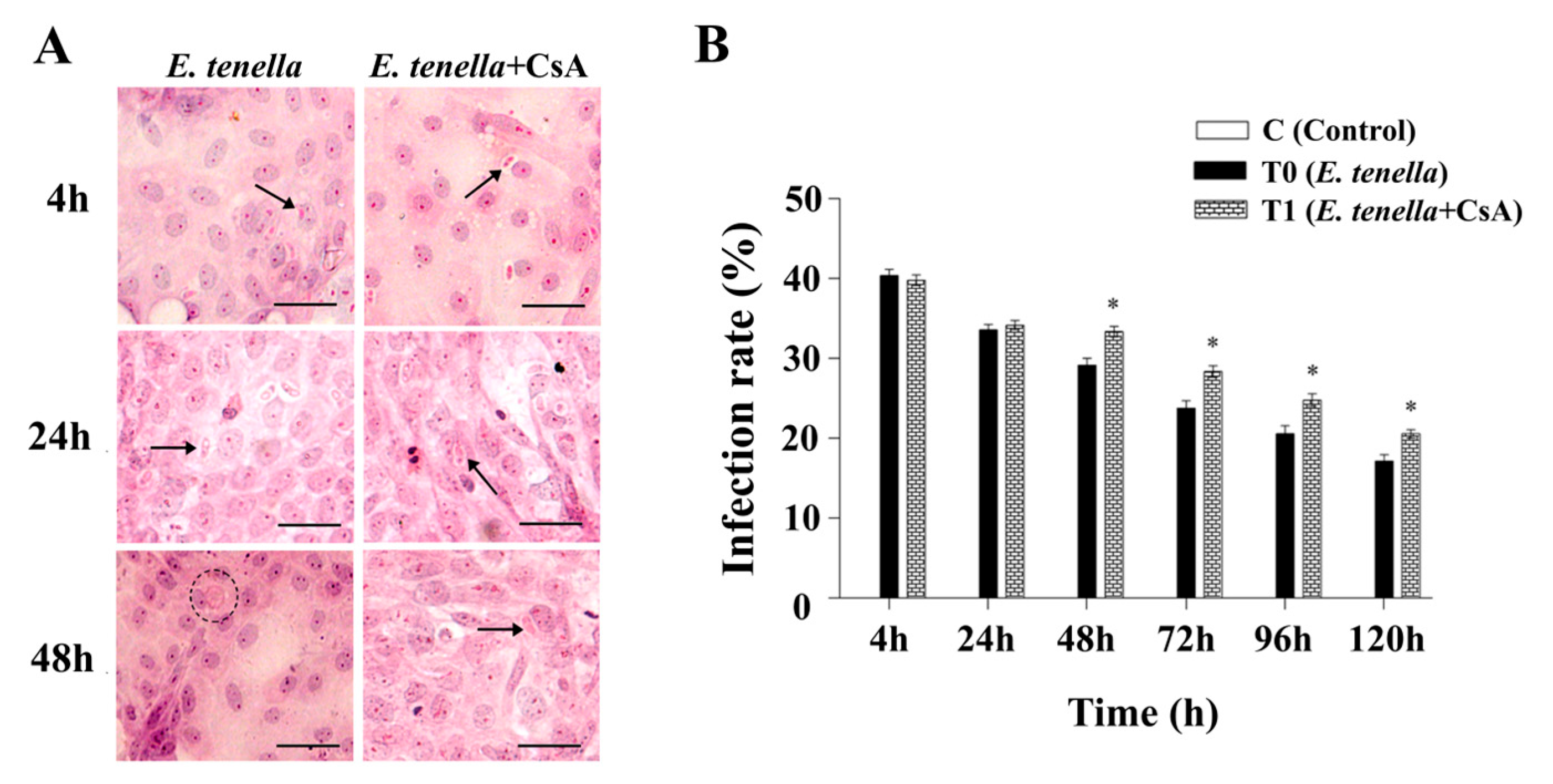
Figure 1. E. tenella infection rates. (A) In vitro development of E. tenella in each group as visualized by haematoxylin and eosin staining. Sporozoites (arrows) and trophozoites (circles) are marked accordingly. Scale bar = 20 μm. (B) Quantification of E. tenella infection rates in host cells under different treatments. C = uninfected control (infection rate = 0% at all time points; displayed as a zero-height bar. Notes: Data are presented as mean ± SD (n = 3). “*” indicates a statistically significant difference between the E. tenella+CsA group and the E. tenella-infected group (p < 0.05).
Microorganisms 13 02139 g001
Figure 2. Effects of E. tenella infection and CsA treatment on MPTP opening in chick embryo caecal epithelial cells. (A) Representative flow cytometry histograms showing changes in MPTP opening at different time points (4, 24, 48, 72, 96, and 120 hpi). (B) Quantification of MFI of calcein-AM at each time point. Notes: Data are presented as mean ± SD (n = 3). “**” indicates a highly significant difference between the E. tenella+CsA group and the E. tenella-infected group (p < 0.01). “+” indicates a statistically significant difference between the E. tenella-infected group and the control group (p < 0.05); “++” indicates a highly significant difference (p < 0.01). The same significance annotations apply throughout the manuscript.
Microorganisms 13 02139 g002
Figure 3. The dynamic changes in Smac levels in E. tenella-infected chicken embryo cecal epithelial cells, measured by ELISA (pg/mL). (A) Effects of E. tenella on total Smac in chicken embryo cecal epithelial cells. (B) Effects of E. tenella on mitochondrial Smac in chicken embryo cecal epithelial cells. (C) Effects of E. tenella on cytoplasmic Smac in chicken embryo cecal epithelial cells. Notes: Data are presented as mean ± SD (n = 3). “*” indicates a statistically significant difference between the E. tenella + CsA group and the E. tenella-infected group (p < 0.05); “**” indicates a highly significant difference (p < 0.01). “+” indicates a statistically significant difference between the E. tenella-infected group and the control group (p < 0.05); “++” indicates a highly significant difference (p < 0.01).
Microorganisms 13 02139 g003
Figure 4. The dynamic changes in Endo G levels in E. tenella-infected chicken embryo cecal epithelial cells, measured by ELISA (pg/mL). (A) Effects of E. tenella on total Endo G in chicken embryo cecal epithelial cells. (B) Effects of E. tenella on mitochondrial Endo G in chicken embryo cecal epithelial cells. (C) Effects of E. tenella on cytoplasmic Endo G in chicken embryo cecal epithelial cells. Notes: Data are presented as mean ± SD (n = 3). “*” indicates a statistically significant difference between the E. tenella + CsA group and the E. tenella-infected group (p < 0.05); “**” indicates a highly significant difference (p < 0.01). “+” indicates a statistically significant difference between the E. tenella-infected group and the control group (p < 0.05); “++” indicates a highly significant difference (p < 0.01).
Microorganisms 13 02139 g004
Figure 5. The dynamic changes in AIF levels in E. tenella-infected chicken embryo cecal epithelial cells, measured by ELISA (pg/mL). (A) Effects of E. tenella on total AIF in chicken embryo cecal epithelial cells. (B) Effects of E. tenella on mitochondrial AIF in chicken embryo cecal epithelial cells. (C) Effects of E. tenella on cytoplasmic AIF in chicken embryo cecal epithelial cells. Notes: Data are presented as mean ± SD (n = 3). “*” indicates a statistically significant difference between the E. tenella + CsA group and the E. tenella-infected group (p < 0.05); “**” indicates a highly significant difference (p < 0.01). “+” indicates a statistically significant difference between the E. tenella-infected group and the control group (p < 0.05); “++” indicates a highly significant difference (p < 0.01).
Microorganisms 13 02139 g005
Disclaimer/Publisher’s Note: The statements, opinions and data contained in all publications are solely those of the individual author(s) and contributor(s) and not of MDPI and/or the editor(s). MDPI and/or the editor(s) disclaim responsibility for any injury to people or property resulting from any ideas, methods, instructions or products referred to in the content.