
Investigating the Antimicrobial Activity of Anuran Toxins

 ,
,  ,
, 
 ,
,  and
and 
Abstract
1. Introduction
2. Anuran Fauna
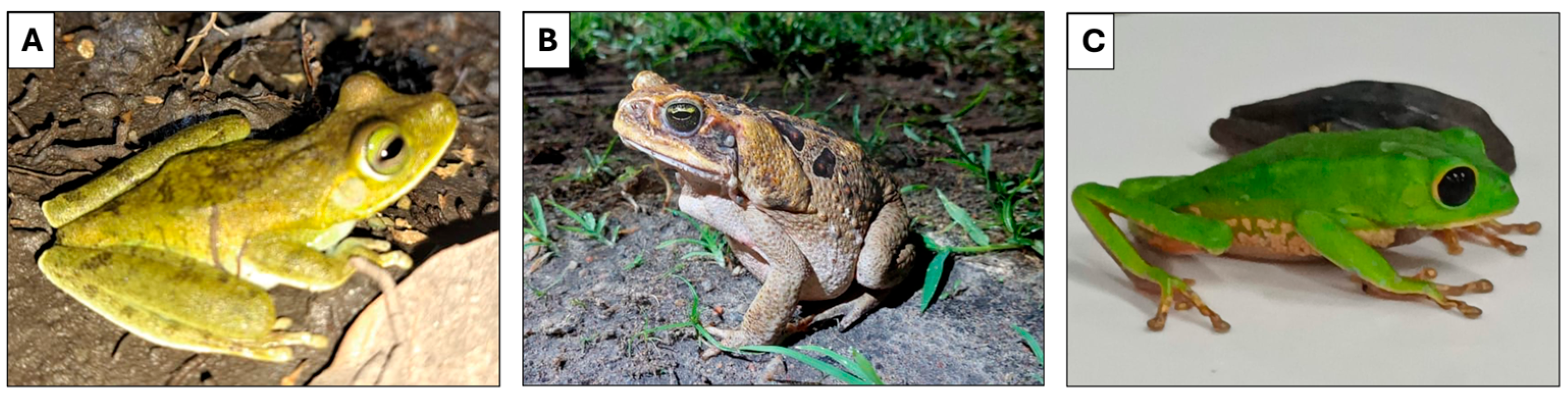
2.1. Toads
2.2. Frogs
2.3. Tree Frogs
3. Secretions from Anuran
4. Antimicrobial Compounds
4.1. Bacteria
4.2. Fungus
4.3. Virus
4.4. Protozoo
5. Advanced Research
6. Conclusions
Author Contributions
Funding
Institutional Review Board Statement
Informed Consent Statement
Data Availability Statement
Conflicts of Interest
References
- Bordon, K.D.C.F.; Cologna, C.T.; Fornari-Baldo, E.C.; Pinheiro-Júnior, E.L.; Cerni, F.A.; Amorim, F.G.; Anjolette, F.A.P.; Cordeiro, F.A.; Wiezel, G.A.; Cardoso, I.A.; et al. From Animal Poisons and Venoms to Medicines: Achievements, Challenges and Perspectives in Drug Discovery. Front. Pharmacol. 2020, 11, 1132. [Google Scholar] [CrossRef]
- García, F.A.; Fuentes, T.F.; Alonso, I.P.; Bosch, R.A.; Brunetti, A.E.; Lopes, N.P. A Comprehensive Review of Patented Antimicrobial Peptides from Amphibian Anurans. J. Nat. Prod. 2024, 87, 600–616. [Google Scholar] [CrossRef]
- Barros, A.L.A.N.; Hamed, A.; Marani, M.; Moreira, D.C.; Eaton, P.; Plácido, A.; Kato, M.J.; Leite, J.R.S.A. The Arsenal of Bioactive Molecules in the Skin Secretion of Urodele Amphibians. Front. Pharmacol. 2021, 12, 810821. [Google Scholar] [CrossRef]
- Barra, D.; Simmaco, M. Amphibian Skin: A Promising Resource for Antimicrobial Peptides. Trends Biotechnol. 1995, 13, 205–209. [Google Scholar] [CrossRef]
- Frost, D.R. Amphibian Species of the World: An Online Reference. 1999. Available online: https://amphibiansoftheworld.amnh.org (accessed on 15 February 2024).
- AmphibiaWeb. AmphibiaWeb 2024. Available online: https://amphibiaweb.org (accessed on 15 February 2024).
- Segalla, M.V.; Berneck, B.; Canedo, C.; Caramaschi, U.; Cruz, C.A.G.; Garcia, P.C.A.; Grant, T.; Haddad, C.F.B.; Lourenço, A.C.C.; Mângia, S.; et al. List of Brazilian Amphibians. Herpetol. Bras. 2021, 10, 121–216. [Google Scholar] [CrossRef]
- Bernarde, P.S. Anfíbios e Répteis: Introdução Ao Estudo da Herpetofauna Brasileira, 1st ed.; Anolis Books: Curitiba, Brazil, 2012; ISBN 978-85-65622-00-4. [Google Scholar]
- Leal, A.; Karnopp, E.; Barreto, Y.C.; Oliveira, R.S.; Rosa, M.E.; Borges, B.T.; Goulart, F.L.; Souza, V.Q.D.; Laikowski, M.M.; Moura, S.; et al. The Insecticidal Activity of Rhinella schneideri (Werner, 1894) Paratoid Secretion in Nauphoeta Cinerea Cocroaches. Toxins 2020, 12, 630. [Google Scholar] [CrossRef] [PubMed]
- Sun, Q.; Zhang, J.; Wang, T.; Xiong, Y.; Zhan, X.; Zhao, H.; Wang, J.; Fan, Y.; Bi, R.; Wang, S.; et al. Cooking Methods Effectively Alter Perfluoroalkyl Substances and Nutrients in Cultured and Wild Bullfrogs. J. Hazard. Mater. 2023, 445, 130555. [Google Scholar] [CrossRef]
- Ribeiro, L.P.; Carvalho, T.; Becker, C.G.; Jenkinson, T.S.; Leite, D.D.S.; James, T.Y.; Greenspan, S.E.; Toledo, L.F. Bullfrog Farms Release Virulent Zoospores of the Frog-Killing Fungus into the Natural Environment. Sci. Rep. 2019, 9, 13422. [Google Scholar] [CrossRef]
- Junior, V.H.; Martins, I.A. KAMBÔ: An Amazonian Enigma. J. Venom Res. 2020, 10, 13–17. [Google Scholar]
- Silva, F.V.A.D.; Monteiro, W.M.; Bernarde, P.S. “Kambô” Frog (Phyllomedusa bicolor): Use in Folk Medicine and Potential Health Risks. Rev. Soc. Bras. Med. Trop. 2019, 52, e20180467. [Google Scholar] [CrossRef]
- Dornelles, M.F.; Marques, M.d.G.B.; Renner, M.F. Revisão Sobre Toxinas de Anura (Tetrapoda, Lissamphibia) e Suas Aplicações Biotecnológicas. Ciência Mov. Rev. Ciências Saúde Educ. 2010, 12, 103–114. [Google Scholar]
- Carrillo, J.F.C.; Boaretto, A.G.; Santana, D.J.; Silva, D.B. Skin Secretions of Leptodactylidae (Anura) and Their Potential Applications. J. Venom. Anim. Toxins Incl. Trop. Dis. 2024, 30, e20230042. [Google Scholar] [CrossRef]
- Xu, X.; Lai, R. The Chemistry and Biological Activities of Peptides from Amphibian Skin Secretions. Chem. Rev. 2015, 115, 1760–1846. [Google Scholar] [CrossRef] [PubMed]
- Qi, J.; Zulfiker, A.H.M.; Li, C.; Good, D.; Wei, M.Q. The Development of Toad Toxins as Potential Therapeutic Agents. Toxins 2018, 10, 336. [Google Scholar] [CrossRef]
- Daly, J.W.; Spande, T.F.; Garraffo, H.M. Alkaloids from Amphibian Skin: A Tabulation of Over Eight-Hundred Compounds. J. Nat. Prod. 2005, 68, 1556–1575. [Google Scholar] [CrossRef]
- Rodríguez, C.; Rollins-Smith, L.; Ibáñez, R.; Durant-Archibold, A.A.; Gutiérrez, M. Toxins and Pharmacologically Active Compounds from Species of the Family Bufonidae (Amphibia, Anura). J. Ethnopharmacol. 2017, 198, 235–254. [Google Scholar] [CrossRef] [PubMed]
- Freitas, C.I.A. Estudo Sobre a Atividade Antimicrobiana de Substâncias Extraídas e Purificadas de Secreções Da Pele de Anfíbios. Ph.D. Thesis, Universidade Federal do Ceará, Fortaleza, Brazil, 2003. [Google Scholar]
- Azevedo Calderon, L.D.; Silva, A.D.A.E.; Ciancaglini, P.; Stábeli, R.G. Antimicrobial Peptides from Phyllomedusa Frogs: From Biomolecular Diversity to Potential Nanotechnologic Medical Applications. Amino Acids 2011, 40, 29–49. [Google Scholar] [CrossRef]
- Huan, Y.; Kong, Q.; Mou, H.; Yi, H. Antimicrobial Peptides: Classification, Design, Application and Research Progress in Multiple Fields. Front. Microbiol. 2020, 11, 582779. [Google Scholar] [CrossRef]
- Pfalzgraff, A.; Brandenburg, K.; Weindl, G. Antimicrobial Peptides and Their Therapeutic Potential for Bacterial Skin Infections and Wounds. Front. Pharmacol. 2018, 9, 281. [Google Scholar] [CrossRef]
- Rollins-Smith, L.A. The Importance of Antimicrobial Peptides (AMPs) in Amphibian Skin Defense. Dev. Comp. Immunol. 2023, 142, 104657. [Google Scholar] [CrossRef]
- König, E.; Bininda-Emonds, O.R.P.; Shaw, C. The Diversity and Evolution of Anuran Skin Peptides. Peptides 2015, 63, 96–117. [Google Scholar] [CrossRef]
- Zhang, Q.-Y.; Yan, Z.-B.; Meng, Y.-M.; Hong, X.-Y.; Shao, G.; Ma, J.-J.; Cheng, X.-R.; Liu, J.; Kang, J.; Fu, C.-Y. Antimicrobial Peptides: Mechanism of Action, Activity and Clinical Potential. Military Med. Res. 2021, 8, 48. [Google Scholar] [CrossRef]
- König, E.; Zhou, M.; Wang, L.; Chen, T.; Bininda-Emonds, O.R.P.; Shaw, C. Antimicrobial Peptides and Alytesin Are Co-Secreted from the Venom of the Midwife Toad, Alytes maurus (Alytidae, Anura): Implications for the Evolution of Frog Skin Defensive Secretions. Toxicon 2012, 60, 967–981. [Google Scholar] [CrossRef]
- Conlon, J.M.; Demandt, A.; Nielsen, P.F.; Leprince, J.; Vaudry, H.; Woodhams, D.C. The Alyteserins: Two Families of Antimicrobial Peptides from the Skin Secretions of the Midwife Toad Alytes obstetricans (Alytidae). Peptides 2009, 30, 1069–1073. [Google Scholar] [CrossRef]
- Yang, X.; Xia, J.; Yu, Z.; Hu, Y.; Li, F.; Meng, H.; Yang, S.; Liu, J.; Wang, H. Characterization of Diverse Antimicrobial Peptides in Skin Secretions of Chungan Torrent Frog Amolops chunganensis. Peptides 2012, 38, 41–53. [Google Scholar] [CrossRef]
- Chen, Z.; Yang, X.; Liu, Z.; Zeng, L.; Lee, W.; Zhang, Y. Two Novel Families of Antimicrobial Peptides from Skin Secretions of the Chinese Torrent Frog, Amolops jingdongensis. Biochimie 2012, 94, 328–334. [Google Scholar] [CrossRef]
- Wang, H.; Ran, R.; Yu, H.; Yu, Z.; Hu, Y.; Zheng, H.; Wang, D.; Yang, F.; Liu, R.; Liu, J. Identification and Characterization of Antimicrobial Peptides from Skin of Amolops ricketti (Anura: Ranidae). Peptides 2012, 33, 27–34. [Google Scholar] [CrossRef]
- Conlon, J.M.; Sonnevend, A.; Davidson, C.; David Smith, D.; Nielsen, P.F. The Ascaphins: A Family of Antimicrobial Peptides from the Skin Secretions of the Most Primitive Extant Frog, Ascaphus Truei. Biochem. Biophys. Res. Commun. 2004, 320, 170–175. [Google Scholar] [CrossRef]
- Wang, X.; Song, Y.; Li, J.; Liu, H.; Xu, X.; Lai, R.; Zhang, K. A New Family of Antimicrobial Peptides from Skin Secretions of Rana pleuraden. Peptides 2007, 28, 2069–2074. [Google Scholar] [CrossRef]
- Lai, R.; Zheng, Y.-T.; Shen, J.-H.; Liu, G.-J.; Liu, H.; Lee, W.-H.; Tang, S.-Z.; Zhang, Y. Antimicrobial Peptides from Skin Secretions of Chinese Red Belly Toad Bombina maxima. Peptides 2002, 23, 427–435. [Google Scholar] [CrossRef]
- Wang, T.; Zhang, J.; Shen, J.-H.; Jin, Y.; Lee, W.-H.; Zhang, Y. Maximins S, a Novel Group of Antimicrobial Peptides from Toad Bombina maxima. Biochem. Biophys. Res. Commun. 2005, 327, 945–951. [Google Scholar] [CrossRef]
- Mignogna, G.; Simmaco, M.; Kreil, G.; Barra, D. Antibacterial and Haemolytic Peptides Containing D-Alloisoleucine from the Skin of Bombina variegata. EMBO J. 1993, 12, 4829–4832. [Google Scholar] [CrossRef]
- Song, Y.; Lu, Y.; Wang, L.; Yang, H.; Zhang, K.; Lai, R. Purification, Characterization and Cloning of Two Novel Tigerinin-like Peptides from Skin Secretions of Fejervarya cancrivora. Peptides 2009, 30, 1228–1232. [Google Scholar] [CrossRef]
- Prates, M.V.; Sforça, M.L.; Regis, W.C.B.; Leite, J.R.S.A.; Silva, L.P.; Pertinhez, T.A.; Araújo, A.L.T.; Azevedo, R.B.; Spisni, A.; Bloch, C. The NMR-Derived Solution Structure of a New Cationic Antimicrobial Peptide from the Skin Secretion of the Anuran Hyla punctata. J. Biol. Chem. 2004, 279, 13018–13026. [Google Scholar] [CrossRef]
- Wu, J.; Liu, H.; Yang, H.; Yu, H.; You, D.; Ma, Y.; Ye, H.; Lai, R. Proteomic Analysis of Skin Defensive Factors of Tree Frog Hyla simplex. J. Proteome Res. 2011, 10, 4230–4240. [Google Scholar] [CrossRef]
- Dong, Z.; Luo, W.; Zhong, H.; Wang, M.; Song, Y.; Deng, S.; Zhang, Y. Molecular Cloning and Characterization of Antimicrobial Peptides from Skin of Hylarana guentheri. Acta Biochim. Biophys. Sin. 2017, 49, 450–457. [Google Scholar] [CrossRef]
- Wang, H.; Yu, Z.; Hu, Y.; Yu, H.; Ran, R.; Xia, J.; Wang, D.; Yang, S.; Yang, X.; Liu, J. Molecular Cloning and Characterization of Antimicrobial Peptides from Skin of the Broad-Folded Frog, Hylarana latouchii. Biochimie 2012, 94, 1317–1326. [Google Scholar] [CrossRef]
- Mishra, B.; Wang, G. Ab Initio Design of Potent Anti-MRSA Peptides Based on Database Filtering Technology. J. Am. Chem. Soc. 2012, 134, 12426–12429. [Google Scholar] [CrossRef]
- Conlon, J.M.; Woodhams, D.C.; Raza, H.; Coquet, L.; Leprince, J.; Jouenne, T.; Vaudry, H.; Rollins-Smith, L.A. Peptides with Differential Cytolytic Activity from Skin Secretions of the Lemur Leaf Frog Hylomantis lemur (Hylidae: Phyllomedusinae). Toxicon 2007, 50, 498–506. [Google Scholar] [CrossRef]
- Castro, M.S.; Ferreira, T.C.G.; Cilli, E.M.; Crusca, E.; Mendes-Giannini, M.J.S.; Sebben, A.; Ricart, C.A.O.; Sousa, M.V.; Fontes, W. Hylin A1, the First Cytolytic Peptide Isolated from the Arboreal South American Frog Hypsiboas albopunctatus (“Spotted Treefrog”). Peptides 2009, 30, 291–296. [Google Scholar] [CrossRef]
- Magalhães, B.S.; Melo, J.A.T.; Leite, J.R.S.A.; Silva, L.P.; Prates, M.V.; Vinecky, F.; Barbosa, E.A.; Verly, R.M.; Mehta, A.; Nicoli, J.R.; et al. Post-Secretory Events Alter the Peptide Content of the Skin Secretion of Hypsiboas raniceps. Biochem. Biophys. Res. Commun. 2008, 377, 1057–1061. [Google Scholar] [CrossRef]
- Wang, L.; Zhou, M.; McGrath, S.; Chen, T.; Gorman, S.P.; Walker, B.; Shaw, C. A Family of Kassinatuerin-2 Related Peptides from the Skin Secretion of the African Hyperoliid Frog, Kassina maculata. Peptides 2009, 30, 1428–1433. [Google Scholar] [CrossRef]
- Chen, H.; Wang, L.; Zeller, M.; Hornshaw, M.; Wu, Y.; Zhou, M.; Li, J.; Hang, X.; Cai, J.; Chen, T.; et al. Kassorins: Novel Innate Immune System Peptides from Skin Secretions of the African Hyperoliid Frogs, Kassina maculata and Kassina senegalensis. Mol. Immunol. 2011, 48, 442–451. [Google Scholar] [CrossRef] [PubMed]
- Conlon, J.M.; Abraham, B.; Galadari, S.; Knoop, F.C.; Sonnevend, A.; Pál, T. Antimicrobial and Cytolytic Properties of the Frog Skin Peptide, Kassinatuerin-1 and Its l- and d-Lysine-Substituted Derivatives. Peptides 2005, 26, 2104–2110. [Google Scholar] [CrossRef]
- Mattute, B.; Knoop, F.C.; Conlon, J.M. Kassinatuerin-1: A Peptide with Broad-Spectrum Antimicrobial Activity Isolated from the Skin of the Hyperoliid Frog, Kassina senegalensis. Biochem. Biophys. Res. Commun. 2000, 268, 433–436. [Google Scholar] [CrossRef]
- Rollins-Smith, L.A.; King, J.D.; Nielsen, P.F.; Sonnevend, A.; Conlon, J.M. An Antimicrobial Peptide from the Skin Secretions of the Mountain Chicken Frog Leptodactylus fallax (Anura:Leptodactylidae). Regul. Pept. 2005, 124, 173–178. [Google Scholar] [CrossRef]
- Conlon, J.M.; Al-Ghaferi, N.; Abraham, B.; Sonnevend, A.; King, J.D.; Nielsen, P.F. Purification and Properties of Laticeptin, an Antimicrobial Peptide from Skin Secretions of the South American Frog Leptodactylus laticeps. Protein Pept. Lett. 2006, 13, 411–415. [Google Scholar] [CrossRef] [PubMed]
- King, J.D.; Al-Ghaferi, N.; Abraham, B.; Sonnevend, A.; Leprince, J.; Nielsen, P.F.; Conlon, J.M. Pentadactylin: An Antimicrobial Peptide from the Skin Secretions of the South American Bullfrog Leptodactylus pentadactylus. Comp. Biochem. Physiol. Part C Toxicol. Pharmacol. 2005, 141, 393–397. [Google Scholar] [CrossRef]
- Nascimento, A.C.C.; Zanotta, L.C.; Kyaw, C.M.; Schwartz, E.N.F.; Schwartz, C.A.; Sebben, A.; Sousa, M.V.; Fontes, W.; Castro, M.S. Ocellatins: New Antimicrobial Peptides from the Skin Secretion of the South American Frog Leptodactylus ocellatus (Anura: Leptodactylidae). Protein J. 2004, 23, 501–508. [Google Scholar] [CrossRef]
- Nascimento, A.; Chapeaurouge, A.; Perales, J.; Sebben, A.; Sousa, M.V.; Fontes, W.; Castro, M.S. Purification, Characterization and Homology Analysis of Ocellatin 4, a Cytolytic Peptide from the Skin Secretion of the Frog Leptodactylus ocellatus. Toxicon 2007, 50, 1095–1104. [Google Scholar] [CrossRef]
- Li, B.; Lyu, P.; Xie, S.; Qin, H.; Pu, W.; Xu, H.; Chen, T.; Shaw, C.; Ge, L.; Kwok, H.F. LFB: A Novel Antimicrobial Brevinin-Like Peptide from the Skin Secretion of the Fujian Large Headed Frog, Limnonectes fujianensi. Biomolecules 2019, 9, 242. [Google Scholar] [CrossRef]
- Conlon, J.M.; Raza, H.; Coquet, L.; Jouenne, T.; Leprince, J.; Vaudry, H.; King, J.D. Purification of Peptides with Differential Cytolytic Activities from the Skin Secretions of the Central American Frog, Lithobates vaillanti (Ranidae). Comp. Biochem. Physiol. Part C Toxicol. Pharmacol. 2009, 150, 150–154. [Google Scholar] [CrossRef]
- Rozek, T.; Wegener, K.L.; Bowie, J.H.; Olver, I.N.; Carver, J.A.; Wallace, J.C.; Tyler, M.J. The Antibiotic and Anticancer Active Aurein Peptides from the Australian Bell Frogs Litoria aurea and Litoria raniformis. Eur. J. Biochem. 2000, 267, 5330–5341. [Google Scholar] [CrossRef]
- Manzo, G.; Ferguson, P.M.; Hind, C.K.; Clifford, M.; Gustilo, V.B.; Ali, H.; Bansal, S.S.; Bui, T.T.; Drake, A.F.; Atkinson, R.A.; et al. Temporin L and Aurein 2.5 Have Identical Conformations but Subtly Distinct Membrane and Antibacterial Activities. Sci. Rep. 2019, 9, 10934. [Google Scholar] [CrossRef]
- Wegener, K.L.; Wabnitz, P.A.; Carver, J.A.; Bowie, J.H.; Chia, B.C.S.; Wallace, J.C.; Tyler, M.J. Host Defence Peptides from the Skin Glands of the Australian Blue Mountains Tree-Frog Litoria citropa. Eur. J. Biochem. 1999, 265, 627–637. [Google Scholar] [CrossRef]
- Steinborner, S.T.; Bowie, J.H.; Tyler, M.J.; Wallace, J.C. An Unusual Combination of Peptides from the Skin Glands of Ewing’s Tree Frog, Litoria Ewingi. Sequence Determination and Antimicrobial Activity. Aust. J. Chem. 1997, 50, 889. [Google Scholar] [CrossRef]
- Jackway, R.J.; Bowie, J.H.; Bilusich, D.; Musgrave, I.F.; Surinya-Johnson, K.H.; Tyler, M.J.; Eichinger, P.C.H. The Fallaxidin Peptides from the Skin Secretion of the Eastern Dwarf Tree Frog Litoria fallax. Sequence Determination by Positive and Negative Ion Electrospray Mass Spectrometry: Antimicrobial Activity and cDNA Cloning of the Fallaxidins. Rapid Commun. Mass Spectrom. 2008, 22, 3207–3216. [Google Scholar] [CrossRef]
- Wong, H.; Bowie, J.H.; Carver, J.A. The Solution Structure and Activity of Caerin 1.1, an Antimicrobial Peptide from the Australian Green Tree Frog, Litoria splendida. Eur. J. Biochem. 1997, 247, 545–557. [Google Scholar] [CrossRef]
- Brinkworth, C.S.; Pukala, T.L.; Bowie, J.H.; Tyler, M.J. Host Defence Peptides from the Skin Glands of Australian Amphibians. Caerulein Neuropeptides and Antimicrobial, Anticancer, and nNOS Inhibiting Citropins from the Glandular Frog Litoria subglandulosa. Aust. J. Chem. 2004, 57, 693–701. [Google Scholar] [CrossRef]
- Yang, H.-L.; Shen, Z.-Q.; Liu, X.; Kong, Y. Two Novel Antimicrobial Peptides from Skin Venoms of Spadefoot Toad Megophrys minor. Chin. J. Nat. Med. 2016, 14, 294–298. [Google Scholar] [CrossRef]
- Lu, Z.; Zhai, L.; Wang, H.; Che, Q.; Wang, D.; Feng, F.; Zhao, Z.; Yu, H. Novel Families of Antimicrobial Peptides with Multiple Functions from Skin of Xizang Plateau Frog, Nanorana parkeri. Biochimie 2010, 92, 475–481. [Google Scholar] [CrossRef]
- Wang, H.; Yu, Z.; Hu, Y.; Li, F.; Liu, L.; Zheng, H.; Meng, H.; Yang, S.; Yang, X.; Liu, J. Novel Antimicrobial Peptides Isolated from the Skin Secretions of Hainan Odorous Frog, Odorrana hainanensis. Peptides 2012, 35, 285–290. [Google Scholar] [CrossRef]
- Abbassi, F.; Galanth, C.; Amiche, M.; Saito, K.; Piesse, C.; Zargarian, L.; Hani, K.; Nicolas, P.; Lequin, O.; Ladram, A. Solution Structure and Model Membrane Interactions of Temporins-SH, Antimicrobial Peptides from Amphibian Skin. A NMR Spectroscopy and Differential Scanning Calorimetry Study. Biochemistry 2008, 47, 10513–10525. [Google Scholar] [CrossRef]
- Abbassi, F.; Raja, Z.; Oury, B.; Gazanion, E.; Piesse, C.; Sereno, D.; Nicolas, P.; Foulon, T.; Ladram, A. Antibacterial and Leishmanicidal Activities of Temporin-SHd, a 17-Residue Long Membrane-Damaging Peptide. Biochimie 2013, 95, 388–399. [Google Scholar] [CrossRef]
- André, S.; Raja, Z.; Humblot, V.; Piesse, C.; Foulon, T.; Sereno, D.; Oury, B.; Ladram, A. Functional Characterization of Temporin-SHe, a New Broad-Spectrum Antibacterial and Leishmanicidal Temporin-SH Paralog from the Sahara Frog (Pelophylax saharicus). Int. J. Mol. Sci. 2020, 21, 6713. [Google Scholar] [CrossRef]
- Abbassi, F.; Lequin, O.; Piesse, C.; Goasdoué, N.; Foulon, T.; Nicolas, P.; Ladram, A. Temporin-SHf, a New Type of Phe-Rich and Hydrophobic Ultrashort Antimicrobial Peptide. J. Biol. Chem. 2010, 285, 16880–16892. [Google Scholar] [CrossRef]
- Pierre, T.N.; Seon, A.A.; Amiche, M.; Nicolas, P. Phylloxin, a Novel Peptide Antibiotic of the Dermaseptin Family of Antimicrobial/Opioid Peptide Precursors. Eur. J. Biochem. 2000, 267, 370–378. [Google Scholar] [CrossRef]
- Batista, C.V.F.; Scaloni, A.; Rigden, D.J.; Silva, L.R.; Rodrigues Romero, A.; Dukor, R.; Sebben, A.; Talamo, F.; Bloch, C. A Novel Heterodimeric Antimicrobial Peptide from the Tree-Frog Phyllomedusa distincta. FEBS Lett. 2001, 494, 85–89. [Google Scholar] [CrossRef]
- Leite, J.R.S.A.; Silva, L.P.; Rodrigues, M.I.S.; Prates, M.V.; Brand, G.D.; Lacava, B.M.; Azevedo, R.B.; Bocca, A.L.; Albuquerque, S.; Bloch, C. Phylloseptins: A Novel Class of Anti-Bacterial and Anti-Protozoan Peptides from the Phyllomedusa Genus. Peptides 2005, 26, 565–573. [Google Scholar] [CrossRef] [PubMed]
- Lequin, O.; Ladram, A.; Chabbert, L.; Bruston, F.; Convert, O.; Vanhoye, D.; Chassaing, G.; Nicolas, P.; Amiche, M. Dermaseptin S9, an α-Helical Antimicrobial Peptide with a Hydrophobic Core and Cationic Termini. Biochemistry 2006, 45, 468–480. [Google Scholar] [CrossRef]
- Zhang, R.; Zhou, M.; Wang, L.; McGrath, S.; Chen, T.; Chen, X.; Shaw, C. Phylloseptin-1 (PSN-1) from Phyllomedusa sauvagei Skin Secretion: A Novel Broad-Spectrum Antimicrobial Peptide with Antibiofilm Activity. Mol. Immunol. 2010, 47, 2030–2037. [Google Scholar] [CrossRef] [PubMed]
- Park, S.-C.; Kim, J.-Y.; Jeong, C.; Yoo, S.; Hahm, K.-S.; Park, Y. A Plausible Mode of Action of Pseudin-2, an Antimicrobial Peptide from Pseudis paradoxa. Biochim. Biophys. Acta (BBA)—Biomembr. 2011, 1808, 171–182. [Google Scholar] [CrossRef]
- Ali, M.F.; Lips, K.R.; Knoop, F.C.; Fritzsch, B.; Miller, C.; Conlon, J.M. Antimicrobial Peptides and Protease Inhibitors in the Skin Secretions of the Crawfish Frog, Rana areolata. Biochim. Biophys. Acta (BBA)—Proteins Proteom. 2002, 1601, 55–63. [Google Scholar] [CrossRef]
- Samgina, T.Y.; Artemenko, K.A.; Gorshkov, V.A.; Ogourtsov, S.V.; Zubarev, R.A.; Lebedev, A.T. Mass Spectrometric Study of Peptides Secreted by the Skin Glands of the Brown Frog Rana Arvalis from the Moscow Region. Rapid Commun. Mass Spectrom. 2009, 23, 1241–1248. [Google Scholar] [CrossRef] [PubMed]
- Conlon, J.M.; Sonnevend, A.; Davidson, C.; Demandt, A.; Jouenne, T. Host-Defense Peptides Isolated from the Skin Secretions of the Northern Red-Legged Frog Rana aurora aurora. Dev. Comp. Immunol. 2005, 29, 83–90. [Google Scholar] [CrossRef]
- Goraya, J.; Wang, Y.; Li, Z.; O’Flaherty, M.; Knoop, F.C.; Platz, J.E.; Conlon, J.M. Peptides with Antimicrobial Activity from Four Different Families Isolated from the Skins of the North American Frogs Rana luteiventris, Rana berlandieri and Rana pipiens. Eur. J. Biochem. 2000, 267, 894–900. [Google Scholar] [CrossRef] [PubMed]
- Goraya, J.; Knoop, F.C.; Conlon, J.M. Ranatuerins: Antimicrobial Peptides Isolated from the Skin of the American Bullfrog, Rana catesbeiana. Biochem. Biophys. Res. Commun. 1998, 250, 589–592. [Google Scholar] [CrossRef]
- Conlon, J.M.; Leprince, J.; Vaudry, H.; Jiansheng, H.; Nielsen, P.F. A Family of Antimicrobial Peptides Related to Japonicin-2 Isolated from the Skin of the Chaochiao Brown Frog Rana chaochiaoensis. Comp. Biochem. Physiol. Part C Toxicol. Pharmacol. 2006, 144, 101–105. [Google Scholar] [CrossRef]
- Shang, D.; Yu, F.; Li, J.; Zheng, J.; Zhang, L.; Li, Y. Molecular Cloning of cDNAs Encoding Antimicrobial Peptide Precursors from the Skin of the Chinese Brown Frog, Rana chensinensis. Zool. Sci. 2009, 26, 220–226. [Google Scholar] [CrossRef]
- Zhao, J.; Sun, Y.; Li, Z.; Su, Q. Molecular Cloning of Novel Antimicrobial Peptide Genes from the Skin of the Chinese Brown Frog, Rana chensinensis. Zool. Sci. 2011, 28, 112–117. [Google Scholar] [CrossRef]
- Ye, Z.; Zhou, X.; Xi, X.; Zai, Y.; Zhou, M.; Chen, X.; Ma, C.; Chen, T.; Wang, L.; Kwok, H.F. In Vitro & In Vivo Studies on Identifying and Designing Temporin-1CEh from the Skin Secretion of Rana chensinensis as the Optimised Antibacterial Prototype Drug. Pharmaceutics 2022, 14, 604. [Google Scholar] [CrossRef]
- Halverson, T.; Basir, Y.J.; Knoop, F.C.; Conlon, J.M. Purification and Characterization of Antimicrobial Peptides from the Skin of the North American Green Frog Rana clamitans. Peptides 2000, 21, 469–476. [Google Scholar] [CrossRef]
- Jin, L.-L.; Li, Q.; Song, S.-S.; Feng, K.; Zhang, D.-B.; Wang, Q.-Y.; Chen, Y.-H. Characterization of Antimicrobial Peptides Isolated from the Skin of the Chinese Frog, Rana dybowskii. Comp. Biochem. Physiol. Part B Biochem. Mol. Biol. 2009, 154, 174–178. [Google Scholar] [CrossRef]
- Simmaco, M.; Mignogna, G.; Barra, D.; Bossa, F. Antimicrobial Peptides from Skin Secretions of Rana esculenta. Molecular Cloning of cDNAs Encoding Esculentin and Brevinins and Isolation of New Active Peptides. J. Biol. Chem. 1994, 269, 11956–11961. [Google Scholar] [CrossRef]
- Mangoni, M.L.; Papo, N.; Mignogna, G.; Andreu, D.; Shai, Y.; Barra, D.; Simmaco, M. Ranacyclins, a New Family of Short Cyclic Antimicrobial Peptides: Biological Function, Mode of Action, and Parameters Involved in Target Specificity. Biochemistry 2003, 42, 14023–14035. [Google Scholar] [CrossRef]
- Conlon, J.M.; Al-Ghaferi, N.; Abraham, B.; Jiansheng, H.; Cosette, P.; Leprince, J.; Jouenne, T.; Vaudry, H. Antimicrobial Peptides from Diverse Families Isolated from the Skin of the Asian Frog, Rana grahami. Peptides 2006, 27, 2111–2117. [Google Scholar] [CrossRef]
- Kim, J.B.; Halverson, T.; Basir, Y.J.; Dulka, J.; Knoop, F.C.; Abel, P.W.; Conlon, J.M. Purification and Characterization of Antimicrobial and Vasorelaxant Peptides from Skin Extracts and Skin Secretions of the North American Pig Frog Rana grylio. Regul. Pept. 2000, 90, 53–60. [Google Scholar] [CrossRef]
- Isaacson, T.; Soto, A.; Iwamuro, S.; Knoop, F.C.; Conlon, J.M. Antimicrobial Peptides with Atypical Structural Features from the Skin of the Japanese Brown Frog Rana japonica. Peptides 2002, 23, 419–425. [Google Scholar] [CrossRef]
- Park, S.; Park, S.-H.; Ahn, H.-C.; Kim, S.; Kim, S.S.; Lee, B.J.; Lee, B.-J. Structural Study of Novel Antimicrobial Peptides, Nigrocins, Isolated from Rana nigromaculata. FEBS Lett. 2001, 507, 95–100. [Google Scholar] [CrossRef]
- Conlon, J.M.; Sonnevend, A.; Jouenne, T.; Coquet, L.; Cosquer, D.; Vaudry, H.; Iwamuro, S. A Family of Acyclic Brevinin-1 Peptides from the Skin of the Ryukyu Brown Frog Rana okinavana. Peptides 2005, 26, 185–190. [Google Scholar] [CrossRef]
- Basir, Y.J.; Knoop, F.C.; Dulka, J.; Conlon, J.M. Multiple Antimicrobial Peptides and Peptides Related to Bradykinin and Neuromedin N Isolated from Skin Secretions of the Pickerel Frog, Rana palustris. Biochim. Biophys. Acta (BBA)—Protein Struct. Mol. Enzymol. 2000, 1543, 95–105. [Google Scholar] [CrossRef]
- Zhou, X.; Shi, D.; Zhong, R.; Ye, Z.; Ma, C.; Zhou, M.; Xi, X.; Wang, L.; Chen, T.; Kwok, H.F. Bioevaluation of Ranatuerin-2Pb from the Frog Skin Secretion of Rana pipiens and Its Truncated Analogues. Biomolecules 2019, 9, 249. [Google Scholar] [CrossRef]
- Conlon, J.M.; Sonnevend, Á.; Patel, M.; Al-Dhaheri, K.; Nielsen, P.F.; Kolodziejek, J.; Nowotny, N.; Iwamuro, S.; Pál, T. A Family of Brevinin-2 Peptides with Potent Activity against Pseudomonas aeruginosa from the Skin of the Hokkaido Frog, Rana pirica. Regul. Pept. 2004, 118, 135–141. [Google Scholar] [CrossRef]
- Park, J.M.; Jung, J.E.; Lee, B.J. Antimicrobial Peptides from the Skin of a Korean Frog, Rana rugosa. Biochem. Biophys. Res. Commun. 1994, 205, 948–954. [Google Scholar] [CrossRef]
- Suzuki, S.; Ohe, Y.; Okubo, T.; Kakegawa, T.; Tatemoto, K. Isolation and Characterization of Novel Antimicrobial Peptides, Rugosins A, B, and C, from the Skin of the Frog, Rana rugosa. Biochem. Biophys. Res. Commun. 1995, 212, 249–254. [Google Scholar] [CrossRef]
- Suzuki, H.; Iwamuro, S.; Ohnuma, A.; Coquet, L.; Leprince, J.; Jouenne, T.; Vaudry, H.; Taylor, C.K.; Abel, P.W.; Conlon, J.M. Expression of Genes Encoding Antimicrobial and Bradykinin-Related Peptides in Skin of the Stream Brown Frog Rana sakuraii. Peptides 2007, 28, 505–514. [Google Scholar] [CrossRef]
- Bevier, C.R.; Sonnevend, A.; Kolodziejek, J.; Nowotny, N.; Nielsen, P.F.; Michael Conlon, J. Purification and Characterization of Antimicrobial Peptides from the Skin Secretions of the Mink Frog (Rana septentrionalis). Comp. Biochem. Physiol. Part C Toxicol. Pharmacol. 2004, 139, 31–38. [Google Scholar] [CrossRef]
- Conlon, J.M.; Sonnevend, A.; Patel, M.; Camasamudram, V.; Nowotny, N.; Zilahi, E.; Iwamuro, S.; Nielsen, P.F.; Pál, T. A Melittin-Related Peptide from the Skin of the Japanese Frog, Rana tagoi, with Antimicrobial and Cytolytic Properties. Biochem. Biophys. Res. Commun. 2003, 306, 496–500. [Google Scholar] [CrossRef]
- Simmaco, M.; Mignogna, G.; Canofeni, S.; Miele, R.; Mangoni, M.L.; Barra, D. Temporins, Antimicrobial Peptides from the European Red Frog Rana temporaria. Eur. J. Biochem. 1996, 242, 788–792. [Google Scholar] [CrossRef]
- Mangoni, M.L.; Maisetta, G.; Di Luca, M.; Gaddi, L.M.H.; Esin, S.; Florio, W.; Brancatisano, F.L.; Barra, D.; Campa, M.; Batoni, G. Comparative Analysis of the Bactericidal Activities of Amphibian Peptide Analogues against Multidrug-Resistant Nosocomial Bacterial Strains. Antimicrob. Agents Chemother. 2008, 52, 85–91. [Google Scholar] [CrossRef] [PubMed]
- Carotenuto, A.; Malfi, S.; Saviello, M.R.; Campiglia, P.; Gomez-Monterrey, I.; Mangoni, M.L.; Gaddi, L.M.H.; Novellino, E.; Grieco, P. A Different Molecular Mechanism Underlying Antimicrobial and Hemolytic Actions of Temporins A and L. J. Med. Chem. 2008, 51, 2354–2362. [Google Scholar] [CrossRef]
- Sai, K.P.; Jagannadham, M.V.; Vairamani, M.; Raju, N.P.; Devi, A.S.; Nagaraj, R.; Sitaram, N. Tigerinins: Novel Antimicrobial Peptides from the Indian Frog Rana tigerina. J. Biol. Chem. 2001, 276, 2701–2707. [Google Scholar] [CrossRef]
- Conlon, J.M.; Al-Ghaferi, N.; Abraham, B.; Sonnevend, A.; Coquet, L.; Leprince, J.; Jouenne, T.; Vaudry, H.; Iwamuro, S. Antimicrobial Peptides from the Skin of the Tsushima Brown Frog Rana tsushimensis. Comp. Biochem. Physiol. Part C Toxicol. Pharmacol. 2006, 143, 42–49. [Google Scholar] [CrossRef]
- Conlon, J.M.; Abraham, B.; Sonnevend, A.; Jouenne, T.; Cosette, P.; Leprince, J.; Vaudry, H.; Bevier, C.R. Purification and Characterization of Antimicrobial Peptides from the Skin Secretions of the Carpenter Frog Rana virgatipes (Ranidae, Aquarana). Regul. Pept. 2005, 131, 38–45. [Google Scholar] [CrossRef]
- Chen, G.; Miao, Y.; Ma, C.; Zhou, M.; Shi, Z.; Chen, X.; Burrows, J.F.; Xi, X.; Chen, T.; Wang, L. Brevinin-2GHk from Sylvirana guentheri and the Design of Truncated Analogs Exhibiting the Enhancement of Antimicrobial Activity. Antibiotics 2020, 9, 85. [Google Scholar] [CrossRef]
- Bradford, A.; Bowie, J.; Tyler, M.; Wallace, J. New Antibiotic Uperin Peptides From the Dorsal Glands of the Australian Toadlet Uperoleia mjobergii. Aust. J. Chem. 1996, 49, 1325. [Google Scholar] [CrossRef]
- Conlon, J.M.; Al-Ghaferi, N.; Ahmed, E.; Meetani, M.A.; Leprince, J.; Nielsen, P.F. Orthologs of Magainin, PGLa, Procaerulein-Derived, and Proxenopsin-Derived Peptides from Skin Secretions of the Octoploid Frog Xenopus amieti (Pipidae). Peptides 2010, 31, 989–994. [Google Scholar] [CrossRef]
- Mechkarska, M.; Eman, A.; Coquet, L.; Jérôme, L.; Jouenne, T.; Vaudry, H.; King, J.D.; Takada, K.; Conlon, J.M. Genome Duplications within the Xenopodinae Do Not Increase the Multiplicity of Antimicrobial Peptides in Silurana paratropicalis and Xenopus andrei Skin Secretions. Comp. Biochem. Physiol. Part D Genom. Proteom. 2011, 6, 206–212. [Google Scholar] [CrossRef]
- Mechkarska, M.; Ahmed, E.; Coquet, L.; Leprince, J.; Jouenne, T.; Vaudry, H.; King, J.D.; Conlon, J.M. Antimicrobial Peptides with Therapeutic Potential from Skin Secretions of the Marsabit Clawed Frog Xenopus borealis (Pipidae). Comp. Biochem. Physiol. Part C Toxicol. Pharmacol. 2010, 152, 467–472. [Google Scholar] [CrossRef]
- Conlon, J.M.; Mechkarska, M.; Ahmed, E.; Leprince, J.; Vaudry, H.; King, J.D.; Takada, K. Purification and Properties of Antimicrobial Peptides from Skin Secretions of the Eritrea Clawed Frog Xenopus clivii (Pipidae). Comp. Biochem. Physiol. Part C Toxicol. Pharmacol. 2011, 153, 350–354. [Google Scholar] [CrossRef]
- Zasloff, M. Magainins, a Class of Antimicrobial Peptides from Xenopus Skin: Isolation, Characterization of Two Active Forms, and Partial cDNA Sequence of a Precursor. Proc. Natl. Acad. Sci. USA 1987, 84, 5449–5453. [Google Scholar] [CrossRef]
- Soravia, E.; Martini, G.; Zasloff, M. Antimicrobial Properties of Peptides from Xenopus granular Gland Secretions. FEBS Lett. 1988, 228, 337–340. [Google Scholar] [CrossRef]
- Mechkarska, M.; Ahmed, E.; Coquet, L.; Leprince, J.; Jouenne, T.; Vaudry, H.; King, J.D.; Conlon, J.M. Peptidomic Analysis of Skin Secretions Demonstrates That the Allopatric Populations of Xenopus muelleri (Pipidae) Are Not Conspecific. Peptides 2011, 32, 1502–1508. [Google Scholar] [CrossRef]
- Ali, M.F.; Soto, A.; Knoop, F.C.; Conlon, J.M. Antimicrobial Peptides Isolated from Skin Secretions of the Diploid Frog, Xenopus tropicalis (Pipidae). Biochim. Biophys. Acta (BBA)—Protein Struct. Mol. Enzymol. 2001, 1550, 81–89. [Google Scholar] [CrossRef]
- Pereira, A.F.M.; Sani, A.A.; Zapata, T.B.; Sousa, D.S.M.d.; Rossini, B.C.; Santos, L.D.d.; Rall, V.L.M.; Riccardi, C.d.S.; Fernandes Júnior, A. Synergistic Antibacterial Efficacy of Melittin in Combination with Oxacillin against Methicillin-Resistant Staphylococcus aureus (MRSA). Microorganisms 2023, 11, 2868. [Google Scholar] [CrossRef]
- Guha, S.; Ferrie, R.P.; Ghimire, J.; Ventura, C.R.; Wu, E.; Sun, L.; Kim, S.Y.; Wiedman, G.R.; Hristova, K.; Wimley, W.C. Applications and Evolution of Melittin, the Quintessential Membrane Active Peptide. Biochem. Pharmacol. 2021, 193, 114769. [Google Scholar] [CrossRef]
- Simmaco, M.; Kreil, G.; Barra, D. Bombinins, Antimicrobial Peptides from Bombina Species. Biochim. Biophys. Acta (BBA)—Biomembr. 2009, 1788, 1551–1555. [Google Scholar] [CrossRef]
- Barnhart, K.; Forman, M.E.; Umile, T.P.; Kueneman, J.; McKenzie, V.; Salinas, I.; Minbiole, K.P.C.; Woodhams, D.C. Identification of Bufadienolides from the Boreal Toad, Anaxyrus Boreas, Active Against a Fungal Pathogen. Microb. Ecol. 2017, 74, 990–1000. [Google Scholar] [CrossRef]
- Artika, I.M.; Pinontoan, S.; Kusrini, M.D. Antifungal Activity of Skin Secretion of Bleeding Toad Leptophryne cruentata and Javan Tree Frog Rhacophorus margaritifer. Am. J. Biochem. Biotechnol. 2015, 11, 5–10. [Google Scholar] [CrossRef]
- Tian, Z.; Liao, A.; Kang, J.; Gao, Y.; Lu, A.; Wang, Z.; Wang, Q. Toad Alkaloid for Pesticide Discovery: Dehydrobufotenine Derivatives as Novel Agents against Plant Virus and Fungi. J. Agric. Food Chem. 2021, 69, 9754–9763. [Google Scholar] [CrossRef]
- Luca, V.; Olivi, M.; Di Grazia, A.; Palleschi, C.; Uccelletti, D.; Mangoni, M.L. Anti-Candida Activity of 1-18 Fragment of the Frog Skin Peptide Esculentin-1b: In Vitro and in Vivo Studies in a Caenorhabditis elegans Infection Model. Cell. Mol. Life Sci. 2014, 71, 2535–2546. [Google Scholar] [CrossRef]
- Ma, Y.; Liu, C.; Liu, X.; Wu, J.; Yang, H.; Wang, Y.; Li, J.; Yu, H.; Lai, R. Peptidomics and Genomics Analysis of Novel Antimicrobial Peptides from the Frog, Rana nigrovittata. Genomics 2010, 95, 66–71. [Google Scholar] [CrossRef]
- Al-Ghaferi, N.; Kolodziejek, J.; Nowotny, N.; Coquet, L.; Jouenne, T.; Leprince, J.; Vaudry, H.; King, J.D.; Conlon, J.M. Antimicrobial Peptides from the Skin Secretions of the South-East Asian Frog Hylarana erythraea (Ranidae). Peptides 2010, 31, 548–554. [Google Scholar] [CrossRef]
- Conlon, J.M.; Kolodziejek, J.; Nowotny, N. Antimicrobial Peptides from Ranid Frogs: Taxonomic and Phylogenetic Markers and a Potential Source of New Therapeutic Agents. Biochim. Biophys. Acta (BBA)—Proteins Proteom. 2004, 1696, 1–14. [Google Scholar] [CrossRef]
- Alangaden, G.J. Nosocomial Fungal Infections: Epidemiology, Infection Control, and Prevention. Infect. Dis. Clin. N. Am. 2011, 25, 201–225. [Google Scholar] [CrossRef]
- VanCompernolle, S.E.; Taylor, R.J.; Oswald-Richter, K.; Jiang, J.; Youree, B.E.; Bowie, J.H.; Tyler, M.J.; Conlon, J.M.; Wade, D.; Aiken, C.; et al. Antimicrobial Peptides from Amphibian Skin Potently Inhibit Human Immunodeficiency Virus Infection and Transfer of Virus from Dendritic Cells to T Cells. J. Virol. 2005, 79, 11598–11606. [Google Scholar] [CrossRef]
- VanCompernolle, S.; Smith, P.B.; Bowie, J.H.; Tyler, M.J.; Unutmaz, D.; Rollins-Smith, L.A. Inhibition of HIV Infection by Caerin 1 Antimicrobial Peptides. Peptides 2015, 71, 296–303. [Google Scholar] [CrossRef]
- Muñoz-Camargo, C.; Méndez, M.C.; Salazar, V.; Moscoso, J.; Narváez, D.; Torres, M.M.; Florez, F.K.; Groot, H.; Mitrani, E. Frog Skin Cultures Secrete Anti-Yellow Fever Compounds. J. Antibiot. 2016, 69, 783–790. [Google Scholar] [CrossRef]
- Bartels, E.J.H.; Dekker, D.; Amiche, M. Dermaseptins, Multifunctional Antimicrobial Peptides: A Review of Their Pharmacology, Effectivity, Mechanism of Action, and Possible Future Directions. Front. Pharmacol. 2019, 10, 1421. [Google Scholar] [CrossRef]
- de Amaral, M.; Ienes-Lima, J. Anurans against SARS-CoV-2: A Review of the Potential Antiviral Action of Anurans Cutaneous Peptides. Virus Res. 2022, 315, 198769. [Google Scholar] [CrossRef]
- Mechlia, M.B.; Belaid, A.; Castel, G.; Jallet, C.; Mansfield, K.L.; Fooks, A.R.; Hani, K.; Tordo, N. Dermaseptins as Potential Antirabies Compounds. Vaccine 2019, 37, 4694–4700. [Google Scholar] [CrossRef]
- Haddad, H.; Tangy, F.; Ouahchi, I.; Sahtout, W.; Ouni, B.; Zaïri, A. Evaluation of the Antiviral Activity of New Dermaseptin Analogs against Zika Virus. Biochem. Biophys. Rep. 2024, 39, 101747. [Google Scholar] [CrossRef]
- Lorin, C.; Saidi, H.; Belaid, A.; Zairi, A.; Baleux, F.; Hocini, H.; Bélec, L.; Hani, K.; Tangy, F. The Antimicrobial Peptide Dermaseptin S4 Inhibits HIV-1 Infectivity in Vitro. Virology 2005, 334, 264–275. [Google Scholar] [CrossRef]
- Chinchar, V.G.; Bryan, L.; Silphadaung, U.; Noga, E.; Wade, D.; Rollins-Smith, L. Inactivation of Viruses Infecting Ectothermic Animals by Amphibian and Piscine Antimicrobial Peptides. Virology 2004, 323, 268–275. [Google Scholar] [CrossRef]
- Chianese, A.; Iovane, V.; Zannella, C.; Capasso, C.; Nastri, B.M.; Monti, A.; Doti, N.; Montagnaro, S.; Pagnini, U.; Iovane, G.; et al. Synthetic Frog-Derived-like Peptides: A New Weapon against Emerging and Potential Zoonotic Viruses. Viruses 2023, 15, 1804. [Google Scholar] [CrossRef]
- Lee, S.H.; Kim, E.H.; O’neal, J.T.; Dale, G.; Holthausen, D.J.; Bowen, J.R.; Quicke, K.M.; Skountzou, I.; Gopal, S.; George, S.; et al. The Amphibian Peptide Yodha Is Virucidal for Zika and Dengue Viruses. Sci. Rep. 2021, 11, 602. [Google Scholar] [CrossRef]
- Bandyopadhyay, S.; Ng, B.Y.; Chong, C.; Lim, M.Z.; Gill, S.K.; Lee, K.H.; Sivaraman, J.; Chatterjee, C. Micelle Bound Structure and DNA Interaction of Brevinin-2-Related Peptide, an Antimicrobial Peptide Derived from Frog Skin. J. Pept. Sci. 2014, 20, 811–821. [Google Scholar] [CrossRef]
- Yasin, B.; Pang, M.; Turner, J.S.; Cho, Y.; Dinh, N.N.; Waring, A.J.; Lehrer, R.I.; Wagar, E.A. Evaluation of the Inactivation of Infectious Herpes simplex Virus by Host-Defense Peptides. Eur. J. Clin. Microbiol. Infect Dis. 2000, 19, 187–194. [Google Scholar] [CrossRef] [PubMed]
- Marcocci, M.E.; Amatore, D.; Villa, S.; Casciaro, B.; Aimola, P.; Franci, G.; Grieco, P.; Galdiero, M.; Palamara, A.T.; Mangoni, M.L.; et al. The Amphibian Antimicrobial Peptide Temporin B Inhibits In Vitro Herpes Simplex Virus 1 Infection. Antimicrob. Agents Chemother. 2018, 62, e02367-17. [Google Scholar] [CrossRef]
- D’Andrea, L.D.; Romanelli, A. Temporins: Multifunctional Peptides from Frog Skin. Int. J. Mol. Sci. 2023, 24, 5426. [Google Scholar] [CrossRef]
- Urmi, U.L.; Vijay, A.K.; Kuppusamy, R.; Islam, S.; Willcox, M.D.P. A Review of the Antiviral Activity of Cationic Antimicrobial Peptides. Peptides 2023, 166, 171024. [Google Scholar] [CrossRef] [PubMed]
- Holthausen, D.J.; Lee, S.H.; Kumar, V.T.; Bouvier, N.M.; Krammer, F.; Ellebedy, A.H.; Wrammert, J.; Lowen, A.C.; George, S.; Pillai, M.R.; et al. An Amphibian Host Defense Peptide Is Virucidal for Human H1 Hemagglutinin-Bearing Influenza Viruses. Immunity 2017, 46, 587–595. [Google Scholar] [CrossRef]
- Albiol Matanic, V.C.; Castilla, V. Antiviral Activity of Antimicrobial Cationic Peptides against Junin Virus and Herpes Simplex Virus. Int. J. Antimicrob. Agents 2004, 23, 382–389. [Google Scholar] [CrossRef] [PubMed]
- Xiong, W.; Li, J.; Feng, Y.; Chai, J.; Wu, J.; Hu, Y.; Tian, M.; Lu, W.; Xu, X.; Zou, M. Brevinin-2GHk, a Peptide Derived from the Skin of Fejervarya Limnocharis, Inhibits Zika Virus Infection by Disrupting Viral Integrity. Viruses 2021, 13, 2382. [Google Scholar] [CrossRef] [PubMed]
- Yang, J.; Zhang, B.; Huang, Y.; Liu, T.; Zeng, B.; Chai, J.; Wu, J.; Xu, X. Antiviral Activity and Mechanism of ESC-1GN from Skin Secretion of Hylarana guentheri against Influenza A Virus. J. Biochem. 2021, 169, 757–765. [Google Scholar] [CrossRef]
- Santana, C.J.C.; Magalhães, A.C.M.; Prías-Márquez, C.A.; Falico, D.A.; Dos Santos Júnior, A.C.M.; Lima, B.D.; Ricart, C.A.O.; de Pilger, D.R.B.; Bonotto, R.M.; Moraes, C.B.; et al. Biological Properties of a Novel Multifunctional Host Defense Peptide from the Skin Secretion of the Chaco Tree Frog, Boana Raniceps. Biomolecules 2020, 10, 790. [Google Scholar] [CrossRef]
- De Angelis, M.; Casciaro, B.; Genovese, A.; Brancaccio, D.; Marcocci, M.E.; Novellino, E.; Carotenuto, A.; Palamara, A.T.; Mangoni, M.L.; Nencioni, L. Temporin G, an Amphibian Antimicrobial Peptide against Influenza and Parainfluenza Respiratory Viruses: Insights into Biological Activity and Mechanism of Action. FASEB J. 2021, 35, e21358. [Google Scholar] [CrossRef]
- Rojas-Pirela, M.; Kemmerling, U.; Quiñones, W.; Michels, P.A.M.; Rojas, V. Antimicrobial Peptides (AMPs): Potential Therapeutic Strategy against Trypanosomiases? Biomolecules 2023, 13, 599. [Google Scholar] [CrossRef]
- McGwire, B.S.; Kulkarni, M.M. Interactions of Antimicrobial Peptides with Leishmania and Trypanosomes and Their Functional Role in Host Parasitism. Exp. Parasitol. 2010, 126, 397–405. [Google Scholar] [CrossRef]
- Ladram, A.; Nicolas, P. Antimicrobial Peptides from Frog Skin: Biodiversity and Therapeutic Promises. Front. Biosci. (Landmark Ed.) 2016, 21, 1341–1371. [Google Scholar] [CrossRef]
- Mangoni, M.L.; Papo, N.; Saugar, J.M.; Barra, D.; Shai, Y.; Simmaco, M.; Rivas, L. Effect of Natural l—to d-Amino Acid Conversion on the Organization, Membrane Binding, and Biological Function of the Antimicrobial Peptides Bombinins H. Biochemistry 2006, 45, 4266–4276. [Google Scholar] [CrossRef] [PubMed]
- Pinto, E.G.; Pimenta, D.C.; Antoniazzi, M.M.; Jared, C.; Tempone, A.G. Antimicrobial Peptides Isolated from Phyllomedusa nordestina (Amphibia) Alter the Permeability of Plasma Membrane of Leishmania and Trypanosoma cruzi. Exp. Parasitol. 2013, 135, 655–660. [Google Scholar] [CrossRef] [PubMed]
- Brand, G.D.; Santos, R.C.; Arake, L.M.; Silva, V.G.; Veras, L.M.C.; Costa, V.; Costa, C.H.N.; Kuckelhaus, S.S.; Alexandre, J.G.; Feio, M.J.; et al. The Skin Secretion of the Amphibian Phyllomedusa nordestina: A Source of Antimicrobial and Antiprotozoal Peptides. Molecules 2013, 18, 7058–7070. [Google Scholar] [CrossRef]
- Brand, G.D.; Leite, J.R.S.A.; Silva, L.P.; Albuquerque, S.; Prates, M.V.; Azevedo, R.B.; Carregaro, V.; Silva, J.S.; Sá, V.C.L.; Brandão, R.A.; et al. Dermaseptins from Phyllomedusa oreades and Phyllomedusa distincta. J. Biol. Chem. 2002, 277, 49332–49340. [Google Scholar] [CrossRef]
- de Moraes, J.; Nascimento, C.; Miura, L.M.C.V.; Leite, J.R.S.A.; Nakano, E.; Kawano, T. Evaluation of the in Vitro Activity of Dermaseptin 01, a Cationic Antimicrobial Peptide, against Schistosoma mansoni. Chem. Biodivers. 2011, 8, 548–558. [Google Scholar] [CrossRef]
- Santana, C.J.C.; Magalhães, A.C.M.; dos Santos Júnior, A.C.M.; Ricart, C.A.O.; Lima, B.D.; Álvares, A.d.C.M.; Freitas, S.M.d.; Pires, O.R.; Fontes, W.; Castro, M.S. Figainin 1, a Novel Amphibian Skin Peptide with Antimicrobial and Antiproliferative Properties. Antibiotics 2020, 9, 625. [Google Scholar] [CrossRef]
- Tempone, A.G.; Pimenta, D.C.; Lebrun, I.; Sartorelli, P.; Taniwaki, N.N.; De Andrade, H.F.; Antoniazzi, M.M.; Jared, C. Antileishmanial and Antitrypanosomal Activity of Bufadienolides Isolated from the Toad Rhinella Jimi Parotoid Macrogland Secretion. Toxicon 2008, 52, 13–21. [Google Scholar] [CrossRef] [PubMed]
- Kückelhaus, S.A.S.; de Aquino, D.S.; Borges, T.K.; Moreira, D.C.; Leite, L.d.M.; Muniz-Junqueira, M.I.; Kückelhaus, C.S.; Romero, G.A.S.; Prates, M.V.; Bloch, C.; et al. Phylloseptin-1 Is Leishmanicidal for Amastigotes of Leishmania amazonensis Inside Infected Macrophages. Int. J. Environ. Res. Public Health 2020, 17, 4856. [Google Scholar] [CrossRef]
- Pinto, E.G.; Antoniazzi, M.M.; Jared, C.; Tempone, A.G. Antileishmanial and Antitrypanosomal Activity of the Cutaneous Secretion of Siphonops annulatus. J. Venom. Anim. Toxins Incl. Trop. Dis. 2014, 20, 50. [Google Scholar] [CrossRef]
- Abbassi, F.; Oury, B.; Blasco, T.; Sereno, D.; Bolbach, G.; Nicolas, P.; Hani, K.; Amiche, M.; Ladram, A. Isolation, Characterization and Molecular Cloning of New Temporins from the Skin of the North African Ranid Pelophylax saharica. Peptides 2008, 29, 1526–1533. [Google Scholar] [CrossRef]
- Tang, D.; Feng, Y.; Lu, J.; Jia, L.; Shen, D.; Shang, J.; Chen, T.; Yin, P.; Chen, J.; Wang, J. Global Trends in Bufalin Application Research for Cancer from 2003 to 2022: A Bibliometric and Visualised Analysis. Heliyon 2024, 10, e24395. [Google Scholar] [CrossRef] [PubMed]
- Genbauffe, A.; Soumoy, L.; Lefranc, F.; Journe, F. Reviewing Clinical Trials and Meta-Analyses to Assess the Efficacy and Safety of Huachansu in Cancer Treatment. J. Biomed. Res. Environ. Sci. 2024, 5, 1132–1139. [Google Scholar] [CrossRef]
- Theuretzbacher, U.; Outterson, K.; Engel, A.; Karlén, A. The Global Preclinical Antibacterial Pipeline. Nat. Rev. Microbiol. 2020, 18, 275–285. [Google Scholar] [CrossRef] [PubMed]
- Rex, J.H.; Outterson, K. Antibiotic Reimbursement in a Model Delinked from Sales: A Benchmark-Based Worldwide Approach. Lancet Infect. Dis. 2016, 16, 500–505. [Google Scholar] [CrossRef]
- Silver, L.L. Challenges of Antibacterial Discovery. Clin. Microbiol. Rev. 2011, 24, 71–109. [Google Scholar] [CrossRef]
- US Food and Drug Administration Memorandum 2010. Available online: https://www.fda.gov/media/83447 (accessed on 18 June 2025).
- Chen, H.-C.; Brown, J.H.; Morell, J.L.; Huang, C.M. Synthetic Magainin Analogues with Improved Antimicrobial Activity. FEBS Lett. 1988, 236, 462–466. [Google Scholar] [CrossRef]
- Christaki, E.; Marcou, M.; Tofarides, A. Antimicrobial Resistance in Bacteria: Mechanisms, Evolution, and Persistence. J. Mol. Evol. 2020, 88, 26–40. [Google Scholar] [CrossRef]
- Magiorakos, A.-P.; Srinivasan, A.; Carey, R.B.; Carmeli, Y.; Falagas, M.E.; Giske, C.G.; Harbarth, S.; Hindler, J.F.; Kahlmeter, G.; Olsson-Liljequist, B.; et al. Multidrug-Resistant, Extensively Drug-Resistant and Pandrug-Resistant Bacteria: An International Expert Proposal for Interim Standard Definitions for Acquired Resistance. Clin. Microbiol. Infect. 2012, 18, 268–281. [Google Scholar] [CrossRef]


| Peptides | Species | Target | Ref. |
|---|---|---|---|
| Alyteserin-1Ma, -1Mb, -2Ma | Alytes maurus | S. aureus and E. coli | [27] |
| Alyteserin-2Mb | Alytes maurus | S. aureus | [27] |
| Alyteserin-1a, -1b, -1c, -2a | Alytes obstetricans | S. aureus and E. coli | [28] |
| Brevinin-1CG1 | Amolops chunganensis | B. licheniformis, R. rhodochrous, and E. coli | [29] |
| Brevinin-1CG2, -1CG3 | Amolops chunganensis | S. aureus, S. carnosus, E. faecalis, B. licheniformis, R. rhodochrous, P. faecalis, S. rubidaea, and E. coli | [29] |
| Brevinin-1CG4 | Amolops chunganensis | S. aureus, S. carnosus, E. faecalis, B. licheniformis, R. rhodochrous, P. faecalis, S. rubidaea, P. aeruginosa and E. coli | [29] |
| Brevinin-1CG5 | Amolops chunganensis | S. aureus, S. carnosus, E. faecalis, B. licheniformis, R. rhodochrous, P. faecalis, S. rubidaea, K. pneumoniae, P. aeruginosa and E. coli | [29] |
| Brevinin-2CG1 | Amolops chunganensis | S. aureus, S. carnosus, B. licheniformis, R. rhodochrous, P. faecalis, S. rubidaea, and E. coli | [29] |
| Esculentin-2CG1, Palustrin-2CG1 | Amolops chunganensis | S. aureus, S. carnosus, E. faecalis, B. licheniformis, R. rhodochrous, P. faecalis, S. rubidaea, K. pneumoniae and E. coli | [29] |
| Temporin-CG1 | Amolops chunganensis | S. aureus, S. carnosus, E. faecalis, B. licheniformis, R. rhodochrous, P. faecalis and S. rubidaea | [29] |
| Temporin-CG2, -CG3 | Amolops chunganensis | S. aureus, S. carnosus, E. faecalis, B. licheniformis, R. rhodochrous and S. rubidaea | [29] |
| Jindongenin-1a | Amolops jingdongensis | S. aureus, E. faecalis, E. cloacae, B. pyocyaneus, S. dysenteriae, K. pneumoniae, P. aeruginosa and E. coli | [30] |
| Palustrin-2AJ1 | Amolops jingdongensis | E. faecalis, B. pyocyaneus, S. dysenteriae, K. pneumoniae, P. aeruginosa and E. coli | [30] |
| Brevinin-1RTa | Amolops ricketti | S. aureus, B. licheniformis, R. rhodochrous and P. faecalis | [31] |
| Brevinin-1RTb, -2RTb | Amolops ricketti | S. aureus, S. carnosus, B. licheniformis, R. rhodochrous and P. faecalis | [31] |
| Brevinin-2RTa | Amolops ricketti | S. aureus, S. carnosus, B. licheniformis, R. rhodochrous, P. faecalis and S. rubidaea | [31] |
| Ascaphin-1 | Ascaphus truei | E. cloacae, K. pneumoniae and E. coli | [32] |
| Ascaphin-3 | Ascaphus truei | E. coli | [32] |
| Ascaphin-5 | Ascaphus truei | S. aureus, S. epidermidis, E. faecalis, E. cloacae, K. pneumoniae, P. aeruginosa and E. coli | [32] |
| Ascaphin-7 | Ascaphus truei | S. aureus and E. coli | [32] |
| Ascaphin-8 | Ascaphus truei | S. aureus, S. epidermidis, E. cloacae, K. pneumoniae, P. aeruginosa and E. coli | [32] |
| Pleurain-A1, -A2 | Babina pleuraden | S. aureus, H. pylori, S. dysenteriae and E. coli | [33] |
| Maximin 1, Maximin 2, Maximin 3, Maximin 4, Maximin 5 | Bombina maxima | S. aureus, B. megaterium, B. pyocyaneus, S. dysenteriae, K. pneumoniae, and E. coli | [34] |
| Maximin H1, Maximin H2, Maximin H3, Maximin H4 | Bombina maxima | S. aureus, B. pyocyaneus and E. coli | [34] |
| Maximin S4 | Bombina maxima | M. humenis and U. urealyticum | [35] |
| Bombinin H1, Bombinin H3, Bombinin H4 | Bombina variegata | S. aureus and E. coli | [36] |
| Tigerinin-RC1, -RC2 | Fejervarya cancrivora | S. aureus, B. subtilis, P. aeruginosa and E. coli | [37] |
| Hylaseptin P1 | Hyla punctata | S. aureus, P. aeruginosa and E. coli | [38] |
| Hylain 1, Hylain 2 | Hyla simplex | S. aureus, B. cereus, B. subtilis, S. dysenteriae, P. aeruginosa and E. coli | [39] |
| Temporin GHa, Temporin GHb, Temporin GHc, Temporin GHd | Hylarana guentheri | S. aureus, MRSA, B. subtilis, V. alginolyticus, P. aeruginosa and E. coli | [40] |
| Brevinin-2LTa, -2LTb | Hylarana latouchii | S. aureus, S. carnosus, B. licheniformis, R. rhodochrous, P. faecalis, S. rubidaea, P. aeruginosa and E. coli | [41] |
| Brevinin-2LTc | Hylarana latouchii | S. aureus, R. rhodochrous and P. faecalis | [41] |
| Esculentin-1LTa | Hylarana latouchii | S. aureus, S. carnosus, B licheniformis, R. rhodochrous, P. faecalis, S. rubidaea and E. coli | [41] |
| Esculentin-2LTa | Hylarana latouchii | S. aureus, S. carnosus, B. licheniformis, R. rhodochrous and P. faecalis | [41] |
| Palustrin-2LTa | Hylarana latouchii | S. aureus, B. licheniformis and P. faecalis | [41] |
| Temporin-LTe | Hylarana latouchii | S. aureus, S. carnosus, B. licheniformis and R. rhodochrous | [41] |
| Temporin PTa | Hylarana picturata | MRSA, B. subtilis and E. coli | [42] |
| Dermaseptin-L1 | Hylomantis lemur | E. coli | [43] |
| Phylloseptin-L1 | Hylomantis lemur | S. aureus | [43] |
| Hylin a1 | Hypsiboas albopunctatus | S. aureus, B. subtilis, E. faecalis, P. aeruginosa and E. coli | [44] |
| Raniseptin-1 | Hypsiboas raniceps | S. aureus, P. aeruginosa and E. coli | [45] |
| Kassinatuerin-2Ma, Kassorin S | Kassina maculata | S. aureus | [46,47] |
| Kassinatuerin-1 | Kassina senegalensis | S. aureus and E. coli | [48,49] |
| Kassorin S | Kassina senegalensis | S. aureus | [47] |
| Fallaxin | Leptodactylus fallax | E. cloacae, P. mirabilis, K. pneumoniae, P. aeruginosa and E. coli | [50] |
| Laticeptin | Leptodactylus laticeps | E. cloacae, K. pneumoniae, P. aeruginosa and E. coli | [51] |
| Pentadactylin | Leptodactylus pentadactylus | S. aureus, S. epidermidis, E. faecalis, S. group B, E. cloacae, K. pneumoniae, P. aeruginosa and E. coli | [52] |
| Ocellatin 1, Ocellatin 2, Ocellatin 3 | Leptodactylus ocellatus | E. coli | [53] |
| Ocellatin 4 | Leptodactylus ocellatus | S. aureus and E. coli | [54] |
| Brevinvin | Limnonectes fujianensi | S. aureus and E. coli | [55] |
| Brevinin-1VLa, -1VLc | Lithobates vaillanti | S. aureus and E. coli | [56] |
| Brevinin-IVLd, -1VLe | Lithobates vaillanti | S. aureus | [56] |
| Ranatuerin-2VLb | Lithobates vaillanti | E. coli | [56] |
| Aurein 2.1 | Litoria aurea | S. epidermidis, B. cereus, M. luteus, S. uberis, L. lactis and L. innocua | [57] |
| Aurein 2.2, Aurein 2.4 | Litoria aurea | S. aureus, S. epidermidis, B. cereus, M. luteus, S. uberis, L. lactis and L. innocua | [57] |
| Aurein 2.3 | Litoria aurea | S. aureus, S. epidermidis, B. cereus, M. luteus, L. lactis and L. innocua | [57] |
| Aurein 2.5 | Litoria aurea | S. aureus, MRSA, S. epidermidis, B. cereus, M. luteus, E. faecalis, Vancomicyn-resistant E. faecalis, L. lactis, L. innocua, A. baumannii, K. pneumoniae, P. aeruginosa and E. coli | [57,58] |
| Aurein 3.1, Aurein 3.2 | Litoria aurea | S. aureus, S. epidermidis, M. luteus, S. uberis, L. lactis and L. innocua | [57] |
| Aurein 5.2 | Litoria aurea | S. uberis and L. lactis | [57] |
| Citropin 1.1 | Litoria citropa | B. cereus, L. lactis, L. innocua, M. luteus, S. aureus, S. epidermidis and S. uberis | [59] |
| Caerin 1.1, Uperin 3.5 | Litoria ewingi | B. cereus, L. lactis, L. innocua, M. luteus, P. multocida, S. aureus, S. epidermidis and S. uberis | [60] |
| Fallaxidin 3.1, Fallaxidin 3.2 | Litoria fallax | E. faecalis and L. lactis | [61] |
| Fallaxidin 4.1 | Litoria fallax | S. epidermidis, S. uberis, M. luteus and L. lactis | [61] |
| Caerin 1.1 | Litoria splendida | B. cereus, L. lactis, L. innocua, M. luteus, P. multocida, S. aureus, S. epidermidis and S. uberis | [62] |
| Aurein 1.1 | Litoria raniformis | B. cereus, S. uberis, L. lactis and L. innocua | [57] |
| Aurein 1.2 | Litoria raniformis | S. aureus, S. epidermidis, B. cereus, M. luteus, S. uberis, L. lactis, P. multocida and L. innocua | [57] |
| Aurein 2.1 | Litoria raniformis | S. epidermidis, B. cereus, M. luteus, S. uberis, L. lactis and L. innocua | [57] |
| Aurein 2.5, Aurein 2.6 | Litoria raniformis | S. aureus, S. epidermidis, B. cereus, M. luteus, L. lactis and L. innocua | [57] |
| Aurein 3.1, Aurein 3.2 | Litoria raniformis | S. aureus, S. epidermidis, Micrococus. luteus, S. uberis, L. lactis and L. innocua | [57] |
| Aurein 3.3 | Litoria raniformis | S. aureus, S. epidermidis, M. luteus, S. uberis and L. lactis | [57] |
| Aurein 5.2 | Litoria raniformis | S. uberis and L. lactis | [57] |
| Caerin 1.1, Citropin 1.1, Citropin 1.2 | Litoria subglandulosa | S. aureus, S. epidermidis, M. luteus, B. cereus, E. faecalis, S. uberis, L. lactis and L. innocua | [63] |
| Megin 1, 2 | Megophrys minor | S. aureus, B. subtilis, S. dysenteriae, and E. coli | [64] |
| Japonicin-1Npa | Nanorana parkeri | S. aureus | [65] |
| Japonicin-1Npb | Nanorana parkeri | S. aureus and N. asteroides | [65] |
| Parkerin | Nanorana parkeri | S. aureus, E. faecium and A. baumannii | [65] |
| Brevinin-1HN1, Brevinin-1V | Odorrana hainanensis | S. aureus, S. carnosus, B. licheniformis, E. faecium, E. faecalis, R. rhodochrous, P. faecalis and E. coli | [66] |
| Brevinin-2HS2, Temporin-HN1 | Odorrana hainanensis | S. aureus, S. carnosus, B. licheniformis, E. faecium, R. rhodochrous and P. faecalis | [66] |
| Temporin-HN2 | Odorrana hainanensis | S. aureus, S. carnosus, B. licheniformis, R. rhodochrous and P. faecalis | [66] |
| Temporin-SHa | Pelophylax saharicus | S. aureus, E. faecalis, B. megaterium, P. aeruginosa and E. coli | [67] |
| Temporin-SHb | Pelophylax saharicus | S. aureus, B. megaterium and E. coli | [67] |
| Temporin-SHc | Pelophylax saharicus | S. aureus and B. megaterium | [67] |
| Temporin-SHd | Pelophylax saharicus | S. aureus, E. faecalis, B. megaterium, L. ivanovii, A. baumannii, K. pneumoniae and E. coli | [68,69] |
| Temporin-SHe | Pelophylax saharicus | S. aureus, E. faecalis, B. megaterium, L. ivanovii, A. baumannii, K. pneumoniae, P. aeruginosa, S. enteritidis and E. coli | [69] |
| Temporin-SHf | Pelophylax saharicus | S. aureus, E. faecalis, B. megaterium and E. coli | [70] |
| Phylloxin | Phyllomedusa bicolor | B. megaterium, M. luteus, C. glutamicum, R. meliloti, and E. coli | [71] |
| Distinctin | Phyllomedusa distincta | S. aureus, E. faecalis, P. aeruginosa and E. coli | [72] |
| Phylloseptin-1 | Phyllomedusa hypochondrialis | S. aureus, E. faecalis, P. aeruginosa and E. coli | [73] |
| Dermaseptin B2 |
Phyllomedusa
sauvagei | S. aureus, S. haemolyticus, B. subtilis, B. megaterium, Listeria monocytogenes, Salmonella enterica serovar Typhimurium, K. pneumoniae and E. coli | [74] |
| Dermaseptin S9 |
Phyllomedusa
sauvagei | S. haemolyticus, B. subtilis, B. megaterium, L. monocytogenes, S. typhimurium, K. pneumoniae and E. coli | [74] |
| Phylloseptin-1 |
Phyllomedusa
sauvagei | S. aureus and E. coli | [75] |
| Pseudin-2 | Pseudis paradoxa | S. aureus, S. epidermidis, B. subtilis, B. megaterium, L. monocytogenes, S. typhimurium, E. cloacae, K. pneumoniae, P. aeruginosa and E. coli | [76] |
| Esculentin-1Ara, -1ARb, Ranatuerin-2ARb, Temporin-1AR | Rana areolata | S. aureus and E. coli | [77] |
| Palustrin-2AR, -3AR, Ranatuerin-2Ara | Rana areolata | E. coli | [77] |
| Brevinin-1AVa | Rana arvalis | L. lactis | [78] |
| Brevinin-1AUa | Rana aurora aurora | S. aureus, S. epidermidis, E. faecalis, S. group B, E. cloacae, K. pneumoniae, P. aeruginosa and E. coli | [79] |
| Brevinin-1AUb | Rana aurora aurora | S. aureus, S. epidermidis, E. faecalis, S. group B, E. cloacae, P. aeruginosa and E. coli | [79] |
| Ranatuerin-2AUa | Rana aurora aurora | S. aureus, S. epidermidis, S. group B, E. cloacae, K. pneumoniae, P. aeruginosa and E. coli | [79] |
| Brevinin-1Ba, -1Bc | Rana berlandieri | S. aureus | [80] |
| Brevinin-1Bb, -1Bd, -1Be, -1Bf, Esculentin-2B, Ranatuerin-2B | Rana berlandieri | S. aureus and E. coli | [80] |
| Ranatuerin-1 | Rana catesbeiana | S. aureus and E. coli | [81] |
| Ranatuerin-2, -3, -4, -6, -7, -8, -9 | Rana catesbeiana | S. aureus | [81] |
| Japonicin-2CHa | Rana chaochiaoensis | S. aureus | [82] |
| Brevinin-1CEa, Temporin-1CEa, -1CEb | Rana chensinensis | S. aureus, B. cereus, Lactococcus lactis and E. coli | [83] |
| Brevinin-2CE, Palustrin-2CE, Temporin-1Cee | Rana chensinensis | S. aureus, Bacillus subtilis, P. aeruginosa and E. coli | [84] |
| Chensinin-1CEb | Rana chensinensis | S. aureus, B. subtilis and E. coli | [84] |
| Chensinin-3CE | Rana chensinensis | S. aureus and E. coli | [84] |
| Temporin-1Ceh | Rana chensinensis | S. aureus, MRSA, E. faecalis, K. pneumoniae, P. aeruginosa and E. coli | [85] |
| Ranalexin-1Ca, Ranatuerin-1C, -2Cb | Rana clamitans | S. aureus and E. coli | [86] |
| Temporin-1Cb, -1Cd, -1Ce | Rana clamitans | S. aureus | [86] |
| Brevinin-1CDYa, Dybowskin-1CDYa, -2CDYa | Rana dybowskii | S. aureus and E. coli | [87] |
| Japonicin-1CDYa | Rana dybowskii | E. coli | [87] |
| Brevinin-1E, -2E, Esculentin-1, -2a | Rana esculenta | S. aureus, B. megaterium, P. aeruginosa and E. coli | [88] |
| Ranacyclin E | Rana esculenta | Staphylococcus lentus, M. luteus, B. megaterium, Y. pseudotuberculosis and P. syringae pv tobaci | [89] |
| Esculentin-1Gra, Brevinin-2GRa, -2GRb, -1GRa, -2GRc, Nigrocin-2GRb | Rana grahami | S. aureus and E. coli | [90] |
| Nigrocin-2GRa, -2GRc | Rana grahami | E. coli | [90] |
| Ranatuerin-1Ga, -2G, Ranalexin-1G | Rana grylio | S. aureus and E. coli | [91] |
| Temporin-1Gb, -1Gc, -1Gd | Rana grylio | S. aureus | [91] |
| Japonicin-1 | Rana japonica | E. coli | [92] |
| Japonicin-2 | Rana japonica | S. aureus and E. coli | [92] |
| Brevinin-1Lb, Esculentin-2L, Ranatuerin-2La, -2Lb, Temporin-1La | Rana luteiventris | S. aureus and E. coli | [80] |
| Temporin-1Lb, -1Lc | Rana luteiventris | S. aureus | [80] |
| Nigrocin-1, -2 | Rana nigromaculata | M. luteus, S. dysentariae, K. pneumoniae, S. typhimurium and P. aeruginosa | [93] |
| Brevinin-1Oka, -1OKc, Ranatuerin-2OK | Rana okinavana | S. aureus and E. coli | [94] |
| Brevinin-1PLb, -1PLc, Esculentin-1Pla, -1PLb, -2Pla, Palustrin-3a | Rana palustris | S. aureus and E. coli | [95] |
| Ranatuerin-2PLb, -2PLc, -2PLd, -2PLf, Palustrin-1b, -2b, -2c, -3b | Rana palustris | E. coli | [95] |
| Temporin-1PLa | Rana palustris | S. aureus | [95] |
| Brevinin-Ipa, -1Pb, -1Pc, -1Pd | Rana pipiens | S. aureus and E. coli | [80] |
| Esculentin-2P | Rana pipiens | E. coli | [80] |
| pLR | Rana pipiens | S. lentus, M. luteus and B. megaterium | [89] |
| Ranatuerin-2P | Rana pipiens | S. aureus and E. coli | [80] |
| Ranatuerin-2Pb | Rana pipiens | S. aureus, MRSA, and E. coli | [96] |
| Temporin-1P | Rana pipiens | S. aureus | [80] |
| Brevinin-1PRa | Rana pirica | S. aureus | [97] |
| Brevinin-2Pra, -2PRb, -2PRc, -2PRd, -2PRe | Rana pirica | S. aureus, E. cloacae, K. pneumoniae, P. aeruginosa and E. coli | [97] |
| Ranatuerin-2Pra | Rana pirica | E. coli | [97] |
| Gaegurin-1 | Rana rugosa | S. epidermidis, B. subtilis, M. luteus, S. dysentariae, K. pneumoniae, P. putida, P. aeruginosa and E. coli | [98] |
| Gaegurin-2, -3, -4, -5 | Rana rugosa | S. epidermidis, B. subtilis, M. luteus, S. dysentariae, K. pneumoniae, S. typhimurium, P. putida, P. aeruginosa and E. coli | [98] |
| Gaegurin-6 | Rana rugosa | S. epidermidis, B. subtilis, M. luteus, S. dysentariae, K. pneumoniae, P. putida, P. aeruginosa and E. coli | [98] |
| Rugosin A | Rana rugosa | S. aureus, B. subtilis, M. luteus, S. pyogenes and E. coli | [99] |
| Rugosin B | Rana rugosa | S. aureus, B. subtilis, M. luteus, S. pyogenes, P. aeruginosa and E. coli | [99] |
| Brevinin-2SKa, Peptide VR-23 | Rana sakuraii | S. aureus and E. coli | [100] |
| Brevinin-2SKb, Ranatuerin-2SKa | Rana sakuraii | E. coli | [100] |
| Temporin-1SKa, -1SKb | Rana sakuraii | S. aureus | [100] |
| Brevinin-1Spa, -1SPb, -1SPd, Temporin-1SPb | Rana septentrionalis | S. aureus and E. coli | [101] |
| Melittin-related peptide | Rana tagoi | S. aureus and E. coli | [102] |
| Ranacyclin T | Rana temporaria | S. lentus, M. luteus, B. megaterium, Y. pseudotuberculosis, P. syringae pv tobaci and E. coli | [89] |
| Temporin A | Rana temporaria | S. aureus, S. epidermidis, Staphylococcus capitis, B. megaterium, Y. pseudotuberculosis, S. pyogenes, E. coli, E. faecium, E. faecalis, S. maltophilia, A. baumannii, P. syringae pv tobaci and P. aeruginosa | [103,104,105] |
| Temporin B | Rana temporaria | S. aureus, B. megaterium, Y. pseudotuberculosis, S. pyogenes, E. coli, E. faecium, S. maltophilia, A. baumannii and P. aeruginosa | [103,104] |
| Temporin G | Rana temporaria | S. aureus, E. faecium, S. maltophilia, A. baumannii and P. aeruginosa | [104] |
| Temporin L | Rana temporaria | S. aureus, MRSA, S. epidermidis, Staphylococcus capitis, E. faecalis, B. megaterium, Vancomicyn-resistant E. faecalis, S. pyogenes, A. baumannii, K. pneumoniae, P. syringae pv tobaci, Y. pseudotuberculosis, P. aeruginosa and E. coli | [58,105] |
| Tigerinin 1, 2, 3 | Rana tigerina | S. aureus, B. subtilis, M. luteus, P. putida and E. coli | [106] |
| Tigerinin 4 | Rana tigerina | S. aureus, P. putida and E. coli | [106] |
| Brevinin-1TSa | Rana tsushimensis | S. aureus, MRSA, S. epidermidis, E. faecalis, S. group B, E. cloacae, P. aeruginosa and E. coli | [107] |
| Brevinin-2TSa | Rana tsushimensis | S. aureus, MRSA, S. epidermidis, E. faecalis, E. cloacae, K. pneumoniae, P. aeruginosa and E. coli | [107] |
| Temporin-1Va | Rana virgatipes | S. aureus, S. epidermidis, E. faecalis, S. group B, K. pneumoniae, E. cloacae, P. aeruginosa and E. coli | [108] |
| Temporin-1Vb | Rana virgatipes | S. aureus, S. epidermidis, E. faecalis and S. group B | [108] |
| Temporin-1Vc | Rana virgatipes | S. aureus, S. epidermidis, E. faecalis, S. group B, K. pneumoniae, E. cloacae and P. aeruginosa | [108] |
| Brevinin-2GHk | Sylvirana guentheri | S. aureus and E. faecalis | [109] |
| Uperin 2.1 | Uperoleia rnjobergii | S. uberis, L. mesenteroides and L. innocua | [110] |
| Uperin 2.8 | Uperoleia rnjobergii | S. epidermidis, B. cereus, S. uberis and L. lactis | [110] |
| Uperin 3.1 | Uperoleia rnjobergii | S. epidermidis, M. luteus, S. uberis and L. lactis | [110] |
| Uperin 3.5 | Uperoleia rnjobergii | S. aureus, S. epidermidis, B. cereus, M. luteus, S. uberis, L. lactis, P. multocida and L. innocua | [110] |
| Uperin 3.6 | Uperoleia rnjobergii | S. aureus, S. epidermidis, B. cereus, M. luteus, S. uberis, L. lactis and L. innocua | [110] |
| CPF-AM1, -AM4, PGLa-AM1 | Xenopus amieti | S. aureus and E. coli | [111] |
| Magainin-AM2 | Xenopus amieti | E. coli | [111] |
| CPF-SP1, -SP2, -SP3 | Xenopus andrei | S. aureus, A. baumannii, K. pneumoniae, P. aeruginosa and E. coli | [112] |
| Magainin-AN2 | Xenopus andrei | K. pneumoniae, P. aeruginosa and E. coli | [112] |
| PGLa-AN2, XPF-AN1 | Xenopus andrei | S. aureus and E. coli | [112] |
| PGLa-SP1, XPF-SP1, -SP2 | Xenopus andrei | A. baumannii and E. coli | [112] |
| CPF-B1, PGLa-B2, XPF-B2 | Xenopus borealis | S. aureus and E. coli | [113] |
| Magainin-B2 | Xenopus borealis | E. coli | [113] |
| CPF-AM1 | Xenopus clivii | A. baumannii, K. pneumoniae and P. aeruginosa | [114] |
| CPF-C1 | Xenopus clivii | S. aureus, A. baumannii, K. pneumoniae, P. aeruginosa and E. coli | [114] |
| CPF-C2 | Xenopus clivii | S. aureus and E. coli | [114] |
| Magainin-C1, -C2, XPF-C1 | Xenopus clivii | E. coli | [114] |
| Peptide XT-7 | Xenopus clivii | A. baumannii, K. pneumoniae and P. aeruginosa | [114] |
| Magainin 1 | Xenopus laevis | S. aureus, S. epidermidis, K. pneumoniae, C. freundii, E. cloacae, S. marcescens, P. putida, P. aeruginosa and E. coli | [115] |
| Magainin 2 | Xenopus laevis | S. aureus, S. epidermidis, S. pyogenes, K. pneumoniae, C. freundii, E. cloacae, S. marcescens, P. putida, P. aeruginosa and E. coli | [115,116] |
| PGLa, XPF | Xenopus laevis | S. aureus, S. pyogenes, P. aeruginosa and E. coli | [116] |
| CPF-M1, -MW1, -MW2, Magainin-M1, PGLa-MW1 | Xenopus muelleri | S. aureus and E. coli | [117] |
| Magainin-MW1 | Xenopus muelleri | E. coli | [117] |
| XT-1, -3, -6, -7 | Xenopus tropicalis | S. aureus and E. coli | [118] |
| XT-2, -4 | Xenopus tropicalis | E. coli | [118] |
| Toxin | Species | Target | Ref. |
|---|---|---|---|
| Arenobufagin, Gamabufotalin, Telocinobufagin | Anaxyrus boreas | Batrachochytrium dendrobatidis | [122] |
| LC6, LC7 | Leptophryne cruentata | Trichophyton mentagrophytes | [123] |
| RJ7, RJ8 | Rhacophorus margaritifer | Trichophyton mentagrophytes | [123] |
| Dehydrobufotenine (alkaloid) | Bufo arenarum | Alternaria solani | [124] |
| Compound 16h (Dehydrobufotenine anolog) | Bufo arenarum | Alternaria solani Rhizoctonia solani Botrytis cinereal Cercospora arachidicola | [124] |
| Compound 16c (Dehydrobufotenine anolog) | Bufo arenarum |
Alternaria solani
Rhizoctonia solani | [124] |
| Compound 16d (Dehydrobufotenine anolog) | Bufo arenarum | Sclerotinia sclerotiorum Alternaria solani Rhizoctonia solani Botrytis cinereal Cercospora arachidicola | [124] |
| Compound 16j (Dehydrobufotenine anolog) | Bufo arenarum | Sclerotinia sclerotiorum Alternaria solani Botrytis cinereal Cercospora arachidicola | [124] |
| Compound 19 (Dehydrobufotenine anolog) | Bufo arenarum |
Alternaria solani
Cercospora arachidicola | [124] |
| Esculentin-1 (Esc-1a(1-21)NH2 and Esc-1b(1-18)NH2) | Pelophylac lessonae/ridibundus | Candida albicans | [125] |
| B2RP, Nigroain, Esculetin-2 | Hylarana erythraea | Candida albicans | [126,127] |
| Brevinin-1 and 2 | Hylarana nigrovittatta | Candida albicans | [128] |
| Toxin | Species | Target | Ref. |
|---|---|---|---|
| AR-23 | Rana tagoi | HSV-1, MeV, HPIV-2, HCoV-229E, SARS-CoV-2, BoHV-1, CpHV-1, CDV, BVDV e SBV | [139] |
| Brevinin-1 | Rana brevipoda porsa | HSV-1 e HSV-2 | [142] |
| Brevinin-2 | Rana septentrionalis | HIV-1 | [141] |
| Brevinin-2GHk | Fejervarya limnocharis | ZIKV | [148] |
| Caerin 1.1 | Litoria splendida | HIV and HPV | [130] |
| Caerin 1.10 | Litoria splendida | HIV | [131] |
| Caerin 1.19 | Litoria gracilenta | HIV | [131] |
| Caerin 1.2 | Litoria caerula | HIV | [131] |
| Caerin 1.20 | Hybrid between the female Litoria splendida and the male Litoria caerulea | HIV | [131] |
| Caerin 1.3 | Litoria caerula | HIV | [131] |
| Caerin 1.4 | Litoria caerula | HIV | [131] |
| Caerin 1.5 | Litoria caerula | HIV | [131] |
| Caerin 1.6 | Litoria xanthomera | HIV | [131] |
| Caerin 1.7 | Litoria xanthomera | HIV | [131] |
| Caerin 1.9 | Litoria chloris | HIV and HPV | [130] |
| Dahlein 5.6 | Litoria dahlii | HIV | [130] |
| Dermaseptin-S1 | Phyllomedusa sauvagii | HIV, FV3, CCV e HSV-1 | [133,134,138] |
| Dermaseptin-S3 | Phyllomedusa sauvagii | RABV | [135] |
| Dermaseptin-S4 | Phyllomedusa sauvagii | HSV-1, HSV-2, ZIKV, HIV e RABV | [133,134,135,136,137] |
| Dermaseptin-S9 | Phyllomedusa sauvagei | HIV-1 e SARS-CoV-2 | [134] |
| Esculentin-1ARb | Rana areolata | HIV | [130] |
| Esculentin-1GN | Hylarana guentheri | H5N1, H1N1, DENV 2 and 3 | [134,149] |
| Esculentin-2P | Rana luteiventris, Rana berlandieri and Rana pipiens | HIV | [130] |
| Figainin 2 | Boana raniceps | CHIKV, YFV, and DENV 4 | [150] |
| Frenatin 2 | Litoria infrafrenata | YFV | [132] |
| Frenatin 2.3S | Sphaenorhynchus lacteus | YFV | [132] |
| Hylin a1 | Hypsiboas albopunctatus | BoHV-1, CpHV-1, CDV, BVDV e SBV HSV-1 e HSV-2 | [139] |
| Maculatin 1.1 | Litoria genimaculate and Litoria eucnemis | HIV and HPV | [130,134] |
| Magainin 1 | Xenopus laevis | HSV-1 e HSV-2 | [147] |
| Magainin 2 | Xenopus laevis | HSV-1 e HSV-2 | [147] |
| Maximin 1 | Bombina maxima | HIV-1 | [34] |
| Maximin 3 | Bombina maxima | HIV | [34] |
| Maximin 4 | Bombina maxima | HIV | [34] |
| Maximin 5 | Bombina maxima | HIV | [34] |
| Palustrin-3AR | Rana areolata | HIV | [130] |
| Ranatuerin 2P | Rana pipiens and Rana pretiosa | HIV | [130] |
| Ranatuerin 6 | Rana catesbeiana | HIV | [130] |
| Ranatuerin 9 | Rana catesbeiana | HIV | [130] |
| RV-23 | Rana draytonii | BoHV-1, CpHV-1, CDV, BVDV e SBV | [139] |
| Temporin A | Rana temporaria | CCV, FV3 | [145] |
| Temporin B | Rana temporaria | HSV-1 | [143,144] |
| Temporin G | Rana temporaria | IAV e VPH | [144,151] |
| Temporin L | Rana temporaria | HSV-1, HSV-2 paramixovírus, IAV and SARS-CoV-2 | [144,145] |
| Temporin-SHa | Pelophylax saharica | HSV-1 | [144] |
| Uperin 3.6 | Uperoleia inundata | HIV | [130] |
| Urumin | Hydrophylax bahuvistara | H1N1 | [134,146] |
| Yodha | Indosylvirana aurantiaca | ZIKV, DENV1, DENV2, DENV3 e DENV4 | [134,140] |
| Toxin | Species | Target | Ref. |
|---|---|---|---|
| Bombinin H2 | Bombina variegata | L. donovani; L. pifanoi | [155] |
| Bombinin H4 | Bombina variegata | L. donovani; L. pifanoi | [155] |
| Dermaseptin 1 | Phyllomedusa nordestina | Trypanosoma cruzi | [156] |
| Dermaseptin 4 | Phyllomedusa nordestina | Trypanosoma cruzi | [156] |
| DRS-H10 | Phyllomedusa nordestina | Leishmania amazonensis; Leishmania infantum | [157] |
| Ds01 | Phyllomedusa oreades | Trypanosoma cruzi; Schistosoma mansoni | [158,159] |
| Figainin 1 | Boana raniceps | Trypanosoma cruzi | [160] |
| Hellebrigenin | Rhinella jimi | Leishmania (L.) chagasi; Trypanosoma cruzi | [161] |
| Phylloseptins PS-4 | Phyllomedusa oreades | Trypanosoma cruzi | [73] |
| Phylloseptins PS-5 | Phyllomedusa oreades | Trypanosoma cruzi | [73] |
| Phylloseptin-1 (PSN-1) | Phyllomedusa azurea | Leishmania amazonensis | [162] |
| Phylloseptin 7 | Phyllomedusa nordestina | Trypanosoma cruzi, Leishmania (L.) infantum | [156] |
| Phylloseptin 8 | Phyllomedusa nordestina | Trypanosoma cruzi | [156] |
| SaFr1 | Siphonops annulatus | Trypanosoma cruzi, Leishmania (L.) infantum | [163] |
| Telocinobufagin | Rhinella jimi | Leishmania (L.) chagasi | [161] |
| Temporin-SHd | Pelophylax saharicus | Leishmania infantum | [68] |
| Temporin-SHe | Pelophylax saharicus | Leishmania infantum | [69] |
| Temporin-1Sa | Pelophylax (Rana) saharica | Leishmania infantum | [164] |
Disclaimer/Publisher’s Note: The statements, opinions and data contained in all publications are solely those of the individual author(s) and contributor(s) and not of MDPI and/or the editor(s). MDPI and/or the editor(s) disclaim responsibility for any injury to people or property resulting from any ideas, methods, instructions or products referred to in the content. |
© 2025 by the authors. Licensee MDPI, Basel, Switzerland. This article is an open access article distributed under the terms and conditions of the Creative Commons Attribution (CC BY) license (https://creativecommons.org/licenses/by/4.0/).
Share and Cite
Pucca, M.B.; Marques, A.G.A.C.; Pereira, A.F.M.; Melo-dos-Santos, G.; Cerni, F.A.; Jacob, B.C.S.; Ferreira, I.G.; Piccolo, R.L.; Sartim, M.A.; Monteiro, W.M.; et al. Investigating the Antimicrobial Activity of Anuran Toxins. Microorganisms 2025, 13, 1610. https://doi.org/10.3390/microorganisms13071610
Pucca MB, Marques AGAC, Pereira AFM, Melo-dos-Santos G, Cerni FA, Jacob BCS, Ferreira IG, Piccolo RL, Sartim MA, Monteiro WM, et al. Investigating the Antimicrobial Activity of Anuran Toxins. Microorganisms. 2025; 13(7):1610. https://doi.org/10.3390/microorganisms13071610
Chicago/Turabian StylePucca, Manuela B., Anne Grace A. C. Marques, Ana Flávia M. Pereira, Guilherme Melo-dos-Santos, Felipe A. Cerni, Beatriz C. S. Jacob, Isabela G. Ferreira, Rafael L. Piccolo, Marco A. Sartim, Wuelton M. Monteiro, and et al. 2025. "Investigating the Antimicrobial Activity of Anuran Toxins" Microorganisms 13, no. 7: 1610. https://doi.org/10.3390/microorganisms13071610
APA StylePucca, M. B., Marques, A. G. A. C., Pereira, A. F. M., Melo-dos-Santos, G., Cerni, F. A., Jacob, B. C. S., Ferreira, I. G., Piccolo, R. L., Sartim, M. A., Monteiro, W. M., & Oliveira, I. S. (2025). Investigating the Antimicrobial Activity of Anuran Toxins. Microorganisms, 13(7), 1610. https://doi.org/10.3390/microorganisms13071610