Abstract
Anurans, commonly known as frogs and toads, comprise a diverse group of amphibians distributed across all continents except Antarctica. This manuscript provides a detailed overview of the global anuran fauna, emphasizing their biology, remarkable adaptations, and ecological importance. A particular focus is placed on their specialized cutaneous glands, which are crucial for defense, communication, and survival. These glands secrete a diverse array of bioactive compounds, including peptides, alkaloids, and other secondary metabolites, shaped by evolutionary pressures. Among these compounds, toxins with potent antimicrobial properties stand out due to their ability to combat a broad spectrum of microbial pathogens. We explore the chemical diversity of these secretions, analyzing their modes of action and their potential applications in combating antibiotic-resistant bacteria and other pathogens. By integrating knowledge, this study underscores the importance of anurans as both ecological keystones and a valuable resource for biotechnological innovations. Furthermore, it highlights the urgent need to conserve anuran biodiversity for harnessing their potential in the development of novel antimicrobial agents to address global health challenges.
1. Introduction
Animal venoms and poisons are widely recognized as rich sources of bioactive molecules with diverse pharmacological properties [1]. Among these, anurans—commonly known as frogs and toads—are particularly notable for their unique skin secretions, which serve as a crucial chemical defense mechanism against predators, microbial infections, and environmental stressors [2]. These amphibians inhabit humid and often microbially dense environments, where they are constantly exposed to potential pathogens. But how do these amphibians manage to survive and thrive in such conditions while exhibiting remarkable resistance to bacterial and fungal infections? This intriguing phenomenon suggests that their skin secretions contain potent antimicrobial compounds, making them an important source of study in the bioprospecting for novel therapeutic agents [3,4]. This review aims to provide a comprehensive analysis of the antimicrobial properties of anuran toxins, summarizing key findings from recent studies, highlighting species known for their antimicrobial activity, and exploring the underlying mechanisms by which these compounds exert their effects. By consolidating current knowledge, we seek to underscore the potential of anuran-derived molecules in the development of next-generation antimicrobial agents and encourage further research into this promising yet underutilized field.
2. Anuran Fauna
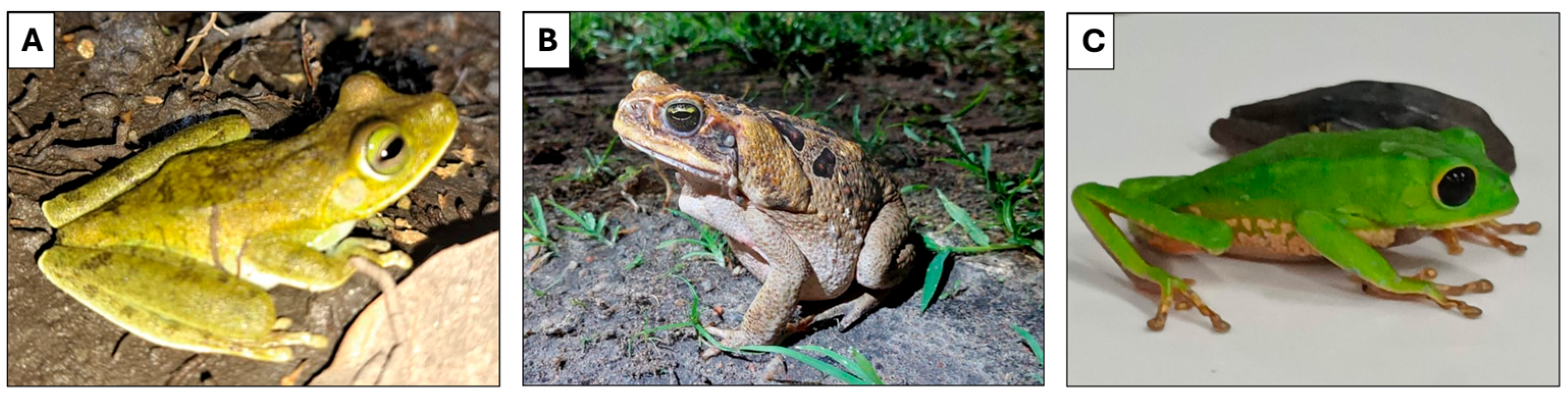
Amphibians of the order Anura are four-legged vertebrate animals (Figure 1). Currently, there are 7664 species distributed in 54 taxonomic families, including toads, frogs, and tree frogs [5,6]. They are found throughout the world, except in the polar regions of Antarctica and Greenland, with the greatest diversity in tropical regions. In general, toads are more commonly found in forested regions, frogs are more prevalent in savannah areas, and tree frogs are most abundant in both forest and Atlantic forest regions [6]. Brazil is one of the richest areas in amphibian species in the world, with around 1200 specimens recorded [6,7].

Figure 1.
Representative anuran species. (A) Boana xerophylla. (B) Rhinella marina. (C) Phyllomedusa bicolor. Photo credits: A and B by Guilherme Melo-dos-Santos; C by Anne Grace A. C. Marques.
2.1. Toads
Toads are widely distributed across the globe, typically found in tropical regions inhabiting terrestrial/aquatic environments, such as lakes, rivers, and marshes. They are characterized by their smooth, dry skin, the presence of paratoid glands, and short legs. They are generally 5 to 20 cm long, but some species can reach up to 30 cm in length [6].
Rhinella schneideri, generally known as the cane toad, is an anuran from the Bufonidae family that has a wide geographic distribution throughout South America, including Brazil, Argentina, Bolivia, Paraguay, and Uruguay. It is the best known of the amphibians, as it appears in backyards and home gardens and can reach up to 25 cm in length. A striking characteristic of the genus Rhinella is the presence of well-developed parotid glands, located dorsally close to the tympanum. These glands are responsible for producing a yellowish, dense, very complex, and variable toxic secretion, depending on the species [8,9].
2.2. Frogs
They are characterized by their moist and smooth skin, the absence of paratoid glands, and long, muscular legs, with fingers without projections and a robust waist. They are generally 5 to 15 cm long, but some species can reach up to 25 cm in length [6,8]. The bullfrog (Rana catesbeiana) is one of the best-known species, as it is widely consumed by humans, promoting the development of aquaculture in several countries [10,11].
2.3. Tree Frogs
Tree frogs generally range in length from 2 to 8 cm, but some species can reach up to 10 cm in length. They are adapted to an arboreal life, have a slender waist, generally smooth skin, long and thin legs, as well as adhesive disks on the tips of their digits, allowing them to climb vertical surfaces. They have paratoid glands located in different parts of the body, depending on the species [6,8].
The most studied genus is Phyllomedusa, for example, Phyllomedusa bicolor from Amazonian forests. Popularly known as “Kambô”, “Kampô”, or “Kampu”, the natives, mainly from Brazil, use the poison secreted by the skin of this animal in traditional medicine, called “frog vaccine”, as a ritual to prevent diseases, and it is considered a physical and mental invigorator [12,13].
3. Secretions from Anuran
Anurans possess two main types of skin glands: mucous glands, associated with moisture maintenance and cutaneous respiration, and granular glands, specialized in secreting chemical substances for defense [14,15]. This duality reflects the direct exposure of their skin to environmental challenges such as desiccation, pathogens, and predators [16]. Thus, anuran skin has evolved to produce a diversity of bioactive compounds, including alkaloids, biogenic amines, steroids, peptides, and proteins, making them valuable models for chemical and biological research [16,17]. The composition of these secretions varies significantly among families and species and will be explored in this review, regarding their antimicrobial ability.
Among the most notable bioactive components of anuran cutaneous secretions are alkaloids, whose discovery and characterization have advanced significantly due to improvements in analytical techniques. Currently, over 800 distinct alkaloids have been identified and characterized in anuran secretions, with new representatives being described annually, expanding our knowledge of this fascinating chemical diversity [18]. These alkaloids can be classified into three main structural groups—guanidinics, lipophilics, and indolics—which present distinct chemical and physiological properties [19]. Some alkaloids deserve special attention for their remarkable pharmacological potential. Concentrated mainly in the Bufonidae family, these compounds have demonstrated in experimental studies muscle-modulating activities, as well as promising anticancer, antiviral, and antimicrobial effects [19]. The structural diversity of these indole alkaloids makes them particularly interesting for investigations in medicinal chemistry and new drug development.
Currently, over 2000 bioactive peptides have been identified in anuran secretions, classified into about 100 distinct families [16]. Present in all species studied, these peptides exhibit remarkable structural diversity that reflects specific adaptations to the different ecological niches occupied by these animals. This molecular variability is directly related to the particular selective pressures of each habitat, including interactions with pathogens, predators, and environmental factors that have driven their molecular evolution [14,16]. Among them, antimicrobial peptides stand out not only for being the most abundant but also for their broad spectrum of activity, acting as a first line of defense against bacteria, fungi, and viruses, and beyond their antimicrobial properties, many of these peptides present multifunctional properties, including neurotoxic, cardiotoxic and myotoxic effects against potential predators, as well as modulatory activities such as analgesic action and immunoregulation [16,20,21].
4. Antimicrobial Compounds
Antimicrobial peptides (AMPs) are commonly found in animal toxins, typically displaying a cationic nature and comprising a small number of amino acids (10 to 50). These peptides tend to adopt an amphipathic α-helix conformation, enabling them to interact effectively with membranes. AMPs showcase a broad spectrum of activity, targeting bacteria, fungi, viruses, and protozoa [22,23]. Within anurans, there exists a diverse array of AMPs that play a crucial role in their innate immune defense. These peptides are synthesized and secreted mainly in the skin, acting as a first line of protection against pathogens through their cytolytic activity. Adapted to various ecological niches, different anuran species have evolved distinct AMPs, serving not only as effective defenses against a wide range of microbial invaders but also reflecting adaptations to their unique habitats [24,25]. Thus, anuran toxins can be applied to combat different microorganisms, which is shown in Figure 2.
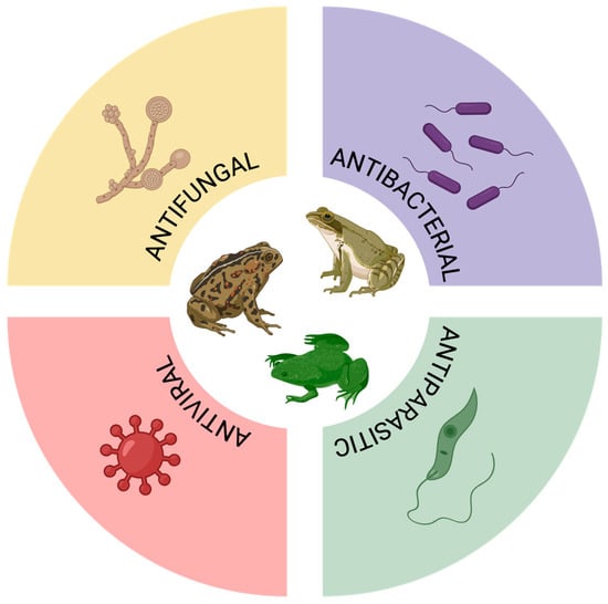
Figure 2.
Antimicrobial properties of anuran toxins.
There are models that explain the actions of AMPs on cell membranes, with the most well-known being the barrel-stave model, toroidal model, and carpet-like model. In the barrel-stave model, peptides form a barrel-shaped pore; the hydrophobic residues of the peptides interact with the lipid part of the membrane, while the hydrophilic residues form the interior of the pore, allowing the release of cytoplasmic contents and leading to cell death [26]. In the toroidal model, peptides align with the phospholipids of the membrane and fold to form hydrophilic pores, enabling membrane permeabilization and disintegration. Finally, in the carpet-like model, peptides accumulate in large amounts and associate parallel to the membrane, creating tension that leads to membrane disruption and cell lysis [25]. In addition, AMPs may also exert intracellular activity, targeting nucleic acids, enzymatic activity, and protein synthesis, among other essential cellular functions [26].
4.1. Bacteria
AMPs derived from various anuran families, including Alytidae, Ranidae, Pipidae, Hylidae, Leptodactylidae, Leiopelmatidae, Hyperoliidae, Bombinatoridae, Dicroglossidae, and Myobatrachidae, exhibit potent activity against both Gram-positive and Gram-negative bacteria, commonly tested with strains such as Staphylococcus aureus (S. aureus) and Escherichia coli (E. coli) [24,25]. Antibacterial activity has been identified in the skin secretions of 78 species of anurans, yielding a total of 323 antimicrobial peptides with antibacterial activity. Detailed information can be found in Table 1.

Table 1.
Anuran antimicrobial peptides with antibacterial activity.
In the Alytidae family, the skin secretions of the anurans Alytes maurus and Alytes obstetricans possess the AMPs alyteserin-1 and alysteserin-2, which have demonstrated effectiveness against S. aureus and E. coli. The peptides from Ascaphus truei, known as ascaphins 1 to 8 and belonging to the Leiopelmatidae family, exhibit antibacterial activity against S. aureus, as well as Gram-negative bacterias such as E. coli, Enterobacter cloacae and Klebsiella pneumoniae [32].
The Ranidae family of anurans, specifically within the genera Amolops, Hylarana, Lithobates, Odorrana, Pelophylax and Rana, express a range of AMPs with broad spectrum of action, including brevinin-1, brevinin-1, esculetin-1, esculetin-2, palustrin-1, palustrin-2, ranacyclin, ranatuerin, and temporin [25,29,30,31,40,41,42,56,66,67,68,69,88,89]. Notably, Rana japonica features japonicin-1 and japonicin-2, while Rana nigromaculata exhibits nigrocin-1 and nigrocin-2 [92,93].
Furthemore, the genus Litoria within the Ranidae family exhibits the peptides aurein, citropin, caerin, uperin, and fallaxidin [57,58,59,60,61,62,63]. Another notable AMP is melittin, derived from the bee venom of Apis mellifera, showcasing efficacy against both Gram-positive and Gram-negative bacteria [119,120]. Within anuran AMPs, melittin-related peptides (MRPs) found in the Ranidae family emulate melittin, demonstrating a broad-spectrum antimicrobial effect against bacteria [25,102].
Bombinin-related peptides (BLPs) from the Bombinatoridae family, specifically bombinins H, exhibit diminished bactericidal efficacy while demonstrating hemolytic activity on erythrocytes [121]. In contrast, other BLPs, including maximins 1 to 5 from Bombina maxima, display robust antibacterial activity [34].
The Dicroglossidae family, represented by Fejervarya cancrivora, possesses the tigerinin peptide, similar to Rana tigerina, demonstrating antibacterial activity against S. aureus, Bacillus subtilis, Pseudomonas aeruginosa, and E. coli [37,106]. In the Hylidae family, found within the genera Hyla, Hylomantis, Phyllomedusa, and Hypsiboas, various naturally occurring peptides such as dermaseptins, dermatoxins, distinctin, hylain, hylin, hylaseptin, phylloseptins, phylloxin, and raniseptin [38,39,43,44,45,71,72,73,74].
Kassinatuerins from the Hyperoliidae family are found in the skin secretion of Kassina senegalensis [48,49] and Kassina maculata [46]. Peptides within the Leptodactylidae family, such as fallaxin (Leptodactylus fallax), laticeptin (Leptodactylus laticeps), pentadactylin (Leptodactylus pentadactylus), and ocellatin (Leptodactylus ocellatus) exhibit action majority for Gram-negative bacteria [50,51,52,53,54]. Addiyionally, uperin from Uperoleia mjobergii (Myobatrachidae) demonstrates efficacy against a great variety of microrganisms S. aureus, Staphylococcus epidermidis, Bacillus cereus, Micrococcus luteus, Streptococcus uberis, Leuconostoc lactis, Pasteurella multocida, and Listeria innocua.
The skin secretion of the genus Xenopus (Xenopus amieti, Xenopus andrei, Xenopus borealis, Xenopus clivii, Xenopus laevis, and Xenopus muelleri) within the Pipidae family reveals AMPs othologous to the magainins, peptides orthologous to peptide glycine-leucine-amide (PGLa), homologous to caerulein-precursor fragments (CPF), and xenopsin precursor fragment (XPF) [111,112,113,114,115,116,117]. Additionally, the XT 1 to 7 peptides from Xenopus tropicalis exhibit some similarities to X. laevis PGLa, procaeruleins, and proxenopsin regions [118].
4.2. Fungus
Frog skin secretions contain potent compounds that exhibit remarkable activity against various fungi, showcasing their natural defense mechanism in combating fungal infections (Table 2). Among these components, peptides are stored in a granular gland located mainly in the skin of the dorsal region. Three compounds (Arenobufagin, Gamabufotalin, and Telocinobufagin) are described as bufadienolides from boreal toad (Anaxyrus boreas) presenting antifungal activities against Batrachochytrium dendrobatidis (Bd) being the Arenobufagin the most effective one since it had the lowest estimated concentration where Bd was maximally inhibited (12.9 μg/mL) [122].
Table 2.
Anuran antifungal toxins.
Anurans endemic in Indonesia were also studied. One is the bleeding toad Leptophryne cruentata and the javan tree frog Rhacophorus margaritifer (also named R. javanus) and their skin secretions demonstrated antifungal activity against the fungus Trichophyton mentagrophytes [123].
Skin secretions of the toad Bufo arenarum demonstrated being a rich source of several components with antifungal activity. One of those is an alkaloid called dehydrobufotenine, which demonstrated being effective against phytopathogenic fungi that affect plants of economic interest. After that, forty-five analogs of dehydrobufotenine were synthetized and evaluated for their activity. Six of these analogs demonstrated similar or higher activity than the control used (carbendazim 50 μg/mL). Dehydrobufotenine and most of its analogs showed higher activity against Alternaria solani (5–100%) than the controls [124].
Candida species are typically harmless fungal microorganisms found in the gastrointestinal and urinary tracts of humans, functioning as commensals. However, under conditions where the immune system is compromised, Candida has the capability to transform into a significant cause of severe mucosal or systemic infections, with Candida albicans (C. albicans). being the most prevalent species [129]. Two peptide components from skin secretion of the European frogs Pelophylac lessonae/ridibundus, named Esculentin-1 derived from its N-terminal part, named Esc-1a(1-21)NH2 and Esc-1b(1-18)NH2 demonstrated being equally active against C. albicans. Also, the smallest component (Esc-1b 1-18)NH2 demonstrated a dose-dependent membrane-perturbing effect on Candida with a kinetic overlapping with killing activity, thus pointing to membrane perturbation as the primary event of its candidacidal activity [125].
Still regarding C. albicans, there are reports of several components active against the fungus, such as synthetic peptides (B2RP) from skin secretions of the Southeast Asian frog Hylarana erythraea as well as Nigroain and esculetin-2 from the same frog [126,127]. Peptides brevinin-1 and 2 from H. nigrovittatta and skin secretion peptide from H. temporalis also demonstrated effective activity against the fungus [128].
4.3. Virus
Among the AMPs with antiviral activity, those effective against viruses of significant public health relevance stand out, such as human immunodeficiency virus (HIV), herpes simplex virus types 1 and 2 (HSV-1 and HSV-2), Zika virus (ZIKV), dengue virus serotypes 1; 2; 3; and 4 (DENV1-4), severe acute respiratory syndrome coronavirus 2 (SARS-CoV-2), and Influenza A virus subtypes H1N1 and H5N1. Maximin 1 to 5 and Maximin H5, present in Bombina maxima, also exhibit antiviral activity against HIV [34].
The genus Litoria stands out for its wide variety of antiviral AMPs. Among them are the peptides Caerin 1.2-1.5 and 4.1 (from Litoria caerulea), Caerin 1.9 (from Litoria chloris), Dahlein 5.6 (from Litoria dahlii), Maculatin 1.1 and 1.3 (from Litoria eucnemis and Litoria genimaculata), Uperin 7.1 (from Litoria ewingi), Caerin 1.19 (from Litoria gracilenta), Caerin 1.1 and 1.10 (from Litoria splendida), and Caerin 1.6 and 1.7 (from Litoria xanthomera), all with proven activity against HIV. Additionally, the peptide Frenatin 2, found in Litoria infrafrenata, has shown efficacy against the yellow fever virus (YFV) [130,131,132].
A relevant family of peptides is the Dermaseptins, found in Phyllomedusa sauvagei. Dermaseptin-S1 acts against HIV, frog virus 3 (FV3), channel catfish virus (CCV), and HSV-1; Dermaseptins-S2 and S3 are effective against HSV-1; Dermaseptin-S4 exhibits anti-ZIKV activity; and Dermaseptin-S9 has demonstrated action against HIV-1 and SARS-CoV-2, standing out as a potential tool in combating the pandemic [133,134,135,136,137,138].
Other notable AMPs include Hylin a1, produced by Hypsiboas albopunctatus, which acts against a wide range of viruses, including bovine herpesvirus type 1 (BoHV-1), caprine herpesvirus type 1 (CpHV-1), canine distemper virus (CDV), bovine viral diarrhea virus (BVDV), Schmallenberg virus (SBV), HSV-1, and HSV-2 [139]. The peptide Yodha, found in Indosylvirana aurantiaca, has shown efficacy against ZIKV and DENV1-4, the viruses responsible for Zika and dengue diseases [134,140].
In the genus Rana, several antimicrobial peptides (AMPs) have been identified, demonstrating antiviral activity against different pathogens. Among them, Esculentin-1ARb, Esculentin-2P, Brevinin-2, Ranatuerin-2P, Ranatuerin-6, and Ranatuerin-9 stand out for their action against HIV [130,141]. Other AMPs, such as Brevinin-1 and Temporin B, are effective against the herpes virus [142,143,144], while Temporin A shows activity against CCV and FV3 [145]. Additionally, Temporin G demonstrates antiviral effects against influenza A virus and human papillomavirus (HPV) [145]. Finally, the peptide AR-23, found in Rana tagoi, has shown activity against an impressive variety of viruses, including SARS-CoV-2, measles virus (MeV), human parainfluenza virus type 2 (HPIV-2), human coronavirus 229E (HCoV-229E), BoHV-1, CpHV-1, CDV, BVDV, and SBV [139].
The Magainin family of peptides, found in Xenopus laevis, acts against HSV-1 and HSV-2, and Urumin, produced by Hydrophylax bahuvistara, has demonstrated efficacy against the H1N1 virus [134,146,147]. The peptide Temporin-SHa, found in Pelophylax saharica, also exhibit antiviral activity against HSV-1 [144].
Of the AMPs already isolated and characterized, only 47 exhibit antiviral activity, with proven efficacy against 26 distinct viruses. Table 3 summarizes the detailed information about these molecules, including their species of origin and the viruses they combat. These examples illustrate the potential of AMPs as a new frontier in combating viral infections.
Table 3.
Antiviral activity of anuran toxins.
4.4. Protozoo
Among the diverse composition of frog skin secretions, several components with anti-parasitic activity have already been identified; this action promoted by AMP’s is being elucidated and has shown promising results against different types of protozoa [152] (Table 4). The mechanism of action responsible for the activity mainly involves the action of these molecules on the permeability of biological membranes, such as Temporins A and B that act on the surface–membrane and also reduce intracellular ATP and dermasipitins responsible for forming amphipathic helices within membranes [153]. In this context, it is possible to highlight how promising the use of these peptides as a treatment for different types of parasitic infections, often neglected but with great potential to cause damage to human health, is promising [154].
Table 4.
Antiparasitic activity of anuran toxins.
5. Advanced Research
Advanced research on anuran toxins has driven significant scientific progress, leading to clinical trials, patents, and the development of commercially available products. Toxins such as bufalin have demonstrated remarkable potential in oncological applications [165]. Clinical investigations, including studies on Huachansu for cancer treatment (breast cancer, gallbladder cancer, gastric cancer, hepatocellular carcinoma, liver cancer, lung cancer, non-small cell lung cancer, and pancreatic cancer), are ongoing and highlight the therapeutic promise of these compounds [166].
Currently, there are no antimicrobial products on the market that are directly derived from anuran toxins. However, many of these toxins exhibit promising antimicrobial properties and remain the focus of extensive research. Anurans produce a diverse array of bioactive peptides in their skin secretions, many of which show activity against bacteria, fungi, and other microorganisms. Examples of antimicrobial peptide classes found in anurans have been previously cited in this review.
Although no direct products derived from anuran toxins have reached the market yet, advancements in biotechnology hold significant promise for enabling new therapeutic applications in the future. However, there are currently no reports of clinical trials directly involving anuran toxins or antimicrobial peptides, such as magainins and dermaseptins, for therapeutic use in humans.
Despite this, numerous studies emphasize the potential of these molecules in combating bacterial, fungal, and other infections. Some of these compounds have already undergone preclinical testing in vitro and in vivo to evaluate their efficacy and safety. For instance, dermaseptins exhibit activity against Gram-positive and Gram-negative bacteria, protozoa, and fungi while demonstrating low toxicity to healthy human cells [133].
The interest in advancing these compounds to clinical trials remains strong. Before an antimicrobial molecule candidate can proceed to in vivo studies and subsequently to clinical trials as an alternative therapeutic agent, it must undergo a comprehensive preclinical evaluation. This includes detailed in vitro assays to determine its antimicrobial potency, spectrum of activity, cytotoxicity against cells, and potential to induce resistance. The compound must also be tested for its mechanism of action and synergy with existing antibiotics. Promising candidates are then advanced to in vivo efficacy studies in relevant infection models to assess pharmacokinetic and pharmacodynamic (PK/PD) parameters, bioavailability, tissue distribution, metabolism, and toxicity. If the in vivo results support safety and efficacy, a regulatory submission—such as an Investigational New Drug (IND) application—must be prepared, including all preclinical data. This step is essential to gain approval from regulatory agencies to initiate Phase I clinical trials focused on evaluating safety, tolerability, and preliminary pharmacokinetics in humans. Such a process ensures that only the most viable and safe antimicrobial candidates progress as potential therapeutic alternatives [167,168,169,170]. So, it is a long lasting protocol that hinges on improvements in formulation, delivery methods, and modifications to their pharmacokinetic and pharmacodynamic properties. Current efforts are focused on developing more stable and less toxic analogs to overcome these limitations and unlock their therapeutic potential, such as magainins and synthetic analogs [171]. Department of Health and Human Services.
6. Conclusions
In recent years, there has been a growing focus on the antimicrobial potential of anuran-derived toxins, especially peptides and alkaloids, which demonstrate broad-spectrum activity against bacteria, fungi, viruses, and other pathogens. This interest is driven largely by the escalating global crisis of antimicrobial resistance [172,173], which urgently demands novel therapeutic agents beyond conventional antibiotics. Anuran secretions, evolved as chemical defense mechanisms, offer a rich and largely untapped reservoir of bioactive molecules with unique structures and modes of action that differ from traditional antimicrobials, making them highly attractive candidates for drug discovery. Despite the promising nature of these compounds, significant knowledge gaps remain. While a handful of species—primarily from well-studied regions—have been the focus of intensive research, the vast majority of anuran biodiversity, especially in ecologically rich yet underexplored habitats such as the Amazon rainforest, Southeast Asian tropical forests, and African wetlands remains uncharacterized. This presents a compelling frontier for future bioprospecting and biodiversity-driven drug discovery efforts. Moreover, understanding the precise mechanisms by which these peptides and alkaloids exert their antimicrobial effects is still incomplete. Insights into their interactions with microbial membranes, immunomodulatory properties, and potential synergy with existing antibiotics could unlock new therapeutic strategies. Additionally, advances in synthetic biology, peptide engineering, and high-throughput screening are enabling the design and optimization of these natural molecules to enhance their stability, reduce toxicity, and improve pharmacokinetic profiles, accelerating their translation from bench to bedside. For researchers, this emerging field highlights the importance of multidisciplinary approaches combining ecology, chemistry, pharmacology, and computational modeling to fully harness anuran toxins’ therapeutic potential. For clinicians and the drug development field, these molecules represent a promising avenue for addressing antimicrobial resistance through novel mechanisms and chemical diversity that could circumvent existing resistance pathways.
In summary, anuran-derived antimicrobial agents not only expand our arsenal against resistant pathogens but also exemplify the value of biodiversity as a critical resource for future drug innovation. Continued exploration, coupled with technological advancements, will be key to realizing their full biomedical potential.
Author Contributions
Investigation, M.B.P., A.G.A.C.M., A.F.M.P., G.M.-d.-S., F.A.C., B.C.S.J., I.G.F., R.L.P., M.A.S. and W.M.M.; writing—original draft preparation, M.B.P., A.G.A.C.M., A.F.M.P., F.A.C., B.C.S.J., I.G.F., R.L.P. and W.M.M.; writing—review and editing, M.B.P. and I.S.O.; supervision, M.B.P. and I.S.O.; project administration, I.S.O. All authors have read and agreed to the published version of the manuscript.
Funding
This research was funded by Conselho Nacional de Desenvolvimento Científico e Tecnológico (CNPq, The National Council for Scientific and Technological Development, scholarships to MBP n. 305778/2023-4, AFMP n. 151190/2023-2, and WMM n. 309207/2020-7), Coordenação de Aperfeiçoamento de Pessoal de Nível Superior (CAPES, the Coordination for the Improvement of Higher Education Personnel, scholarships to GMS), and Fundação de Amparo à Pesquisa do Estado de São Paulo (FAPESP, São Paulo Research Foundation, scholarships to BCSJ n. 2024/17849-3, IGF n. 2023/11264-0, and n. 2024/14618-0, and ISO n. 2020/13176-3, and grant to AFMP n. 2023/09921-3), acknowledges funding support from Fundação de Amparo à Pesquisa do Estado do Amazonas (AGACM, Programa UNIVERSAL, FAPEAM 20 ANOS, Edital n.º 001/2023) and MAS (EDITAL N. 020/2024—PRODUTIVIDADE EM CT&I).
Institutional Review Board Statement
Not applicable.
Informed Consent Statement
Not applicable.
Data Availability Statement
No new data were created or analyzed in this study.
Conflicts of Interest
The authors declare no conflicts of interest.
References
- Bordon, K.D.C.F.; Cologna, C.T.; Fornari-Baldo, E.C.; Pinheiro-Júnior, E.L.; Cerni, F.A.; Amorim, F.G.; Anjolette, F.A.P.; Cordeiro, F.A.; Wiezel, G.A.; Cardoso, I.A.; et al. From Animal Poisons and Venoms to Medicines: Achievements, Challenges and Perspectives in Drug Discovery. Front. Pharmacol. 2020, 11, 1132. [Google Scholar] [CrossRef]
- García, F.A.; Fuentes, T.F.; Alonso, I.P.; Bosch, R.A.; Brunetti, A.E.; Lopes, N.P. A Comprehensive Review of Patented Antimicrobial Peptides from Amphibian Anurans. J. Nat. Prod. 2024, 87, 600–616. [Google Scholar] [CrossRef]
- Barros, A.L.A.N.; Hamed, A.; Marani, M.; Moreira, D.C.; Eaton, P.; Plácido, A.; Kato, M.J.; Leite, J.R.S.A. The Arsenal of Bioactive Molecules in the Skin Secretion of Urodele Amphibians. Front. Pharmacol. 2021, 12, 810821. [Google Scholar] [CrossRef]
- Barra, D.; Simmaco, M. Amphibian Skin: A Promising Resource for Antimicrobial Peptides. Trends Biotechnol. 1995, 13, 205–209. [Google Scholar] [CrossRef]
- Frost, D.R. Amphibian Species of the World: An Online Reference. 1999. Available online: https://amphibiansoftheworld.amnh.org (accessed on 15 February 2024).
- AmphibiaWeb. AmphibiaWeb 2024. Available online: https://amphibiaweb.org (accessed on 15 February 2024).
- Segalla, M.V.; Berneck, B.; Canedo, C.; Caramaschi, U.; Cruz, C.A.G.; Garcia, P.C.A.; Grant, T.; Haddad, C.F.B.; Lourenço, A.C.C.; Mângia, S.; et al. List of Brazilian Amphibians. Herpetol. Bras. 2021, 10, 121–216. [Google Scholar] [CrossRef]
- Bernarde, P.S. Anfíbios e Répteis: Introdução Ao Estudo da Herpetofauna Brasileira, 1st ed.; Anolis Books: Curitiba, Brazil, 2012; ISBN 978-85-65622-00-4. [Google Scholar]
- Leal, A.; Karnopp, E.; Barreto, Y.C.; Oliveira, R.S.; Rosa, M.E.; Borges, B.T.; Goulart, F.L.; Souza, V.Q.D.; Laikowski, M.M.; Moura, S.; et al. The Insecticidal Activity of Rhinella schneideri (Werner, 1894) Paratoid Secretion in Nauphoeta Cinerea Cocroaches. Toxins 2020, 12, 630. [Google Scholar] [CrossRef] [PubMed]
- Sun, Q.; Zhang, J.; Wang, T.; Xiong, Y.; Zhan, X.; Zhao, H.; Wang, J.; Fan, Y.; Bi, R.; Wang, S.; et al. Cooking Methods Effectively Alter Perfluoroalkyl Substances and Nutrients in Cultured and Wild Bullfrogs. J. Hazard. Mater. 2023, 445, 130555. [Google Scholar] [CrossRef]
- Ribeiro, L.P.; Carvalho, T.; Becker, C.G.; Jenkinson, T.S.; Leite, D.D.S.; James, T.Y.; Greenspan, S.E.; Toledo, L.F. Bullfrog Farms Release Virulent Zoospores of the Frog-Killing Fungus into the Natural Environment. Sci. Rep. 2019, 9, 13422. [Google Scholar] [CrossRef]
- Junior, V.H.; Martins, I.A. KAMBÔ: An Amazonian Enigma. J. Venom Res. 2020, 10, 13–17. [Google Scholar]
- Silva, F.V.A.D.; Monteiro, W.M.; Bernarde, P.S. “Kambô” Frog (Phyllomedusa bicolor): Use in Folk Medicine and Potential Health Risks. Rev. Soc. Bras. Med. Trop. 2019, 52, e20180467. [Google Scholar] [CrossRef]
- Dornelles, M.F.; Marques, M.d.G.B.; Renner, M.F. Revisão Sobre Toxinas de Anura (Tetrapoda, Lissamphibia) e Suas Aplicações Biotecnológicas. Ciência Mov. Rev. Ciências Saúde Educ. 2010, 12, 103–114. [Google Scholar]
- Carrillo, J.F.C.; Boaretto, A.G.; Santana, D.J.; Silva, D.B. Skin Secretions of Leptodactylidae (Anura) and Their Potential Applications. J. Venom. Anim. Toxins Incl. Trop. Dis. 2024, 30, e20230042. [Google Scholar] [CrossRef]
- Xu, X.; Lai, R. The Chemistry and Biological Activities of Peptides from Amphibian Skin Secretions. Chem. Rev. 2015, 115, 1760–1846. [Google Scholar] [CrossRef] [PubMed]
- Qi, J.; Zulfiker, A.H.M.; Li, C.; Good, D.; Wei, M.Q. The Development of Toad Toxins as Potential Therapeutic Agents. Toxins 2018, 10, 336. [Google Scholar] [CrossRef]
- Daly, J.W.; Spande, T.F.; Garraffo, H.M. Alkaloids from Amphibian Skin: A Tabulation of Over Eight-Hundred Compounds. J. Nat. Prod. 2005, 68, 1556–1575. [Google Scholar] [CrossRef]
- Rodríguez, C.; Rollins-Smith, L.; Ibáñez, R.; Durant-Archibold, A.A.; Gutiérrez, M. Toxins and Pharmacologically Active Compounds from Species of the Family Bufonidae (Amphibia, Anura). J. Ethnopharmacol. 2017, 198, 235–254. [Google Scholar] [CrossRef] [PubMed]
- Freitas, C.I.A. Estudo Sobre a Atividade Antimicrobiana de Substâncias Extraídas e Purificadas de Secreções Da Pele de Anfíbios. Ph.D. Thesis, Universidade Federal do Ceará, Fortaleza, Brazil, 2003. [Google Scholar]
- Azevedo Calderon, L.D.; Silva, A.D.A.E.; Ciancaglini, P.; Stábeli, R.G. Antimicrobial Peptides from Phyllomedusa Frogs: From Biomolecular Diversity to Potential Nanotechnologic Medical Applications. Amino Acids 2011, 40, 29–49. [Google Scholar] [CrossRef]
- Huan, Y.; Kong, Q.; Mou, H.; Yi, H. Antimicrobial Peptides: Classification, Design, Application and Research Progress in Multiple Fields. Front. Microbiol. 2020, 11, 582779. [Google Scholar] [CrossRef]
- Pfalzgraff, A.; Brandenburg, K.; Weindl, G. Antimicrobial Peptides and Their Therapeutic Potential for Bacterial Skin Infections and Wounds. Front. Pharmacol. 2018, 9, 281. [Google Scholar] [CrossRef]
- Rollins-Smith, L.A. The Importance of Antimicrobial Peptides (AMPs) in Amphibian Skin Defense. Dev. Comp. Immunol. 2023, 142, 104657. [Google Scholar] [CrossRef]
- König, E.; Bininda-Emonds, O.R.P.; Shaw, C. The Diversity and Evolution of Anuran Skin Peptides. Peptides 2015, 63, 96–117. [Google Scholar] [CrossRef]
- Zhang, Q.-Y.; Yan, Z.-B.; Meng, Y.-M.; Hong, X.-Y.; Shao, G.; Ma, J.-J.; Cheng, X.-R.; Liu, J.; Kang, J.; Fu, C.-Y. Antimicrobial Peptides: Mechanism of Action, Activity and Clinical Potential. Military Med. Res. 2021, 8, 48. [Google Scholar] [CrossRef]
- König, E.; Zhou, M.; Wang, L.; Chen, T.; Bininda-Emonds, O.R.P.; Shaw, C. Antimicrobial Peptides and Alytesin Are Co-Secreted from the Venom of the Midwife Toad, Alytes maurus (Alytidae, Anura): Implications for the Evolution of Frog Skin Defensive Secretions. Toxicon 2012, 60, 967–981. [Google Scholar] [CrossRef]
- Conlon, J.M.; Demandt, A.; Nielsen, P.F.; Leprince, J.; Vaudry, H.; Woodhams, D.C. The Alyteserins: Two Families of Antimicrobial Peptides from the Skin Secretions of the Midwife Toad Alytes obstetricans (Alytidae). Peptides 2009, 30, 1069–1073. [Google Scholar] [CrossRef]
- Yang, X.; Xia, J.; Yu, Z.; Hu, Y.; Li, F.; Meng, H.; Yang, S.; Liu, J.; Wang, H. Characterization of Diverse Antimicrobial Peptides in Skin Secretions of Chungan Torrent Frog Amolops chunganensis. Peptides 2012, 38, 41–53. [Google Scholar] [CrossRef]
- Chen, Z.; Yang, X.; Liu, Z.; Zeng, L.; Lee, W.; Zhang, Y. Two Novel Families of Antimicrobial Peptides from Skin Secretions of the Chinese Torrent Frog, Amolops jingdongensis. Biochimie 2012, 94, 328–334. [Google Scholar] [CrossRef]
- Wang, H.; Ran, R.; Yu, H.; Yu, Z.; Hu, Y.; Zheng, H.; Wang, D.; Yang, F.; Liu, R.; Liu, J. Identification and Characterization of Antimicrobial Peptides from Skin of Amolops ricketti (Anura: Ranidae). Peptides 2012, 33, 27–34. [Google Scholar] [CrossRef]
- Conlon, J.M.; Sonnevend, A.; Davidson, C.; David Smith, D.; Nielsen, P.F. The Ascaphins: A Family of Antimicrobial Peptides from the Skin Secretions of the Most Primitive Extant Frog, Ascaphus Truei. Biochem. Biophys. Res. Commun. 2004, 320, 170–175. [Google Scholar] [CrossRef]
- Wang, X.; Song, Y.; Li, J.; Liu, H.; Xu, X.; Lai, R.; Zhang, K. A New Family of Antimicrobial Peptides from Skin Secretions of Rana pleuraden. Peptides 2007, 28, 2069–2074. [Google Scholar] [CrossRef]
- Lai, R.; Zheng, Y.-T.; Shen, J.-H.; Liu, G.-J.; Liu, H.; Lee, W.-H.; Tang, S.-Z.; Zhang, Y. Antimicrobial Peptides from Skin Secretions of Chinese Red Belly Toad Bombina maxima. Peptides 2002, 23, 427–435. [Google Scholar] [CrossRef]
- Wang, T.; Zhang, J.; Shen, J.-H.; Jin, Y.; Lee, W.-H.; Zhang, Y. Maximins S, a Novel Group of Antimicrobial Peptides from Toad Bombina maxima. Biochem. Biophys. Res. Commun. 2005, 327, 945–951. [Google Scholar] [CrossRef]
- Mignogna, G.; Simmaco, M.; Kreil, G.; Barra, D. Antibacterial and Haemolytic Peptides Containing D-Alloisoleucine from the Skin of Bombina variegata. EMBO J. 1993, 12, 4829–4832. [Google Scholar] [CrossRef]
- Song, Y.; Lu, Y.; Wang, L.; Yang, H.; Zhang, K.; Lai, R. Purification, Characterization and Cloning of Two Novel Tigerinin-like Peptides from Skin Secretions of Fejervarya cancrivora. Peptides 2009, 30, 1228–1232. [Google Scholar] [CrossRef]
- Prates, M.V.; Sforça, M.L.; Regis, W.C.B.; Leite, J.R.S.A.; Silva, L.P.; Pertinhez, T.A.; Araújo, A.L.T.; Azevedo, R.B.; Spisni, A.; Bloch, C. The NMR-Derived Solution Structure of a New Cationic Antimicrobial Peptide from the Skin Secretion of the Anuran Hyla punctata. J. Biol. Chem. 2004, 279, 13018–13026. [Google Scholar] [CrossRef]
- Wu, J.; Liu, H.; Yang, H.; Yu, H.; You, D.; Ma, Y.; Ye, H.; Lai, R. Proteomic Analysis of Skin Defensive Factors of Tree Frog Hyla simplex. J. Proteome Res. 2011, 10, 4230–4240. [Google Scholar] [CrossRef]
- Dong, Z.; Luo, W.; Zhong, H.; Wang, M.; Song, Y.; Deng, S.; Zhang, Y. Molecular Cloning and Characterization of Antimicrobial Peptides from Skin of Hylarana guentheri. Acta Biochim. Biophys. Sin. 2017, 49, 450–457. [Google Scholar] [CrossRef]
- Wang, H.; Yu, Z.; Hu, Y.; Yu, H.; Ran, R.; Xia, J.; Wang, D.; Yang, S.; Yang, X.; Liu, J. Molecular Cloning and Characterization of Antimicrobial Peptides from Skin of the Broad-Folded Frog, Hylarana latouchii. Biochimie 2012, 94, 1317–1326. [Google Scholar] [CrossRef]
- Mishra, B.; Wang, G. Ab Initio Design of Potent Anti-MRSA Peptides Based on Database Filtering Technology. J. Am. Chem. Soc. 2012, 134, 12426–12429. [Google Scholar] [CrossRef]
- Conlon, J.M.; Woodhams, D.C.; Raza, H.; Coquet, L.; Leprince, J.; Jouenne, T.; Vaudry, H.; Rollins-Smith, L.A. Peptides with Differential Cytolytic Activity from Skin Secretions of the Lemur Leaf Frog Hylomantis lemur (Hylidae: Phyllomedusinae). Toxicon 2007, 50, 498–506. [Google Scholar] [CrossRef]
- Castro, M.S.; Ferreira, T.C.G.; Cilli, E.M.; Crusca, E.; Mendes-Giannini, M.J.S.; Sebben, A.; Ricart, C.A.O.; Sousa, M.V.; Fontes, W. Hylin A1, the First Cytolytic Peptide Isolated from the Arboreal South American Frog Hypsiboas albopunctatus (“Spotted Treefrog”). Peptides 2009, 30, 291–296. [Google Scholar] [CrossRef]
- Magalhães, B.S.; Melo, J.A.T.; Leite, J.R.S.A.; Silva, L.P.; Prates, M.V.; Vinecky, F.; Barbosa, E.A.; Verly, R.M.; Mehta, A.; Nicoli, J.R.; et al. Post-Secretory Events Alter the Peptide Content of the Skin Secretion of Hypsiboas raniceps. Biochem. Biophys. Res. Commun. 2008, 377, 1057–1061. [Google Scholar] [CrossRef]
- Wang, L.; Zhou, M.; McGrath, S.; Chen, T.; Gorman, S.P.; Walker, B.; Shaw, C. A Family of Kassinatuerin-2 Related Peptides from the Skin Secretion of the African Hyperoliid Frog, Kassina maculata. Peptides 2009, 30, 1428–1433. [Google Scholar] [CrossRef]
- Chen, H.; Wang, L.; Zeller, M.; Hornshaw, M.; Wu, Y.; Zhou, M.; Li, J.; Hang, X.; Cai, J.; Chen, T.; et al. Kassorins: Novel Innate Immune System Peptides from Skin Secretions of the African Hyperoliid Frogs, Kassina maculata and Kassina senegalensis. Mol. Immunol. 2011, 48, 442–451. [Google Scholar] [CrossRef] [PubMed]
- Conlon, J.M.; Abraham, B.; Galadari, S.; Knoop, F.C.; Sonnevend, A.; Pál, T. Antimicrobial and Cytolytic Properties of the Frog Skin Peptide, Kassinatuerin-1 and Its l- and d-Lysine-Substituted Derivatives. Peptides 2005, 26, 2104–2110. [Google Scholar] [CrossRef]
- Mattute, B.; Knoop, F.C.; Conlon, J.M. Kassinatuerin-1: A Peptide with Broad-Spectrum Antimicrobial Activity Isolated from the Skin of the Hyperoliid Frog, Kassina senegalensis. Biochem. Biophys. Res. Commun. 2000, 268, 433–436. [Google Scholar] [CrossRef]
- Rollins-Smith, L.A.; King, J.D.; Nielsen, P.F.; Sonnevend, A.; Conlon, J.M. An Antimicrobial Peptide from the Skin Secretions of the Mountain Chicken Frog Leptodactylus fallax (Anura:Leptodactylidae). Regul. Pept. 2005, 124, 173–178. [Google Scholar] [CrossRef]
- Conlon, J.M.; Al-Ghaferi, N.; Abraham, B.; Sonnevend, A.; King, J.D.; Nielsen, P.F. Purification and Properties of Laticeptin, an Antimicrobial Peptide from Skin Secretions of the South American Frog Leptodactylus laticeps. Protein Pept. Lett. 2006, 13, 411–415. [Google Scholar] [CrossRef] [PubMed]
- King, J.D.; Al-Ghaferi, N.; Abraham, B.; Sonnevend, A.; Leprince, J.; Nielsen, P.F.; Conlon, J.M. Pentadactylin: An Antimicrobial Peptide from the Skin Secretions of the South American Bullfrog Leptodactylus pentadactylus. Comp. Biochem. Physiol. Part C Toxicol. Pharmacol. 2005, 141, 393–397. [Google Scholar] [CrossRef]
- Nascimento, A.C.C.; Zanotta, L.C.; Kyaw, C.M.; Schwartz, E.N.F.; Schwartz, C.A.; Sebben, A.; Sousa, M.V.; Fontes, W.; Castro, M.S. Ocellatins: New Antimicrobial Peptides from the Skin Secretion of the South American Frog Leptodactylus ocellatus (Anura: Leptodactylidae). Protein J. 2004, 23, 501–508. [Google Scholar] [CrossRef]
- Nascimento, A.; Chapeaurouge, A.; Perales, J.; Sebben, A.; Sousa, M.V.; Fontes, W.; Castro, M.S. Purification, Characterization and Homology Analysis of Ocellatin 4, a Cytolytic Peptide from the Skin Secretion of the Frog Leptodactylus ocellatus. Toxicon 2007, 50, 1095–1104. [Google Scholar] [CrossRef]
- Li, B.; Lyu, P.; Xie, S.; Qin, H.; Pu, W.; Xu, H.; Chen, T.; Shaw, C.; Ge, L.; Kwok, H.F. LFB: A Novel Antimicrobial Brevinin-Like Peptide from the Skin Secretion of the Fujian Large Headed Frog, Limnonectes fujianensi. Biomolecules 2019, 9, 242. [Google Scholar] [CrossRef]
- Conlon, J.M.; Raza, H.; Coquet, L.; Jouenne, T.; Leprince, J.; Vaudry, H.; King, J.D. Purification of Peptides with Differential Cytolytic Activities from the Skin Secretions of the Central American Frog, Lithobates vaillanti (Ranidae). Comp. Biochem. Physiol. Part C Toxicol. Pharmacol. 2009, 150, 150–154. [Google Scholar] [CrossRef]
- Rozek, T.; Wegener, K.L.; Bowie, J.H.; Olver, I.N.; Carver, J.A.; Wallace, J.C.; Tyler, M.J. The Antibiotic and Anticancer Active Aurein Peptides from the Australian Bell Frogs Litoria aurea and Litoria raniformis. Eur. J. Biochem. 2000, 267, 5330–5341. [Google Scholar] [CrossRef]
- Manzo, G.; Ferguson, P.M.; Hind, C.K.; Clifford, M.; Gustilo, V.B.; Ali, H.; Bansal, S.S.; Bui, T.T.; Drake, A.F.; Atkinson, R.A.; et al. Temporin L and Aurein 2.5 Have Identical Conformations but Subtly Distinct Membrane and Antibacterial Activities. Sci. Rep. 2019, 9, 10934. [Google Scholar] [CrossRef]
- Wegener, K.L.; Wabnitz, P.A.; Carver, J.A.; Bowie, J.H.; Chia, B.C.S.; Wallace, J.C.; Tyler, M.J. Host Defence Peptides from the Skin Glands of the Australian Blue Mountains Tree-Frog Litoria citropa. Eur. J. Biochem. 1999, 265, 627–637. [Google Scholar] [CrossRef]
- Steinborner, S.T.; Bowie, J.H.; Tyler, M.J.; Wallace, J.C. An Unusual Combination of Peptides from the Skin Glands of Ewing’s Tree Frog, Litoria Ewingi. Sequence Determination and Antimicrobial Activity. Aust. J. Chem. 1997, 50, 889. [Google Scholar] [CrossRef]
- Jackway, R.J.; Bowie, J.H.; Bilusich, D.; Musgrave, I.F.; Surinya-Johnson, K.H.; Tyler, M.J.; Eichinger, P.C.H. The Fallaxidin Peptides from the Skin Secretion of the Eastern Dwarf Tree Frog Litoria fallax. Sequence Determination by Positive and Negative Ion Electrospray Mass Spectrometry: Antimicrobial Activity and cDNA Cloning of the Fallaxidins. Rapid Commun. Mass Spectrom. 2008, 22, 3207–3216. [Google Scholar] [CrossRef]
- Wong, H.; Bowie, J.H.; Carver, J.A. The Solution Structure and Activity of Caerin 1.1, an Antimicrobial Peptide from the Australian Green Tree Frog, Litoria splendida. Eur. J. Biochem. 1997, 247, 545–557. [Google Scholar] [CrossRef]
- Brinkworth, C.S.; Pukala, T.L.; Bowie, J.H.; Tyler, M.J. Host Defence Peptides from the Skin Glands of Australian Amphibians. Caerulein Neuropeptides and Antimicrobial, Anticancer, and nNOS Inhibiting Citropins from the Glandular Frog Litoria subglandulosa. Aust. J. Chem. 2004, 57, 693–701. [Google Scholar] [CrossRef]
- Yang, H.-L.; Shen, Z.-Q.; Liu, X.; Kong, Y. Two Novel Antimicrobial Peptides from Skin Venoms of Spadefoot Toad Megophrys minor. Chin. J. Nat. Med. 2016, 14, 294–298. [Google Scholar] [CrossRef]
- Lu, Z.; Zhai, L.; Wang, H.; Che, Q.; Wang, D.; Feng, F.; Zhao, Z.; Yu, H. Novel Families of Antimicrobial Peptides with Multiple Functions from Skin of Xizang Plateau Frog, Nanorana parkeri. Biochimie 2010, 92, 475–481. [Google Scholar] [CrossRef]
- Wang, H.; Yu, Z.; Hu, Y.; Li, F.; Liu, L.; Zheng, H.; Meng, H.; Yang, S.; Yang, X.; Liu, J. Novel Antimicrobial Peptides Isolated from the Skin Secretions of Hainan Odorous Frog, Odorrana hainanensis. Peptides 2012, 35, 285–290. [Google Scholar] [CrossRef]
- Abbassi, F.; Galanth, C.; Amiche, M.; Saito, K.; Piesse, C.; Zargarian, L.; Hani, K.; Nicolas, P.; Lequin, O.; Ladram, A. Solution Structure and Model Membrane Interactions of Temporins-SH, Antimicrobial Peptides from Amphibian Skin. A NMR Spectroscopy and Differential Scanning Calorimetry Study. Biochemistry 2008, 47, 10513–10525. [Google Scholar] [CrossRef]
- Abbassi, F.; Raja, Z.; Oury, B.; Gazanion, E.; Piesse, C.; Sereno, D.; Nicolas, P.; Foulon, T.; Ladram, A. Antibacterial and Leishmanicidal Activities of Temporin-SHd, a 17-Residue Long Membrane-Damaging Peptide. Biochimie 2013, 95, 388–399. [Google Scholar] [CrossRef]
- André, S.; Raja, Z.; Humblot, V.; Piesse, C.; Foulon, T.; Sereno, D.; Oury, B.; Ladram, A. Functional Characterization of Temporin-SHe, a New Broad-Spectrum Antibacterial and Leishmanicidal Temporin-SH Paralog from the Sahara Frog (Pelophylax saharicus). Int. J. Mol. Sci. 2020, 21, 6713. [Google Scholar] [CrossRef]
- Abbassi, F.; Lequin, O.; Piesse, C.; Goasdoué, N.; Foulon, T.; Nicolas, P.; Ladram, A. Temporin-SHf, a New Type of Phe-Rich and Hydrophobic Ultrashort Antimicrobial Peptide. J. Biol. Chem. 2010, 285, 16880–16892. [Google Scholar] [CrossRef]
- Pierre, T.N.; Seon, A.A.; Amiche, M.; Nicolas, P. Phylloxin, a Novel Peptide Antibiotic of the Dermaseptin Family of Antimicrobial/Opioid Peptide Precursors. Eur. J. Biochem. 2000, 267, 370–378. [Google Scholar] [CrossRef]
- Batista, C.V.F.; Scaloni, A.; Rigden, D.J.; Silva, L.R.; Rodrigues Romero, A.; Dukor, R.; Sebben, A.; Talamo, F.; Bloch, C. A Novel Heterodimeric Antimicrobial Peptide from the Tree-Frog Phyllomedusa distincta. FEBS Lett. 2001, 494, 85–89. [Google Scholar] [CrossRef]
- Leite, J.R.S.A.; Silva, L.P.; Rodrigues, M.I.S.; Prates, M.V.; Brand, G.D.; Lacava, B.M.; Azevedo, R.B.; Bocca, A.L.; Albuquerque, S.; Bloch, C. Phylloseptins: A Novel Class of Anti-Bacterial and Anti-Protozoan Peptides from the Phyllomedusa Genus. Peptides 2005, 26, 565–573. [Google Scholar] [CrossRef] [PubMed]
- Lequin, O.; Ladram, A.; Chabbert, L.; Bruston, F.; Convert, O.; Vanhoye, D.; Chassaing, G.; Nicolas, P.; Amiche, M. Dermaseptin S9, an α-Helical Antimicrobial Peptide with a Hydrophobic Core and Cationic Termini. Biochemistry 2006, 45, 468–480. [Google Scholar] [CrossRef]
- Zhang, R.; Zhou, M.; Wang, L.; McGrath, S.; Chen, T.; Chen, X.; Shaw, C. Phylloseptin-1 (PSN-1) from Phyllomedusa sauvagei Skin Secretion: A Novel Broad-Spectrum Antimicrobial Peptide with Antibiofilm Activity. Mol. Immunol. 2010, 47, 2030–2037. [Google Scholar] [CrossRef] [PubMed]
- Park, S.-C.; Kim, J.-Y.; Jeong, C.; Yoo, S.; Hahm, K.-S.; Park, Y. A Plausible Mode of Action of Pseudin-2, an Antimicrobial Peptide from Pseudis paradoxa. Biochim. Biophys. Acta (BBA)—Biomembr. 2011, 1808, 171–182. [Google Scholar] [CrossRef]
- Ali, M.F.; Lips, K.R.; Knoop, F.C.; Fritzsch, B.; Miller, C.; Conlon, J.M. Antimicrobial Peptides and Protease Inhibitors in the Skin Secretions of the Crawfish Frog, Rana areolata. Biochim. Biophys. Acta (BBA)—Proteins Proteom. 2002, 1601, 55–63. [Google Scholar] [CrossRef]
- Samgina, T.Y.; Artemenko, K.A.; Gorshkov, V.A.; Ogourtsov, S.V.; Zubarev, R.A.; Lebedev, A.T. Mass Spectrometric Study of Peptides Secreted by the Skin Glands of the Brown Frog Rana Arvalis from the Moscow Region. Rapid Commun. Mass Spectrom. 2009, 23, 1241–1248. [Google Scholar] [CrossRef] [PubMed]
- Conlon, J.M.; Sonnevend, A.; Davidson, C.; Demandt, A.; Jouenne, T. Host-Defense Peptides Isolated from the Skin Secretions of the Northern Red-Legged Frog Rana aurora aurora. Dev. Comp. Immunol. 2005, 29, 83–90. [Google Scholar] [CrossRef]
- Goraya, J.; Wang, Y.; Li, Z.; O’Flaherty, M.; Knoop, F.C.; Platz, J.E.; Conlon, J.M. Peptides with Antimicrobial Activity from Four Different Families Isolated from the Skins of the North American Frogs Rana luteiventris, Rana berlandieri and Rana pipiens. Eur. J. Biochem. 2000, 267, 894–900. [Google Scholar] [CrossRef] [PubMed]
- Goraya, J.; Knoop, F.C.; Conlon, J.M. Ranatuerins: Antimicrobial Peptides Isolated from the Skin of the American Bullfrog, Rana catesbeiana. Biochem. Biophys. Res. Commun. 1998, 250, 589–592. [Google Scholar] [CrossRef]
- Conlon, J.M.; Leprince, J.; Vaudry, H.; Jiansheng, H.; Nielsen, P.F. A Family of Antimicrobial Peptides Related to Japonicin-2 Isolated from the Skin of the Chaochiao Brown Frog Rana chaochiaoensis. Comp. Biochem. Physiol. Part C Toxicol. Pharmacol. 2006, 144, 101–105. [Google Scholar] [CrossRef]
- Shang, D.; Yu, F.; Li, J.; Zheng, J.; Zhang, L.; Li, Y. Molecular Cloning of cDNAs Encoding Antimicrobial Peptide Precursors from the Skin of the Chinese Brown Frog, Rana chensinensis. Zool. Sci. 2009, 26, 220–226. [Google Scholar] [CrossRef]
- Zhao, J.; Sun, Y.; Li, Z.; Su, Q. Molecular Cloning of Novel Antimicrobial Peptide Genes from the Skin of the Chinese Brown Frog, Rana chensinensis. Zool. Sci. 2011, 28, 112–117. [Google Scholar] [CrossRef]
- Ye, Z.; Zhou, X.; Xi, X.; Zai, Y.; Zhou, M.; Chen, X.; Ma, C.; Chen, T.; Wang, L.; Kwok, H.F. In Vitro & In Vivo Studies on Identifying and Designing Temporin-1CEh from the Skin Secretion of Rana chensinensis as the Optimised Antibacterial Prototype Drug. Pharmaceutics 2022, 14, 604. [Google Scholar] [CrossRef]
- Halverson, T.; Basir, Y.J.; Knoop, F.C.; Conlon, J.M. Purification and Characterization of Antimicrobial Peptides from the Skin of the North American Green Frog Rana clamitans. Peptides 2000, 21, 469–476. [Google Scholar] [CrossRef]
- Jin, L.-L.; Li, Q.; Song, S.-S.; Feng, K.; Zhang, D.-B.; Wang, Q.-Y.; Chen, Y.-H. Characterization of Antimicrobial Peptides Isolated from the Skin of the Chinese Frog, Rana dybowskii. Comp. Biochem. Physiol. Part B Biochem. Mol. Biol. 2009, 154, 174–178. [Google Scholar] [CrossRef]
- Simmaco, M.; Mignogna, G.; Barra, D.; Bossa, F. Antimicrobial Peptides from Skin Secretions of Rana esculenta. Molecular Cloning of cDNAs Encoding Esculentin and Brevinins and Isolation of New Active Peptides. J. Biol. Chem. 1994, 269, 11956–11961. [Google Scholar] [CrossRef]
- Mangoni, M.L.; Papo, N.; Mignogna, G.; Andreu, D.; Shai, Y.; Barra, D.; Simmaco, M. Ranacyclins, a New Family of Short Cyclic Antimicrobial Peptides: Biological Function, Mode of Action, and Parameters Involved in Target Specificity. Biochemistry 2003, 42, 14023–14035. [Google Scholar] [CrossRef]
- Conlon, J.M.; Al-Ghaferi, N.; Abraham, B.; Jiansheng, H.; Cosette, P.; Leprince, J.; Jouenne, T.; Vaudry, H. Antimicrobial Peptides from Diverse Families Isolated from the Skin of the Asian Frog, Rana grahami. Peptides 2006, 27, 2111–2117. [Google Scholar] [CrossRef]
- Kim, J.B.; Halverson, T.; Basir, Y.J.; Dulka, J.; Knoop, F.C.; Abel, P.W.; Conlon, J.M. Purification and Characterization of Antimicrobial and Vasorelaxant Peptides from Skin Extracts and Skin Secretions of the North American Pig Frog Rana grylio. Regul. Pept. 2000, 90, 53–60. [Google Scholar] [CrossRef]
- Isaacson, T.; Soto, A.; Iwamuro, S.; Knoop, F.C.; Conlon, J.M. Antimicrobial Peptides with Atypical Structural Features from the Skin of the Japanese Brown Frog Rana japonica. Peptides 2002, 23, 419–425. [Google Scholar] [CrossRef]
- Park, S.; Park, S.-H.; Ahn, H.-C.; Kim, S.; Kim, S.S.; Lee, B.J.; Lee, B.-J. Structural Study of Novel Antimicrobial Peptides, Nigrocins, Isolated from Rana nigromaculata. FEBS Lett. 2001, 507, 95–100. [Google Scholar] [CrossRef]
- Conlon, J.M.; Sonnevend, A.; Jouenne, T.; Coquet, L.; Cosquer, D.; Vaudry, H.; Iwamuro, S. A Family of Acyclic Brevinin-1 Peptides from the Skin of the Ryukyu Brown Frog Rana okinavana. Peptides 2005, 26, 185–190. [Google Scholar] [CrossRef]
- Basir, Y.J.; Knoop, F.C.; Dulka, J.; Conlon, J.M. Multiple Antimicrobial Peptides and Peptides Related to Bradykinin and Neuromedin N Isolated from Skin Secretions of the Pickerel Frog, Rana palustris. Biochim. Biophys. Acta (BBA)—Protein Struct. Mol. Enzymol. 2000, 1543, 95–105. [Google Scholar] [CrossRef]
- Zhou, X.; Shi, D.; Zhong, R.; Ye, Z.; Ma, C.; Zhou, M.; Xi, X.; Wang, L.; Chen, T.; Kwok, H.F. Bioevaluation of Ranatuerin-2Pb from the Frog Skin Secretion of Rana pipiens and Its Truncated Analogues. Biomolecules 2019, 9, 249. [Google Scholar] [CrossRef]
- Conlon, J.M.; Sonnevend, Á.; Patel, M.; Al-Dhaheri, K.; Nielsen, P.F.; Kolodziejek, J.; Nowotny, N.; Iwamuro, S.; Pál, T. A Family of Brevinin-2 Peptides with Potent Activity against Pseudomonas aeruginosa from the Skin of the Hokkaido Frog, Rana pirica. Regul. Pept. 2004, 118, 135–141. [Google Scholar] [CrossRef]
- Park, J.M.; Jung, J.E.; Lee, B.J. Antimicrobial Peptides from the Skin of a Korean Frog, Rana rugosa. Biochem. Biophys. Res. Commun. 1994, 205, 948–954. [Google Scholar] [CrossRef]
- Suzuki, S.; Ohe, Y.; Okubo, T.; Kakegawa, T.; Tatemoto, K. Isolation and Characterization of Novel Antimicrobial Peptides, Rugosins A, B, and C, from the Skin of the Frog, Rana rugosa. Biochem. Biophys. Res. Commun. 1995, 212, 249–254. [Google Scholar] [CrossRef]
- Suzuki, H.; Iwamuro, S.; Ohnuma, A.; Coquet, L.; Leprince, J.; Jouenne, T.; Vaudry, H.; Taylor, C.K.; Abel, P.W.; Conlon, J.M. Expression of Genes Encoding Antimicrobial and Bradykinin-Related Peptides in Skin of the Stream Brown Frog Rana sakuraii. Peptides 2007, 28, 505–514. [Google Scholar] [CrossRef]
- Bevier, C.R.; Sonnevend, A.; Kolodziejek, J.; Nowotny, N.; Nielsen, P.F.; Michael Conlon, J. Purification and Characterization of Antimicrobial Peptides from the Skin Secretions of the Mink Frog (Rana septentrionalis). Comp. Biochem. Physiol. Part C Toxicol. Pharmacol. 2004, 139, 31–38. [Google Scholar] [CrossRef]
- Conlon, J.M.; Sonnevend, A.; Patel, M.; Camasamudram, V.; Nowotny, N.; Zilahi, E.; Iwamuro, S.; Nielsen, P.F.; Pál, T. A Melittin-Related Peptide from the Skin of the Japanese Frog, Rana tagoi, with Antimicrobial and Cytolytic Properties. Biochem. Biophys. Res. Commun. 2003, 306, 496–500. [Google Scholar] [CrossRef]
- Simmaco, M.; Mignogna, G.; Canofeni, S.; Miele, R.; Mangoni, M.L.; Barra, D. Temporins, Antimicrobial Peptides from the European Red Frog Rana temporaria. Eur. J. Biochem. 1996, 242, 788–792. [Google Scholar] [CrossRef]
- Mangoni, M.L.; Maisetta, G.; Di Luca, M.; Gaddi, L.M.H.; Esin, S.; Florio, W.; Brancatisano, F.L.; Barra, D.; Campa, M.; Batoni, G. Comparative Analysis of the Bactericidal Activities of Amphibian Peptide Analogues against Multidrug-Resistant Nosocomial Bacterial Strains. Antimicrob. Agents Chemother. 2008, 52, 85–91. [Google Scholar] [CrossRef] [PubMed]
- Carotenuto, A.; Malfi, S.; Saviello, M.R.; Campiglia, P.; Gomez-Monterrey, I.; Mangoni, M.L.; Gaddi, L.M.H.; Novellino, E.; Grieco, P. A Different Molecular Mechanism Underlying Antimicrobial and Hemolytic Actions of Temporins A and L. J. Med. Chem. 2008, 51, 2354–2362. [Google Scholar] [CrossRef]
- Sai, K.P.; Jagannadham, M.V.; Vairamani, M.; Raju, N.P.; Devi, A.S.; Nagaraj, R.; Sitaram, N. Tigerinins: Novel Antimicrobial Peptides from the Indian Frog Rana tigerina. J. Biol. Chem. 2001, 276, 2701–2707. [Google Scholar] [CrossRef]
- Conlon, J.M.; Al-Ghaferi, N.; Abraham, B.; Sonnevend, A.; Coquet, L.; Leprince, J.; Jouenne, T.; Vaudry, H.; Iwamuro, S. Antimicrobial Peptides from the Skin of the Tsushima Brown Frog Rana tsushimensis. Comp. Biochem. Physiol. Part C Toxicol. Pharmacol. 2006, 143, 42–49. [Google Scholar] [CrossRef]
- Conlon, J.M.; Abraham, B.; Sonnevend, A.; Jouenne, T.; Cosette, P.; Leprince, J.; Vaudry, H.; Bevier, C.R. Purification and Characterization of Antimicrobial Peptides from the Skin Secretions of the Carpenter Frog Rana virgatipes (Ranidae, Aquarana). Regul. Pept. 2005, 131, 38–45. [Google Scholar] [CrossRef]
- Chen, G.; Miao, Y.; Ma, C.; Zhou, M.; Shi, Z.; Chen, X.; Burrows, J.F.; Xi, X.; Chen, T.; Wang, L. Brevinin-2GHk from Sylvirana guentheri and the Design of Truncated Analogs Exhibiting the Enhancement of Antimicrobial Activity. Antibiotics 2020, 9, 85. [Google Scholar] [CrossRef]
- Bradford, A.; Bowie, J.; Tyler, M.; Wallace, J. New Antibiotic Uperin Peptides From the Dorsal Glands of the Australian Toadlet Uperoleia mjobergii. Aust. J. Chem. 1996, 49, 1325. [Google Scholar] [CrossRef]
- Conlon, J.M.; Al-Ghaferi, N.; Ahmed, E.; Meetani, M.A.; Leprince, J.; Nielsen, P.F. Orthologs of Magainin, PGLa, Procaerulein-Derived, and Proxenopsin-Derived Peptides from Skin Secretions of the Octoploid Frog Xenopus amieti (Pipidae). Peptides 2010, 31, 989–994. [Google Scholar] [CrossRef]
- Mechkarska, M.; Eman, A.; Coquet, L.; Jérôme, L.; Jouenne, T.; Vaudry, H.; King, J.D.; Takada, K.; Conlon, J.M. Genome Duplications within the Xenopodinae Do Not Increase the Multiplicity of Antimicrobial Peptides in Silurana paratropicalis and Xenopus andrei Skin Secretions. Comp. Biochem. Physiol. Part D Genom. Proteom. 2011, 6, 206–212. [Google Scholar] [CrossRef]
- Mechkarska, M.; Ahmed, E.; Coquet, L.; Leprince, J.; Jouenne, T.; Vaudry, H.; King, J.D.; Conlon, J.M. Antimicrobial Peptides with Therapeutic Potential from Skin Secretions of the Marsabit Clawed Frog Xenopus borealis (Pipidae). Comp. Biochem. Physiol. Part C Toxicol. Pharmacol. 2010, 152, 467–472. [Google Scholar] [CrossRef]
- Conlon, J.M.; Mechkarska, M.; Ahmed, E.; Leprince, J.; Vaudry, H.; King, J.D.; Takada, K. Purification and Properties of Antimicrobial Peptides from Skin Secretions of the Eritrea Clawed Frog Xenopus clivii (Pipidae). Comp. Biochem. Physiol. Part C Toxicol. Pharmacol. 2011, 153, 350–354. [Google Scholar] [CrossRef]
- Zasloff, M. Magainins, a Class of Antimicrobial Peptides from Xenopus Skin: Isolation, Characterization of Two Active Forms, and Partial cDNA Sequence of a Precursor. Proc. Natl. Acad. Sci. USA 1987, 84, 5449–5453. [Google Scholar] [CrossRef]
- Soravia, E.; Martini, G.; Zasloff, M. Antimicrobial Properties of Peptides from Xenopus granular Gland Secretions. FEBS Lett. 1988, 228, 337–340. [Google Scholar] [CrossRef]
- Mechkarska, M.; Ahmed, E.; Coquet, L.; Leprince, J.; Jouenne, T.; Vaudry, H.; King, J.D.; Conlon, J.M. Peptidomic Analysis of Skin Secretions Demonstrates That the Allopatric Populations of Xenopus muelleri (Pipidae) Are Not Conspecific. Peptides 2011, 32, 1502–1508. [Google Scholar] [CrossRef]
- Ali, M.F.; Soto, A.; Knoop, F.C.; Conlon, J.M. Antimicrobial Peptides Isolated from Skin Secretions of the Diploid Frog, Xenopus tropicalis (Pipidae). Biochim. Biophys. Acta (BBA)—Protein Struct. Mol. Enzymol. 2001, 1550, 81–89. [Google Scholar] [CrossRef]
- Pereira, A.F.M.; Sani, A.A.; Zapata, T.B.; Sousa, D.S.M.d.; Rossini, B.C.; Santos, L.D.d.; Rall, V.L.M.; Riccardi, C.d.S.; Fernandes Júnior, A. Synergistic Antibacterial Efficacy of Melittin in Combination with Oxacillin against Methicillin-Resistant Staphylococcus aureus (MRSA). Microorganisms 2023, 11, 2868. [Google Scholar] [CrossRef]
- Guha, S.; Ferrie, R.P.; Ghimire, J.; Ventura, C.R.; Wu, E.; Sun, L.; Kim, S.Y.; Wiedman, G.R.; Hristova, K.; Wimley, W.C. Applications and Evolution of Melittin, the Quintessential Membrane Active Peptide. Biochem. Pharmacol. 2021, 193, 114769. [Google Scholar] [CrossRef]
- Simmaco, M.; Kreil, G.; Barra, D. Bombinins, Antimicrobial Peptides from Bombina Species. Biochim. Biophys. Acta (BBA)—Biomembr. 2009, 1788, 1551–1555. [Google Scholar] [CrossRef]
- Barnhart, K.; Forman, M.E.; Umile, T.P.; Kueneman, J.; McKenzie, V.; Salinas, I.; Minbiole, K.P.C.; Woodhams, D.C. Identification of Bufadienolides from the Boreal Toad, Anaxyrus Boreas, Active Against a Fungal Pathogen. Microb. Ecol. 2017, 74, 990–1000. [Google Scholar] [CrossRef]
- Artika, I.M.; Pinontoan, S.; Kusrini, M.D. Antifungal Activity of Skin Secretion of Bleeding Toad Leptophryne cruentata and Javan Tree Frog Rhacophorus margaritifer. Am. J. Biochem. Biotechnol. 2015, 11, 5–10. [Google Scholar] [CrossRef]
- Tian, Z.; Liao, A.; Kang, J.; Gao, Y.; Lu, A.; Wang, Z.; Wang, Q. Toad Alkaloid for Pesticide Discovery: Dehydrobufotenine Derivatives as Novel Agents against Plant Virus and Fungi. J. Agric. Food Chem. 2021, 69, 9754–9763. [Google Scholar] [CrossRef]
- Luca, V.; Olivi, M.; Di Grazia, A.; Palleschi, C.; Uccelletti, D.; Mangoni, M.L. Anti-Candida Activity of 1-18 Fragment of the Frog Skin Peptide Esculentin-1b: In Vitro and in Vivo Studies in a Caenorhabditis elegans Infection Model. Cell. Mol. Life Sci. 2014, 71, 2535–2546. [Google Scholar] [CrossRef]
- Ma, Y.; Liu, C.; Liu, X.; Wu, J.; Yang, H.; Wang, Y.; Li, J.; Yu, H.; Lai, R. Peptidomics and Genomics Analysis of Novel Antimicrobial Peptides from the Frog, Rana nigrovittata. Genomics 2010, 95, 66–71. [Google Scholar] [CrossRef]
- Al-Ghaferi, N.; Kolodziejek, J.; Nowotny, N.; Coquet, L.; Jouenne, T.; Leprince, J.; Vaudry, H.; King, J.D.; Conlon, J.M. Antimicrobial Peptides from the Skin Secretions of the South-East Asian Frog Hylarana erythraea (Ranidae). Peptides 2010, 31, 548–554. [Google Scholar] [CrossRef]
- Conlon, J.M.; Kolodziejek, J.; Nowotny, N. Antimicrobial Peptides from Ranid Frogs: Taxonomic and Phylogenetic Markers and a Potential Source of New Therapeutic Agents. Biochim. Biophys. Acta (BBA)—Proteins Proteom. 2004, 1696, 1–14. [Google Scholar] [CrossRef]
- Alangaden, G.J. Nosocomial Fungal Infections: Epidemiology, Infection Control, and Prevention. Infect. Dis. Clin. N. Am. 2011, 25, 201–225. [Google Scholar] [CrossRef]
- VanCompernolle, S.E.; Taylor, R.J.; Oswald-Richter, K.; Jiang, J.; Youree, B.E.; Bowie, J.H.; Tyler, M.J.; Conlon, J.M.; Wade, D.; Aiken, C.; et al. Antimicrobial Peptides from Amphibian Skin Potently Inhibit Human Immunodeficiency Virus Infection and Transfer of Virus from Dendritic Cells to T Cells. J. Virol. 2005, 79, 11598–11606. [Google Scholar] [CrossRef]
- VanCompernolle, S.; Smith, P.B.; Bowie, J.H.; Tyler, M.J.; Unutmaz, D.; Rollins-Smith, L.A. Inhibition of HIV Infection by Caerin 1 Antimicrobial Peptides. Peptides 2015, 71, 296–303. [Google Scholar] [CrossRef]
- Muñoz-Camargo, C.; Méndez, M.C.; Salazar, V.; Moscoso, J.; Narváez, D.; Torres, M.M.; Florez, F.K.; Groot, H.; Mitrani, E. Frog Skin Cultures Secrete Anti-Yellow Fever Compounds. J. Antibiot. 2016, 69, 783–790. [Google Scholar] [CrossRef]
- Bartels, E.J.H.; Dekker, D.; Amiche, M. Dermaseptins, Multifunctional Antimicrobial Peptides: A Review of Their Pharmacology, Effectivity, Mechanism of Action, and Possible Future Directions. Front. Pharmacol. 2019, 10, 1421. [Google Scholar] [CrossRef]
- de Amaral, M.; Ienes-Lima, J. Anurans against SARS-CoV-2: A Review of the Potential Antiviral Action of Anurans Cutaneous Peptides. Virus Res. 2022, 315, 198769. [Google Scholar] [CrossRef]
- Mechlia, M.B.; Belaid, A.; Castel, G.; Jallet, C.; Mansfield, K.L.; Fooks, A.R.; Hani, K.; Tordo, N. Dermaseptins as Potential Antirabies Compounds. Vaccine 2019, 37, 4694–4700. [Google Scholar] [CrossRef]
- Haddad, H.; Tangy, F.; Ouahchi, I.; Sahtout, W.; Ouni, B.; Zaïri, A. Evaluation of the Antiviral Activity of New Dermaseptin Analogs against Zika Virus. Biochem. Biophys. Rep. 2024, 39, 101747. [Google Scholar] [CrossRef]
- Lorin, C.; Saidi, H.; Belaid, A.; Zairi, A.; Baleux, F.; Hocini, H.; Bélec, L.; Hani, K.; Tangy, F. The Antimicrobial Peptide Dermaseptin S4 Inhibits HIV-1 Infectivity in Vitro. Virology 2005, 334, 264–275. [Google Scholar] [CrossRef]
- Chinchar, V.G.; Bryan, L.; Silphadaung, U.; Noga, E.; Wade, D.; Rollins-Smith, L. Inactivation of Viruses Infecting Ectothermic Animals by Amphibian and Piscine Antimicrobial Peptides. Virology 2004, 323, 268–275. [Google Scholar] [CrossRef]
- Chianese, A.; Iovane, V.; Zannella, C.; Capasso, C.; Nastri, B.M.; Monti, A.; Doti, N.; Montagnaro, S.; Pagnini, U.; Iovane, G.; et al. Synthetic Frog-Derived-like Peptides: A New Weapon against Emerging and Potential Zoonotic Viruses. Viruses 2023, 15, 1804. [Google Scholar] [CrossRef]
- Lee, S.H.; Kim, E.H.; O’neal, J.T.; Dale, G.; Holthausen, D.J.; Bowen, J.R.; Quicke, K.M.; Skountzou, I.; Gopal, S.; George, S.; et al. The Amphibian Peptide Yodha Is Virucidal for Zika and Dengue Viruses. Sci. Rep. 2021, 11, 602. [Google Scholar] [CrossRef]
- Bandyopadhyay, S.; Ng, B.Y.; Chong, C.; Lim, M.Z.; Gill, S.K.; Lee, K.H.; Sivaraman, J.; Chatterjee, C. Micelle Bound Structure and DNA Interaction of Brevinin-2-Related Peptide, an Antimicrobial Peptide Derived from Frog Skin. J. Pept. Sci. 2014, 20, 811–821. [Google Scholar] [CrossRef]
- Yasin, B.; Pang, M.; Turner, J.S.; Cho, Y.; Dinh, N.N.; Waring, A.J.; Lehrer, R.I.; Wagar, E.A. Evaluation of the Inactivation of Infectious Herpes simplex Virus by Host-Defense Peptides. Eur. J. Clin. Microbiol. Infect Dis. 2000, 19, 187–194. [Google Scholar] [CrossRef] [PubMed]
- Marcocci, M.E.; Amatore, D.; Villa, S.; Casciaro, B.; Aimola, P.; Franci, G.; Grieco, P.; Galdiero, M.; Palamara, A.T.; Mangoni, M.L.; et al. The Amphibian Antimicrobial Peptide Temporin B Inhibits In Vitro Herpes Simplex Virus 1 Infection. Antimicrob. Agents Chemother. 2018, 62, e02367-17. [Google Scholar] [CrossRef]
- D’Andrea, L.D.; Romanelli, A. Temporins: Multifunctional Peptides from Frog Skin. Int. J. Mol. Sci. 2023, 24, 5426. [Google Scholar] [CrossRef]
- Urmi, U.L.; Vijay, A.K.; Kuppusamy, R.; Islam, S.; Willcox, M.D.P. A Review of the Antiviral Activity of Cationic Antimicrobial Peptides. Peptides 2023, 166, 171024. [Google Scholar] [CrossRef] [PubMed]
- Holthausen, D.J.; Lee, S.H.; Kumar, V.T.; Bouvier, N.M.; Krammer, F.; Ellebedy, A.H.; Wrammert, J.; Lowen, A.C.; George, S.; Pillai, M.R.; et al. An Amphibian Host Defense Peptide Is Virucidal for Human H1 Hemagglutinin-Bearing Influenza Viruses. Immunity 2017, 46, 587–595. [Google Scholar] [CrossRef]
- Albiol Matanic, V.C.; Castilla, V. Antiviral Activity of Antimicrobial Cationic Peptides against Junin Virus and Herpes Simplex Virus. Int. J. Antimicrob. Agents 2004, 23, 382–389. [Google Scholar] [CrossRef] [PubMed]
- Xiong, W.; Li, J.; Feng, Y.; Chai, J.; Wu, J.; Hu, Y.; Tian, M.; Lu, W.; Xu, X.; Zou, M. Brevinin-2GHk, a Peptide Derived from the Skin of Fejervarya Limnocharis, Inhibits Zika Virus Infection by Disrupting Viral Integrity. Viruses 2021, 13, 2382. [Google Scholar] [CrossRef] [PubMed]
- Yang, J.; Zhang, B.; Huang, Y.; Liu, T.; Zeng, B.; Chai, J.; Wu, J.; Xu, X. Antiviral Activity and Mechanism of ESC-1GN from Skin Secretion of Hylarana guentheri against Influenza A Virus. J. Biochem. 2021, 169, 757–765. [Google Scholar] [CrossRef]
- Santana, C.J.C.; Magalhães, A.C.M.; Prías-Márquez, C.A.; Falico, D.A.; Dos Santos Júnior, A.C.M.; Lima, B.D.; Ricart, C.A.O.; de Pilger, D.R.B.; Bonotto, R.M.; Moraes, C.B.; et al. Biological Properties of a Novel Multifunctional Host Defense Peptide from the Skin Secretion of the Chaco Tree Frog, Boana Raniceps. Biomolecules 2020, 10, 790. [Google Scholar] [CrossRef]
- De Angelis, M.; Casciaro, B.; Genovese, A.; Brancaccio, D.; Marcocci, M.E.; Novellino, E.; Carotenuto, A.; Palamara, A.T.; Mangoni, M.L.; Nencioni, L. Temporin G, an Amphibian Antimicrobial Peptide against Influenza and Parainfluenza Respiratory Viruses: Insights into Biological Activity and Mechanism of Action. FASEB J. 2021, 35, e21358. [Google Scholar] [CrossRef]
- Rojas-Pirela, M.; Kemmerling, U.; Quiñones, W.; Michels, P.A.M.; Rojas, V. Antimicrobial Peptides (AMPs): Potential Therapeutic Strategy against Trypanosomiases? Biomolecules 2023, 13, 599. [Google Scholar] [CrossRef]
- McGwire, B.S.; Kulkarni, M.M. Interactions of Antimicrobial Peptides with Leishmania and Trypanosomes and Their Functional Role in Host Parasitism. Exp. Parasitol. 2010, 126, 397–405. [Google Scholar] [CrossRef]
- Ladram, A.; Nicolas, P. Antimicrobial Peptides from Frog Skin: Biodiversity and Therapeutic Promises. Front. Biosci. (Landmark Ed.) 2016, 21, 1341–1371. [Google Scholar] [CrossRef]
- Mangoni, M.L.; Papo, N.; Saugar, J.M.; Barra, D.; Shai, Y.; Simmaco, M.; Rivas, L. Effect of Natural l—to d-Amino Acid Conversion on the Organization, Membrane Binding, and Biological Function of the Antimicrobial Peptides Bombinins H. Biochemistry 2006, 45, 4266–4276. [Google Scholar] [CrossRef] [PubMed]
- Pinto, E.G.; Pimenta, D.C.; Antoniazzi, M.M.; Jared, C.; Tempone, A.G. Antimicrobial Peptides Isolated from Phyllomedusa nordestina (Amphibia) Alter the Permeability of Plasma Membrane of Leishmania and Trypanosoma cruzi. Exp. Parasitol. 2013, 135, 655–660. [Google Scholar] [CrossRef] [PubMed]
- Brand, G.D.; Santos, R.C.; Arake, L.M.; Silva, V.G.; Veras, L.M.C.; Costa, V.; Costa, C.H.N.; Kuckelhaus, S.S.; Alexandre, J.G.; Feio, M.J.; et al. The Skin Secretion of the Amphibian Phyllomedusa nordestina: A Source of Antimicrobial and Antiprotozoal Peptides. Molecules 2013, 18, 7058–7070. [Google Scholar] [CrossRef]
- Brand, G.D.; Leite, J.R.S.A.; Silva, L.P.; Albuquerque, S.; Prates, M.V.; Azevedo, R.B.; Carregaro, V.; Silva, J.S.; Sá, V.C.L.; Brandão, R.A.; et al. Dermaseptins from Phyllomedusa oreades and Phyllomedusa distincta. J. Biol. Chem. 2002, 277, 49332–49340. [Google Scholar] [CrossRef]
- de Moraes, J.; Nascimento, C.; Miura, L.M.C.V.; Leite, J.R.S.A.; Nakano, E.; Kawano, T. Evaluation of the in Vitro Activity of Dermaseptin 01, a Cationic Antimicrobial Peptide, against Schistosoma mansoni. Chem. Biodivers. 2011, 8, 548–558. [Google Scholar] [CrossRef]
- Santana, C.J.C.; Magalhães, A.C.M.; dos Santos Júnior, A.C.M.; Ricart, C.A.O.; Lima, B.D.; Álvares, A.d.C.M.; Freitas, S.M.d.; Pires, O.R.; Fontes, W.; Castro, M.S. Figainin 1, a Novel Amphibian Skin Peptide with Antimicrobial and Antiproliferative Properties. Antibiotics 2020, 9, 625. [Google Scholar] [CrossRef]
- Tempone, A.G.; Pimenta, D.C.; Lebrun, I.; Sartorelli, P.; Taniwaki, N.N.; De Andrade, H.F.; Antoniazzi, M.M.; Jared, C. Antileishmanial and Antitrypanosomal Activity of Bufadienolides Isolated from the Toad Rhinella Jimi Parotoid Macrogland Secretion. Toxicon 2008, 52, 13–21. [Google Scholar] [CrossRef] [PubMed]
- Kückelhaus, S.A.S.; de Aquino, D.S.; Borges, T.K.; Moreira, D.C.; Leite, L.d.M.; Muniz-Junqueira, M.I.; Kückelhaus, C.S.; Romero, G.A.S.; Prates, M.V.; Bloch, C.; et al. Phylloseptin-1 Is Leishmanicidal for Amastigotes of Leishmania amazonensis Inside Infected Macrophages. Int. J. Environ. Res. Public Health 2020, 17, 4856. [Google Scholar] [CrossRef]
- Pinto, E.G.; Antoniazzi, M.M.; Jared, C.; Tempone, A.G. Antileishmanial and Antitrypanosomal Activity of the Cutaneous Secretion of Siphonops annulatus. J. Venom. Anim. Toxins Incl. Trop. Dis. 2014, 20, 50. [Google Scholar] [CrossRef]
- Abbassi, F.; Oury, B.; Blasco, T.; Sereno, D.; Bolbach, G.; Nicolas, P.; Hani, K.; Amiche, M.; Ladram, A. Isolation, Characterization and Molecular Cloning of New Temporins from the Skin of the North African Ranid Pelophylax saharica. Peptides 2008, 29, 1526–1533. [Google Scholar] [CrossRef]
- Tang, D.; Feng, Y.; Lu, J.; Jia, L.; Shen, D.; Shang, J.; Chen, T.; Yin, P.; Chen, J.; Wang, J. Global Trends in Bufalin Application Research for Cancer from 2003 to 2022: A Bibliometric and Visualised Analysis. Heliyon 2024, 10, e24395. [Google Scholar] [CrossRef] [PubMed]
- Genbauffe, A.; Soumoy, L.; Lefranc, F.; Journe, F. Reviewing Clinical Trials and Meta-Analyses to Assess the Efficacy and Safety of Huachansu in Cancer Treatment. J. Biomed. Res. Environ. Sci. 2024, 5, 1132–1139. [Google Scholar] [CrossRef]
- Theuretzbacher, U.; Outterson, K.; Engel, A.; Karlén, A. The Global Preclinical Antibacterial Pipeline. Nat. Rev. Microbiol. 2020, 18, 275–285. [Google Scholar] [CrossRef] [PubMed]
- Rex, J.H.; Outterson, K. Antibiotic Reimbursement in a Model Delinked from Sales: A Benchmark-Based Worldwide Approach. Lancet Infect. Dis. 2016, 16, 500–505. [Google Scholar] [CrossRef]
- Silver, L.L. Challenges of Antibacterial Discovery. Clin. Microbiol. Rev. 2011, 24, 71–109. [Google Scholar] [CrossRef]
- US Food and Drug Administration Memorandum 2010. Available online: https://www.fda.gov/media/83447 (accessed on 18 June 2025).
- Chen, H.-C.; Brown, J.H.; Morell, J.L.; Huang, C.M. Synthetic Magainin Analogues with Improved Antimicrobial Activity. FEBS Lett. 1988, 236, 462–466. [Google Scholar] [CrossRef]
- Christaki, E.; Marcou, M.; Tofarides, A. Antimicrobial Resistance in Bacteria: Mechanisms, Evolution, and Persistence. J. Mol. Evol. 2020, 88, 26–40. [Google Scholar] [CrossRef]
- Magiorakos, A.-P.; Srinivasan, A.; Carey, R.B.; Carmeli, Y.; Falagas, M.E.; Giske, C.G.; Harbarth, S.; Hindler, J.F.; Kahlmeter, G.; Olsson-Liljequist, B.; et al. Multidrug-Resistant, Extensively Drug-Resistant and Pandrug-Resistant Bacteria: An International Expert Proposal for Interim Standard Definitions for Acquired Resistance. Clin. Microbiol. Infect. 2012, 18, 268–281. [Google Scholar] [CrossRef]
Disclaimer/Publisher’s Note: The statements, opinions and data contained in all publications are solely those of the individual author(s) and contributor(s) and not of MDPI and/or the editor(s). MDPI and/or the editor(s) disclaim responsibility for any injury to people or property resulting from any ideas, methods, instructions or products referred to in the content. |
© 2025 by the authors. Licensee MDPI, Basel, Switzerland. This article is an open access article distributed under the terms and conditions of the Creative Commons Attribution (CC BY) license (https://creativecommons.org/licenses/by/4.0/).