Article

Familial Reclassification Within Order Lysobacterales and Proposal of Four Novel Species

1
Peanut Research Institute, Henan Academy of Agricultural Sciences, Zhengzhou 450002, China
2
Key Laboratory of Biodiversity Conservation of National Forestry and Grassland Administration, Ecology and Nature Conservation Institute, Chinese Academy of Forestry, Beijing 100091, China
*
Authors to whom correspondence should be addressed.
These authors contributed equally to this work.
Microorganisms 2025, 13(6), 1212; https://doi.org/10.3390/microorganisms13061212
Submission received: 17 April 2025 / Revised: 22 May 2025 / Accepted: 23 May 2025 / Published: 26 May 2025
(This article belongs to the Section Molecular Microbiology and Immunology)

Abstract

:
The order Lysobacterales consists of three families (Rhodanobacteraceae, Lysobacteraceae and Marinicellaceae), many members of which are important pathogenic and beneficial bacteria. Previous classifications of members within order Lysobacterales have relied heavily on 16S rRNA gene sequences, leading to taxonomic ambiguities at the familial level. With the advancement of sequencing technologies, an increasing number of whole-genome sequences have been available, providing an opportunity to revisit the taxonomy of families in Lysobacterales. In this study, we revisited the taxonomy of Lysobacterales by focusing on family-level reclassification based on phylogenomic frameworks. A total of 218 genome sequences, including 217 strains from Lysobacterales and 1 from Nevskiales (used as an outgroup), were collected for phylogenetic analysis. Phylogenetic relationships were inferred based on UBCG (up-to-date bacterial core gene) approach using 92 core genes and a concatenated protein phylogeney based on 227 single-copy orthologous proteins. Additionally, genomic similarity metrics, including average nucleotide identity (ANI), digital DNA–DNA hybridization (dDDH), average amino acid identity (AAI) and core-proteome average amino acid identity (cpAAI), were employed to assess the taxonomy of order Lysobacterales. Our results support the proposal of one novel family and the reassignment of six genera across different families within Lysobacterales, emphasizing the need for a refined family-level taxonomy. In addition, four novel species belonging to the family Lysobacteraceae were also confirmed. This study provides an updated familial framework for Lysobacterales, laying a robust foundation for future detailed taxonomic revisions at the genus and species levels.

1. Introduction

The order Lysobacterales (formerly Xanthomonadales) taxonomically belongs to the class Gammaproteobacteria [1] and currently comprises 3 families, 31 genera and 267 species with validly published names. The family Lysobacteraceae includes 18 genera and 200 species; Rhodanobacteraceae contains 15 genera and 74 species; and Marinicellaceae, proposed recently, consists of 1 genus and 7 species with validly published names (LSPN database). Historically, the order Lysobacterales included five families: Nevskiaceae, Xanthomonadaceae, Algiphilaceae, Solimonadaceae and Sinobacteraceae. In 2015, Nevskiaceae, Algiphilaceae, Solimonadaceae and Sinobacteraceae were transferred to a newly established order, Nevskiales, while the remaining members were reclassified into two families, Lysobacteraceae and Rhodanobacteraceae, based on phylogenetic analyses of 25 conserved proteins and conserved signature indels [2]. Recently, two additional families (Ahniellaceae, with not yet validly published names, and Marinicellaceae, with validly published names) were proposed according to the Genome Taxonomy Database (GTDB) [3]. However, Marinicellaceae should belong to a different order outside Lysobacterales. Therefore, in the subsequent analyses of this study, we focused only on three families within Lysobacterales: Ahniellaceae, Lysobacteraceae and Rhodanobacteraceae. Moreover, due to the limited availability of genomic data, several genera within Lysobacterales have not been properly classified at the family level in previous studies, resulting in misclassifications.
Although there is no consensus on a standardized metric for genus- or higher-level classification, several phylogenomic indices (ANI, AAI, cpAAI) could be chosen as supplementary evidence to support the creation of new genera (or higher levels) [4,5]. Genome-based phylogeny has been regarded as a more effective approach for genus- or family-level classification [6], and it is recommended that at least 30 single-copy orthologous genes be used in phylogenomic analysis [7]. In this paper, phylogenetic analyses based on 16S rRNA gene, 92 core genes (UBCG) and the concatenated alignment of 227 single-copy orthologous proteins, combined with ANI, AAI and cpAAI metrics, were conducted to investigate the taxonomic relationships of families within Lysobacterales. Additionally, this study also describes the isolation, identification and description of four novel species. These reclassifications not only resolve previous taxonomic inconsistencies but also provide a clearer framework for understanding the ecological roles of Lysobacterales members, including their potential as plant pathogens or beneficial microbes in biocontrol applications.

2. Materials and Methods

2.1. Strain and Culture Conditions

Strain MHLX1AT was isolated from leaf spot disease of Quercus dentata collected from Beijing, China (40°00′14″ N, 116°14′24″ E); strain BDR2-5T was isolated from bark sample of Quercus acutissima collected from Anhui, China (31°43′15″ N, 117°0′45″ E); and strains XNQY3-4T and Y-2-3-4FT were isolated from bark canker of Populus euramericana collected from Qinghai, China (36°36′26″ N, 101°46′39″ E), and Ningxia, China (37°31 55″ N, 105°5′46″ E), respectively. Isolation followed previously established protocols [8]. In brief, samples were successively washed with 70% ethanol and 4% sodium hypochlorite to eliminate surface contaminants, then ground in a sterile mortar using a pestle. After incubating at 30 °C on TSA plates for two days, individual colonies were selected, transferred to a new plate and stored at −80 °C in 20% (v/v) glycerol.

2.2. Genomic Data

The genomes of the four novel strains were sequenced with Illumina NovaSeq PE150 (Illumina, San Diego, CA, USA). To ensure high-quality data, raw sequence reads were processed by filtering out low-quality reads, followed by de novo assembly using several bioinformatics tools, including SOAPdenovo (version 2.04) [9,10], SPAdes [11] and ABySS [12]. The assembled results were integrated with CISA [13], and gaps were filled using GapCloser (version 1.12). The whole genome sequences of the four novel strains were uploaded to the NCBI database (Table S1).
The genome sequences of reference type strains used in present study were obtained from NCBI database. These genomes were subjected to quality control procedures using CheckM [14], excluding those with contamination levels exceeding 5% or completeness below 95%. As a result, a total of 218 high-quality genome sequences, including the type strains of 217 Lysobacterales and 1 Nevskiales, were retained for further analysis, as listed in Table S1.

2.3. Phylogenetic Analyses

The full-length 16S rRNA gene sequences were extracted from the genome sequences via RNAmmer 1.2 [15] for phylogenetic analysis. All of the extracted full-length 16S rRNA gene sequences were aligned using CLUSTAL W, and the full-length 16S rRNA gene tree was constructed with MEGA X via maximum-parsimony, neighbor-joining and maximum-likelihood methods [16]. Tree reliability was assessed with 1000 bootstrap replicates.
Phylogenomic analysis, particularly through concatenated core gene trees, has become a standard approach for inferring evolutionary relationships due to its high-resolution capabilities [17]. Core genes from the species analyzed in this study were identified using UBCG [18], yielding 92 core genes via the command “java -jar UBCG.jar extract”. The phylogenomic tree was constructed via the command “java -jar UBCG.jar align”.
In addition, a concatenated protein phylogeny was also performed with OrthoFinder [19] and FastTree [20]. In brief, the whole genome protein sequences of 217 Lysobacterales type strains were first predicted by prodigal [21], then the single-copy orthologous proteins were extracted from the sequences by OrthoFinder with default parameters, and 227 single-copy orthologous proteins were obtained. Finaly a concatenated alignment of the 227 single-copy proteins was used to construct the phylogenetic tree with FastTree. The visualization and editing of all phylogenetic trees were completed with iTOL [22].

2.4. Genome-Based Metrics Analyses

Average nucleotide identity (ANI) values serve as a key metric for species classification and were calculated using pyani [23]. Along with ANI, the digital DNA–DNA hybridization (dDDH) values, another metric for species classification, were calculated with the GGDC (http://ggdc.dsmz.de, accessed on 16 July 2024). For higher taxonomic ranks, average amino acid identity (AAI) was determined by using CompareM [24]. In addition, we also calculated the core-proteome average amino acid identity (cpAAI) of 217 Lysobacterales type strains as described previously [24]. In brief, each set of the 227 single-copy orthologous protein sequences obtained using OrthoFinder was first aligned with MAFFT [25] to the pre-computed, then a concatenated alignment of the proteins was generated with the command “genome2cpAAI.py”; finally, the cpAAI values were calculated by a custom R script from the ‘ape’ package [26]. The numerical distribution of AAI, ANI and cpAAI were visualized by using R package ggplot2 (Version: 3.5.0).

2.5. Chemotaxonomy and Physiology

The four novel strains were characterized through a series of chemotaxonomic and physiological analyses to determine its biochemical profile and assess its growth conditions. Strains were cultured in tryptic soy broth (Difco) for 24 h at 30 °C. Harvested cells were analyzed for polar lipids and respiratory quinones following established protocols [27,28,29,30]. The extraction of cellular fatty acids followed the method by Kuykendall [31] and then analyzed with the Sherlock Microbial Identification System (MIDI) [32].
Growth conditions, including pH, temperature and salinity, were optimized following methods by Li et al. [33]. Growth temperatures ranged from 4 °C to 50 °C (intervals: 4, 10, 15, 20, 25, 28, 30, 37, 41, 45 and 50 °C). pH was adjusted between 4 and 11 using specific buffers [34,35]: Na2HPO4/NaOH (pH 10.0–11.0), tris (pH 8.0–9.0), phosphate (pH 6.0–7.0) and citrate/Na2HPO4 (pH 4.0–5.0). Salinity effects were tested at 1–9% (w/v, 1% intervals). Gram staining followed Jenkins’ protocol [36]. An anaerobic jar was used to assess the anaerobic growth of the four novel strains over one week [33]. Catalase and oxidase activities were evaluated via methods from Smibert and Krieg [37]. The assessment of carbon source utilization, enzyme activity and acid production was conducted using API-ZYM, API 20NE and API 50 CH kits (bioMérieux, Craponne, France).

3. Results

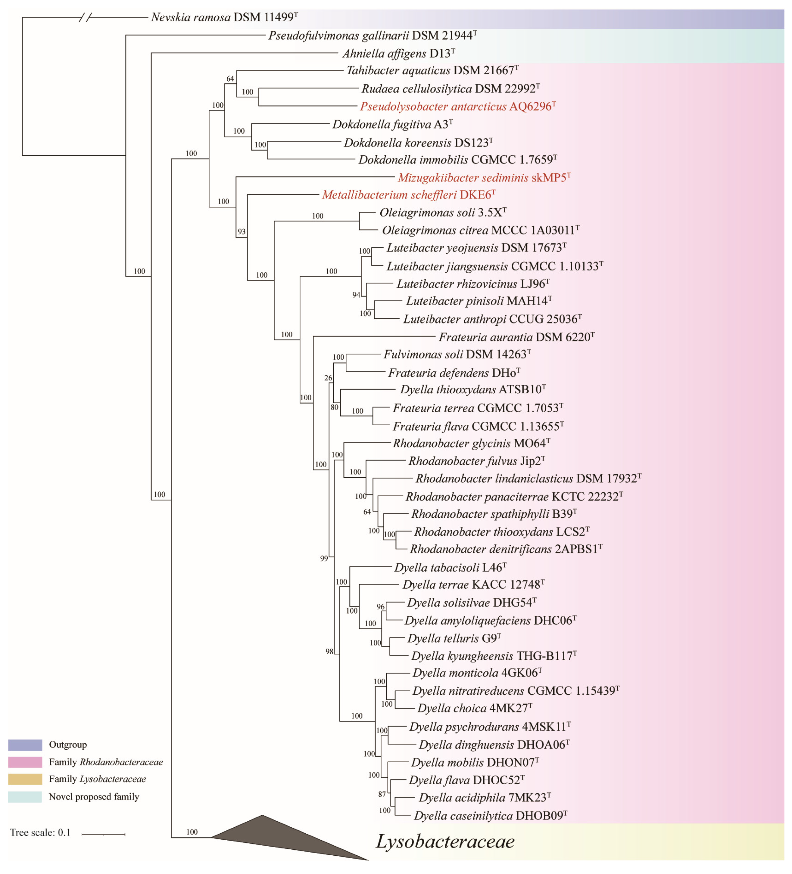
The concatenated proteins and UBCG phylogenetic trees exhibited a similar overall phylogenetic backbone, with most species in order Lysobacterales consistently clustering into similar monophyletic clades with high bootstrap support values. The phylogenetic trees based on concatenated proteins are shown in Figure 1 and Figure 2, while the full details of the concatenated proteins and UBCG phylogenetic trees were provided in Figures S1 and S2. The 16S rRNA gene phylogenetic tree is presented in Figure S3. However, it displayed low resolution, and the distribution of strains among closely related genera appeared amphibolous, consistent with previous findings [38]. Therefore, in the present study, genome-based phylogenetic trees were primarily used to infer the taxonomic relationships of species within the order Lysobacterales.
Until now, there has been no consensus on a standardized metric for family-level classification, but species within the same family are generally more similar to each other than to species from different families [39]. For example, genomic metrics such as AAI and ANI can serve as complementary tools for prokaryotic classification [40,41]. In addition to these metrics, cpAAI has recently been proposed as a reliable demarcation method among several prokaryotic families [42,43]. Therefore, in the present study, cpAAI values were also calculated as supplementary evidence for the reclassification of the order Lysobacterales.

3.1. Proposal for New Family

All phylogenetic trees (Figure 1 and Figure 2, Figures S1 and S2) consistently showed that the order Lysobacterales was divided into four monophyletic branches: two corresponding to the families Rhodanobacteraceae and Lysobacteraceae and two composed separately of A. affigens D13T and Pseudofulvimonas gallinarii DSM 21944T. A. affigens D13T and P. gallinarii DSM 21944T formed two distinct, strongly supported branched outside of the clades of Rhodanobacteraceae and Lysobacteraceae, suggesting that they represent two novel families.
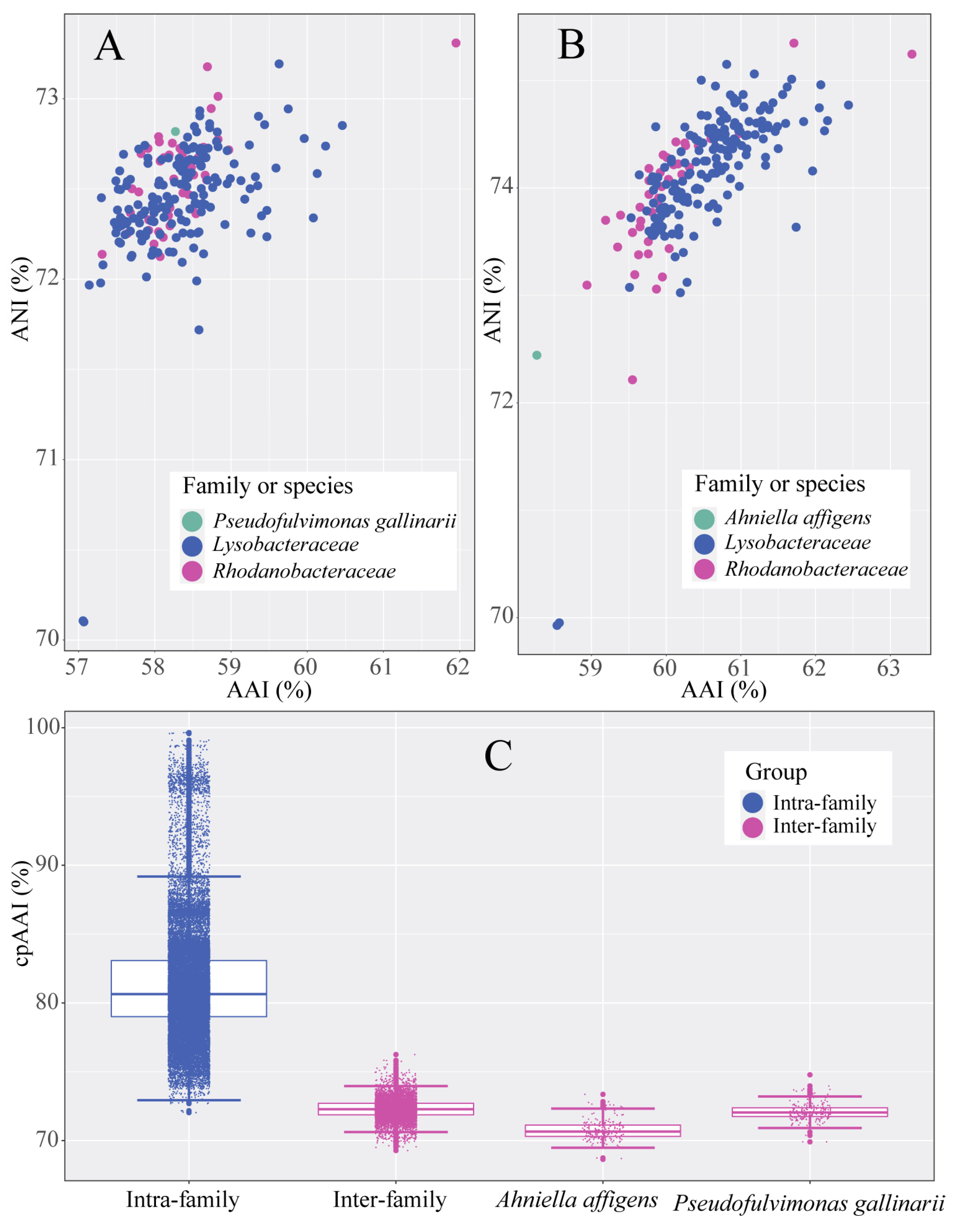
AAI and ANI analyses (Figure 3A) also revealed that P. gallinarii DSM 21944T did not cluster with any known families within Lysobacterales, supporting its placement into a novel family. The cpAAI results (Figure 3C) showed that intra-family and inter-family species could generally be clearly distinguished, although a few overlaps were observed, likely due to differences in evolutionary and ecological rates [39]. Specifically, P. gallinarii DSM 21944T exhibited lower cpAAI values (69.09–74.78%) when compared with species from known families, even lower than the inter-family cpAAI values observed within Lysobacterales (72.03–99.65%) (Figure 3C and Figure 4).
Furthermore, according to the EzBioCloud database [44], P. gallinarii DSM 21944T shared less than 93.4% 16S rRNA gene sequences similarity with all type strains in the order Lysobacterales, indicating that it cannot be assigned to any existing family within the order. According to the previous literature, the standardized 16S rRNA gene sequence threshold for defining novel families is typically 86.5% [45]. Although the similarity observed here is above this threshold, it still falls significantly below the common inter-family similarity range within Lysobacterales, suggesting substantial phylogenetic distinctiveness. Moreover, a recent study indicates that thresholds based on 16S rRNA gene sequences can overlap between taxa, and that higher-resolution genomic indices and comprehensive phylogenomic analysis are necessary for robust taxonomic delineation [46]. Therefore, based on all available data, we proposed that P. gallinarii DSM 21944T should be classified into a novel family within the order Lysobacterales.
Similarly, AAI and ANI analyses (Figure 3B) showed that A. affigens D13T did not belong to any known family within Lysobacterales, suggesting that it should also be placed into a novel family. The majority of cpAAI values between A. affigens D13T and other members of Lysobacterales were also lower than the inter-family cpAAI values observed within the order (Figure 3C and Figure 4). These results corroborate recent findings that A. affigens D13T belongs to a novel family [47].
Additionally, the polar lipids of P. gallinarii DSM 21944T were diphosphatidylglycerol (DPG) and phosphatidylmethylethanolamine (PME), and the major fatty acids of P. gallinarii DSM 21944T were iso-C15:0, iso-C17:1 ω9c and iso-C17:0. Significant differences in physicochemical properties between P. gallinarii DSM 21944T and the type genera of existing families in Lysobacterales (Table S1) further support its classification into a separate family. In summary, we propose that P. gallinarii DSM 21944T should represent a distinct novel family within the order Lysobacterales.

3.2. Proposal for the Transfer of Genera Between Families Lysobacteraceae and Rhodanobacteraceae

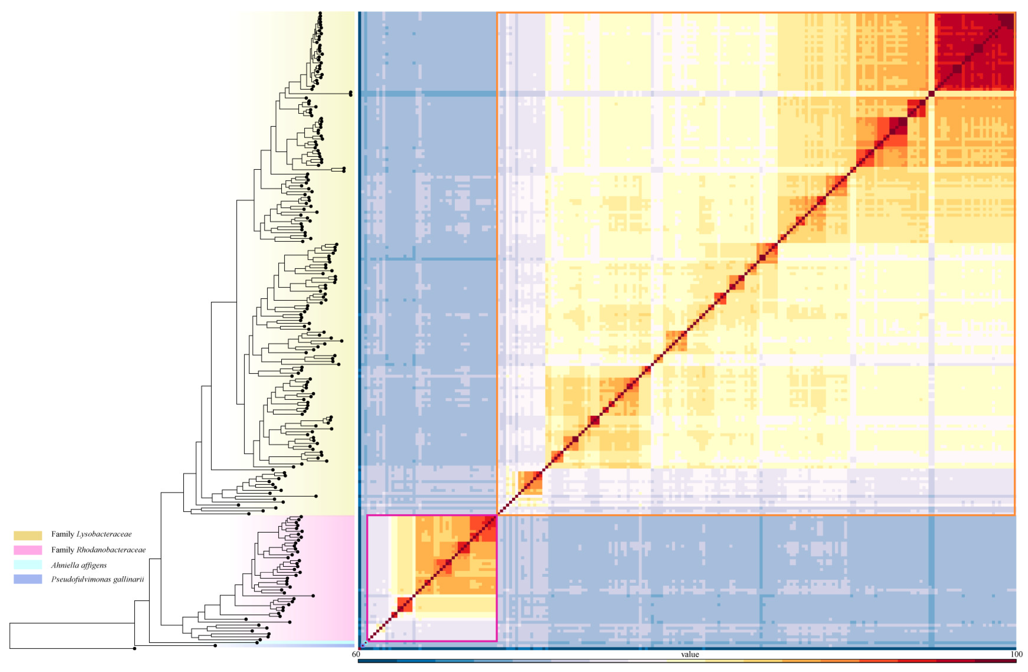
Pseudolysobacter antarcticus AQ6-296T, Mizugakiibacter sediminis skMP5T and Metallibacterium scheffleri DKE6T, currently classified under Lysobacteraceae, were consistently clustered within the family Rhodanobacteraceae across all phylogenetic trees (Figure 1, Figures S1 and S2), suggesting that they should be reclassified into Rhodanobacteraceae. Similarly, Chiayiivirga flava DSM 24163T, Aquimonas voraii DSM 16957T and Rehaibacterium terrae DSM 25897T, current placed under Rhodanobacteraceae, were clustered within Lysobacteraceae (Figure 2, Figures S1 and S2), indicating that they should be transferred to Lysobacteraceae. These reassignments are further supported by the heatmap of cpAAI values (Figure 4), which are consistent with the phylogenomic trees. Based on these findings, we propose the following reclassification that P. antarcticus AQ6-296T, M. sediminis skMP5T M. scheffleri DKE6T should be transferred to family Rhodanobacteraceae and C. flava DSM 24163T, A. voraii DSM 16957T and R. terrae DSM 25897T be transferred to family Lysobacteraceae.

3.3. Proposal for Four Novel Species

3.3.1. Genome-Based Phylogenetic and Metrics Analyses

In the concatenated protein and UBCG phylogenetic trees (Figure 2, Figures S1 and S2), strains Y-2-3-4FT, MHLX1AT, BDR2-5T and XNQY3-4T formed distinct strongly branches within the genus Luteimonas. These trees suggested that the four strains represent four novel species within Luteimonas.
Based on 16S rRNA gene sequences similarity, strains Y-2-3-4FT and MHLX1AT were most closely related to Luteimonas huabeiensis HB2T (99.0%) and Luteimonas yindakui S-1072T (98.7%), respectively, consistent with the phylogenetic analyses. Strain BDR2-5T was closely related to Luteimonas chenhongjianii 100111T (98.3%) and Luteimonas aestuarii B9T (98.2%), and strain XNQY3-4T was closely related to L. chenhongjianii 100111T (98.6%) and Luteimonas terrae HG-MD21T (98.3%). ANI and dDDH values, golden standards for species delineation [6], were calculated between the four strains and their closest reference strains, with all results falling below the recommended species boundary thresholds (ANI, 95–96%, and dDDH, 70%) (Table 1). These results confirm that the four strains represent four novel species within the genus Luteimonas.

3.3.2. Chemotaxonomic and Physiological Analysis

Although the polar lipids of the four strains shared several major components, including diphosphatidylglycerol (DPG), phosphatidyl ethanolamine (PE) and phosphatidylglycerol (PG), certain lipids (PL1, AL, APL1) were able to distinguish the strains from each other (Figure S4). Additionally, differences in the polar lipids profiles also separated these strains from related species within Luteimonas. The presence of four unidentified lipids (L) differentiated strain XNQY3-4T from its relatives. Strain MHLX1AT could be distinguished from L. yindakuii S-1072T its higher PE content and absence of unidentified aminophospholipid (APL) and unidentified glycolipid (GL) could distinguish strain MHLX1AT from L. yindakuii S-1072T. The sole respiratory quinone detected in all four strains was Q-8, which is consistent with their assignment to the genus Luteimonas.
The main fatty acids of strain Y-2-3-4FT were similar to those of L. huabeiensis HB2T, but it had significantly higher proportions of iso-C16:0 and summed features 9. Strain MHLX1AT exhibited notably greater levels of iso-C15:0 and iso-C17:0, and a lower amount of iso-C16:0 compared to its relatives. The absence of iso-C10:0 and higher levels of iso-C15:0 and iso-C17:0 distinguish strain BDR2-5T from L. chenhongjianii 100111T and L. aestuarii B9T. Likewise, strain XNQY3-4T differed from L. chenhongjianii 100111T and L. terrae THG-MD21T by its larger amounts of summed features 9 and iso-C17:0 and lower levels of iso-C10:0 (Table S2).
On TSA medium, all four strains produced yellow, viscous colonies. Despite sharing some basic features, they differed in optimal growth pH, temperature and NaCl tolerance. They were also distinguishable from each other and from related species based on substrate utilization and enzyme activity profiles. The phenotypic characteristics are summarized in Table 2 and detailed in the strain descriptions. Altogether, these chemotaxonomic and physiological features confirmed that the four strains represent four novel species within the genus Luteimonas.

4. Discussion and Conclusions

The increasing availability of genomic data have provided a more comprehensive framework for understanding the taxonomy of families within the order Lysobacterales. Although genomic metrics such as AAI, ANI and POCP values [53,54] are not absolute standards for classifications above the genus level, higher whole-genome similarities are generally observed within genera or closely related groups [39,55]. In our analysis, the AAI, ANI and POCP values between the family Marinicellaceae and members of Lysobacterales were significantly lower than those between Nevskiales and Lysobacterales, suggesting that Marinicellaceae may belong to a different, albeit closely related, order of Lysobacterales. Moreover, the clustering of Marinicellaceae outside Lysobacterales in previous studies [3] supports its placement in a distinct order.
Because single-copy core genes can vary greatly among families or orders [24,43], and to ensure phylogenetic accuracy, Marinicellaceae was excluded from this study. We focused instead on two main families within Lysobacterales: Lysobacteraceae and Rhodanobacteraceae. Based on genome-based phylogenetic and phylogenomic analyses, our results support that the families within Lysobacterales are monophyletic. We propose that the order Lysobacterales should be divided into four families, Ahniellaceae, Lysobacteraceae, Rhodanobacteraceae and Pseudofulvimonadaceae (the latter proposed in this study).
The phylogenetic trees constructed using concatenated core proteins and UBCG markers also indicate that the taxonomy of several genera within Lysobacterales requires revision. The genus Lysobacter consistently separated into several well-supported clades, suggesting the need for recognition of new genera. A recent study proposed splitting Lysobacter into multiple genera based on AAI values and phylogenomic analyses, suggesting an AAI threshold of 69.5–76.0% for genus-level delimitation within the family Lysobacteraceae. However, previous research highlighted the limitations of AAI values in genus-level classification [24,52,53,54,55]. For example, AAI values within Luteimonas range from 69.0% to 90.1%, and the newly proposed genus Cognatiluteimonas shares AAI values of 69–73.9% with Luteimonas [3]. Therefore, additional evidence will be needed before formally reclassifying Lysobacter. The genera Xanthomonas, Pseudoxanthomonas and Stenotrophomonas may also require taxonomic revisions, and a recent study also confirmed that these three genera should merged into genus Xanthomonas [56]. Nevertheless, such comprehensive taxonomic revisions are beyond the scope of the current study.
A total of 218 genome sequences (217 Lysobacterales and 1 Nevskiales) were analyzed. Genome-based phylogenetic and phylogenomic metrics revealed the need to revise the family-level taxonomy within Lysobacterales. Phylogenetic analyses using UBCG and concatenated proteins, along with ANI, AAI and cpAAI values, demonstrated that Pseudofulvimonas gallinarii DSM 21944T should be assigned to a distinct novel family, and that six genera should be reclassified across existing families. Furthermore, four novel species within the family Lysobacteraceae were identified based on phylogenetic analysis, physiological and biochemical characteristics and genomic data and were described in this study.

4.1. Taxonomic Levels: New Family

Description of Pseudofulvimonadaceae fam. nov.

Pseu.do.ful.vi.mo.na.da’ce.ae (N.L. fem. n. Pseudofulvimonas, type genus of the family; -aceae, ending to denote a family; N.L. fem. pl. n. Pseudofulvimonadaceae, the Pseudofulvimonas family).
Cells are Gram-strain-negative, aerobic, non-motile, rod-shaped. The predominant respiratory quinone is Q-8. The major cellular fatty acids are usually iso-C15:0, iso-C17:1 ω9c and iso-C17:0. The DNA G+C content is 67.6 mol %. The family is defined based on phylogenetic analyses of UBCG and concatenated protein phylogenetic trees, phylogenomic metric analyses of AAI, ANI and cpAAI. The description of the family is as given for Pseudofulvimonas [57], which is the type and currently the only genus of the family.

4.2. The Transfer for the Member of Family Rhodanobacteraceae and Lysobacteraceae

Pseudolysobacter antarcticus, Mizugakiibacter sediminis and Metallibacterium scheffleri were transferred to family Rhodanobacteraceae, and Chiayiivirga flava, Aquimonas voraii, Rehaibacterium terrae were transferred to family Lysobacteraceae.

4.3. Taxonomic Levels: New Species

4.3.1. Description of Alterluteimonas quercicellularis sp. nov.

Alterluteimonas quercicellularis (quer.ci.cel.lu.la’ris. N.L. gen. fem. n. quercus, of quercus tree; L. fem. n. cellula, cell; N.L. fem. n. quercicellularis, a bacterium isolated from quercus tree).
Cells are Gram-stain-negative, aerobic, motile with a single polar flagellum, catalase- and oxidase-positive, rod-shaped, 1.2–1.8 mm in length and 0.6–0.8 mm in width. Colonies are yellow, circular, viscous, smooth, with entire margins after incubation for 2 days at 28 °C on TSA. The strain can grow at 10–41 °C (optimum, 28–30 °C) at pH 6–9 (optimum, pH 7–8). Growth occurs at a concentration of 0–5% (w/v) NaCl. Positive for alkaline phosphatase, leucine arylamidase, trypsin, α-chymotrypsin, acid phosphatase, naphthol-AS-BI-phosphohydrolase, α-glucosidase; weakly positive for esterase lipase (C8), esterase (C4), lipase (C14), valine arylamidase, β-galactosidase, β-glucosidase; negative for N-acetyl-β-glucosaminidase, cystine arylamidase, α-galactosidase, β-glucuronidase, α-mannosidase, α-fucosidase (API ZYM). Positive for reduction of nitrates to nitrites, esculin, gelatin hydrolysis, N-acetyl-glucosamine, D-maltose, malic acid; weakly positive for D-glucose, D-mannose; and negative for the rest tests in API 20NE.
Positive results in tests of using the following carbon sources: D-glucose, amygdalin, esculin ferric citrate, D-cellobiose, D-maltose, D-trehalose, D-melezitose, glycogen; weakly positive for D-xylose, D-lactose, starch, gentiobiose and D-lyxose, and negative for the rest tests in API 50 CH. The polar lipids were DPG, PE, PG and four unidentified lipids (L). The respiratory quinones were Q-8. The predominant fatty acids were iso-C15:0, iso-C16:0 and summed features 3 (C16:1 ω7c and/or C16:1 ω6c). The type strain is Y-2-3-4FT (= CFCC 15605T = LMG 32557T), isolated from Ningxia, China. The strain Y-2-3-4FT was predicted to have 3911 coding genes, 52 tRNA genes, 3 rRNA genes and 7 other RNA genes, and the DNA G+C content was 73.0 mol %.

4.3.2. Description of Alterluteimonas muca sp. nov.

Alterluteimonas muca (mu’ca. L. fem. adj. muca, mucus, referring to the characteristics of the colonies).
Cells are Gram-stain-negative, aerobic, motile with a single polar flagellum, catalase- and oxidase-positive, rod-shaped, 1.5–2.0 mm in length and 0.5–0.8 mm in width. Colonies are pale yellow, circular, viscous, smooth, with entire margins after incubation for 2 days at 28 °C on TSA. The strain can grow at 10–37 °C (optimum, 28 °C) at pH 6–8.5 (optimum, pH 7–7.5). Growth occurs at a concentration of 0–4% (w/v) NaCl. Positive for alkaline phosphatase, esterase lipase (C8), esterase (C4), leucine arylamidase, valine arylamidase, trypsin, α-chymotrypsin, acid phosphatase, naphthol-AS-BI-phosphohydrolase; weakly positive for lipase (C14) and cystine arylamidase; negative for the rest tests in API ZYM. Positive for esculin, gelatin hydrolysis, and negative for the rest tests in API 20NE. Positive results in tests of using the following carbon sources: D-glucose, D-fructose, D-mannose, salicin, D-lactose, D-melibiose and D-trehalose, and negative for the rest tests in API 50 CH. The polar lipids were DPG, PE, PG, PL1, AL and four unidentified lipids (L). The respiratory quinones were Q-8. The predominant fatty acids were iso-C15:0, iso-C17:0 and summed features 3 (C16:1 ω7c and/or C16:1 ω6c). The type strain is MHLX1AT (= CFCC 16400T = LMG 32554T), isolated from Beijing, China. The strain MHLX1AT was predicted to have 3009 coding genes, 45 tRNA genes, 3 rRNA genes and 4 other RNA genes, and the DNA G+C content was 69.1 mol %.

4.3.3. Description of Proluteimonas luteida sp. nov.

Proluteimonas luteida (Lu’tei.da. L. fem. adj. luteida, yellow, referring to the color of the colonies).
Cells are Gram-stain-negative, aerobic, motile with a single polar flagellum, catalase- and oxidase-positive, rod-shaped, 1.5–1.8 mm in length and 0.5–0.7 mm in width. Colonies are yellow, moist, viscid with irregular margins after incubation for 2 days at 28 °C on TSA. The strain can grow at 10–37 °C (optimum, 28 °C) at pH 6–8.5 (optimum, pH 7–7.5). Growth occurs at a concentration of 0–4% (w/v) NaCl. Positive for alkaline phosphatase, esterase lipase (C8), esterase (C4), leucine arylamidase, trypsin, α-chymotrypsin, acid phosphatase and naphthol-AS-BI-phosphohydrolase; weakly positive for valine arylamidase; and negative for the rest tests in API ZYM. Positive for esculin, gelatin hydrolysis and malic acid; weakly positive for D-glucose assimilation and D-mannose; and negative for the rest tests in API 20NE. Positive results in tests of using the following carbon sources: salicin and negative for the rest tests in API 50 CH. The polar lipids were DPG, PE, PG, PL1, APL1 and two unidentified lipids (L). The respiratory quinones were Q-8. The predominant fatty acids were iso-C15:0, iso-C17:0 and summed features 3 (C16:1 ω7c and/or C16:1 ω6c). The type strain is BDR2-5T (= CFCC 16401T = LMG 32545T), isolated from Anhui, China. The strain BDR2-5T was predicted to have 3520 coding genes, 20 tRNA genes, 7 rRNA genes and 5 other RNA genes, and the DNA G+C content was 69.7 mol %.

4.3.4. Description of Proluteimonas flavola sp. nov.

Proluteimonas flavola (fla.vo.la. L. masc. adj. flavus, yellow; L. fem. adj. flavola, referring to the color of the colonies).
Cells are Gram-stain-negative, aerobic, motile with a single polar flagellum, catalase- and oxidase-positive, rod-shaped, 1.6–2.0 mm in length and 0.6–0.8 mm in width. Colonies are yellow, circular, viscous, smooth, with entire margins after incubation for 2 days at 28 °C on TSA. The strain can grow at 10–37 °C (optimum, 25–28 °C) at pH 6–9 (optimum, pH 7–8). Growth occurs at a concentration of 0–3% (w/v) NaCl. Positive for alkaline phosphatase, esterase lipase (C8), esterase (C4), leucine arylamidase, valine arylamidase, trypsin, α-chymotrypsin, acid phosphatase and naphthol-AS-BI-phosphohydrolase; negative for the rest tests in API ZYM. Positive for esculin, gelatin hydrolysis, D-maltose and malic acid; weakly positive for D-glucose assimilation; and negative for the rest tests in API 20NE. Positive results in tests of using the following carbon sources: D-fructose, salicin, D-maltose, D-lactose, D-melibiose and D-turanose, and negative for the rest tests in API 50 CH. The polar lipids were DPG, PE, PG, PL and four unidentified lipids (L). The respiratory quinones were Q-8. The predominant fatty acids were iso-C15:0, iso-C16:0, iso-C17:0 and summed features 3 (C16:1 ω7c and/or C16:1 ω6c). The type strain is XNQY3-4T (= CFCC 16543T = LMG 32556T), isolated from Qinghai, China. The strain XNQY3-4T was predicted to have 3648 coding genes, 48 tRNA genes, 5 rRNA genes and 4 other RNA genes, and the DNA G+C content was 68.3 mol %.

Supplementary Materials

The following supporting information can be downloaded at: https://www.mdpi.com/article/10.3390/microorganisms13061212/s1. Table S1: the pairwise values of AAI, ANI and cpAAI and phenotypic comparisons within the species. Supplementary File S1: Supplementary Figures S1–S5 and Table S2.

Author Contributions

Conceptualization, T.M. and H.L.; methodology, T.M.; software, Y.C. and H.X.; validation, J.L.; formal analysis, C.P.; resources, T.M.; writing—original draft preparation, T.M.; writing—review and editing H.L., Y.L. and R.X.; visualization, T.M. and R.X.; project administration, T.M. All authors have read and agreed to the published version of the manuscript.

Funding

This research was funded by the National Modern Agricultural Industry Technology System Project of the Ministry of Finance and the Ministry of Agriculture and Rural Affairs, grant number CARS-13; the Major Emergency Response Project for Agricultural Production in Henan Province, grant number 2024ZDYJ001; the Key R&D Special Project of Henan Province, grant number 241111112700; and the Henan Province Science and Technology Research Project 252102111078, 242102111072.

Institutional Review Board Statement

Not applicable.

Informed Consent Statement

Not applicable.

Data Availability Statement

The GenBank/EMBL/DDBJ accession numbers for the 16S rRNA gene are OK287346–OK287349, and for the genome sequences of strains BDR2-5T, XNQY3-4T, Y-2-3-4FT and MHLX1AT are JAIWPT000000000, JAIWPU000000000, JAIWPR000000000 and JAIWPS000000000, respectively.

Acknowledgments

The authors would particularly like to thank Ning Jiang, China Forestry Culture Collection Center, China, for verifying of the proposed Latin name for the novel species.

Conflicts of Interest

The authors declare no conflicts of interest.

Abbreviations

The following abbreviations are used in this manuscript:
AAIaverage amino acid identity
ANIaverage nucleotide identity
cpAAIcore-proteome average amino acid identity
dDDHdigital DNA–DNA hybridization

References

  1. Kumar, S.; Bansal, K.; Patil, P.P.; Patil, P.B. Phylogenomics insights into order and families of Lysobacterales. Access Microbiol. 2019, 1, e000015. [Google Scholar] [CrossRef] [PubMed]
  2. Naushad, S.; Adeolu, M.; Wong, S.; Sohail, M.; Schellhorn, H.E.; Gupta, R.S. A phylogenomic and molecular marker based taxonomic framework for the order Xanthomonadales: Proposal to transfer the families Algiphilaceae and Solimonadaceae to the order Nevskiales ord. nov. and to create a new family within the order Xanthomonadales, the family Rhodanobacteraceae fam. nov., containing the genus Rhodanobacter and its closest relatives. Antonie Van Leeuwenhoek 2015, 107, 467–485. [Google Scholar] [CrossRef] [PubMed]
  3. Lu, H.; Chen, L.; Wang, Y.; Xing, P.; Wu, Q. Luteimonas flava sp. nov. and Aquilutibacter rugosus gen. nov., sp. nov., isolated from freshwater environments in China and re-examining the taxonomic status of genera Luteimonas and Lysobacter. Int. J. Syst. Evol. Microbiol. 2024, 74, 006585. [Google Scholar] [CrossRef] [PubMed]
  4. Romanenko, L.; Otstavnykh, N.; Tanaka, N.; Kurilenko, V.; Svetashev, V.; Tekutyeva, L.; Mikhailov, V.; Isaeva, M. Characterization and genomic analysis of Fererhizobium litorale gen. nov., sp. nov., isolated from the sandy sediments of the sea of Japan seashore. Microorganisms 2023, 11, 2385. [Google Scholar] [CrossRef]
  5. Yin, J.; He, M.; Liu, X.X.; Ren, C.B.; Liu, H.H.; Luo, H.; Chen, G.; Wang, Z.F.; Debnath, S.C.; Wang, P.M.; et al. Peteryoungia algae sp. nov. isolated from seaweeds of Gouqi Island, China, and its unique genetic features among Peteryoungia strains. Antonie Van Leeuwenhoek 2024, 117, 112. [Google Scholar] [CrossRef]
  6. De Lajudie, P.M.; Andrews, M.; Ardley, J.; Eardly, B.; Jumas-Bilak, E.; Kuzmanović, N.; Lassalle, F.; Lindström, K.; Mhamdi, R.; Martínez-Romero, E. Minimal standards for the description of new genera and species of rhizobia and agrobacteria. Int. J. Syst. Evol. Microbiol. 2019, 69, 1852–1863. [Google Scholar] [CrossRef]
  7. Parks, D.H.; Chuvochina, M.; Waite, D.W.; Rinke, C.; Skarshewski, A.; Chaumeil, P.-A.; Hugenholtz, P. A standardized bacterial taxonomy based on genome phylogeny substantially revises the tree of life. Nat. Biotechnol. 2018, 36, 996–1004. [Google Scholar] [CrossRef]
  8. Ma, T.; Xue, H.; Piao, C.; Liu, C.; Yang, M.; Bian, D.; Li, Y. Reclassification of 11 members of the family Rhodobacteraceae at genus and species levels and proposal of Pseudogemmobacter hezensis sp. nov. Front. Microbiol. 2022, 13, 849695. [Google Scholar] [CrossRef]
  9. Li, R.; Li, Y.; Kristiansen, K.; Wang, J. SOAP: Short oligonucleotide alignment program. Bioinformatics 2008, 24, 713–714. [Google Scholar] [CrossRef]
  10. Li, R.; Zhu, H.; Ruan, J.; Qian, W.; Fang, X.; Shi, Z.; Li, Y.; Li, S.; Shan, G.; Kristiansen, K. De novo assembly of human genomes with massively parallel short read sequencing. Genome Res. 2010, 20, 265–272. [Google Scholar] [CrossRef]
  11. Bankevich, A.; Nurk, S.; Antipov, D.; Gurevich, A.A.; Dvorkin, M.; Kulikov, A.S.; Lesin, V.M.; Nikolenko, S.I.; Pham, S.; Prjibelski, A.D. SPAdes: A new genome assembly algorithm and its applications to single-cell sequencing. J. Comput. Biol. 2012, 19, 455–477. [Google Scholar] [CrossRef]
  12. Simpson, J.T.; Wong, K.; Jackman, S.D.; Schein, J.E.; Jones, S.J.M.; Birol, I. ABySS: A parallel assembler for short read sequence data. Genome Res. 2009, 19, 1117–1123. [Google Scholar] [CrossRef] [PubMed]
  13. Lin, S.H.; Liao, Y.C. CISA: Contig integrator for sequence assembly of bacterial genomes. PLoS ONE 2013, 8, e60843. [Google Scholar] [CrossRef] [PubMed]
  14. Parks, D.H.; Imelfort, M.; Skennerton, C.T.; Hugenholtz, P.; Tyson, G.W. CheckM: Assessing the quality of microbial genomes recovered from isolates, single cells, and metagenomes. Genome Res. 2015, 25, 1043–1055. [Google Scholar] [CrossRef]
  15. Lagesen, K.; Hallin, P.; Rødland, E.A.; Stærfeldt, H.H.; Rognes, T.; Ussery, D.W. RNAmmer: Consistent and rapid annotation of ribosomal RNA genes. Nucleic Acids Res. 2007, 35, 3100–3108. [Google Scholar] [CrossRef]
  16. Kumar, S.; Stecher, G.; Li, M.; Knyaz, C.; Tamura, K. MEGA X: Molecular evolutionary genetics analysis across computing platforms. Mol. Biol. Evol. 2018, 35, 1547. [Google Scholar] [CrossRef]
  17. Kim, J.; Na, S.I.; Kim, D.; Chun, J. UBCG2: Up-to-date bacterial core genes and pipeline for phylogenomic analysis. J. Microbiol. 2021, 59, 609–615. [Google Scholar] [CrossRef] [PubMed]
  18. Na, S.I.; Kim, Y.O.; Yoon, S.H.; Ha, S.M.; Baek, I.; Chun, J. UBCG: Up-to-date bacterial core gene set and pipeline for phylogenomic tree reconstruction. J. Microbiol. 2018, 56, 280–285. [Google Scholar] [CrossRef]
  19. Emms, D.M.; Kelly, S. OrthoFinder: Phylogenetic orthology inference for comparative genomics. Genome Biol. 2019, 20, 238. [Google Scholar] [CrossRef]
  20. Price, M.N.; Dehal, P.S.; Arkin, A.P. FastTree: Computing large minimum evolution trees with profiles instead of a distance matrix. Mol. Biol. Evol. 2009, 26, 1641–1650. [Google Scholar] [CrossRef]
  21. Hyatt, D.; Chen, G.L.; LoCascio, P.F.; Land, M.L.; Larimer, F.W.; Hauser, L.J. Prodigal: Prokaryotic gene recognition and translation initiation site identification. BMC Bioinform. 2010, 11, 119. [Google Scholar] [CrossRef]
  22. Letunic, I.; Bork, P. Interactive Tree of Life (iTOL) v5: An online tool for phylogenetic tree display and annotation. Nucleic Acids Res. 2021, 49, W293–W296. [Google Scholar] [CrossRef]
  23. Pritchard, L.; Cock, P.; Esen, Ö. Pyani, version 0.2.8. Average Nucleotide Identity (ANI) and Related Measures for Whole Genome Comparisons. Zenodo: San Francisco, CA, USA, 2019. [CrossRef]
  24. Ma, T.; Xue, H.; Piao, C.; Jiang, N.; Li, Y. Genome-based analyses of family Oxalobacteraceae reveal the taxonomic classification. Res. Microbiol. 2023, 7, 104076. [Google Scholar] [CrossRef] [PubMed]
  25. Katoh, K.; Standley, D.M. MAFFT multiple sequence alignment software version 7: Improvements in performance and usability. Mol. Biol. Evol. 2013, 30, 772–780. [Google Scholar] [CrossRef] [PubMed]
  26. Paradis, E.; Schliep, K. ape 5.0: An environment for modern phylogenetics and evolutionary analyses in R. Bioinformatics 2019, 35, 526–528. [Google Scholar] [CrossRef] [PubMed]
  27. Minnikin, D.E.; O’Donnell, A.G.; Goodfellow, M.; Alderson, G.; Athalye, M.; Schaal, A.; Parlett, J.H. An integrated procedure for the extraction of bacterial isoprenoid quinones and polar lipids. J. Microbiol. Meth. 1984, 2, 233–241. [Google Scholar] [CrossRef]
  28. Collins, M.D.; Pirouz, T.; Goodfellow, M.; Minnikin, D.E. Distribution of menaquinones in actinomycetes and corynebacteria. Microbiology 1977, 100, 221–230. [Google Scholar] [CrossRef]
  29. Groth, I.; Schumann, P.; Rainey, F.A.; Martin, K.; Schuetze, B.; Augsten, K. Demetria terragena gen. nov., sp. nov., a new genus of Actinomycetes isolated from compost soil. Int. J. Syst. Evol. Microbiol. 1997, 47, 1129–1133. [Google Scholar] [CrossRef] [PubMed]
  30. Du, H.J.; Zhang, Y.Q.; Liu, H.Y.; Su, J.; Wei, Y.Z.; Ma, B.P.; Guo, B.L.; Yu, L.Y. Allonocardiopsis opalescens gen. nov., sp. nov., a new member of the suborder Streptosporangineae, from the surface-sterilized fruit of a medicinal plant. Int. J. Syst. Evol. Microbiol. 2013, 63, 900–904. [Google Scholar] [CrossRef]
  31. Kuykendall, L.D.; Roy, M.A.; O’Neill, J.J.; Devine, T.E. Fatty acids, antibiotic resistance, and deoxyribonucleic acid homology groups of Bradyrhizobium japonicum. Int. J. Syst. Evol. Microbiol. 1988, 38, 358–361. [Google Scholar] [CrossRef]
  32. Sasser, M. Identification of Bacteria by Gas Chromatography of Cellular Fatty Acids; MIDI Inc.: Newark, NJ, USA, 1990; pp. 1–6. [Google Scholar]
  33. Li, Y.; Song, L.M.; Guo, M.W.; Wang, L.F.; Liang, W.X. Sphingobacterium populi sp. nov., isolated from bark of Populus × euramericana. Int. J. Syst. Evol. Microbiol. 2016, 66, 3456–3462. [Google Scholar] [CrossRef] [PubMed]
  34. Delory, G.E.; King, E.J. A sodium carbonate-bicarbonate buffer for alkaline phosphatases. Biochem. J. 1945, 39, 245. [Google Scholar] [CrossRef]
  35. Gomori, G. Preparation of buffers for use in enzyme studies. In Methods in Enzymology; Academic Press: New York, NY, USA, 1955; pp. 138–146. [Google Scholar] [CrossRef]
  36. Jenkins, D.; Richard, M.G.; Daigger, G.T. Manual on the Causes and Control of Activated Sludge Bulking, Foaming, and Other Solids Separation Problems; CRC Press: Boca Raton, FL, USA, 2003; pp. 9–56. [Google Scholar] [CrossRef]
  37. Smibert, R.M. Phenotypic characterization. In Methods for General and Molecular Bacteriology, 1st ed.; Gerhardt, P., Murray, R.G.E., Eds.; American Society for Microbiology: Washington, DC, USA, 1993; pp. 607–654. [Google Scholar]
  38. Zayulina, K.S.; Prokofeva, M.I.; Elcheninov, A.G.; Voytova, M.P.; Novikov, A.A.; Kochetkova, T.V.; Kublanov, I.V. Arenimonas fontis sp. nov., a bacterium isolated from Chukotka hot spring, Arctic region, Russia. Int. J. Syst. Evol. Microbiol. 2020, 70, 2726–2731. [Google Scholar] [CrossRef]
  39. Liang, K.Y.H.; Orata, F.D.; Boucher, Y.F.; Case, R.J. Roseobacters in a sea of poly-and paraphyly: Whole genome-based taxonomy of the family Rhodobacteraceae and the proposal for the split of the “Roseobacter clade” into a novel family, Roseobacteraceae fam. nov. Front. Microbiol. 2021, 12, 1635. [Google Scholar] [CrossRef]
  40. Xu, Z.; Masuda, Y.; Wang, X.; Ushijima, N.; Shiratori, Y.; Senoo, K.; Itoh, H. Genome-Based taxonomic rearrangement of the order Geobacterales including the description of Geomonas azotofigens sp. nov. and Geomonas diazotrophica sp. nov. Front. Microbiol. 2021, 12, 737531. [Google Scholar] [CrossRef] [PubMed]
  41. Gupta, R.S.; Lo, B.; Son, J. Phylogenomics and comparative genomic studies robustly support division of the genus Mycobacterium into an emended genus Mycobacterium and four novel genera. Front. Microbiol. 2018, 9, 67. [Google Scholar] [CrossRef] [PubMed]
  42. Wirth, J.S.; Whitman, W.B. Phylogenomic analyses of a clade within the roseobacter group suggest taxonomic reassignments of species of the genera Aestuariivita, Citreicella, Loktanella, Nautella, Pelagibaca, Ruegeria, Thalassobius, Thiobacimonas and Tropicibacter, and the proposal of six novel genera. Int. J. Syst. Evol. Microbiol. 2018, 68, 2393–2411. [Google Scholar] [CrossRef]
  43. Kuzmanović, N.; Fagorzi, C.; Mengoni, A.; Lassalle, F. Taxonomy of Rhizobiaceae revisited: Proposal of a new framework for genus delimitation. Int. J. Syst. Evol. Microbiol. 2022, 72, 005243. [Google Scholar] [CrossRef]
  44. Yoon, S.H.; Ha, S.M.; Kwon, S.; Lim, J.; Kim, Y.; Seo, H.; Chun, J. Introducing EzBioCloud: A taxonomically united database of 16S rRNA gene sequences and whole-genome assemblies. Int. J. Syst. Evol. Microbiol. 2017, 67, 1613. [Google Scholar] [CrossRef]
  45. Yarza, P.; Yilmaz, P.; Pruesse, E.; Glöckner, F.O.; Ludwig, W.; Schleifer, K.H.; Whitman, W.B.; Euzéby, J.; Amann, R.; Rosselló-Móra, R. Uniting the classification of cultured and uncultured bacteria and archaea using 16S rRNA gene sequences. Nat. Rev. Microbiol. 2014, 12, 635–645. [Google Scholar] [CrossRef]
  46. Hackmann, T.J. Setting new boundaries of 16S rRNA gene identity for prokaryotic taxonomy. Int. J. Syst. Evol. Microbiol. 2025, 75, 006747. [Google Scholar] [CrossRef]
  47. Chuvochina, M.; Mussig, A.J.; Chaumeil, P.; Skarshewski, A.; Rinke, C.; Parks, D.H.; Hugenholtz, P. Proposal of names for 329 higher rank taxa defined in the Genome Taxonomy Database under two prokaryotic codes. FEMS Microbiol. Lett. 2023, 370, fnad071. [Google Scholar] [CrossRef] [PubMed]
  48. Wu, G.; Liu, Y.; Li, Q.; Du, H.; You, J.; Li, H.; Ke, C.; Zhang, X.; Yu, J.; Zhao, T. Luteimonas huabeiensis sp. nov., isolated from stratum water. Int. J. Syst. Evol. Microbiol. 2013, 63, 3352–3357. [Google Scholar] [CrossRef] [PubMed]
  49. Zhang, S.; Wang, X.; Yang, J.; Lu, S.; Lai, X.H.; Jin, D.; Pu, J.; Li, J.; Huang, Y.; Zhang, G. Luteimonas yindakuii sp. nov. isolated from the leaves of Dandelion (Taraxacum officinale) on the Qinghai-Tibetan Plateau. Int. J. Syst. Evol. Microbiol. 2020, 70, 1007–1014. [Google Scholar] [CrossRef]
  50. Zhang, G.; Lai, X.H.; Yang, J.; Jin, D.; Pu, J.; Xiong, Y.; Yang, C.; Dong, K.; Huang, Y.; Luo, X. Luteimonas chenhongjianii, a novel species isolated from rectal contents of Tibetan Plateau pika (Ochotona curzoniae). Int. J. Syst. Evol. Microbiol. 2020, 70, 3186–3193. [Google Scholar] [CrossRef]
  51. Ngo, H.T.T.; Yin, C.S. Luteimonas terrae sp. nov., isolated from rhizosphere soil of Radix ophiopogonis. Int. J. Syst. Evol. Microbiol. 2016, 66, 1920–1925. [Google Scholar] [CrossRef]
  52. Roh, S.W.; Kim, K.H.; Nam, Y.D.; Chang, H.W.; Kim, M.S.; Yoon, J.H.; Oh, H.M.; Bae, J.W. Luteimonas aestuarii sp. nov., isolated from tidal flat sediment. J. Microbiol. 2008, 46, 525–529. [Google Scholar] [CrossRef] [PubMed]
  53. Riesco, R.; Trujillo, M.E. Update on the proposed minimal standards for the use of genome data for the taxonomy of prokaryotes. Int. J. Syst. Evol. Microbiol. 2024, 74, 006300. [Google Scholar] [CrossRef] [PubMed]
  54. Qin, Q.L.; Xie, B.B.; Zhang, X.Y.; Chen, X.L.; Zhou, B.C.; Zhou, J.; Oren, A.; Zhang, Y.Z. A proposed genus boundary for the prokaryotes based on genomic insights. J. Bacteriol. 2014, 12, 2210–2215. [Google Scholar] [CrossRef]
  55. Ma, T.; Xue, H.; Piao, C.; Jiang, N.; Li, Y. Phylogenomic reappraisal of the family Rhizobiaceae at the genus and species levels, including the description of Ectorhizobium quercum gen. nov., sp. nov. Front. Microbiol. 2023, 14, 1207256. [Google Scholar] [CrossRef]
  56. Bansal, K.; Kumar, S.; Singh, A.; Chaudhary, A.; Patil, P.B. Redefining the taxonomic boundaries of genus Xanthomonas. Taxonomy 2023, 3, 452–465. [Google Scholar] [CrossRef]
  57. Kämpfer, P.; Martin, E.; Lodders, N.; Langer, S.; Schumann, P.; Jäckel, U.; Busse, H.J. Pseudofulvimonas gallinarii gen. nov., sp. nov., a new member of the family Xanthomonadaceae. Int. J. Syst. Evol. Microbiol. 2010, 60, 1427–1431. [Google Scholar] [CrossRef] [PubMed]
Figure 1. The first part of the concatenated protein phylogenetic tree among strains in order Lysobacterales based on a concatenated alignment of 227 ubiquitous single-copy proteins, focusing on family Rhodanobacteraceae. For details and abbreviations see Figure S1. The scale bar corresponds to 0.1 substitutions per amino acid position.
Microorganisms 13 01212 g001
Figure 2. The second part of the concatenated protein phylogenetic tree among strains in order Lysobacterales based on a concatenated alignment of 227 ubiquitous single-copy proteins, focusing on part of family Lysobacteraceae. For details and abbreviations see Figure S1. The scale bar corresponds to 0.1 substitutions per amino acid position.
Microorganisms 13 01212 g002
Figure 3. AAI and ANI distribution between Ahniella affigens D13T and other members within order Lysobacterales are shown in plot (A); AAI and ANI distribution between Pseudofulvimonas gallinarii DSM 21944T and other members within order Lysobacterales are shown in plot (B). Box plot indicates pairwise cpAAI values within and between families of order Lysobacterales; pairwise cpAAI values between Ahniella affigens D13T and other members within order Lysobacterales; and pairwise cpAAI values between Pseudofulvimonas gallinarii DSM 21944T and other members within order Lysobacterales (C).
Microorganisms 13 01212 g003
Figure 4. Heatmap of pairwise cpAAI within order Lysobacterales; the proposed genera are marked with quadrangles and the pairwise cpAAI values are provided in Table S1. The phylogenetic tree on the left of the plot is the concatenated protein tree, and different proposed families are shown in different colors.
Microorganisms 13 01212 g004
Table 1. Digital DNA–DNA hybridization (dDDH) values, average nucleotide identity (ANI) among the four novel strains and closely related type strains.
StrainY-2-3-4FTMHLX1ATBDR2-5TXNQY3-4T
ANIdDDHANIdDDHANIdDDHANIdDDH
MHLX1AT81.5224.5
BDR2-5T79.4322.977.8421.6
XNQY3-4T78.0721.677.9221.382.2025.7
Luteimonas huabeiensis HB2T90.7841.987.2724.279.5022.777.8521.6
Luteimonas yindakuii S-1072T81.7424.587.7334.177.9421.877.2421.2
Luteimonas chenhongjianii 100111T77.4221.276.9720.881.1724.284.2528.0
Luteimonas terrae THG-MD21T77.8021.576.6120.881.5524.688.5330.0
Luteimonas aestuarii B9T77.9221.877.3221.478.2022.077.3021.2
Table 2. Differential characteristics of the four novel strains and closely related type strains. Strains: 1, Y-2-3-4FT; 2, MHLX1AT; 3, BDR2-5T; 4, XNQY3-4T; 5, Luteimonas huabeiensis HB2T; 6, Luteimonas yindakuii S-1072T; 7, Luteimonas chenhongjianii 100111T; 8, Luteimonas terrae THG-MD21T; 9, Luteimonas aestuarii B9T. Data in column 5 are from [48], data in column 6 are from [49], data in column 7 are from [50], data in column 8 are from [51] and data in column 9 are from [52]. +, positive; −, negative; W, weakly positive.
Characteristic123456789
Cell size (µm)0.6–0.8 × 1.2–1.8 0.5–0.8 × 1.5–20.5–0.7 × 1.5–1.80.6–0.8 × 1.6–2.00.4–0.5 × 0.9–1.60.7–1 × 1.7–2.81.1–1.4 × 1.5–1.80.4–0.5 × 1.1–1.70.5 × 1.5–2.0
pH (optimum pH)6–9
(7–8)
6–8.5
(7–7.5)
6–8.5
(7–7.5)
6–9
(7–8)
6–11
(7)
6.5–9.5
(7)
6–10
(7–8)
6.5–8
(7–7.5)
6.5–11
(8)
Temperature
(Optimum temperature °C)
10–41
(28–30)
10–37
(28)
10–37
(28)
10–37
(25–28)
20–45
(30)
4–40
(28)
22–40
(35–37)
4–45
(25–30)
15–40
(34–37)
NaCl range (%, w/v)0–50–40–40–30–50–2.50–3.50–5.50–4
Reduction of nitrate++
Utilization of:
 D-mannose+++
 D-mannitol++
Methyl α-D-mannopyranoside+
 D-maltose++++++
 Glycogen++W
 Xylitol+
Enzyme activities:
 Esterase (C4)W++++++W
 Lipase (C14)WW+
 Valine arylamidaseW+W++++
 Cystine arylamidaseW+WW
 Trypsin+++++++
α-chymotrypsin++++++++
α-glucosidase++W
 N-acetyl-β-glucosaminidase+++
Hydrolysis from:
 Urease+++
 Aesculin++++++++
 Gelatin+++++++
G+C content (mol %)73.069.169.768.367.069.268.364.464.7
Disclaimer/Publisher’s Note: The statements, opinions and data contained in all publications are solely those of the individual author(s) and contributor(s) and not of MDPI and/or the editor(s). MDPI and/or the editor(s) disclaim responsibility for any injury to people or property resulting from any ideas, methods, instructions or products referred to in the content.