Article

In-Feed vs. In-Water Chlortetracycline Administration on the Fecal Prevalence of Virulence Genes and Pathotypes of Escherichia coli Involved in Enteric Colibacillosis in Piglets

by
Ramya Kalam
1,
Raghavendra G. Amachawadi
1,*,
Xiaorong Shi
2,
Jianfa Bai
3,
Mina Abbasi
2,
Mike D. Tokach
4 and
Tiruvoor G. Nagaraja
2
1
Department of Clinical Sciences, College of Veterinary Medicine, Kansas State University, Manhattan, KS 66506, USA
2
Department of Diagnostic Medicine/Pathobiology, College of Veterinary Medicine, Kansas State University, Manhattan, KS 66506, USA
3
Kansas State Veterinary Diagnostic Laboratory, College of Veterinary Medicine, Kansas State University, Manhattan, KS 66506, USA
4
Department of Animal Sciences and Industry, College of Agriculture, Kansas State University, Manhattan, KS 66506, USA
*
Author to whom correspondence should be addressed.
Microorganisms 2025, 13(6), 1185; https://doi.org/10.3390/microorganisms13061185
Submission received: 5 May 2025 / Revised: 19 May 2025 / Accepted: 19 May 2025 / Published: 22 May 2025
(This article belongs to the Special Issue Advances in Veterinary Microbiology)

Abstract

:
Colibacillosis in nursery pigs, caused by Escherichia coli (ETEC, EPEC, and STEC pathotypes), remains a major economic concern in the swine industry. This study evaluated the effects of in-feed or in-water chlortetracycline (CTC) administration on the fecal prevalence of virulence genes and pathotypes associated with colibacillosis. A total of 1296 weaned piglets (21 days old) were allocated to 48 pens (16 pens/treatment; 27 piglets/pen) and assigned randomly to no CTC, in-feed CTC, or in-water CTC groups. CTC was administered from days 0 to 14. Fecal samples from five piglets per pen on days 0, 14, and 28 were enriched, screened by 11-plex PCR, cultured for pathotypes, and tested for CTC susceptibility and tetracycline resistance genes. None of the 360 fecal samples or 3267 E. coli isolates were positive for bfpA or aggA. Prevalence of estB (96.9%) and astA (92.8%) was highest. ETEC was the dominant pathotype (41.2%), with astA (29%) and estB (21.9%) as predominant enterotoxin genes. CTC administration had no significant effect on fecal prevalence of virulence genes or pathotypes (p > 0.05). stx2 and STEC were detected only at day 28, all harboring stx2e. All pathotypes were CTC-resistant, with tetA as the predominant resistance gene.

1. Introduction

Enteric infections caused by Escherichia coli in swine, particularly in nursery pigs, are of significant economic importance in the swine industry [1]. Three diseases, neonatal enteritis, post-weaning diarrhea, and edema disease, collectively called ‘colibacillosis’, caused by different pathotypes of E. coli are common in neonatal and weaned piglets. The economic impacts of colibacillosis are because of mortality, morbidity, and costs associated with treatment and vaccinations [2,3]. The pathotypes involved in colibacillosis are enterotoxigenic (ETEC), enteropathogenic (EPEC), and Shiga toxigenic (STEC) E. coli. The major exotoxins produced by the three pathotypes responsible for the enteric infections include heat-labile and heat-stable enterotoxins (mainly by ETEC), enteroaggregative heat-stable toxins (EASTs; by ETEC and EPEC), Shiga toxins (mainly by STEC), and hemolysins (mainly by EPEC and STEC). The STEC pathotype produces a subtype of Shiga toxin (Stx), called Stx2e, which causes edema disease in weaned piglets [3]. In addition, there are hybrid pathotypes, which can produce a combination of toxins [3]. Also, certain serogroups and serotypes of E. coli within each pathotype, based on O, H, and F antigens, are more prevalent than others in causing the diseases [4,5]. The early detection, diagnosis, treatment, and prevention of E. coli infections are critically important to reduce their economic impact [3,6].
Although E. coli is a commensal in the gut of pigs, certain serotypes and strains possess specific virulence genes to initiate infection because of the influence of predisposing factors. In addition, E. coli, like many other bacterial species, communicate and coordinate certain biological functions, including virulence gene expressions, through cell-density-dependent quorum sensing [7]. Undoubtedly, quorum sensing to express adhesins, including pili, and exotoxins, like hemolysins and enterotoxins, play a critical role in initiating the infection.
Feed- and water-based antimicrobials and antimicrobial alternatives are commonly employed for mitigation of enteric infections in piglets. Chlortetracycline (CTC), a broad-spectrum and medically important antibiotic, is widely used for the treatment of respiratory and enteric diseases [8,9]. Chlortetracycline can be administered orally, either in feed or in water, or as an injectable. The oral route of administration is by far the most common because of ease and convenience [5]. In a nursery pig trial, the effects of chlortetracycline (CTC), with or without direct-fed microbials (DFMs), on growth performance and antimicrobial resistance (AMR) in fecal Escherichia coli were assessed. CTC, alone or combined with DFMs, improved pig growth but increased AMR to tetracycline and ceftiofur in E. coli isolates, although this resistance generally declined over time [10]. CTC supplementation was significantly associated with an increase in tetracycline resistance in a randomized controlled trial evaluating the effects of CTC and copper in weaned pigs. This finding suggests that routine use of CTC in feed may contribute to the selection and proliferation of tetracycline-resistant bacterial populations in swine production systems [11]. Because of public health concerns associated with the development of AMR, there is considerable interest and effort in identifying feeding and management practices to minimize the prevalence of AMR in bacteria. Our objectives of the study were to (1) develop and validate a multiplex PCR assay to identify major virulence genes of the enteric pathotypes of E. coli in swine feces, (2) assess the impact of route of administration, in feed or in water, of CTC on prevalence of the pathotypes (ETEC, EPEC, and STEC) of E. coli involved in enteric colibacillosis in nursery pigs, and (3) compare the phenotypic and genotypic antimicrobial susceptibilities of E. coli isolates to CTC.

2. Materials and Methods

2.1. Animals and Study Design

A total of 1296 piglets (21 days of age) were housed in 48 pens with 27 piglets per pen, arranged in a randomized complete block design. Each pen was randomly assigned to one of the three treatment groups: control, in-feed CTC (22 mg/kg BW), and in-water CTC (22 mg/kg BW). The study was conducted in an enclosed commercial research nursery barn. Pens were equipped with slatted flooring, six-hole stainless steel self-feeders, and pan waterers to provide ad libitum access to feed and water. Fresh fecal samples were collected randomly from 5 of 27 piglets from each pen on days 0 (pre-treatment), 14 (treatment), and 28 (post-treatment).

2.2. Escherichia coli Enrichment

Approximately 1 g of fecal sample was suspended in 9 mL of Escherichia coli (EC) broth (Becton Dickinson and Co., Sparks, MD, USA) and vortexed for 1 min; the fecal suspension was incubated at 40 °C for 6 h. After incubation, 1 mL of the enriched fecal suspension was pipetted into a 2 mL centrifuge tube and subjected to DNA extraction [9].

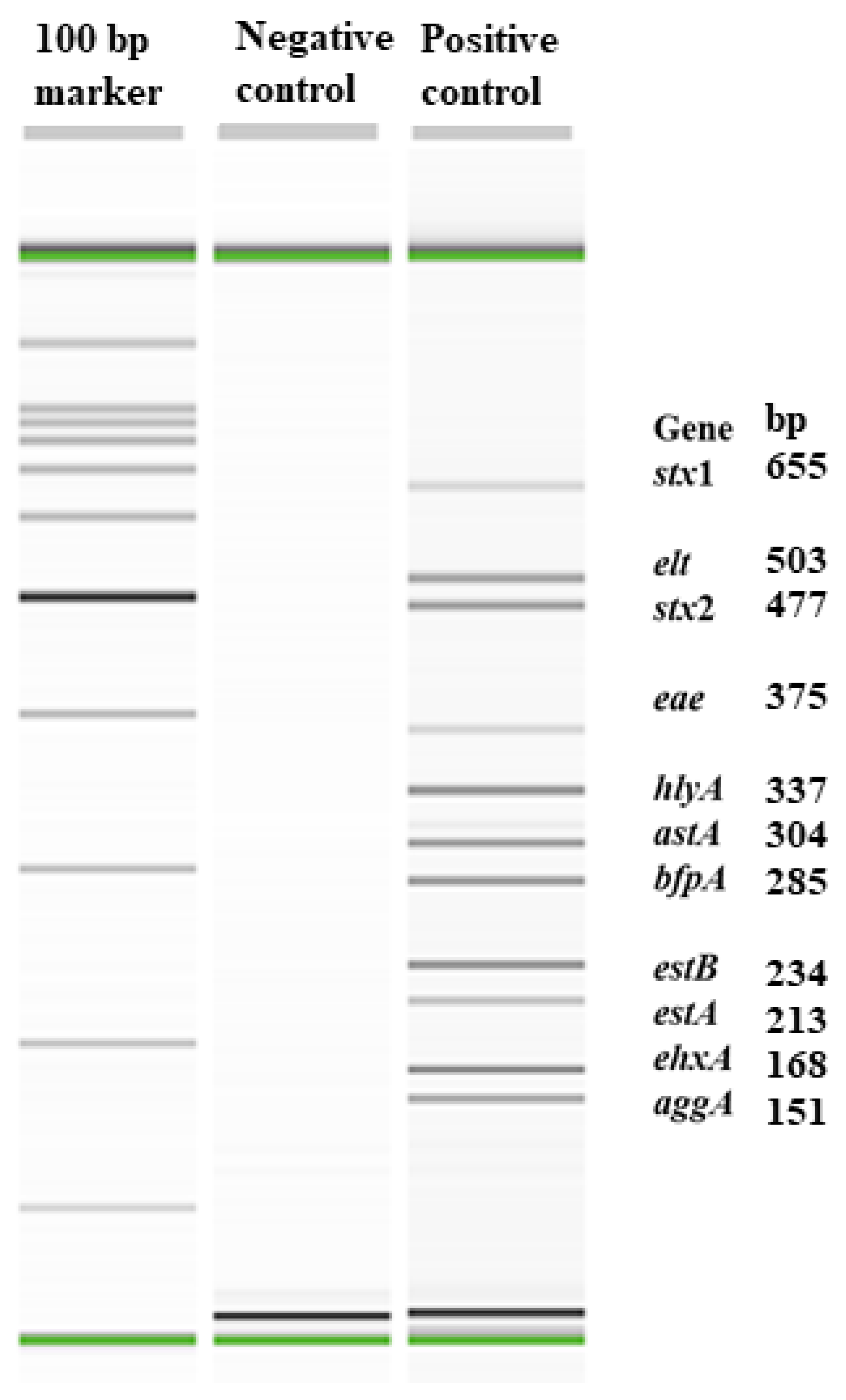
2.3. Development and Validation of an Eleven-Plex PCR to Detect Major Virulence Genes of Enteric E. coli Pathotypes

2.3.1. Gene Targets

The virulence genes were selected to identify four enteric pathotypes of E. coli, ETEC, EPEC, STEC, and enteroaggregative E. coli (EAEC), in swine feces. The targets included four enterotoxin genes, estA, estB, elt, and astA, which encode for heat-stable A, heat-stable B, heat-labile, and enteroaggregative heat-stable enterotoxins, respectively; two Shiga toxins, stx1 and stx2; adhesion factor genes, aggA, eae, and bfpA, which encode for subunits of enteroaggregative fimbriae, intimin, and bundle-forming pili subunit A, respectively; and two hemolysin genes, ehxA and hlyA, which encode for enterohemolysin and hemolysin A. The primers for stx1, stx2, and ehxA were from a previous study [12]; the adhesin factor genes primers are also from other studies [13,14,15]. The primers to amplify genes estA, estB, elt, astA, and hlyA were designed from the analyses of all available target sequences in this study. In addition to being specific to the target genes, the primers were designed to have a similar annealing temperature (~60 °C) and to have amplicon sizes that were well separated. The information on targeted genes and primer sequences are in Table 1.

2.3.2. Assay Conditions

The working concentration of the primer mix was 4.5 pM/µL of each primer. The reaction mix, with a total volume of 20 µL, consisted of 10 µL of IQ Multiplex Powermix (Bio-Rad, Hercules, CA, USA), 7 µL of nuclease-free water (Promega, Madison, WI, USA), 1 µL of primer mix, and 2 µL of DNA extracted from enriched fecal samples. The PCR running conditions for fecal samples included initial denaturation for 2 min at 94 °C, followed by 35 cycles of 15 s denaturation at 94 °C, annealing and extension at 68 °C for 80 s, and a final extension at 68 °C for 2 min. The E. coli strains H10407 and sPRH-20 (ETEC), 2348-69 (EPEC), 17-2 (EAEC), and ATCC 43894 (STEC) served as our reference positive controls in the PCR assay. The PCR running conditions for pure culture isolates were the same as those for fecal samples, except the number of amplification cycles was 25.

2.3.3. Sensitivity of the Assay

Escherichia coli strains H10407, sPRH-20, 2348-69, 17-2, and ATCC 43894 were used in pure cultures and to spike fecal samples to determine the sensitivity of the assay. A single colony of each strain was grown individually in 10 mL Luria–Bertani broth (LB; Becton Dickinson, Sparks, MD, USA) for 16 h at 37 °C. Then, 100 μL of each strain was added to another 10 mL of LB broth and incubated for 4 h at 37 °C. The bacterial cell concentrations of individual strains were determined by spread-plate count. All five strains were mixed together and seven 10-fold serial dilutions, were prepared. Each dilution was subjected to DNA extraction and PCR assay. Then 1 mL of each mixed-culture dilution (10−1 to 10−7) was added to 9 mL of EC broth suspended with swine feces, which was confirmed to be negative for most of the target genes. The spiked samples were incubated for 6 h at 40 °C, and 1 mL of each swine fecal broth suspension was collected before and after 6 h enrichment, boiled for 10 min, and centrifuged at 9300× g for 5 min. The supernatant of boiled broth suspension of each dilution was purified with a GeneClean Turbo kit (MP Biomedicals, Solon, OH, USA). Two microliters of purified DNA was used for the PCR assay. The experiment was repeated with the different fecal samples collected.

2.4. Isolation of E. coli by Direct Plating of Enriched Fecal Samples

Enriched fecal samples that were positive for one or more of the targeted virulence genes (enterotoxins, Shiga toxins, intimin) were streaked with a sterile loop onto MacConkey (MAC) agar plates (Becton Dickinson and Company, Sparks, MD, USA). Additionally, samples were diluted (1 in 100 dilution) in buffered peptone water, and 100 μL of the diluted fecal suspension was spread-plated onto MAC plates. The plates were incubated at 37 °C for 18 to 24 h. After incubation, 10 presumptive E. coli colonies (round, smooth, and pink-colored), positive for indole production by spot indole test, were selected and streaked onto blood agar plates (Remel, Lenexa, KS, USA) and incubated at 37 °C for 18 to 24 h. The colonies from each pie of blood agar plate were confirmed as E. coli by a three-plex PCR assay that targeted uidA, clbB, and ybb genes, which encode for beta-glucoronidase, casinolytic protease B (a heat shock protein), and putative allantoin permease, respectively. The primer sequences and amplicon size are shown in Table 2. An isolate positive for any one of the three genes was considered to be E. coli. The colonies of ten isolates were suspended in 100 μL of distilled water, boiled for 5 min at 95 °C, and centrifuged at 9300× g for 5 min, and the lysate was subjected to the 11-plex PCR assay. If pooled colonies were positive for any one or more of the virulence genes, then the ten colonies were tested individually by the 11-plex PCR to identify pure cultures positive for the virulence genes. If an isolate was positive for any of the 11 genes, it was stored using cryo-protect beads (Key Scientific Products, Stamford, TX, USA) in a −80 °C freezer.

2.5. Subtyping of stx2e Gene in STEC Isolates

A single-plex PCR assay was used to determine whether the stx2 gene was of the subtype 2e, which is involved in the edema disease, in all the STEC isolates (n = 238). The primer sequences designed were forward, CCACCAGGAAGTTATATTTCCGTA, and reverse, AACTTCACCTGGGCAAAGC. The primers’ working concentrations were 10 pM/μL. The reaction consisted of 1 μL of forward primer and 1 μL of reverse primer, 10 μL of Hot StartTaq plus Master mix (Qiagen, Hilden, Germany), 6 μL of nuclease-free water (Promega Corp., Madison, WI, USA), and 2 μL of DNA template. The total reaction volume was 20 μL. The PCR running program included an initial denaturation at 94 °C for 5 min, followed by 25 cycles of denaturation at 94 °C for 30 s, annealing at 60 °C for 30 s, extension at 67 °C for 75 s, and a final step of extension at 68 °C for 7 min. The PCR product was separated, and amplicon size was determined by using a capillary electrophoresis system, QIAxcel Advanced System, with a QIAxcel DNA Screening Kit (Qiagen, Germantown, MD, USA). The amplicon size was 1031 bp.

2.6. Detection of Fimbrial Genes by PCR

The purified DNA from E. coli isolates positive for one or more of the four enterotoxin genes was used to identify the fimbrial genes. A mPCR assay was used to detect genes that encoded for F6, F18, and F41 fimbriae, which are commonly associated with ETEC pathotypes involved in enteritis or post-weaning diarrhea. The assay also targeted putative glycosyl transferases of serogroups O8/O9, which are the main serogroups carrying the F5 gene. The primer sequences designed to amplify the genes and amplicon sizes are shown in Table 2. The assay reaction mixture included a working concentration of primer mix 1 and primer mix 2 (10 pM/uL of each primer) in a 20 µL reaction mix containing 10 µL of IQ Multiplex Powermix, 6 µL of nuclease-free water, 1 µL of primer mix 1, 1 µL of primer mix 2, and 2 µL of DNA extracted from positive isolates. The PCR running conditions were an initial denaturation for 5 min at 94 °C, followed by 25 cycles of denaturation at 94 °C for 30 s, annealing at 62 °C for 30 s, extension at 68 °C for 75 s, and final extension at 68 °C for 7 min.

2.7. Phenotypic and Genotypic Antimicrobial Susceptibility Determinations

A subset of isolates positive for one or more virulence genes were subjected to antimicrobial susceptibility testing of CTC by microbroth dilution method to determine the minimum inhibitory concentrations (MIC) according to the Clinical Laboratory and Standards Institute (CLSI). Stock solution of CTC antibiotic was prepared by adding sterile distilled water and the final concentration was adjusted to 1000 µg/mL. Chlortetracycline was tested at the concentrations of 100, 50, 25, 12.5, 6.25, 3.125, 1.56, 0.78, 0.39, and 0.195 µg/mL. The concentration of bacterial inoculum was adjusted to 0.5 McFarland turbidity standards (Remel Company, Lenexa, KS, USA) by adding individual bacterial colonies from the blood agar plate into 10 mL of Mueller–Hinton broth (MH; Becton, Dickinson and Co., Sparks, MD, USA) and vortexing. Then, 100 µL of the MH broth containing bacterial inoculum was dispensed into 96-well microtiter plates (Corning Incorporated, Corning, NY, USA) followed by the addition of 1:100 dilution of the culture. The microtiter plates were incubated at 37 °C for 20 to 24 h and the results were recorded as either growth or no growth. A mPCR assay was employed to detect tetA, tetB, tetC, and tetD genes [16,17].

2.8. Statistical Analysis

The data analysis was carried out using SAS (v. 9.4; Cary, NC, USA). Pen was considered as an experimental unit. Bivariate descriptive statistics of all the virulence genes by treatment and sampling day were carried out before building a final model. The PROC MIXED procedure with RFML (residual maximum likelihood) was used to evaluate the prevalence of virulence genes, pathotypes, their association with fimbrial genes, and tetracycline resistance genes. The final model included the fixed effects of treatment with sampling day as a random effect. The outcome variables, modeled using a binary outcome, consisted of the number of positive samples within each pen divided by the total number of samples tested per pen. The least square means were generated using the PDIFF option with a Tukey–Kramer adjustment for multiple comparisons by treatment, sampling day, and their interaction. The MIC values were log-transformed using PROC RANK and analysis was performed on ranked values. Results were considered significant at a p-value of <0.05.

3. Results

The optimization of the mPCR assay was achieved with DNA from two ETEC strains (H10407 and sPRH), one EPEC strain (2348-69), one EAEC strain (17-2), and one STEC strain (ATCC 43894). The amplicon sizes of the 11 virulence genes ranged from 151 to 655 bp (Figure 1). The detection limit of the assay with pooled pure cultures was 6.2×10⁴ CFU/mL and in fecal samples spiked with serially diluted, pooled pure cultures, the detection limit of the assay was 6.2×10⁵ CFU/g. A total of 360 fecal samples were analyzed, which represented 40 fecal samples from each of the three treatment groups (control, in-feed CTC, and in-water CTC) at each of the three sampling days (days 0, 14, and 28). Fecal samples were enriched in E. coli broth (6 h) before determining the prevalence of 11 virulence genes by mPCR assay and isolating E. coli positive for one or more of the virulence genes.

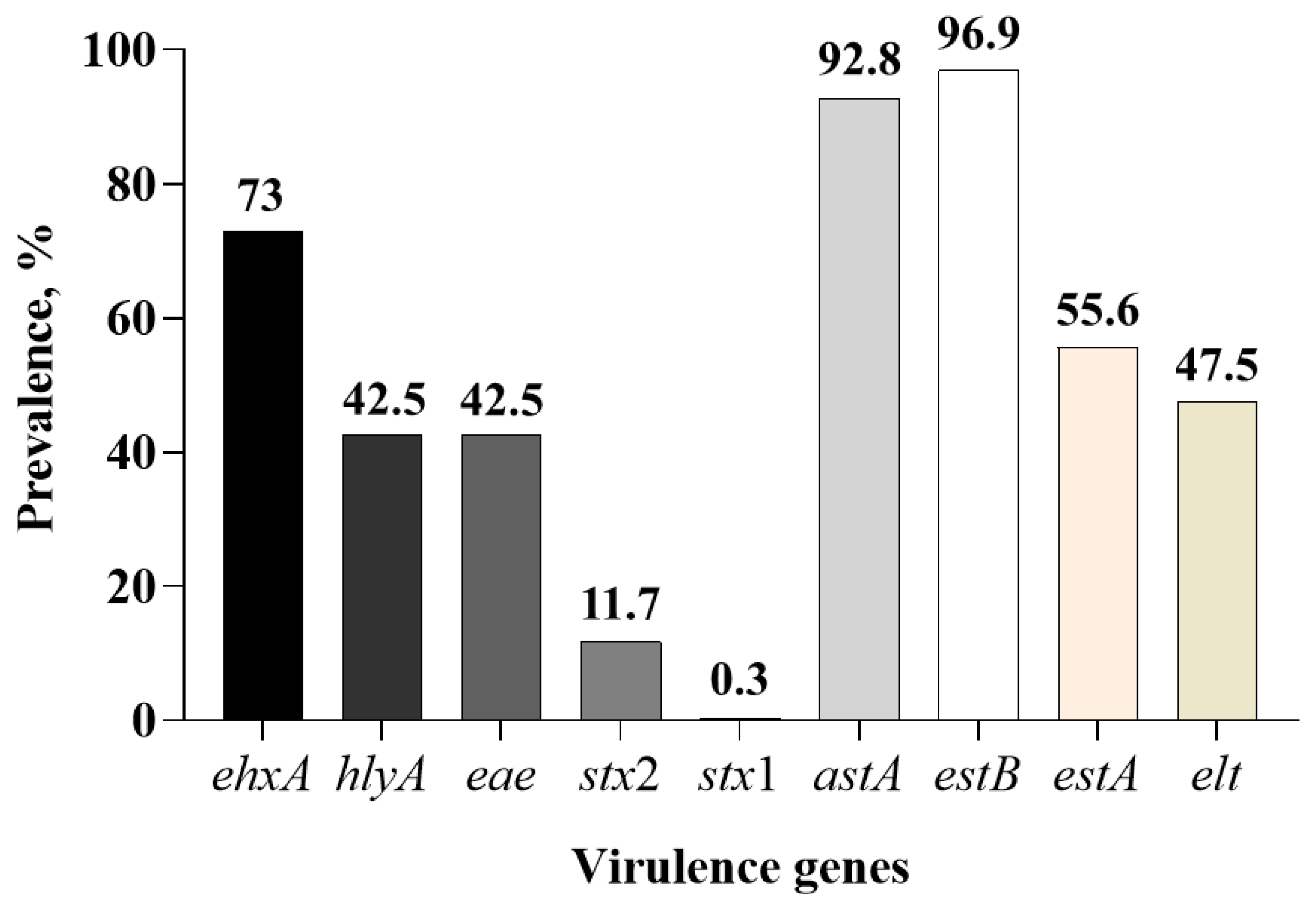
3.1. Prevalence of Virulence Genes in Fecal Samples

The 11 virulence genes tested were categorized into three broad groups based on their role in virulence: colonization factors (aggA, bfpA, and eae), enterotoxins (elt, estA, estB, and astA), and cytotoxins (stx1, stx2, hlyA, and ehxA) (Table 3). None of the 360 enriched fecal samples tested by the eleven-plex PCR assay were positive for the aggA or bfpA gene, which encode for the subunit of the enteroaggregative adherence fimbria AAF/1 and bundle-forming pilus protein, respectively. Only 4 fecal samples were negative for all 11 genes and the remaining 356 out of 360 (98.8%) samples were positive for one or more of the nine virulence genes. Overall, the prevalence of the estB gene (96.9%; 349/360) that encodes for the heat-stable enterotoxin B and astA (92.8%; 334/360) that encodes for the enteroaggregative heat-stable enterotoxin (EAST) was higher compared to the other toxin genes across all treatment groups and sampling days (Figure 2).
The prevalence of the heat-stable enterotoxin A gene, estA, was low (2.5%) in day 0 samples and increased to 84.2% and 80% in fecal samples collected on days 14 and 28, respectively (Table 3). Interestingly, the heat-labile enterotoxin gene, elt, was present in fecal samples of all three treatment groups collected on day 14 and was absent in fecal samples collected on days 0 and 28. Both stx1 and stx2 were absent in the fecal samples collected on days 0 and 14. Only one fecal sample in day 28 in the in-feed CTC group was positive for the Shiga toxin 1 gene (stx1). stx2 was prevalent in piglets of all three treatment groups and the total prevalence of stx2 in day 28 fecal samples was 35% (Table 3). Between the two hemolysin genes, ehxA, which encodes for enterohemolysin, was more prevalent (73%) than the hlyA gene (42.5%).
The pen-based prevalence of the nine virulence genes is shown in Table 4. In-feed or in-water CTC administration had no effect on the prevalence of the virulence genes. With the exception of stx1, the sampling day had significant effects (p < 0.05) on the prevalence of the other eight genes. However, there was no significant treatment by sampling day interaction (Table 4). The pen-level prevalence of estB and astA was 100% across all three sampling days and treatment groups. Similarly, treatment phase (day 14) and post-treatment phase (day 28) had 100% pen prevalence for ehxA, estA, and hlyA virulence genes. The prevalence of genes, except for estB and astA, was less than 100% in the pretreatment phase of the study (day 0). The stx1 gene was detected in only one pen in the in-feed CTC group. The stx2 gene was present in all the three treatment groups in day 28 samples, and the pen-based prevalence ranged from 37.5 to 62.5%.

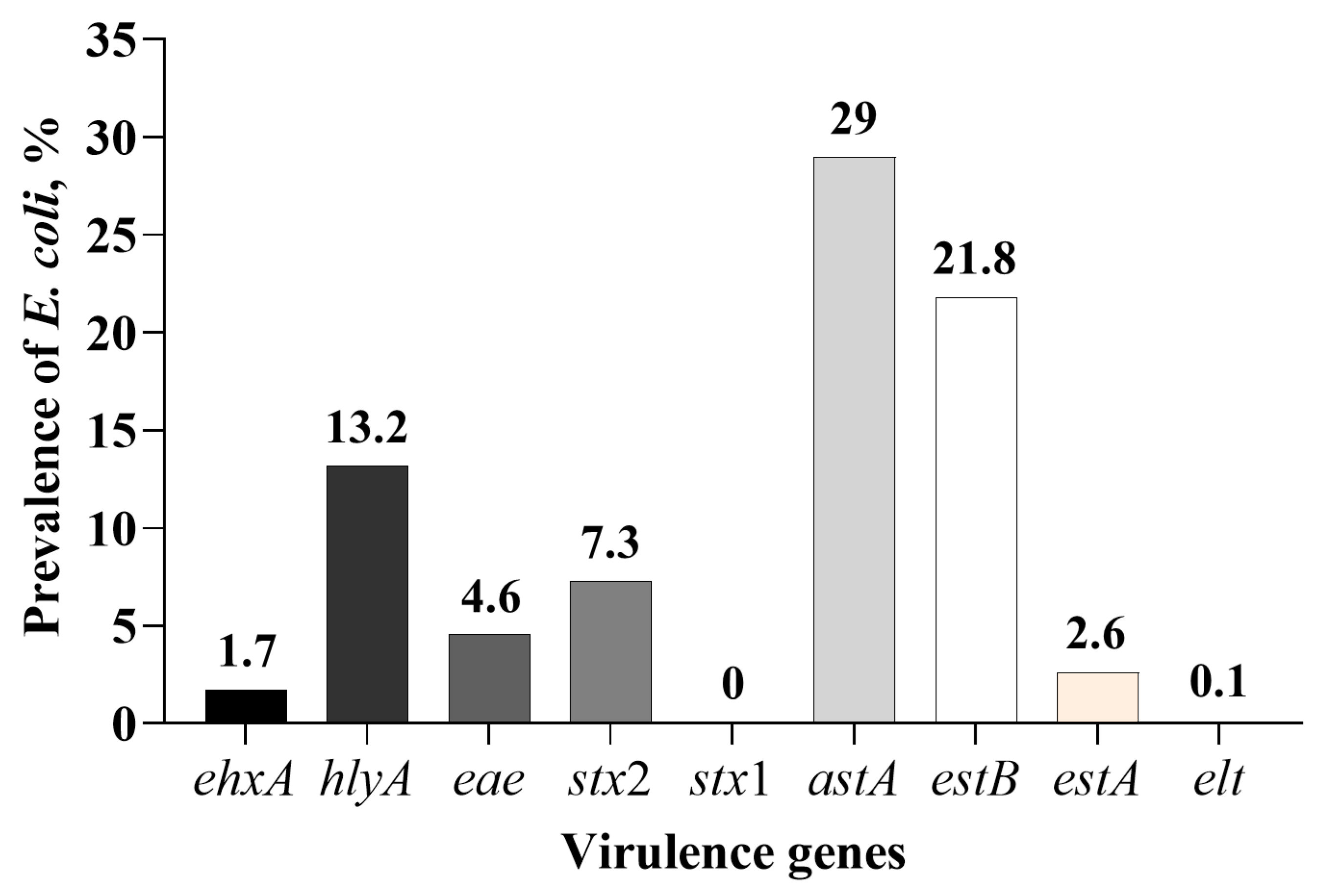
3.2. Isolation of E. coli Positive for One or More of the Nine Virulence Genes

A total of 3263 E. coli isolates positive for one or more of the nine virulence genes were obtained from fecal samples collected from piglets in the three treatment groups at the three sampling days (Table 5). Of those, 1923 isolates (58.9%) were positive for more than one gene. E. coli isolates positive for the two enterotoxin genes, astA (947/3263; 29%) and estB (714/3263; 21.8%), were higher than the other two enterotoxin genes, elt and estA (Figure 3). Only a small number of isolates were positive for the estA gene (84/3263; 2.6%). Only two isolates were positive for the elt enterotoxin gene. None of the isolates were positive for the stx1 gene. Shiga toxigenic E. coli were isolated from samples collected on day 28, the total number of isolates obtained was 238 out of 3263 isolates tested (7.3%), and all the isolates were positive for the stx2 gene only. Only a small proportion of the isolates were positive for the intimin gene, eae (4.6%), and none of them were positive for the stx2 gene. Between the two hemolysin genes tested, more isolates were positive for the hlyA (13.2%) than the ehxA (1.7%) gene.
E. coli isolates positive for one or more of the nine virulence genes based on pen-level prevalence are shown in Table 6. There were no treatment effects or treatment by sampling day interaction in the number of E. coli isolates positive for one or more of the nine virulence genes. However, except for elt and estA genes, the isolates positive for the other six genes were affected by sampling day. Between the two major enterotoxin-positive E. coli isolates, astA-positive isolates were higher in samples on day 28 than day 0 or 14, and in contrast, estB-positive isolates were higher in fecal samples collected on days 0 and 14 than on day 28 (Table 5 and Table 6). Shiga toxin 2-positive E. coli were isolated only in day 28 fecal samples.

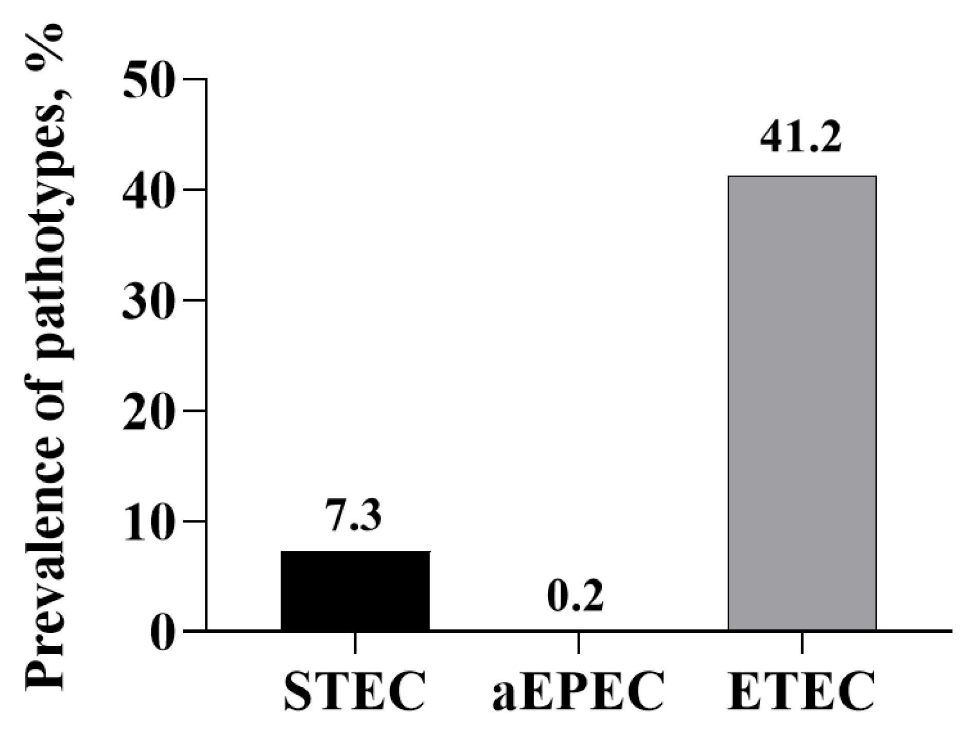
3.3. Prevalence of E. coli Pathotypes

Based on the virulence genes, E. coli isolates were grouped into the following three pathotypes: E. coli positive for one or more of the four enterotoxin genes (elt, estA, estB, and astA) and negative for eae as enterotoxigenic E. coli (ETEC); E. coli positive for eae and one or more of the four enterotoxin genes and negative for Shiga toxin gene (stx1 or stx2) as atypical enteropathogenic E. coli (aEPEC); and E. coli isolates positive for Shiga toxin gene (stx1 or stx2) as Shiga toxin-producing E. coli (STEC) (Table 7).
Because of the total absence of bfpA and aggA, none of the pathotypes belonged to enteropathogenic (EPEC) or enteroaggregative E. coli. Also, none of the isolates were of the hybrid pathotype. The ETEC pathotype was the most dominant of the three pathotypes detected (1344/3263; 41.2%; Figure 4). The ETEC pathotype, containing the astA gene, was the most dominant (70.5% of the ETEC isolates), followed by estB (53.1%; Figure 5). None of the isolates contained all four enterotoxin genes, and 19 (1.4%) contained the three enterotoxin genes (estA, estB, and astA). Among the ETEC containing two enterotoxin genes, more isolates contained estB and astA genes (23.9% of ETEC isolates). The number of ETEC isolates obtained increased with the sampling day. Only a small number of isolates were characterized as aEPEC (5/3263; 0.2%), and of the 5, 2 were positive for estB, 1 was positive for astA and 2 were positive for both estB and astA genes (Table 7). The STEC pathotypes were only present in day 28 samples, and all of them contained the stx2 gene and none were positive for stx1. Based on the single-plex PCR assay, all stx2-positive isolates contained the subtype stx2e. The pen level-based prevalence of the three pathotypes is shown in Table 8. There was no treatment effect nor treatment by sampling time interaction.

3.4. Prevalence of Fimbrial Genes

The ETEC strains (n = 1344) isolated across all treatment groups and sampling days were subjected to the mPCR assay targeting F4, F5, F6, F18, and F41 fimbriae genes (Table 9). None of the isolates was positive for F5, F6, and F41 genes. Only 11 strains were positive for fimbrial genes; 9 were positive for F4 and 2 were positive for F18. The nine F4-positive strains harbored estA and estB genes and two F18-positive strains possessed estB and elt genes.

3.5. Antimicrobial Susceptibility Testing

E. coli isolates positive for either two or more virulence genes (n = 165) were randomly selected from the three treatment groups across the three sampling days for antimicrobial susceptibility testing of CTC by microbroth dilution method. A strain with an MIC of ≥16 µg/mL was considered resistant. All the strains tested were resistant to CTC with MIC values of 50 µg/mL (CI = 37.5 to 62.5), 50 µg/mL (CI = 44.7 to 60.5), and 62.5 µg/mL (CI = 50 to 75) in the control, in-feed CTC, and in-water CTC groups, respectively (Table 10).

3.6. Prevalence of Tetracycline Resistance Genes

E. coli isolates (n = 165) used for antimicrobial susceptibility testing were further tested by PCR for the detection of tetA, tetB, tetC, and tetD genes (Table 10). None of the isolates tested positive for the tetC gene. tetA was the most predominant gene detected (151/165; 91.5%), followed by tetB with 16.9% prevalence (28/165), and the least dominant was tetD (12.7%: 21/165).

4. Discussion

Escherichia coli that causes enteric infections in animals and humans belong to six pathotypes, but four pathotypes, enterotoxigenic, enteropathogenic, Shigatoxigenic and enteroaggregative E. coli, are considered more common than the other two, enteroinvasive and diffusely adherent E. coli [18]. In order to determine the prevalence of E. coli pathotypes in healthy weaned piglets, a multiplex PCR assay targeting 11 major genes associated with the four pathotypes was designed and validated. Besides genes that encode for the four enterotoxin genes (heat-labile, heat-stable A, heat-stable B, and enteroaggregative heat-stable), two Shiga toxins (1 and 2), and two hemolysins (hemolysin A and enterohemolysin), eae, which encodes for intimin, a non-fimbrial adhesin, bfpA, which encodes for the type IV bundle-forming pili [19,20], and aggA, which encodes for fimbrial subunit of aggregative adherence fimbriae 1(AAF-1) [21], were included. The presence of bfpA and aggA in E. coli strains is indicative of EPEC and EAEC pathotypes, respectively. The two hemolysin genes were included because previous studies have shown an association between hemolytic activities and other virulence factors [22,23]. The hlyA gene encodes for alpha-hemolysin, a pore-forming cytotoxin belonging to the RTX family of toxins, and is cytotoxic to a variety of cells besides erythrocytes [24]. Although it is predominantly detected in extraintestinal pathogenic E. coli, it is also a major marker of swine ETEC strains, particularly those involved in post-weaning diarrhea, and is considered to enhance virulence and colonization [25]. ehxA, which encodes for enterohemolysin, is plasmid-borne and is widely distributed in STEC strains [26]. The presence of ehxA correlates with that of the Shiga toxin; therefore, it has been suggested as an epidemiological marker for STEC strains [27].
A number of PCR assays, either single-, two-, or multiplex, targeting a variety of virulence genes (toxins, fimbriae, and non-fimbrial adhesins) associated with swine pathogenic E. coli, have been reported [28,29,30,31,32,33,34,35]. Many of the assays have included the four enterotoxin genes and the five fimbrial genes (F4, F5, F6, F18, and F41) associated with ETEC and EPEC pathotypes. The novelty of our mPCR assay was its inclusion of genes that would facilitate identification of the four major pathotypes involved in enteric infections.
None of the fecal samples (n = 360) collected from weaned piglets and the E. coli isolates obtained in pure cultures (n = 3263) in the control or CTC-administered group were positive for either bfpA or aggA, which are characteristic of EPEC and EAEC, respectively. Strains of EPEC carrying the bfpA gene, which is located on a plasmid called the EPEC adherence factor plasmid, are described as typical EPEC because of their characteristic adherence to enterocytes, called local adherence, in which bacterial cells form clusters, called microcolonies [20]. In contrast, strains of EPEC that do not carry bfpA but carry the eae gene are described as atypical EPEC (aEPEC) [36]. In this study, only a small number of E. coli isolates were identified as aEPEC. The aggA is one of four genes (aggA to aggD) in a cluster that encodes for type I aggregative adherence fimbriae (AAF/1) required for the phenotypic expression of an aggregative adhesion pattern called ‘stacked brick’ adherence [37,38]. The EAEC is typically a human diarrheagenic E. coli recognized for causing persistent diarrhea in children and traveler’s diarrhea and diarrhea in immunocompromised adults [39,40]. Although the pathotype is widely prevalent in water and human food sources, it is not a pathotype generally associated with animals [41]. In a comparative genetic characterization of E. coli strains isolated from non-clinical samples (feces from healthy humans, companion animals, and swine) and confirmed as EAEC based on phenotypic adherence pattern, none were positive for the aggA gene [41]. In a comparative genomic analysis of 127 E. coli strains isolated from domestic animals, including swine, it was reported that 66.9% of isolates belonged to the EAEC pathotype based on a number of virulence genes (csgA, aggR, fimA, astA, etc.). Despite the prevalence of EAEC by genomic analysis, there has been no report of isolation of the EAEC pathotype from healthy or diarrheic swine [42]. However, the enterotoxin gene, astA, which codes for enteroaggregative heat-stable toxins has been reported in a number of studies that have characterized virulence genes in E. coli associated with enteric colibacillosis [22,43,44,45,46,47]. The astA gene of swine isolates has more than 98% homology with the nucleotide sequence of the corresponding gene from human isolates [48]. The astA gene is more prevalent in atypical EPEC than in typical EPEC strains implicated in diarrhea [49]. The astA was the second most prevalent virulence gene detected in feces at the sample level (92.8%). However, among the isolated E. coli strains, astA was the dominant enterotoxin gene (29%), followed by the estB gene (21.8%), and among the 947 isolates positive for the astA gene, 625 (65.6%) isolates were negative for the other three enterotoxin genes.
In this study, 41.2% (1351) of isolated E. coli strains were identified as ETEC (positive for one more of the four enterotoxin genes). The predominant enterotoxin genes in E. coli isolates were estB and astA, which agrees with a report of virotypes reported in weaned piglets with diarrhea or edema disease [33,34,50,51,52,53]. Only a small number of ETEC isolates (n = 9) carried fimbrial genes, faeG (F4; n = 9) and fedA (F18; n = 2). The nine F4-positive ETEC isolates carried estA and estB genes and the two F18-positive ETEC isolates carried elt and estB genes. The low prevalence of fimbrial genes in E. coli isolates is likely because the piglets were healthy, and none had diarrhea or edema disease. Also, it is possible that E. coli isolates had non-fimbrial adhesion factors, such as adhesin involved in diffuse adherence (AIDA) and porcine attaching effacing factor (paa) [51,53,54]. The prevalence of the aidA gene has been shown to be positively associated with the presence of astA in E. coli isolates in pigs with post-weaning diarrhea or edema disease [55]. In the study that reported E. coli pathotypes in piglets in South Korea, the predominant virotype was estB + astA + aida (10.4% of the ETEC isolates). The paa, which encodes for porcine attaching and effacing-associated protein, was first identified in EPEC isolates from swine, and is identical to the Paa protein described in E. coli O157:H7. The inactivation of paa leads to a loss of attaching and effacing activity [56].
It is interesting that Shiga toxin genes were not detected in fecal samples and no Shiga toxin-positive E. coli were isolated until day 28 samples. Except for one fecal sample from the in-feed CTC group, all the positive fecal samples and the isolates obtained were positive for the Shiga toxin 2 gene. Many of the published mPCR assays have targeted a subtype of the Shiga toxin 2 gene, stx2e, which targets endothelial cells of blood vessels, resulting in edema at specific locations [57]. The primers designed for Shiga toxin 1 and 2 genes were nonspecific to any particular subtype [58]. The intent was also to identify STEC that could also be of food safety importance [59]. Therefore, a single-plex PCR assay targeting the stx2e subtype was designed to test the stx2-postive E. coli isolates obtained (238/3263 isolates) and all the isolates were positive for the stx2e subtype. The absence of stx2e in the first 14 days of the study period is surprising because edema is typically observed in the first two weeks of the post-weaning period [60]. Normally, newborn piglets pick up the STEC strains from the sows, and it is possible that the sows in the herd were negative.
The presence of CTC resistance among these E. coli isolates (including the control group) suggests a high baseline prevalence of tetracycline resistance, which is attributed to the widespread use of tetracycline antibiotics in animal-based food production. The CTC supplementation was significantly associated with increased tetracycline resistance, with 99% (95% CI: 98–100%) of isolates exhibiting resistance compared to 95% (95% CI: 94–97%) in the control group [11]. This indicates that therapeutic use of CTC may further amplify existing resistance levels in fecal E. coli populations. Another study conducted at a U.S. research farm with no antimicrobial exposure for over five years found that E. coli isolates from weaned pigs exhibited higher levels of resistance to tetracycline, sulfisoxazole, and streptomycin compared to isolates from most older age groups. This finding suggests that age-related factors, such as immune system maturity or microbial community structure, may influence the persistence and distribution of antimicrobial resistance even in the absence of recent antimicrobial use [61]. Besides determining prevalence of virulence genes and pathotypes associated with enteric colibacillosis in healthy pigs, the study was designed to assess the impact of in-feed and in-water administration of CTC. The rationale for comparing in-feed and in-water administration is that the distribution of CTC in the gut is likely to be different between the two methods of administration. The distribution of in-water-administered CTC is likely to be more uniform compared to in-feed administration; therefore, the impact is likely to be different between the two groups. Neither in-feed or in-water administration of CTC had any impact on the prevalence of virulence genes or pathotypes, likely because all isolates of E. coli were resistant to CTC. The genotypic characterization revealed the high prevalence of tetA, followed by tetB and tetD. Even in previous studies, tetracycline resistance is the most prevalent resistant phenotype [62]. Tetracyclines are widely used because of their therapeutic benefits.

5. Conclusions

In conclusion, the use of 11-plex PCR for the detection of major virulence genes allowed us to determine the prevalence of virulence genes and pathotypes of E. coli associated with neonatal diarrhea, post-weaning diarrhea, and edema diseases in weaned piglets. The predominant pathotype detected was ETEC with astA and estB as the predominant enterotoxin genes. The second pathotype was stx2e-positive STEC and the prevalence of aEPEC was low (0.2%). Only a small number of ETEC isolates carried the fimbrial genes, likely because the piglets were healthy. All the E. coli isolates were resistant to chlortetracycline, with tetA being the predominant tetracycline-resistant gene. Overall, neither in-feed nor in-water CTC administration had any effects on the fecal prevalence of virulence genes and pathotypes implicated in swine colibacillosis.

Author Contributions

Conceptualization, R.G.A. and T.G.N.; Methodology, X.S. and J.B.; Formal analysis, R.K., R.G.A. and X.S.; Investigation, R.K., R.G.A., M.A., M.D.T. and T.G.N.; Resources, R.G.A.; Writing—original draft, R.K., R.G.A. and T.G.N.; Writing—review & editing, X.S., J.B., M.A. and M.D.T.; Supervision, R.G.A. and T.G.N.; Project administration, R.G.A.; Funding acquisition, R.G.A., M.D.T. and T.G.N. All authors have read and agreed to the published version of the manuscript.

Funding

This research was funded partially by the National Pork Board, grant number 19-028. This research was also supported in part by the USDA National Institute of Food and Agriculture, Hatch/Multistate Project 1014385.

Institutional Review Board Statement

The animal study protocol was approved by the Kansas State University Institutional Animal Care and Use Committee (IACUC) (protocol code #4033 dated 4 January 2018).

Informed Consent Statement

Not applicable.

Data Availability Statement

The original contributions presented in this study are included in the article. Further inquiries can be directed to the corresponding author.

Acknowledgments

This publication is listed as contribution number 25-046-J from the Kansas Agricultural Experiment Station, Manhattan, KS, USA.

Conflicts of Interest

The authors declare no conflicts of interest.

Abbreviations

The following abbreviations are used in this manuscript:
E. coliEscherichia coli
ETECEnterotoxigenic Escherichia coli
EPECEnteropathogenic Escherichia coli
STECShigatoxigenic Escherichia coli
aEPECAtypical enteropathogenic Escherichia coli
CTCChlortetracycline
PCRPolymerase chain reaction
tetTetracycline
AMRAntimicrobial resistance
MACMacConkey
mPCRMultiplex polymerase chain reaction
MICMinimum inhibitory concentration
CLSIClinical Laboratory and Standards Institute
PWDPost-weaning diarrhea
eltHeat-labile enterotoxin
estAHeat-stable enterotoxin A
estBHeat-stable enterotoxin B
astAEnteroaggregative heat-stable enterotoxin (EAST)
stx 1Shiga toxin 1
stx 2Shiga toxin 2
eaeIntimin
hlyAHemolysin
ehxAEnterohemolysin
aggASubunit of enteroaggregative adherence fimbria AAF/1
bfpABundle-forming pilus protein

References

  1. Moxley, R.A.; Duhamel, G.E. Comparative pathology of bacterial enteric diseases of swine. Adv. Exp. Med. Biol. 1999, 473, 83–101. [Google Scholar] [PubMed]
  2. Holland, R.E. Some infectious causes of diarrhea in young farm animals. Clin. Microbiol. Rev. 1990, 3, 345–375. [Google Scholar] [CrossRef]
  3. Garcia-Menino, I.; Garcia, V.; Mora, A.; Diaz-Jimenez, D.; Flament-Simon, S.C.; Alonso, M.P.; Blanco, J.E.; Blanco, M.; Blanco, J. Swine enteric colibacillosis in Spain: Pathogenic potential of mcr-1 ST10 and ST131 E. coli isolates. Front. Microbiol. 2018, 9, 2659. [Google Scholar] [CrossRef]
  4. Nordeste, R.; Tessema, A.; Sharma, S.; Kovac, Z.; Wang, C.; Morales, R.; Griffiths, M.W. Molecules produced by probiotics prevent enteric colibacillosis in pigs. BMC Vet. Res. 2017, 13, 335. [Google Scholar] [CrossRef]
  5. Wills, R.W. Diarrhea in growing-finishing swine. Vet. Clin. N. Am. Food. Anim. Pract. 2000, 16, 135–161. [Google Scholar] [CrossRef]
  6. Sacristan, R.D.P.; Rodriguez, A.L.; Sierens, A.; Vranckx, K.; Boyen, F.; Dereu, A.; Haesebrouck, F.; Maes, D.G.D. Efficacy of in-feed medication with chlortetracycline in a farrow-to-finish herd against a clinical outbreak of respiratory disease in fattening pigs. Vet. Rec. 2012, 171, 645. [Google Scholar] [CrossRef]
  7. Mayer, C.; Borges, A.; Flament-Simon, S.-C.; Simões, M. Quorum sensing architecture network in Escherichia coli virulence and pathogenesis. FEMS Microbiol. Rev. 2023, 47, fuad031. [Google Scholar] [CrossRef]
  8. Holman, D.B.; Bearson, B.L.; Allen, H.K.; Shippy, D.C.; Loving, C.L.; Kerr, B.J.; Bearson, S.M.D.; Brunelle, B.W. Chlortetracycline enhances tonsil colonization and fecal shedding of multidrug-resistant Salmonella enterica serovar Typhimurium DT104 without major alterations to the porcine tonsillar and intestinal microbiota. Appl. Environ. Microbiol. 2019, 85, e02354-18. [Google Scholar] [CrossRef]
  9. Remfry, S.E.; Amachawadi, R.G.; Shi, X.; Bai, J.; Woodworth, J.C.; Tokach, M.D.; Dritz, S.S.; Goodband, R.D.; Derouchey, J.M.; Nagaraja, T.G. Polymerase chain reaction-based prevalence of serogroups of Escherichia coli known to carry Shiga toxin genes in feces of finisher pigs. Foodborne Pathog. Dis. 2020, 17, 782–791. [Google Scholar] [CrossRef]
  10. Williams, H.E.; Tokach, M.D.; Dritz, S.S.; Woodworth, J.C.; DeRouchey, J.M.; Nagaraja, T.G.; Goodband, R.D.; Pluske, J.R.; Chitakasempornkul, K.; Bello, N.M.; et al. Effects of chlortetracycline alone or in combination with direct-fed microbials on nursery pig growth performance and antimicrobial resistance of fecal Escherichia coli. J. Anim. Sci. 2018, 96, 5166–5178. [Google Scholar] [CrossRef]
  11. Agga, G.E.; Scott, H.M.; Amachawadi, R.G.; Nagaraja, T.G.; Vinasco, J.; Bai, J.; Norby, B.; Renter, D.G.; Dritz, S.S.; Nelssen, J.L.; et al. Effects of chlortetracycline and copper supplementation on antimicrobial resistance of fecal Escherichia coli from weaned pigs. Prev. Vet. Med. 2014, 114, 303–308. [Google Scholar] [CrossRef] [PubMed]
  12. Bai, J.; Shi, X.; Nagaraja, T.G. A multiplex PCR procedure for the detection of six major virulence genes in Escherichia coli O157:H7. J. Microbiol. Methods. 2010, 82, 85–89. [Google Scholar] [CrossRef]
  13. Paddock, Z.; Bai, J.; Shi, X.; Renter, D.G.; Nagaraja, T.G. Detection of Escherichia coli O104 in the feces of feedlot cattle by a multiplex PCR assay designed to target major genetic traits of the virulent hybrid strain responsible for the 2011 German outbreak. Appl. Environ. Microbiol. 2013, 79, 3522–3525. [Google Scholar] [CrossRef]
  14. Noll, L.W.; Shridhar, P.B.; Dewsbury, D.M.; Shi, X.; Cernicchiaro, N.; Renter, D.G.; Nagaraja, T.G. A comparison of culture and PCR based methods to detect six major non-O157 serogroups of Shiga toxin-producing Escherichia coli in cattle feces. PLoS ONE 2015, 10, e0135446. [Google Scholar] [CrossRef]
  15. Shridhar, P.B.; Noll, L.W.; Shi, X.; Cernicchiaro, N.; Renter, D.G.; Bai, J.; Nagaraja, T.G. Escherichia coli O104 in feedlot cattle feces: Prevalence, isolation and characterization. PLoS ONE 2016, 11, e0152101. [Google Scholar] [CrossRef]
  16. Ng, L.K.; Martin, I.; Alfa, M.; Mulvey, M. Multiplex PCR for the detection of tetracycline resistant genes. Mol. Cell. Probes 2001, 15, 209–215. [Google Scholar] [CrossRef]
  17. Aminov, R.I.; Chee-Sanford, J.C.; Garrigues, N.; Teferedegne, B.; Krapac, I.J.; White, B.A. Development, validation, and application of PCR Primers for detection of tetracycline efflux genes of gram-negative bacteria. Appl. Environ. Microbiol. 2002, 68, 1786–1793. [Google Scholar] [CrossRef]
  18. Kaper, J.B.; Nataro, J.P.; Mobley, H.L.T. Pathogenic Escherichia coli. Nat. Rev. Microbiol. 2004, 2, 123–140. [Google Scholar] [CrossRef]
  19. Nataro, J.P.; Kaper, J.B. Diarrheagenic Escherichia coli. Clin. Microbiol. Rev. 1998, 11, 142. [Google Scholar] [CrossRef]
  20. Blank, T.E.; Donnenberg, M.S. Novel topology of BfpE, a cytoplasmic membrane protein required for type IV fimbrial biogenesis in enteropathogenic Escherichia coli. J. Bacteriol. 2001, 183, 4435–4450. [Google Scholar] [CrossRef]
  21. Elias, W.P.; Czeczulin, J.R.; Henderson, I.R.; Trabulsi, L.R.; Nataro, J.P. Organization of biogenesis genes for aggregative adherence fimbria II defines a virulence gene cluster in enteroaggregative Escherichia coli. J. Bacteriol. 1999, 181, 1779–1785. [Google Scholar] [CrossRef] [PubMed]
  22. Frydendahl, K. Prevalence of serogroups and virulence genes in Escherichia coli associated with postweaning diarrhoea and edema disease in pigs and a comparison of diagnostic approaches. Vet. Microbiol. 2002, 85, 169–182. [Google Scholar] [CrossRef] [PubMed]
  23. Fairbrother, J.M.; Nadeau, E. Colibacillosis. In Diseases of Swine, 11th ed.; Zimmerman, J.J., Karriker, L.A., Ramirez, A., Schwartz, K.J., Stevenson, G.W., Zhang, J., Eds.; Wiley Blackwell: Hoboken, NJ, USA, 2019; pp. 807–834. [Google Scholar]
  24. Fairbrother, J.M.; Nadeau, E.; Gyles, C.L. Escherichia. coli in postweaning diarrhea in pigs: An update on bacterial types, pathogenesis, and prevention strategies. Anim. Health Res. Rev. 2005, 6, 17–39. [Google Scholar] [CrossRef]
  25. Smith, H.W.; Linggood, M.A. Observations on the pathogenic properties of the K88, Hly and Ent plasmids of Escherichia coli with particular reference to porcine diarrhoea. J. Med. Microbiol. 1971, 4, 467–485. [Google Scholar] [CrossRef]
  26. Lorenz, S.C.; Son, I.; Maounounen-Laasri, A.; Lin, A.; Fischer, M.; Kase, J.A. Prevalence of hemolysin genes and comparison of ehxA subtype patterns in Shiga Toxin-Producing Escherichia coli (STEC) and Non-STEC Strains from clinical, food, and animal sources. Appl. Environ. Microbiol. 2013, 79, 6301–6311. [Google Scholar] [CrossRef]
  27. Beutin, L.; Montenegro, M.A.; Orskov, I.; Orskov, F.; Prada, J.; Zimmermann, S.; Stephan, R. Close association of verotoxin (Shiga-like toxin) production with enterohemolysin production in strains of Escherichia coli. J. Clin. Microbiol. 1989, 27, 2559–2564. [Google Scholar] [CrossRef]
  28. Frydendahl, K.; Imberechts, H.; Lehmann, S. Automated 5’ nuclease assay for detection of virulence factors in porcine Escherichia coli. Mol. Cell. Probes 2001, 15, 151–160. [Google Scholar] [CrossRef]
  29. Do, T.; Stephens, C.; Townsend, K.; Wu, X.; Chapman, T.; Chin, J.; McCormick, B.; Bara, M.; Trott, D. Rapid identification of virulence genes in enterotoxigenic Escherichia coli isolates associated with diarrhea in Queensland piggeries. Aust. Vet. J. 2005, 83, 293–299. [Google Scholar] [CrossRef]
  30. Taras, D.; Vahjen, W.; Macha, M.; Simon, O. Performance, diarrhea incidence, and occurrence of Escherichia coli virulence genes during long-term administration of a probiotic Enterococcus faecium strain to sows and piglets. J. Anim. Sci. 2006, 84, 608–617. [Google Scholar] [CrossRef]
  31. Wu, X.Y.; Chapman, T.; Trott, D.J.; Bettelheim, K.; Do, T.N.; Driesen, S.; Walker, M.J.; Chin, J. Comparative analysis of virulence genes, genetic diversity, and phylogeny of commensal and enterotoxigenic Escherichia coli isolates from weaned pigs. Appl. Environ. Microbiol. 2007, 73, 83–91. [Google Scholar] [CrossRef]
  32. Lee, S.I.; Kang, S.G.; Kang, M.L.; Yoo, H.S. Development of multiplex polymerase chain reaction assays for detecting enterotoxigenic Escherichia coli and their application to field isolates from piglets with diarrhoea. J. Vet. Diagn. Investig. 2008, 20, 492–496. [Google Scholar] [CrossRef] [PubMed]
  33. Casey, T.A.; Bosworth, B.T. Design and evaluation of a multiplex polymerase chain reaction assay for the simultaneous identification of genes for nine different virulence factors associated with Escherichia coli that cause diarrhea and edema disease in swine. J. Vet. Diagn. Investig. 2009, 21, 25–30. [Google Scholar] [CrossRef]
  34. Mohlatlole, R.P.; Madoroba, E.; Muchadeyi, F.C.; Chimonyo, M.; Kanengoni, A.T.; Dzomba, E.F. Virulence profiles of enterotoxigenic, Shiga toxin and enteroaggregative Escherichia coli in South African pigs. Trop. Anim. Health Prod. 2013, 45, 1399–1405. [Google Scholar] [CrossRef]
  35. Luppi, A.; Gibellini, M.; Gin, T.; Vangroenweghe, F.; Vandenbroucke, V.; Bauerfeind, R.; Bonilauri, P.; Labarque, G.; Hidalgo, A. Prevalence of virulence factors in enterotoxigenic Escherichia coli isolated from pigs with post-weaning diarrhoea in Europe. Porc. Health Manag. 2016, 2, 20. [Google Scholar] [CrossRef]
  36. Chen, H.D.; Frankel, G. Enteropathogenic Escherichia coli: Unravelling pathogenesis. FEMS Microbiol. Rev. 2005, 29, 83–98. [Google Scholar] [CrossRef]
  37. Suzart, S.; Guth, B.E.; Pedroso, M.Z.; Okafor, U.M.; Gomes, T.A. Diversity of surface structures and virulence genetic markers among enteroaggregative Escherichia coli (EAEC) strains with and without the EAEC DNA probe sequence. FEMS Microbiol. Lett. 2001, 201, 163–168. [Google Scholar] [CrossRef]
  38. Jonsson, R.; Struve, C.; Boisen, N.; Mateiu, R.V.; Santiago, A.E.; Jenssen, H.; Nataro, J.P.; Krogfelt, K.A. Novel aggregative adherence fimbria variant of enteroaggregative Escherichia coli. Infect. Immun. 2015, 83, 1396–1405. [Google Scholar] [CrossRef]
  39. Okeke, I.N.; Nataro, J.P. Enteroaggregative Escherichia coli. Lancet Infect. Dis. 2001, 1, 304–313. [Google Scholar] [CrossRef]
  40. Croxen, M.A.; Law, R.J.; Scholz, R.; Keeney, K.M.; Wlodarska, M.; Finlay, B.B. Recent advances in understanding enteric pathogenic Escherichia coli. Clin. Microbiol. Rev. 2013, 26, 822–880. [Google Scholar] [CrossRef]
  41. Zhang, R.; Gu, D.; Huang, Y.; Chan, E.W.; Chen, G.; Chen, S. Comparative genetic characterization of enteroaggregative Escherichia coli strains recovered from clinical and non-clinical settings. Sci. Rep. 2016, 6, 24321. [Google Scholar] [CrossRef]
  42. Tang, F.; Wang, J.; Li, D.; Gao, S.; Ren, J.; Ma, L.; Liu, F.; Zhuge, X.; Yan, G.; Lu, Y.; et al. Comparative genomic analysis of 127 Escherichia coli strains isolated from domestic animals with diarrhea in China. BMC Genom. 2019, 20, 212. [Google Scholar] [CrossRef] [PubMed]
  43. Yamamoto, T.; Nakazawa, M. Detection and sequences of the enteroaggregative Escherichia coli heat-stable enterotoxin 1 gene in enterotoxigenic E. coli strains isolated from piglets and calves with diarrhea. J. Clin. Microbiol. 1997, 35, 223–227. [Google Scholar] [CrossRef]
  44. Choi, C.; Cho, W.S.; Chung, H.K.; Jung, T.; Kim, J.; Chae, C. Prevalence of the enteroaggregative Escherichia coli heat-stable enterotoxin 1 (EAST1) gene in isolates in weaned pigs with diarrhea and/or edema disease. Vet. Microbiol. 2001, 81, 65–71. [Google Scholar] [CrossRef]
  45. Menard, L.P.; Dubreuil, J.D. Enteroaggregative Escherichia coli heat-stable enterotoxin 1 (EAST1): A new toxin with an old twist. Crit. Rev. Microbiol. 2002, 28, 43–60. [Google Scholar] [CrossRef]
  46. Osek, J. Detection of the enteroaggregative Escherichia coli heat-stable enterotoxin 1 (EAST1) gene and its relationship with fimbrial and enterotoxin markers in E. coli isolates from pigs with diarrhoea. Vet. Micrbiol. 2003, 91, 65–72. [Google Scholar] [CrossRef]
  47. Turner, S.M.; Scott-Tucker, A.; Cooper, L.M.; Henderson, I.R. Weapons of mass destruction: Virulence factors of the global killer enterotoxigenic Escherichia coli. FEMS Microbiol. Lett. 2006, 263, 10–20. [Google Scholar] [CrossRef]
  48. Berberov, E.M.; Zhou, Y.; Francis, D.H.; Scott, M.A.; Kachman, S.D.; Moxley, R.A. Relative importance of heat-labile enterotoxin in the causation of severe diarrheal disease in the gnotobiotic piglet model by a strain of enterotoxigenic Escherichia coli that produces multiple enterotoxins. Infect. Immun. 2004, 72, 3914–3924. [Google Scholar] [CrossRef]
  49. Silva, L.E.P.; Souza, T.B.; Silva, N.P.; Scaletsky, I.C.A. Detection and genetic analysis of the enteroaggregative Escherichia coli heat-stable enterotoxin (EAST1) gene in clinical isolates of enteropathogenic Escherichia coli (EPEC) strains. BMC Microbiol. 2014, 14, 135. [Google Scholar] [CrossRef]
  50. Blanco, M.; Lazo, L.; Blanco, J.E.; Dahbi, G.; Mora, A.; Lopez, C.; Gonzalez, E.A.; Blanco, J. Serotypes, virulence genes, and PFGE patterns of enteropathogenic Escherichia coli isolated from Cuban pigs with diarrhea. Int. Microbiol. 2006, 9, 53–60. [Google Scholar]
  51. Zhang, W.; Zhao, M.; Ruesch, L.; Omot, A.; Francis, D. Prevalence of virulence genes in Escherichia coli strains recently isolated from young pigs with diarrhea in the US. Vet. Microbiol. 2007, 123, 145–152. [Google Scholar] [CrossRef]
  52. Wang, X.M.; Liao, X.P.; Liu, S.G.; Zhang, W.J.; Jiang, H.X.; Zhang, M.J.; Zhu, H.Q.; Sun, Y.; Sun, J.; Li, A.X.; et al. Serotypes, virulence genes, and antimicrobial susceptibility of Escherichia coli isolates from pigs. Foodborne Pathog. Dis. 2011, 8, 687–692. [Google Scholar] [CrossRef]
  53. Byun, J.W.; Moon, B.Y.; Do, K.H.; Lee, K.H.; Lee, Y.; Kim, W.I.; So, B.J.; Lee, W.K. O-serogroups and pathovirotypes of Escherichia coli Isolated from post-weaning piglets showing diarrhoea and/or oedema in South Korea. Vet. Sci. 2022, 9, 1. [Google Scholar] [CrossRef]
  54. Tesh, V.L.; O’Brien, A.D. Adherence and colonization mechanisms of enteropathogenic and enterohemorrhagic Escherichia coli. Microb. Pathog. 1992, 12, 245–254. [Google Scholar] [CrossRef] [PubMed]
  55. Ha, S.K.; Choi, C.; Jung, K.; Kim, J.; Han, D.U.; Ha, Y.; Lee, S.D.; Kim, S.H.; Chae, C. Genotypic prevalence of the adhesin involved in diffuse adherence in Escherichia coli isolates in pre-weaned pigs with diarrhoea in Korea. J. Vet. Med. B 2004, 51, 166–168. [Google Scholar] [CrossRef] [PubMed]
  56. An, H.; Fairbrother, J.M.; Desautels, C.; Harel, J. Distribution of a novel locus called Paa (porcine attaching and effacing associated) among enteric Escherichia coli. Adv. Exp. Med. Biol. 1999, 473, 179–184. [Google Scholar]
  57. Macleod, D.L.; Gyles, C.L. Immunization of pigs with a purified Shiga-like toxin II variant toxoid. Vet. Microbiol. 1991, 29, 309–318. [Google Scholar] [CrossRef]
  58. Shridhar, P.B.; Siepker, C.; Noll, L.W.; Shi, X.; Nagaraja, T.G.; Bai, J. Shiga toxin subtypes of non-O157 Escherichia coli serogroups isolated from cattle feces. Front. Cell. Infect. Microbiol. 2017, 7, 121. [Google Scholar] [CrossRef]
  59. Tseng, M.; Fratamico, P.M.; Bagi, L.; Manzinger, D.; Funk, J.A. Shiga toxin producing E. coli (STEC) in swine: Prevalence over the finishing period and characteristics of the STEC isolates. Epidemiol. Infect. 2014, 143, 505–514. [Google Scholar] [CrossRef]
  60. Moxley, R.A. Edema disease. Vet. Clin. N. Am. Food Anim. Pract. 2000, 16, 175–185. [Google Scholar] [CrossRef]
  61. Langlois, B.; Dawson, K.; Leak, I.; Aaron, D. Effect of age and housing location on antibiotic resistance of fecal coliforms from pigs in a non-antibiotic-exposed herd. Appl. Environ. Microbiol. 1988, 54, 1341–1344. [Google Scholar] [CrossRef]
  62. Tadesse, D.A.; Zhao, S.; Tong, E.; Ayers, S.; Singh, A.; Bartholomew, M.J.; McDermott, P.F. Antimicrobial drug resistance in Escherichia coli from humans and food animals, United States, 1950–2002. Emerg. Infect. Dis. 2012, 18, 741–749. [Google Scholar] [CrossRef]
Figure 1. QIAxcel image of the eleven virulence genes of enteric pathotypes of Escherichia coli implicated in swine colibacillosis.
Microorganisms 13 01185 g001
Figure 2. Overall sample level-based prevalence of the major virulence genes of Escherichia coli pathotypes involved in swine colibacillosis in fecal samples collected from piglets administered with or without in-feed or in-water chlortetracycline.
Microorganisms 13 01185 g002
Figure 3. Overall sample level-based prevalence of Escherichia coli isolates positive for one or more of the virulence genes involved in swine colibacillosis in fecal samples collected from piglets administered with or without in-feed or in-water chlortetracycline.
Microorganisms 13 01185 g003
Figure 4. Overall sample level-based prevalence of Escherichia coli pathotypes involved in swine colibacillosis in fecal samples collected from piglets administered with or without in-feed or in-water chlortetracycline.
Microorganisms 13 01185 g004
Figure 5. Overall sample level-based prevalence of enterotoxigenic Escherichia coli in fecal samples collected from piglets administered with or without in-feed or in-water chlortetracycline.
Microorganisms 13 01185 g005
Table 1. Targeted virulence genes and virulence factors associated with enteric Escherichia coli pathotypes, primers, primer sequences, and amplicon size.
Virulence GenesVirulence FactorsPrimersPrimer Sequences (5′ to 3′)Amplicon Size, bpSource
aggASubunit of enteroaggregative adherence fimbria AAF/1aggA-F1CGTTACAAATGATTGTCCTGTTACTAT151Paddock et al., 2013 [13]
aggA-R1ACCTGTTCCCCATAACCAGAC
ehxAEnterohemolysinehxA-FGCGAGCTAAGCAGCTTGAAT168Bai et al., 2010 [12]
ehxA-RCTGGAGGCTGCACTAACTCC
estAHeat-stable enterotoxin AestA-F2CATGACGGGAGGTAACATGA213This study
estA-R2GGATTACAACAAAGTTCACAGCA
estBHeat-stable enterotoxin BestB-F2CTTGACTCATATAAAAGCCCACTG234This study
estB-R2GCAGTACCATCTCTAACCCCTAAA
bfpABundle-forming pilus proteinbfpA-F2CAGAAGTAATGAGCGCAACG285Shridhar et al., 2016 [15]
bfpA-R2CGTAGCCTTTCGCTGAAGTA
astAEnteroaggregative heat-stable enterotoxin (EAST)astA-FGGCTCAATGTGCTGACTGAA304This study
astA-RTGCCAGCTTCGGCTTATC
hlyAHemolysinhlyA-F4ACGAAAGTACTGGGTAATGTTGG337This study
hlyA-R4ATGTCGTTGCAGCAGCACT
eaeIntimineae-F2TACGCGAAAGATACCGCTCT375Noll et al., 2015 [14]
eae-R2CATGCGGAAATAGCCGTTA
stx2Shiga toxin 2stx1-FCCATGACAACGGACAGCAGTT477Bai et al., 2010 [12]
stx1-RTGTCGCCAGTTATCTGACATTC
eltHeat-labile enterotoxinelt-F2TTATGATCACGCGAGAGGAA503This study
elt-R2TTGTGCTCAGATTCTGGGTCT
stx1Shiga toxin 1stx1-FTGTCGCATAGTGGAACCTCA655Bai et al., 2010 [12]
stx1-RTGCGCACTGAGAAGAAGAGA
Table 2. Genes targeted for species confirmation of E. coli and fimbriae associated with enterotoxigenic Escherichia coli pathotypes, primer sequences, and amplicon sizes.
Genes
Targeted
Encoded ProteinPrimersPrimer SequencesAmplicon Size, bpSource
clpBCaseinolytic protease B
(A heat-shock protein)
clpB-F#CATACGAATGCTGGATGCTG449This
study
clpB-RTTTGAAGAACGTTTAAAAGGCG
uidABeta-glucuronidaseuidA-FACCACGGTGATATCGTCCAC449This study
uidA-RTACAAGAAAGCCGGGCAAT
ybbWPutative allantoin permeaseybbW-FAATCTGGCCGGGATTTTT449This study
ybbW-RTGGCTCCGGCAATAATACAT
faeGF4 fimbrial adhesinF4-FATTTCAATGGTTCGGTCGAT416This study
F4-RCGCAGAAGTAACCCCACCT
fanCF5 fimbrial adhesinF5-FCAGGAAATACTGCTGCTAAAG150This study
F5-RGCTGGGCTGAATAGTTAAATGAC
fasAF6 fimbrial adhesinF6-FACCAGCCAGGCAAATTTAGA492This study
F6-RTGTACCTGCTGAACGAATAGTCA
fedAF18 fimbrial adhesinF18-FCAGCAAGGGGATGTTAAATTC218This study
F18-RAACTGCCCGCTCCAAGTTA
f41F41 fimbrial adhesinF41-FTGATTGGACGGAAGGTCAAC561This study
F41-RCCTGGCATTAACTTTTCTACATAACC
_Putative glycosyltransferasesO8/O9-FGTCTTCATCCGGGACATAGC735This study
O8/O9-RCGTGAAATCGAAGAGCTGAA
Table 3. Sample level-based prevalence of the major virulence genes of Escherichia coli pathotypes involved in swine colibacillosis in fecal samples collected from piglets administered with or without in-feed or in-water chlortetracycline (CTC).
Virulence FactorDay 0Day 14Day 28
Control (n = 40)In-Feed CTC (n = 40)In-Water CTC
(n = 40)
Total, n = 120 (%)Control (n = 40)In-Feed CTC (n = 40)In-Water CTC
(n = 40)
Total, n = 120 (%)Control (n = 40)In-Feed CTC (n = 40)In-Water CTC
(n = 40)
Total,
n = 120 (%)
aggA000 000 0000
bfpA000 000 0000
eae53210 (8.3%)31293595
(79.2%)
15151848
(40%)
elt000 15231957 (47.5%)0000
estA2103
(2.5%)
362837101 (84.2%)33323196
(80%)
estB383736111 (92.5%)404040120 (100%)394039118 (98.3%)
astA394039118 (98.3%)333833104 (86.7%)373837112 (93.3%)
stx1000000000101
(0.83%)
stx20000000013161342
(35%)
hlyA109928
(23.3%)
31293595
(79.2%)
15151848
(40%)
ehxA1415938
(31.7%)
404040120 (100%)333437104 (86.7%)
Sample-level prevalence calculated as the number of samples positive for the gene out of the total number of samples collected within each treatment group.
Table 4. Pen level-based prevalence of the major virulence genes of Escherichia coli pathotypes involved in swine colibacillosis in fecal samples collected from piglets administered with or without in-feed or in-water chlortetracycline (CTC).
Virulence FactorDay 0Day 14Day 28TrtDayTrt × Day
Control
(n = 8)
In-Feed CTC (n = 8)In-Water CTC
(n = 8)
Control
(n = 8)
In-Feed CTC (n = 8)In-Water CTC
(n = 8)
Control
(n = 8)
In-Feed CTC (n = 8)In-Water CTC
(n = 8)
Colonization factor
eae4 (50%)2 (25%)2 (25%)8 (100%)8 (100%)8 (100%)6 (75%)6 (75%)6 (75%)0.658<0.0010.769
Enterotoxins
elt0005 (62.5%)7 (87.5%)7 (87.5%)0000.522<0.0010.623
estA1 (12.5%)1 (12.5%)08 (100%)7 (87.5%)8 (100%)8 (100%)8 (100%)8 (100%)0.301<0.0010.311
estB8 (100%)8 (100%)8 (100%)8 (100%)8 (100%)8 (100%)8 (100%)8 (100%)8 (100%)---
astA8 (100%)8 (100%)8 (100%)8 (100%)8 (100%)8 (100%)8 (100%)8 (100%)8 (100%)---
Cytotoxins
stx100000001 (12.5)00.3760.3770.418
stx20000004 (50%)5 (62.5%)3 (37.5%)0.926<0.0010.989
hlyA6 (75%)5 (62.5%)8 (100%)8 (100%)8 (100%)8 (100%)8 (100%)7 (87.5%)8 (100%)0.707<0.0010.982
ehxA5 (62.5%)8 (100%)6 (75%)8 (100%)8 (100%)8 (100%)8 (100%)8 (100%)8 (100%)0.920<0.0010.337
Pen-level prevalence calculated as the number of pens that had at least one positive sample for the gene divided by the total number of pens sampled per treatment group.
Table 5. Sample level-based prevalence of Escherichia coli positive for the virulence genes associated with enteric pathotypes involved in swine colibacillosis in fecal samples from piglets administered with or without in-feed or in-water chlortetracycline (CTC).
Virulence FactorDay 0Day 14Day 28
Control (n = 40)In-Feed CTC (n = 40)In-Water CTC
(n = 40)
Total,
(n = 120)
Control (n = 40)In-Feed CTC (n = 40)In-Water CTC
(n = 40)
Total (n = 120)Control (n = 40)In-Feed CTC (n = 40)In-Water CTC
(n = 40)
Total (n = 120)
No. of isolates31028027986939440040011944004004001200
eae8
(2.6) a
21 (7.5)41 (14.7)70 (8.1)21
(5.3)
23
(5.8)
22 (5.5)66
(5.5)
8
(2)
2
(0.5)
5
(1.3)
15
(1.3)
elt00002
(0.5)
002
(0.2)
0000
estA7
(2.3)
007 (0.8)21
(5.3)
12
(3)
16
(4)
49
(4.1)
3
(0.8)
12
(3)
13 (3.3)28
(2.3)
estB99 (31.9)112 (40)83 (29.7)294 (33.8)78
(19.8)
154 (38.5)145 (36.3)377 (31.6)2
(0.5)
16
(4)
25 (6.3)43
(6)
astA98
(32)
102 (36.4)92
(33)
292 (33.6)31
(7.9)
40
(10)
34 (8.5)105 (8.8)163 (40.8)209
(52.3)
178 (44.5)550 (45.8)
stx1000000000000
stx20000000090
(22.5)
75
(18.7)
73 (18.2)238 (19.8)
hlyA15
(4.8)
9
(3.2)
9
(3.2)
33 (3.8)73
(18.5)
29
(7.3)
35 (8.8)137 (11.5)97
(24.3)
87
(21.8)
76
(19)
260 (21.6)
ehxA12
(3.9)
0012 (1.4)9
(2.3)
15
(3.8)
10 (2.5)34
(2.8)
5
(1.3)
1
(0.3)
4
(1)
10
(0.8)
a Numbers in parentheses are percentage of isolates positive for the virulence gene out of the total isolates obtained in each treatment group.
Table 6. Pen level-based prevalence of Escherichia coli positive for the virulence genes associated with enteric pathotypes involved in swine colibacillosis in fecal samples from piglets received CTC via feed, water, or no treatment.
Virulence GenesDay 0Day 14Day 28p-value
ControlIn-Feed CTCIn-
Water CTC
ControlIn-Feed CTCIn-
Water CTC
ControlIn-Feed CTCIn-
Water CTC
TrtDayTrt × Day
Colonization factor
eae2 (25%)3 (37.5%)4 (50%)6 (75%)6 (75%)6 (75%)3 (37.5%)2 (25%)2 (25%)0.2570.0300.102
Enterotoxins
elt0001 (12.5%)000000.3770.3770.418
estA1 (12.5%)005 (62.5%)4 (50%)6 (75%)3 (37.5%)3 (37.5%)2 (25%)0.8810.0540.525
estB6 (75%)7 (87.5%)6 (75%)6 (75%)7 (87.5%)7 (87.5%)2 (25%)3 (37.5%)4 (50%)0.268< 0.0010.829
astA6 (75%)7 (87.5%)6 (75%)6 (75%)6 (75%)5(62.5%)7 (87.5%)7 (87.5%)7 (87.5%)0.737< 0.0010.976
Cytotoxins
stx1000000000000
stx20000003 (37.5%)3 (37.5%)3 (37.5%)0.9640.0010.997
hlyA2 (25%)3 (37.5%)2 (25%)8 (100%)4 (50%)6 (75%)6 (75%)5 (62.5%)6 (75%)0.5760.0160.963
ehxA2 (25%)004 (50%)6 (75%)4 (50%)2 (25%)1 (12.5%)2 (25%)0.3870.0690.256
Pen-level prevalence calculated as the number of pens that had at least one positive sample for the gene divided by the total number of pens sampled per treatment group (n = 8).
Table 7. Sample level-based prevalence of the Escherichia coli pathotypes involved in swine colibacillosis in fecal samples collected from piglets receiving CTC via feed or water or no treatment.
PathotypesDay 0Day 14Day 28
Control (n = 40)In-Feed CTC (n = 40)In-Water CTC
(n = 40)
Total (n = 120)Control (n = 40)In-Feed CTC (n = 40)In-Water CTC
(n = 40)
Total (n = 120)Control (n = 40)In-Feed CTC (n = 40)In-Water CTC
(n = 40)
Total (n = 120)
No. of isolates31028027986939440040011944004004001200
ETEC a10711193311104190152446168220199587
Heat-labile (elt)000020020000
Heat-stable A (estA)7007211216493121328
Heat-stable B (estB)9911183294781541453772162543
Enteroaggregative heat-stable (astA)9810292292314034105163209178550
elt + estB000020020000
estA + estB700721111648081018
estA + astA0000211408715
estB + astA901028227445233208715
estA + estB + astA0000211408715
aEPEC b000011240101
STEC c00000000907573238
a ETEC pathotype: Isolates positive for one or more of the four enterotoxin genes (elt, estA, estB, and astA). b Atypical EPEC pathotype: Isolates positive for eae gene, negative for Shiga toxin genes (stx1 or stx2), and positive for one or more of the four enterotoxin genes (elt, estA, estB, and astA). c STEC pathotype: Isolates positive for one or both of the Shiga toxin genes (stx1 and stx2).
Table 8. Pen level-based prevalence of Escherichia coli pathotypes implicated in swine colibacillosis in fecal samples collected from piglets administered with or without in-feed or in-water chlortetracycline (CTC) and enriched in E. coli broth.
PathotypeDay 0Day 14Day 28TrtDayTrt × Day
Control
(n = 8)
In-Feed CTC (n = 8)In-Water CTC
(n = 8)
Control
(n = 8)
In-Feed CTC (n = 8)In-Water CTC
(n = 8)
Control
(n = 8)
In-Feed CTC (n = 8)In-Water CTC
(n = 8)
Enterotoxigenic
E. coli a
6
(75%)
7
(87.5%)
6
(75%)
7
(87.5%)
7
(87.5%)
7
(87.5%)
7
(87.5%)
7
(87.5%)
7
(87.5%)
0.9290.6400.977
Atypical Enteropathogenic E. coli b0001
(12.5%)
1
(12.5%)
2
(25%)
01
(12.5%)
00.8150.0040.895
Shigatoxigenic
E. coli c
0000003
(37.5%)
3
(37.5%)
3
(37.5%)
1.000<0.0011.000
a ETEC pathotype: Isolates positive for one or more of the four enterotoxin genes (elt, estA, estB, and astA). b Atypical EPEC pathotype: Isolates positive for eae gene, negative for Shiga toxin genes (stx1 or stx2) and positive for one or more of the four enterotoxin genes (elt, estA, estB, and astA). c STEC pathotype: Isolates positive for one or both of the Shiga toxin genes (stx1 and stx2).
Table 9. Prevalence of fimbrial genes in enterotoxigenic Escherichia coli isolated from fecal samples collected from piglets received CTC via feed or water or no treatment and enriched in E. coli broth.
Fimbriae GenesDay 0Day 14Day 28Overall Total, n = 1344
Control (n = 107)In-Feed CTC (n = 111)In-Water CTC
(n = 93)
Total, n = 311Control (n = 104)In-Feed CTC (n = 190)In-Water CTC
(n = 152)
Total, n = 446Control (n = 168)In-Feed CTC (n = 220)In-Water CTC
(n = 199)
Total, n = 587
F4-faeG7007110200009 *
F5-fanC0000000000000
F6-fasA0000000000000
F18-fedA0000200200002 **
F41-f410000000000000
* Nine F4-positive isolates were positive for virulence genes estA and estB. ** Two F18-positive isolates were also positive for virulence genes estB and elt.
Table 10. Minimum inhibitory concentrations (MICs) of chlortetracycline (CTC) for Escherichia coli (n = 165) isolated from fecal samples collected from piglets administered with or without in-feed or in-water CTC.
Treatment GroupsPathotypesNo. of IsolatesChlortetracycline, µg/mLTetracycline Resistance Genes (%, Proportion)
MIC95% Confidence IntervaltetAtetBtetD
ControlETEC a3554.2[44.9–63.6]31 (88.5%)5 (14.3%)4 (11.4%)
STEC b345.8[27.9–63.7]03 (100%)0
Others c1462.2[43.3–81.2]14 (100%)1 (7.1%)4 (28.5%)
Total5250[37.5–62.5]45 (86.5%)9 (17.3%)8 (15.4%)
In-feed CTCETEC a4153.9[46.6–61.2]40 (97.5%)5 (12.2%)3 (7.3%)
STEC b343.7[28.2–59.3]03 (100%)0
Others c1375.5[58.3–92.8]13 (100%)1 (7.7%)2 (15.4%)
Total5750[44.7–60.5]53 (93%)9 (15.8%)5 (8.7%)
In-water CTCETEC a3658.8[49.6–68.0]35 (97.2%)5 (13.8%)5 (13.8%)
STEC b345.8[36.8–54.7]2 (66.6%)3 (100%)0
Others c1774.4[58.2–90.6]16 (94.1%)2 (11.7%)3 (17.6%)
Total5662.5[50–75]53 (94.6%)10 (17.8%)8 (14.3%)
MIC breakpoint for CTC resistance is ≥16 µg/mL. a ETEC pathotype: Isolates positive for one or more of the four enterotoxin genes (elt, estA, estB, and astA). b STEC pathotype: Isolates positive for one or both of the Shiga toxin genes (stx1 and stx2). c Others: Isolates positive for both ETEC and aEPEC pathotypes (4) and remaining 40 isolates, which are positive for the eae gene.
Disclaimer/Publisher’s Note: The statements, opinions and data contained in all publications are solely those of the individual author(s) and contributor(s) and not of MDPI and/or the editor(s). MDPI and/or the editor(s) disclaim responsibility for any injury to people or property resulting from any ideas, methods, instructions or products referred to in the content.

