Article

Diversity of Antifungal Properties in Bacterial Isolates from Different Plant Species Growing Across Uzbekistan

by
Mukhlisa K. Shodmonova
1,
Dono A. Muhammadova
1,
Ilkham S. Aytenov
1,
Marufbek Z. Isokulov
1,
Tohir A. Bozorov
1,*,
Daoyuan Zhang
2,3,
Ozodbek S. Abduraimov
4,
Sojida M. Murodova
1,
Fazliddin A. Melikuziev
1,
Bekhruz O. Ochilov
1 and
Sodir K. Meliev
1
1
Laboratory of Molecular and Biochemical Genetics, Institute of Genetics and Plants Experimental Biology, Uzbek Academy of Sciences, Tashkent Region, Yukori-Yuz, Kibray 111226, Uzbekistan
2
Key Laboratory of Ecological Safety and Sustainable Development in Arid Lands, Xinjiang Institute of Ecology and Geography, Chinese Academy of Sciences, Urumqi 830011, China
3
Xinjiang Key Laboratory of Conservation and Utilization of Plant Gene Resources, Xinjiang Institute of Ecology and Geography, Chinese Academy of Sciences, Urumqi 830011, China
4
Laboratory of the Cadastre of Natural Plant Resources and Population Biology, Institute of Botany, Uzbek Academy of Sciences, Tashkent 100125, Uzbekistan
*
Author to whom correspondence should be addressed.
Microorganisms 2025, 13(5), 1161; https://doi.org/10.3390/microorganisms13051161
Submission received: 21 April 2025 / Revised: 16 May 2025 / Accepted: 16 May 2025 / Published: 20 May 2025
(This article belongs to the Section Environmental Microbiology)

Abstract

Plant-associated bacteria play a crucial role in protecting plants from pathogens, yet the diversity and antagonistic potential of these bacteria across different plant species remain underexplored, especially in central Asia. To investigate the competitive dynamics between phytopathogenic fungi and plant-associated bacteria, we collected stem and root samples from 50 plant species across nine regions of Uzbekistan. A total of 3355 bacterial isolates were obtained (1896 from roots and 1459 from shoots) and screened for antifungal activity against six fungal pathogens, resulting in 432 antagonistic isolates. These were identified through 16S rDNA sequencing, revealing 65 bacterial species across three phyla: Firmicutes, Proteobacteria, and Actinobacteria, predominantly in the respective families Bacillaceae, Pseudomonadaceae, and Caryophanaceae. The plant Salsola vvedenskii hosted the highest diversity of antagonists (26 species), while other species harbored fewer. Plant species showed strong associations with specific bacterial communities, with 14 plant species each hosting unique antagonists. Enzymatic profiling revealed functional diversity, with Bacillus species producing protease, cellulase, and lipase activities, while Pseudomonas species excelled in xylanase, glucanase, and cellobiase production. B. mojavensis 9r-29 stood out by producing all six enzymes. These findings underscore the ecological diversity and biocontrol potential of plant-associated bacteria in natural ecosystems, offering promising candidates for sustainable plant protection strategies.

1. Introduction

Plants are constantly exposed to a wide range of fungal pathogens that can cause significant damage to agricultural crops and can disrupt ecosystems [1]. Fungal pathogens are responsible for diseases that reduce crop quality, diminish yields, and in some cases, lead to complete crop failure. In response to these threats, plants have evolved sophisticated immune systems and established relationships with beneficial microorganisms, including antagonistic bacteria.
Antagonistic bacteria play a crucial role in maintaining plant health and ecological balance by naturally suppressing the growth of harmful phytopathogens [2]. These beneficial microbes, residing in niches such as the rhizosphere (below ground), phyllosphere (above ground), and as endophytes within plant tissues [3], exert their protective effects through various mechanisms such as competition for nutrients and space, production of antimicrobial compounds, including antibiotics, lipopeptides (e.g., surfactin, iturin, and fengycin), and polyketides, and secretion of hydrolytic enzymes that degrade pathogen cell walls [4,5]. These bioactive compounds can inhibit fungal growth by disrupting fungal cell membranes, interfering with key metabolic processes, or blocking spore germination [6]. Unlike chemical fungicides, which often have harmful environmental and health impacts, antagonistic bacteria provide a safer alternative. In agriculture, the use of antagonistic bacteria presents a sustainable alternative to chemical pesticides, helping to reduce environmental pollution and the development of pathogen resistance [7]. Moreover, many of these bacteria promote plant growth by enhancing nutrient uptake, producing phytohormones, and improving soil structure [8]. These bacteria play a crucial role in protecting plants by suppressing fungal pathogens and promoting overall plant health through various biological mechanisms [9,10].
In addition to producing antifungal compounds, antagonistic bacteria also compete with fungal pathogens for nutrients and space, a process known as competitive exclusion. This mechanism is particularly important in controlling soil-borne pathogens, where the availability of resources is essential for pathogen establishment [6]. Furthermore, antagonistic bacteria can enhance plant immunity by triggering systemic resistance (ISR), a phenomenon where the plant’s immune system is primed to respond more effectively to future pathogen attacks. This process leads to enhanced plant resistance not only against fungal pathogens but also against other types of pathogens. For example, Pseudomonas fluorescens has been shown to induce ISR in plants, improving their resistance against Fusarium and Phytophthora species [9]. Additionally, antagonistic bacteria produce lytic enzymes, such as chitinases and glucanases, that break down fungal cell walls, weakening the pathogen and inhibiting its growth. Some bacteria also release volatile organic compounds (VOCs), which act at a distance to suppress fungal growth in both soil and aerial environments [9,11,12].
As concerns about the negative impacts of chemical fungicides grow, antagonistic bacteria have become an attractive and sustainable alternative for managing plant diseases. The use of biocontrol agents, such as antagonistic bacteria, offers several benefits over traditional chemical treatments, including target specificity, environmental safety, and reduced pathogen resistance. Advances in genomic and metagenomic techniques have further expanded the discovery of novel antagonistic bacteria with broad-spectrum activity, opening up new opportunities for biocontrol applications. The incorporation of these bacteria into integrated pest management (IPM) systems can help manage fungal diseases more effectively while reducing reliance on harmful chemical fungicides [13,14,15].
The potential of antagonistic bacteria extends beyond disease control; these bacteria also play an important role in promoting plant growth and enhancing tolerance to biotic and abiotic stresses. The growing interest in sustainable pest management strategies has led to an increased focus on biopesticides. For instance, certain species of Bacillus, Pseudomonas, and Streptomyces not only protect plants from fungal pathogens but also support plant growth by improving nutrient uptake and promoting resilience to environmental stressors [16,17]. The widespread use of Bacillus species in biocontrol, with a significant percentage of commercially available biopesticides containing strains of this genus, highlights their importance in sustainable agriculture. Approximately 74% of commercially available biopesticides used to control plant diseases contain Bacillus strains. For example, Bacillus subtilis has shown efficacy in controlling fungal pathogens, including Rhizoctonia, Rosellinia, and Fusarium species [18]. These advancements underscore the potential of harnessing beneficial microorganisms like antagonistic bacteria to develop more sustainable and eco-friendly pest management strategies, which are essential for maintaining crop health and ensuring long-term agricultural productivity [6,9,19,20].
In this study, we investigated the antagonistic properties of bacteria associated with plants from different regions of Uzbekistan, specifically against various pathogenic fungi. The aim of this study was to identify antagonistic Bacillus spp. and Pseudomonas spp., along with their characteristics, relative abundance, strain-specific antagonistic metabolites, and their potential for use as biofertilizers. Based on their biocontrol potential, Bacillus spp. were identified as effective against phytopathogens affecting key agricultural crops, and their properties described in detail. Our research involves several steps: isolating bacteria from different parts of collected plants, assessing their antagonistic activity against plant pathogens, performing molecular identification of the antagonistic bacteria, and studying the enzymatic activities of these bacteria. This approach contributes to sustainable agriculture by reducing the reliance on chemical fertilizers and limiting the impact of disease-causing pathogens on plant health and productivity through the use of beneficial microorganisms.

2. Materials and Methods

2.1. Sample Location, Plant Materials, and Fungal Pathogens

Plant samples were collected in 2022 from various ecological environments across different regions of Uzbekistan, including areas affected by drought, salinity, and upland soils. GPS coordinates and elevations were recorded. Uzbekistan has a continental climate, as classified by the Koppen system [21]. This means the country experiences cold winters and long, hot, and dry summers. The southern areas have a hot and dry climate, while the mountainous and sub-mountainous regions receive more rainfall and have relatively cooler temperatures.
Both leaf and root organs were collected, placed in sterile bags, transported to the laboratory, and stored at 4 °C for future use. Several phytopathogenic fungi, including Rhizoctonia solani, Aspergillus terreus, Aspergillus flavus, Fusarium solani, Fusarium graminearum, and Fusarium culmorum were obtained from the “Unique Collections of Phytopathogens and Other Microorganisms” at the Institute of Genetics and Plant Experimental Biology, Academy of Sciences of Uzbekistan.

2.2. Isolation and Purification of Bacteria from the Plant

Bacterial isolation was undertaken following our previous study [22]. Briefly, the collected plant samples were homogenized, and 1 mL of sterilized phosphate-buffered saline (PBS) buffer (137 mM NaCl, 2.7 mM KCl, 1 mM Na2HPO4, and 1.8 mM KH2PO4; pH 7.4) was added and mixed thoroughly. The solution was serially diluted up to 10−6 using the same sterile buffer. Each diluted sample was plated onto nutrient agar (NA) (0.5% peptone, 0.3% beef extract, 1.5% agar, pH 6.8) (Coolaber, Beijing, China) under a laminar flow cabinet. The plates were incubated in a thermostatic chamber at 28 °C for 48 to 96 h, until bacterial colonies were observed. Each serially diluted solution was repeated three times. Pure cultures were obtained by re-growing the samples on NA plates. The bacterial colonies displayed distinct morphological characteristics on the agar, including form, size, margin, and elevation. Each colony differed in its morphological features. The isolation of antagonistic bacteria followed the methodology in our earlier work [22]. The study assessed the inhibition of fungal mycelial growth by antagonistic bacteria by measuring the distance between the edges of bacterial and fungal growth. This was achieved using the following formula described by Alenezi, et al. [23]:
I (%) = (1 − a/b) × 100
In this formula, ‘a’ denotes the distance from the center of the fungal colony to the edge of bacterial growth, while ‘b’ refers to the radius of the fungal colony in the control group.

2.3. DNA Extraction and Molecular Identification

PCR, sequencing and analyzing was achieved following [22]. Sequence assembly and analysis were performed using SeqMan software (V. 7.2.1) from the DNASTAR Lasergene package (v7). The bacterial 16S rRNA sequences were compared with those of related bacterial species in GenBank using the BLASTN algorithm, and sequences with high identity were selected. These sequences were then aligned using CLUSTALW (MEGA7). A maximum likelihood (ML) phylogenetic tree was constructed using the neighbor-joining algorithm, based on the Tajima–Nei model, with 5000 bootstrap replicates in MEGA11.

2.4. Determination of Enzymatic Activities

Bacterial cellulase activity was evaluated using a carboxymethylcellulose (CMC)-containing medium [24]. Bacterial isolates were cultured on this medium for 96 h, with CMC as the sole carbon source. After incubation, the colonies were rinsed with water, and Congo red solution (0.5%) was applied for 30 min to stain the agar plates until the CMC became dye-bound [25,26]. To fix the coloration, the plates were rinsed with 1 M NaCl for 5 min and then washed with water. As Congo red binds to cellulose, decolorized zones around the colonies indicated microbial cellulose degradation [26].
Proteolytic activity was assessed on an agar plate [27]. Bacterial isolates were cultured on casein agar plates and incubated at 28 °C for 72 h. Protease activity was observed as clear halos around the colonies, indicating the hydrolysis of proteins by the bacteria.
To assess the bacteria’s lipolytic activity, we used 15 mL of nutrient agar. Cool-filtered Tween 85 was added to the pre-cooled medium and mixed thoroughly. Bacteria were cultured at 30 °C for 96 h. Clear halos surrounding the bacterial colonies were observed, indicating lipolytic activity [28].
Antagonistic bacteria were isolated from the tested bacterial samples. The selected isolates were re-inoculated onto the previously mentioned standard media and grown under the described conditions until healthy colonies appeared. Each of the 30 bacterial isolates was transferred to separate Petri dishes. Substrate solutions of glucanase (4-nitrophenyl β-D-glucopyranoside—30.125 mg), xylanase (4-nitrophenyl β-D-xylopyranoside—27.122 mg), and cellobiase (4-nitrophenyl β-D-cellobioside—46.339 mg) were dissolved in 10 mM PBS buffer (pH 5.0) and sprayed onto the colonies (0.3 µM enzyme solution for each cultivated bacteria) until they were completely covered. The plates were incubated at room temperature for 8 h, with monitoring every 15 min. Substrate hydrolysis and the resulting release of 4-nitrophenyl phosphate (4NP) were indicated by a yellow color. Isolates were scored qualitatively as “0” if no color change occurred, and “1”, “2”, or “3” if coloration appeared within 1, 2, or more hours, respectively. The activities of xylanase, cellobiase, and glucanase were determined by assessing the coloration of their respective substrates [25,29].

2.5. Data Analysis

A Sankey plot was generated using the online tool SankeyMATIC. Heatmaps were constructed with the SRPlot online platform (bioinformatics.com.cn) (accessed on 10 March 2025). Quantitative analysis of endophytic bacterial counts and species diversity was performed using Microsoft Excel 2019. All experiments were conducted in three independent replicates. Data are presented as mean ± standard deviation (SD). The geographical coordinates (GPS) of plant collection sites were recorded. Antagonistic bacterial strains were isolated and tested against six phytopathogenic fungi. Their inhibitory effects were assessed by measuring the diameter of the inhibition zones, while enzymatic activity was evaluated based on the size of halo zones formed around the bacterial colonies. For molecular analysis, MEGA software (version 11) was used for sequence alignment and cluster analysis.

3. Result

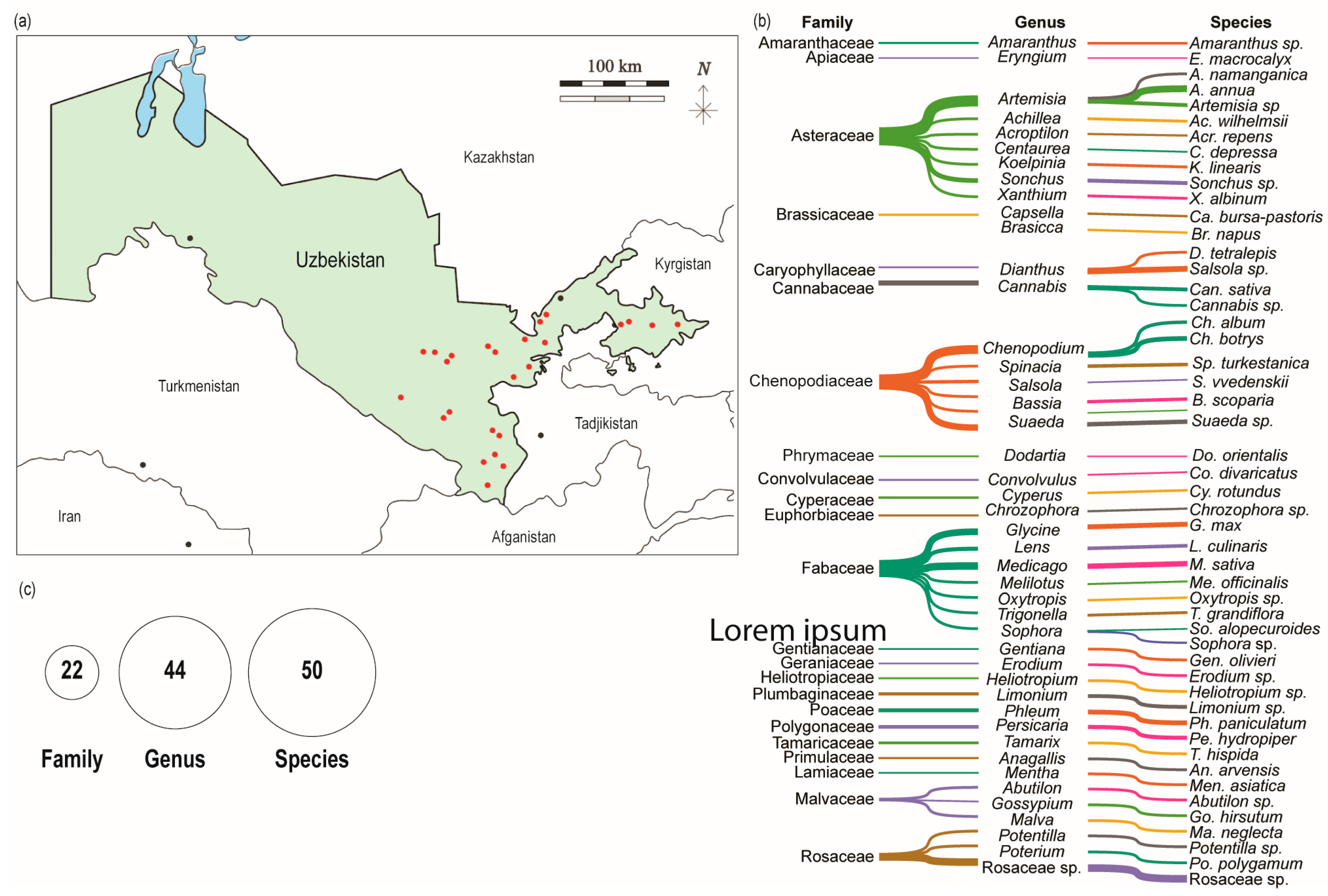
3.1. Research Site and Plant Collection

To explore the competition between phytopathogenic fungi and plant-associated bacteria, we utilized a range of molecular, microbiological, and analytical techniques. A stepwise approach, based on the method by Aytenov, Bozorov, Zhang, Samadiy, Muhammadova, Isokulov, Murodova, Zakirova, Chinikulov and Sherimbetov [22], was used to identify the bacteria and screen them for antifungal properties against various phytopathogenic fungi, as well as for other relevant activities, as illustrated in Figure 1.
Following this workflow, stem (twig) and root samples were collected from different regions of the country, and pure cultures were isolated from each plant part. Each bacterial isolate was then tested for antifungal activity against six selected fungi. The bacteria that exhibited antagonistic effects were further subjected to molecular identification. Finally, the bacteria showing antifungal activity were assessed for additional enzymatic activities.
A total of fifty randomly selected plant species, representing families such as Amaranthaceae, Apiaceae, Asteraceae, Brassicaceae, Caryophyllaceae, Cannabaceae, Chenopodiaceae, Phrymaceae, Convolvulaceae, Cyperaceae, Euphorbiaceae, Fabaceae, Geraniaceae, Gentianaceae, Heliotropiaceae, Plumbaginaceae, Poaceae, Polygonaceae, Tamaricaceae, Primulaceae, Lamiaceae, Malvaceae, and Rosaceae, were collected from nine regions of Uzbekistan: Andijan, Jizzakh, Namangan, Navoiy, Kashkadarya, Samarkand, Surkhandarya, Sirdarya, and Tashkent (Figure 2a,b; Table 1). The elevation of the sampling sites ranged from 241 to 1702 m above sea level, reflecting a diversity of ecological environments (Table 1). The collected species were classified into 21 families and 44 genera, as shown in Figure 2c.

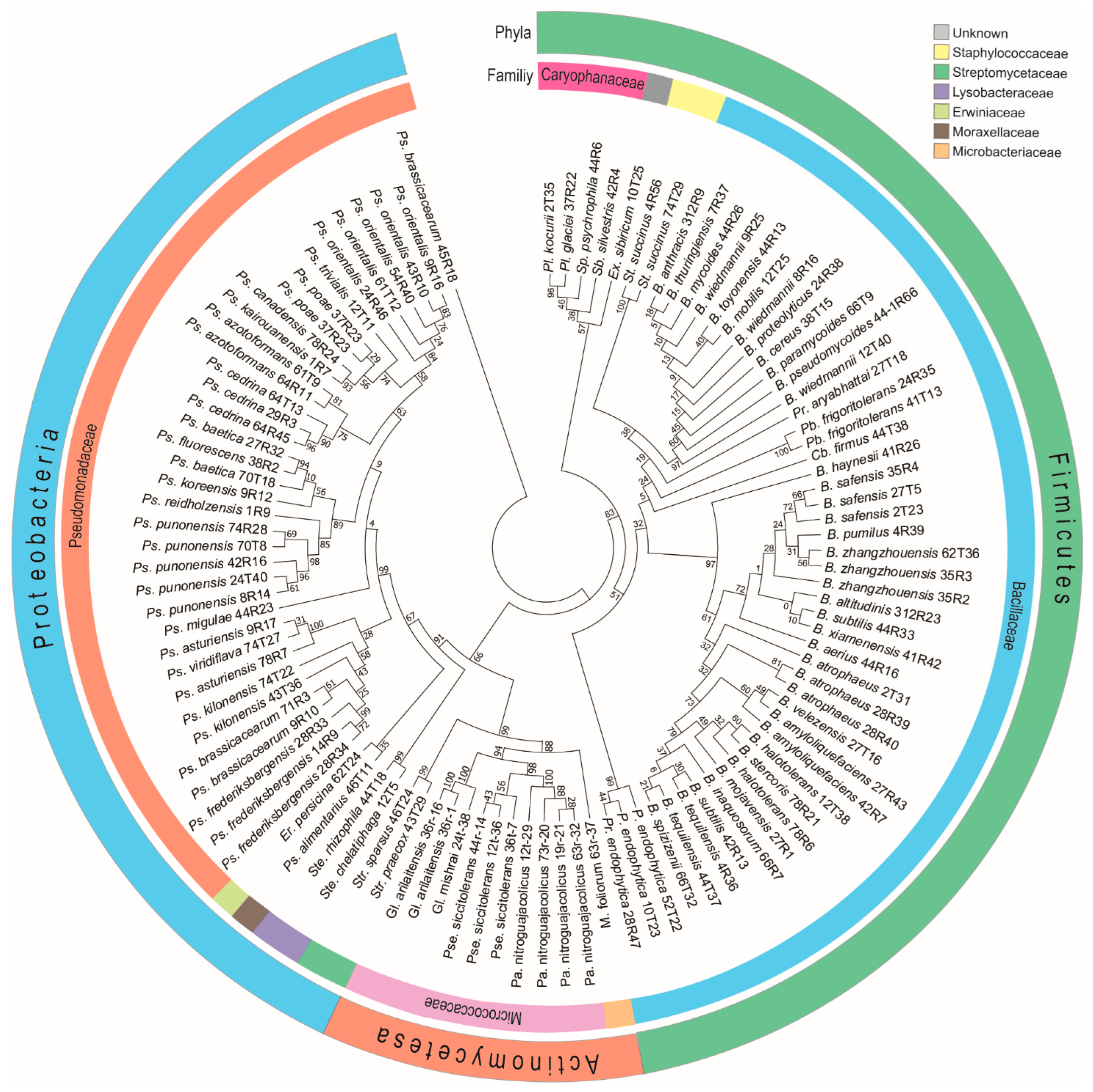
3.2. Identification of Cultivable Plant Associated Antagonistic Bacteria

Bacterial colonies were randomly selected from various plant parts. Pure isolates were obtained from both the phyllosphere and rhizosphere of different plant species. In total, 3355 bacterial isolates were obtained, with 1896 derived from roots and 1459 from shoots (Table 2). Using a co-cultivation method, 432 out of the 3355 isolates exhibited antifungal activity against six fungal pathogens in vitro (Figure 3). The antagonistic bacterial isolates were subjected to molecular identification through analysis of the 16S rDNA gene fragment. Sequences obtained from these bacteria were compared against publicly available databases using BLAST 1.4.0 searches in GenBank. Analysis revealed that some antagonistic bacterial isolates were capable of inhibiting up to five different pathogenic fungi (Figure 3b). Among the tested fungi, R. solani was the most susceptible to bacterial inhibition, whereas A. terreus and F. solani were the most tolerant (Figure 3b). The results reveal that the most frequently identified bacterial genera were Bacillus and Pseudomonas. Several individual strains from these genera were capable of inhibiting up to five fungal pathogens. For instance, B. atrophaeus 66T1 and B. velezensis 27T16 showed antagonistic activity against five fungal pathogens, while B. atrophaeus 28R2 inhibited four. Among Pseudomonas species, Ps. orientalis 54R40 was effective against five fungal pathogens, and Ps. cedrina 64R45 inhibited four.

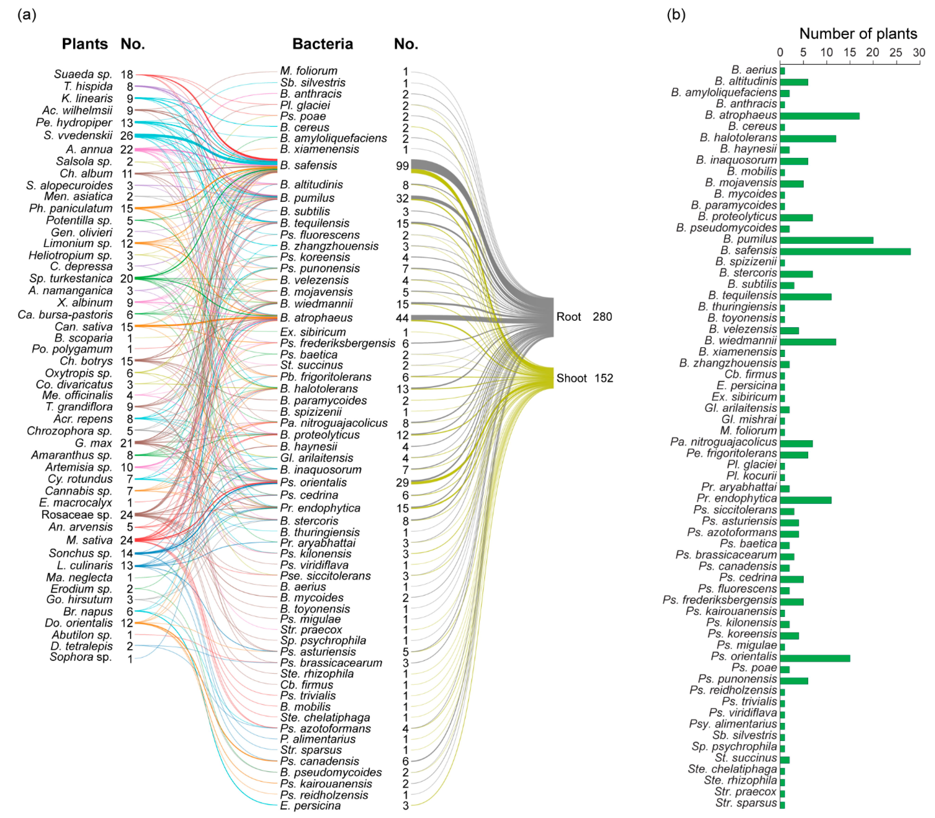
3.3. Diversity of Antagonistic Bacteria

Out of the 3328 bacterial isolates, only 432 demonstrated antagonistic activity against various pathogenic fungi. Genomic DNA was extracted individually from each of these isolates, and the 16S rRNA gene region was amplified for preliminary identification. Sequencing was performed using the traditional Sanger method. The resulting sequences were analyzed to determine the phylogenetic origin of the endophytic bacteria (Figure 4). A total of 65 bacterial species were identified, spanning three phyla—Firmicutes, Proteobacteria, and Actinobacteria; three classes—Bacilli, Gammaproteobacteria, and Actinomycetia; and seven orders—Micrococcales, Kitasatosporales, Bacillales, Enterobacterales, Lysobacterales, Moraxellales, and Pseudomonadales. These were further grouped into ten families (Microbacteriaceae, Micrococcaceae, Streptomycetaceae, Bacillales, Caryophanaceae, Staphylococcaceae, Erwiniaceae, Lysobacteraceae, Moraxellaceae, and Pseudomonadaceae. In total, 18 genera were represented among the antagonistic isolates, including Bacillus, Cytobacillus, Erwinia, Exiguobacterium, Glutamicibacter, Microbacterium, Paenarthrobacter, Peribacillus, Planococcus, Priestia, Pseudarthrobacter, Pseudomonas, Psychrobacter, Solibacillus, Sporosarcina, Staphylococcus, Stenotrophomonas, and Streptomyces (Table S1).
Phylogenetic analysis of the sequenced bacterial species demonstrated that the three largest clades that were most dominant were Firmicutes followed by Proteobacteria and Actinomycetes. The Bacillaceae family was dominant, followed by Pseudomonadaceae, Caryophanaceae, Micrococcaceae, Streptomycetaceae, Staphylococcaceae, Lysobacteraceae, Microbacteriaceae, Erwiniaceae, Moraxellaceae, and an unknown family (Figure 4).
The analysis of plant sources revealed that the majority of antagonistic bacterial isolates are associated with S. vvedenskii, which harbored 26 bacteria antagonists. This was followed by M. sativa (25), Rosaceae sp. (24), A. annua (22), G. max (21), Sp. turkestanica (20), Suaeda sp. (18), Can. sativa (15), Ch. botrys (15), Pa. paniculatum (15), Sonchus sp. (15), L. culinaris (13), Pe. hydropiper (13), Do. orientalis (12), Limonium sp. (12), Ch. album (11), Artemisia sp. (10), Ac. wilhelmsii (9), K. linearis (9), T. grandiflora (9), X. albinum (9), Acr. repens (8), Amaranthus sp. (8), T. hispida (8), Cannabis sp. (7), Cy. rotundus (7), Br. napus (6), Ca. bursa-pastoris (6), Oxytropis sp. (6), An. arvensis (5), Chrozophora sp. (5), Potentilla sp. (5), Me. officinalis (4), A. namanganica (3), C. depressa (3), Co. divaricatus (3), Go. hirsutum (3), Heliotropium sp. (3), S. alopecuroides (3), Abutilon sp. (2), D. tetralepis (2), Erodium sp. (2), Gen. olivieri (2), Men. asiatica (2), Salsola sp. (2), B. scoparia (1), E. macrocalyx (1), Po. polygamum (1), and Sophora sp. (1) (Figure 5). It appears that the majority of antagonistic bacteria were found in the roots compared with the shoots. Antagonistic bacteria were detected in the shoots of only nine plant species, whereas thirteen were identified in the roots (Figure 5).
Analysis of the distribution across plant organs revealed 280 isolates from the roots and 152 from the shoots, indicating that the roots harbor a higher abundance of antagonistic bacteria. Among the antagonistic bacteria, 21 species—including Bacillus aerius, B. amyloliquefaciens, B. anthracis, B. haynesii, B. mycoides, B. subtilis, B. thuringiensis, B. toyonensis, B. xiamenensis, M. foliorum, Pl. glaciei, Ps. brassicacearum, Ps. canadensis, Ps. frederiksbergensis, Ps. kairouanensis, Ps. koreensis, Ps. migulae, Ps. poae, Ps. reidholzensis, Sp. silvestris, and Sp. psychrophila—were exclusively found in roots. In contrast, 17 species—including Bacillus cereus, B. mobilis, B. paramycoides, B. spizizenii, Cohnella firmus, Erwinia persicina, Ex. sibiricum, Gl. mishrai, Pl. kocurii, Ps. kilonensis, Ps. trivialis, Ps. viridiflava, Psy. alimentarius, Ste. chelatiphaga, Ste. rhizophila, Str. praecox, and Str. sparsus—were identified in shoots (Figure 6a). Apparently, the highest number of antagonistic bacteria (26 in total) was found in S. vvedenskii, which harbored nine different bacterial species. In contrast, the lowest number—only one bacterial species—was observed in B. scoparia, E. macrocalyx, M. neglecta, Po. polygamum, and Sophora sp. (Figure 6a,b). B. safensis was the most widely distributed bacterial species, found in association with 27 different plant species (Figure 6b). Fourteen plant species each harbored distinct bacterial species not found in others (Figure 6a). Most bacterial species, according to plant family, also appeared to be distinct. For example, Ps. migulae, Ste. rhizophila, B. toyonensis, Cb. firmus, B. mycoides, B. haynesii, and B. aerius were found to be associated with Rosaceae species. B. mobilis, Ps. trivialis, Ste. chelatiphaga, E. persicina, and Ps. viridiflava were found in Fabaceae. Pl. kocurii, Pl. glaciei, B. paramycoides, and B. spizizenii were detected in Chenopodiaceae, while B. anthracis, B. cereus, Gl. mishrai, Str. praecox, Ps. kairouanensis, and Ps. reidholzensis were found to be associated with Phrymaceae. In Asteraceae, M. foliorum and Str. sparsus were identified. Additionally, B. thuringiensis was specific to Cyperaceae, and Ex. sibiricum to Brassicaceae.

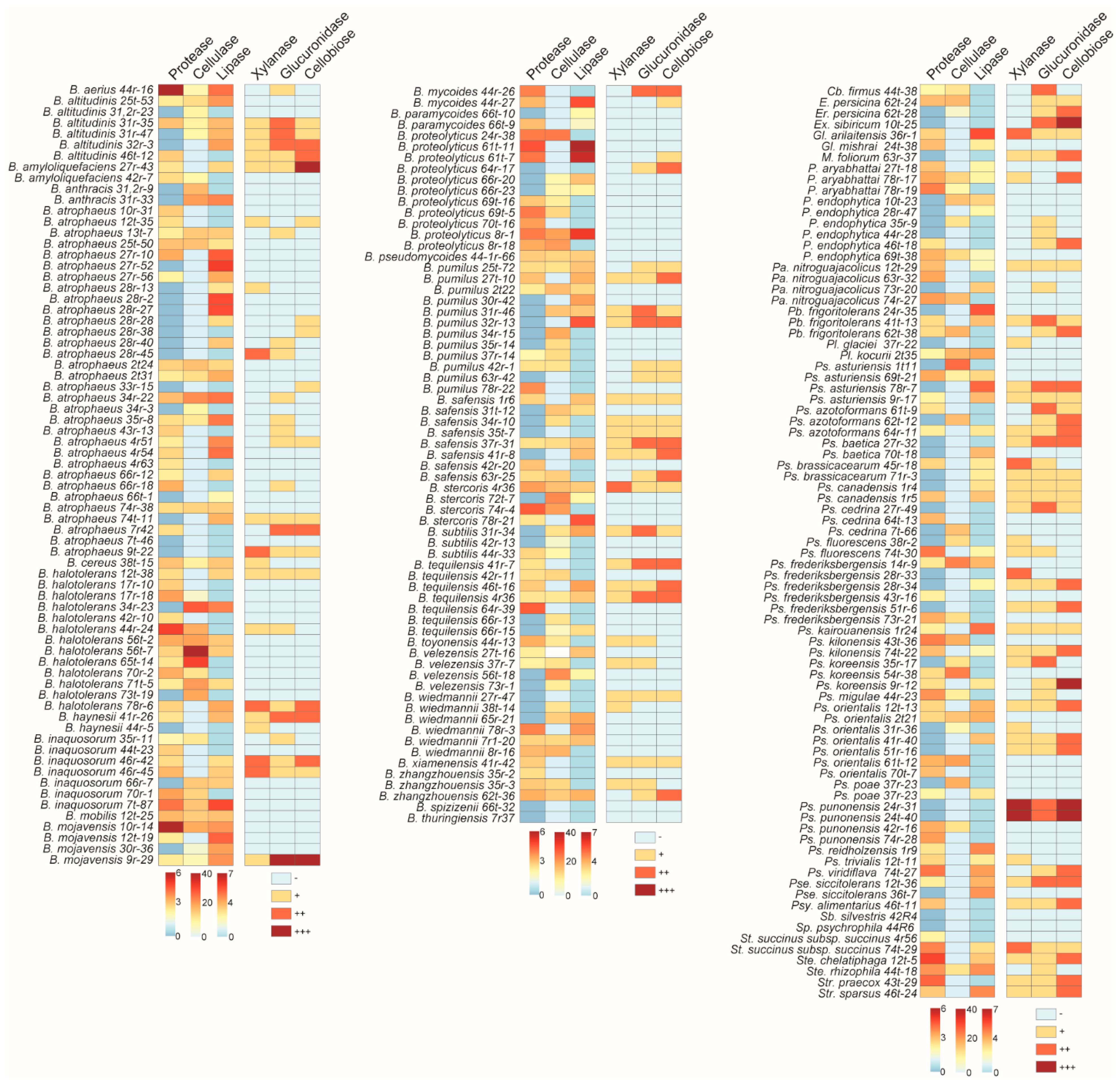
3.4. Determination of the Enzymatic Activities of the Antagonistic Bacteria

The enzymatic properties of antagonistic bacteria isolated from plant roots and shoots were determined, with a focus on their ability to break down cell wall components, as well as degrade lipids and proteins. Among the bacterial genera, Bacillus species exhibited the strongest protease, cellulase, and lipase activities, surpassing other genera in these enzymatic functions. Interestingly, the Bacillus strains demonstrated variability in their enzymatic degradation capabilities. For instance, B. mojavensis 10r-14 displayed the highest protease activity among the strains tested, highlighting its potential as a robust proteolytic agent. On the other hand, B. halotolerans 56t-7 showed significant cellulase activity, suggesting its ability to break down complex polysaccharides, while B. atrophaeus 27r-52 was particularly effective in lipase activity, which may aid in lipid degradation (Figure 7).
In contrast, Pseudomonas species were more effective in exhibiting xylanase, glucanase, and cellobiase activities compared with other bacterial genera. This indicates their potential in breaking down lignocellulosic materials, an important characteristic for bioremediation and plant protection. Similar to Bacillus species, Pseudomonas strains also exhibited varying levels of enzymatic activity, indicating differences in their metabolic capabilities. For example, Ps. punonensis 24r-31 and Ps. punonensis 24t-40 showed particularly strong enzymatic activity across multiple enzymes, suggesting a broad range of degradative functions. Additionally, Ps. koreensis 9r-12 demonstrated superior cellobiase activity when compared with other Pseudomonas strains, highlighting its specific role in breaking down cellulose (Figure 7).
Among all the antagonistic bacteria tested, B. mojavensis 9r-29 stood out by demonstrating all six enzymatic activities—protease, cellulase, lipase, xylanase, glucanase, and cellobiase. This versatile enzyme profile positions B. mojavensis 9r-29 as a particularly promising candidate for further studies in biocontrol and agricultural applications, where multiple forms of enzymatic degradation are beneficial for plant protection and growth promotion.

4. Discussion

The ecological benefits of antagonistic bacteria extend beyond crop productivity, as they contribute to biodiversity, soil fertility, and the resilience of agroecosystems against climate stress and disease outbreaks. As such, antagonistic bacteria represent a valuable resource for developing eco-friendly strategies in modern agriculture and environmental management. The diversity of plant species sampled across nine regions in Uzbekistan, from 241 to 1702 m above sea level, highlights the vast ecological variation within the country. A wide range of plant families were represented, including Amaranthaceae, Apiaceae, Asteraceae, and many others, which emphasizes the extensive potential sources for isolating plant-associated bacteria. The distribution of bacterial isolates from both the phyllosphere and rhizosphere further underlines the significance of plant-associated microorganisms in diverse ecological niches. The finding that roots generally harbor a higher abundance of antagonistic bacteria is consistent with previous research, which suggests that the rhizosphere provides a more nutrient-rich environment conducive to microbial growth and activity [30]. Furthermore, the study also highlights the ecological and phylogenetic diversity of antagonistic bacteria across different plant species and families. Of the 50 identified antagonistic bacterial strains, 37 were found to be associated with more than two plant hosts. This suggests that many bacterial species across the region are capable of colonizing multiple plant types simultaneously. In contrast, some strains exhibited host specificity. For instance, certain bacterial species were found to be specific to particular plant families, such as Ps. migulae and B. aerius, which were found to be associated with Rosaceae species. This observation suggests that plant species and their associated bacteria may have co-evolved, with certain bacterial strains becoming highly specialized for specific plant hosts. This specialized association between plant species and their microbial communities could have important implications for understanding the dynamics of plant–microbe interactions and for developing targeted biocontrol strategies.
In total, 432 bacterial isolates exhibited antifungal activity against six phytopathogenic fungi, with some bacteria showing the ability to inhibit up to five different fungi. Notably, R. solani was the most susceptible to bacterial inhibition, while As. terreus and F. solani showed greater tolerance. This differential inhibition suggests that certain bacterial strains possess a broader spectrum of antifungal activity, which could be exploited for developing biocontrol agents. The molecular identification of the antagonistic isolates revealed a rich diversity of bacterial species, including those from the Firmicutes, Proteobacteria, and Actinobacteria phyla, with Bacillus, Pseudomonas, and Streptomyces being the most commonly encountered genera. The dominance of Bacillus species in this study is particularly noteworthy, as these bacteria are well-known for their antagonistic properties against plant pathogens [22,31,32,33,34]. The wide range of antibacterial compounds synthesized by Bacillus species makes them excellent candidates for use as biological control agents (BCAs) in agriculture. Their key antimicrobial metabolites include lipopeptides, siderophores, non-ribosomally synthesized peptides (NRSPs), polyketides, and bacteriocins [6]. Among the Bacillus species tested, B. atrophaeus exhibited the strongest antifungal activity against most fungal pathogens, followed by B. safensis, B. velezensis, B. pumilus, and B. proteolyticus, with most of these producing antifungal compounds such as iturin A, safencin E, cyclo-(D-phenylalanyl-D-prolyl), fengycin, surfactin, bacillaene, macrolactin, difficidin, bacilysin and siderophores (bacillibactin) [35,36,37]. These Bacillus strains have been previously reported as promising biological control agents [38,39,40,41,42]. Notably, the B. atrophaeus 66T1 strain demonstrated strong inhibitory activity against several Fusarium pathogens, including F. graminearum, F. solani, and F. culmorum, consistent with previous studies [43,44]. Guo et al. demonstrated the efficient inhibition of F. pseudograminearum, which causes crown rot in wheat, with the disease severity index reduced 76% in a greenhouse pot trial. Since then, the Fusarium species is considered to be composed of destructive pathogens in agriculture, and eliminating this pathogen by biological control agents rather than via chemical fungicides is ecologically friendly. Pseudomonas species were the second most frequently identified antagonistic microbes associated with plants. For instance, Ps. orientalis strain 54R40 exhibited antifungal activity, effectively inhibiting five different fungal pathogens. Another strain, P. orientalis F9, is known for its antibacterial properties against Er. amylovora, the causative agent of fire blight in apples. This strain produces a range of antibiotics, including safracin and phenazine. Notably, phenazine and its acidic or amidated derivatives are recognized for their potent antifungal activity [29,34,45,46,47]. Another species, Ps. cedrina strain 64R45, also exhibited strong antifungal activity, though with a distinct inhibition spectrum. According to Goryluk-Salmonowicz, et al. [48], P. cedrina demonstrated significant antifungal effects among thirteen medicinal plant species. The same study also identified other potent antifungal endophytes, including P. azotoformans, B. subtilis, and E. persicina, all of which are referenced in the current study.
One of the most interesting findings of this study was the variation in the enzymatic activities of the antagonistic bacteria. Bacillus species exhibited strong protease, cellulase, and lipase activities, surpassing other genera in these enzymatic functions. These activities are essential for breaking down complex macromolecules in the plant cell wall, lipids, and proteins, making Bacillus species particularly effective in both biocontrol and plant growth promotion [49,50,51]. Furthermore, certain strains of Bacillus demonstrated variability in their enzymatic degradation capabilities, suggesting that different strains might be specialized for distinct types of macromolecular degradation. It has been reported to be an enzymatically diverse bacteria [52,53]. Pseudomonas species, on the other hand, are particularly proficient at breaking down lignocellulosic materials, with notable xylanase, glucanase, and cellobiase activities. This ability positions Pseudomonas strains as valuable candidates for bioremediation and sustainable agricultural practices.
In conclusion, our study provides valuable insights into the diversity and antifungal potential of plant-associated bacteria. The findings suggest that these bacteria, particularly from genera such as Bacillus (B. atrophaeus, B. safensis, B. velezensis, B. pumilus, and B. proteolyticus) and Pseudomonas (Ps. sedrina, Ps. kilonensis, and Ps. orientalis), hold significant promise for biological control applications. Their diverse enzymatic profiles further enhance their potential in various agricultural and bioremediation practices. The discovery of the B. mojavensis 9r-29 strain as a particularly versatile bacterium capable of demonstrating all six tested enzymatic activities—protease, cellulase, lipase, xylanase, glucanase, and cellobiase—makes it an exceptional candidate for future bioremediation practices. The broad enzymatic profile of this strain suggests that it could play a significant role in degrading a wide range of plant pathogens and promoting plant health, making it a promising candidate for agricultural applications. Future studies should focus on the in-depth exploration of the mechanisms of action of these bacteria, as well as on their interactions with phytopathogens and their efficacy in field conditions.

Supplementary Materials

The following supporting information can be downloaded at: https://www.mdpi.com/article/10.3390/microorganisms13051161/s1, Table S1: Identified bacterial isolates from plant species by 16S RNA sequence analysis and their taxonomic status.

Author Contributions

M.K.S. and T.A.B. wrote the manuscript and prepared all of the tables and figures. I.S.A., and T.A.B. collected plant samples. M.K.S., D.A.M., I.S.A., and M.Z.I., contributed to bacterial isolation and enzymatic and co-cultivation analyses. I.S.A., M.K.S., and T.A.B. carried out molecular identification. O.S.A., S.M.M., F.A.M., B.O.O., and S.K.M. assisted in the preparation procedures for microbiological and chemical work. I.S.A., T.A.B. and D.Z. contributed to the writing of the manuscript and corrections. T.A.B. and D.Z. supervised the experiment and manuscript writing. All authors reviewed the manuscript. All authors have read and agreed to the published version of the manuscript.

Funding

This study was funded by a fundamental research grant of the Ministry of Innovative Development of Uzbekistan (Grant number: PZ-20200929166).

Institutional Review Board Statement

Not applicable.

Informed Consent Statement

Not applicable.

Data Availability Statement

The authors declare that the experimental data published in this paper. will be made accessible upon request for interested readers. All 16S rRNA gene sequences of the new strains can be found under accession numbers: starting from PV653521 to PV653563; and PV653596 to PV653651.

Acknowledgments

We appreciate the support provided by Sirojbek Isokulov from the Institute of Genetics and Experimental Biology of Plants, Uzbek Academy of Sciences, for his assistance with timely chemical ordering, and Bakhodir Chinkukulov for plant sampling.

Conflicts of Interest

The authors declare that they have no competing interests.

References

  1. Fisher, M.C.; Gurr, S.J.; Cuomo, C.A.; Blehert, D.S.; Jin, H.; Stukenbrock, E.H.; Stajich, J.E.; Kahmann, R.; Boone, C.; Denning, D.W.; et al. Threats Posed by the Fungal Kingdom to Humans, Wildlife, and Agriculture. mBio 2020, 11, e00449-20. [Google Scholar] [CrossRef] [PubMed]
  2. Wei, X.; Xie, B.; Wan, C.; Song, R.; Zhong, W.; Xin, S.; Song, K.D. Enhancing soil health and plant growth through microbial fertilizers: Mechanisms, benefits, and sustainable agricultural practices. Agronomy 2024, 14, 609. [Google Scholar] [CrossRef]
  3. Bonaterra, A.; Badosa, E.; Daranas, N.; Francés, J.; Roselló, G.; Montesinos, E. Bacteria as biological control agents of plant diseases. Microorganisms 2022, 10, 1759. [Google Scholar] [CrossRef]
  4. Chepsergon, J.; Moleleki, L.N. Rhizosphere bacterial interactions and impact on plant health. Curr. Opin. Microbiol. 2023, 73, 102297. [Google Scholar] [CrossRef]
  5. Wang, C.; Kuzyakov, Y. Mechanisms and implications of bacterial-fungal competition for soil resources. ISME J. 2024, 18, wrae073. [Google Scholar] [CrossRef]
  6. Compant, S.; Duffy, B.; Nowak, J.; Clement, C.; Barka, E.A. Use of plant growth-promoting bacteria for biocontrol of plant diseases: Principles, mechanisms of action, and future prospects. Appl. Environ. Microbiol. 2005, 71, 4951–4959. [Google Scholar] [CrossRef]
  7. Kohl, J.; Kolnaar, R.; Ravensberg, W.J. Mode of Action of Microbial Biological Control Agents Against Plant Diseases: Relevance Beyond Efficacy. Front. Plant Sci. 2019, 10, 845. [Google Scholar] [CrossRef]
  8. Beneduzi, A.; Ambrosini, A.; Passaglia, L.M. Plant growth-promoting rhizobacteria (PGPR): Their potential as antagonists and biocontrol agents. Genet. Mol. Biol. 2012, 35, 1044–1051. [Google Scholar] [CrossRef]
  9. Bakker, P.A.; Doornbos, R.F.; Zamioudis, C.; Berendsen, R.L.; Pieterse, C.M. Induced systemic resistance and the rhizosphere microbiome. Plant Pathol. J. 2013, 29, 136–143. [Google Scholar] [CrossRef]
  10. Pieterse, C.M.; Poelman, E.H.; Van Wees, S.C.; Dicke, M. Induced plant responses to microbes and insects. Front. Plant Sci. 2013, 4, 475. [Google Scholar] [CrossRef]
  11. Rajkumar, M.; Lee, K.J.; Freitas, H. Effects of chitin and salicylic acid on biological control activity of Pseudomonas spp. against damping off of pepper. S. Afr. J. Bot. 2008, 74, 268–273. [Google Scholar] [CrossRef]
  12. Srikamwang, C.; Onsa, N.E.; Sunanta, P.; Sangta, J.; Chanway, C.P.; Thanakkasaranee, S.; Sommano, S.R. Role of Microbial Volatile Organic Compounds in Promoting Plant Growth and Disease Resistance in Horticultural Production. Plant Signal Behav. 2023, 18, 2227440. [Google Scholar] [CrossRef] [PubMed]
  13. He, D.C.; He, M.H.; Amalin, D.M.; Liu, W.; Alvindia, D.G.; Zhan, J. Biological Control of Plant Diseases: An Evolutionary and Eco-Economic Consideration. Pathogens 2021, 10, 1311. [Google Scholar] [CrossRef] [PubMed]
  14. Ayaz, M.; Li, C.H.; Ali, Q.; Zhao, W.; Chi, Y.K.; Shafiq, M.; Ali, F.; Yu, X.Y.; Yu, Q.; Zhao, J.T.; et al. Bacterial and Fungal Biocontrol Agents for Plant Disease Protection: Journey from Lab to Field, Current Status, Challenges, and Global Perspectives. Molecules 2023, 28, 6735. [Google Scholar] [CrossRef]
  15. Haddoudi, I.; Cabrefiga, J.; Mora, I.; Mhadhbi, H.; Montesinos, E.; Mrabet, M. Biological control of Fusarium wilt caused by Fusarium equiseti in Vicia faba with broad spectrum antifungal plant-associated Bacillus spp. Biol. Control 2021, 160, 104671. [Google Scholar] [CrossRef]
  16. Alattas, H.; Glick, B.R.; Murphy, D.V.; Scott, C. Harnessing Pseudomonas spp. for sustainable plant crop protection. Front. Microbiol. 2024, 15, 1485197. [Google Scholar] [CrossRef]
  17. Khan, S.; Srivastava, S.; Karnwal, A.; Malik, T. Streptomyces as a promising biological control agents for plant pathogens. Front. Microbiol. 2023, 14, 1285543. [Google Scholar] [CrossRef]
  18. Etesami, H.; Jeong, B.R.; Glick, B.R. Biocontrol of plant diseases by Bacillus spp. Physiol. Mol. Plant Pathol. 2023, 126, 02048. [Google Scholar] [CrossRef]
  19. Lahlali, R.; Ezrari, S.; Radouane, N.; Kenfaoui, J.; Esmaeel, Q.; El Hamss, H.; Belabess, Z.; Barka, E.A. Biological Control of Plant Pathogens: A Global Perspective. Microorganisms 2022, 10, 596. [Google Scholar] [CrossRef]
  20. Ji, C.; Chen, Z.; Kong, X.; Xin, Z.; Sun, F.; Xing, J.; Li, C.; Li, K.; Liang, Z.; Cao, H. Biocontrol and plant growth promotion by combined Bacillus spp. inoculation affecting pathogen and AMF communities in the wheat rhizosphere at low salt stress conditions. Front. Plant Sci. 2022, 13, 1043171. [Google Scholar] [CrossRef]
  21. Peel, M.F.; Finlayson, B.L.; McMahon, B.T. Updated world map of the Köppen-Geiger climate classification. Hydrol. Earth Syst. Sci. Discuss. 2007, 4, 439–473. [Google Scholar] [CrossRef]
  22. Aytenov, I.S.; Bozorov, T.A.; Zhang, D.; Samadiy, S.A.; Muhammadova, D.A.; Isokulov, M.Z.; Murodova, S.M.; Zakirova, O.R.; Chinikulov, B.K.; Sherimbetov, A.G. Uncovering the Antifungal Potential of Plant-Associated Cultivable Bacteria from the Aral Sea Region against Phytopathogenic Fungi. Pathogens 2024, 13, 585. [Google Scholar] [CrossRef] [PubMed]
  23. Alenezi, F.N.; Rekik, I.; Bełka, M.; Ibrahim, A.F.; Luptakova, L.; Jaspars, M.; Woodward, S.; Belbahri, L. Strain-level diversity of secondary metabolism in the biocontrol species Aneurinibacillus migulanus. Microbiol. Res. 2016, 182, 116–124. [Google Scholar] [CrossRef]
  24. Bozorov, T.A.; Rasulov, B.A.; Zhang, D. Characterization of the gut microbiota of invasive Agrilus mali Matsumara (Coleoptera: Buprestidae) using high-throughput sequencing: Uncovering plant cell-wall degrading bacteria. Sci. Rep. 2019, 9, 4923. [Google Scholar] [CrossRef]
  25. Vargas-Asensio, G.; Pinto-Tomas, A.; Rivera, B.; Hernandez, M.; Hernandez, C.; Soto-Montero, S.; Murillo, C.; Sherman, D.H.; Tamayo-Castillo, G. Uncovering the cultivable microbial diversity of costa rican beetles and its ability to break down plant cell wall components. PLoS ONE 2014, 9, e113303. [Google Scholar] [CrossRef]
  26. Teather, R.M.; Wood, P.J. Use of Congo red-polysaccharide interactions in enumeration and characterization of cellulolytic bacteria from the bovine rumen. Appl. Environ. Microbiol. 1982, 43, 777–780. [Google Scholar] [CrossRef]
  27. Mayerhofer, H.J.; Marshall, R.T.; White, C.H.; Lu, M. Characterization of a heat-stable protease of Pseudomonas fluorescens P26. Appl. Microbiol. 1973, 25, 44–48. [Google Scholar] [CrossRef]
  28. Kumar, D.; Kumar, L.; Nagar, S.; Raina, C.; Parshad, R.; Gupta, V.K. Screening, isolation and production of lipase/esterase producing Bacillus sp. strain DVL2 and its potential evaluation in esterification and resolution reactions. Arch. Appl. Sci. Res. 2012, 4, 1763–1770. [Google Scholar]
  29. Bozorov, T.A.; Toshmatov, Z.O.; Kahar, G.; Zhang, D.; Shao, H.; Gafforov, Y. Wild apple-associated fungi and bacteria compete to colonize the larval gut of an invasive wood-borer Agrilus mali in Tianshan forests. Front. Microbiol. 2021, 12, 743831. [Google Scholar] [CrossRef]
  30. Dong, C.J.; Wang, L.L.; Li, Q.; Shang, Q.M. Bacterial communities in the rhizosphere, phyllosphere and endosphere of tomato plants. PLoS ONE 2019, 14, e0223847. [Google Scholar] [CrossRef]
  31. Cao, Y.; Pi, H.; Chandrangsu, P.; Li, Y.; Wang, Y.; Zhou, H.; Xiong, H.; Helmann, J.D.; Cai, Y. Antagonism of Two Plant-Growth Promoting Bacillus velezensis Isolates Against Ralstonia solanacearum and Fusarium oxysporum. Sci. Rep. 2018, 8, 4360. [Google Scholar] [CrossRef] [PubMed]
  32. Abbas, A.; Khan, S.U.; Khan, W.U.; Saleh, T.A.; Khan, M.H.U.; Ullah, S.; Ali, A.; Ikram, M. Antagonist effects of strains of Bacillus spp. against Rhizoctonia solani for their protection against several plant diseases: Alternatives to chemical pesticides. Comptes Rendus Biol. 2019, 342, 124–135. [Google Scholar] [CrossRef] [PubMed]
  33. Zalila-Kolsi, I.; Ben Mahmoud, A.; Ali, H.; Sellami, S.; Nasfi, Z.; Tounsi, S.; Jamoussi, K. Antagonist effects of Bacillus spp. strains against Fusarium graminearum for protection of durum wheat (Triticum turgidum L. subsp. durum). Microbiol. Res. 2016, 192, 148–158. [Google Scholar] [CrossRef]
  34. Bozorov, T.A.; Toshmatov, Z.O.; Kahar, G.; Muhammad, S.M.; Liu, X.; Zhang, D.; Aytenov, I.S.; Turakulov, K.S. Uncovering the antifungal activities of wild apple-associated bacteria against two canker-causing fungi, Cytospora mali and C. parasitica. Sci. Rep. 2024, 14, 6307. [Google Scholar] [CrossRef]
  35. Kamilari, E.; O’Connor, P.M.; Farias, F.M.d.; Johnson, C.N.; Buttimer, C.; Deliephan, A.; Hill, D.; Fursenko, O.; Wiese, J.; Stanton, C.; et al. Bacillus safensis APC 4099 has broad-spectrum antimicrobial activity against both bacteria and fungi and produces several antimicrobial peptides, including the novel circular bacteriocin safencin E. Appl. Environ. Microbiol. 2024, 91, e01942-24. [Google Scholar] [CrossRef]
  36. Rong, S.; Xu, H.; Li, L.; Chen, R.; Gao, X.; Xu, Z. Antifungal activity of endophytic Bacillus safensis B21 and its potential application as a biopesticide to control rice blast. Pestic. Biochem. Physiol. 2020, 162, 69–77. [Google Scholar] [CrossRef]
  37. Bidima, M.G.S.; Chtaina, N.; Ezzahiri, B.; El Guilli, M.; Barakat, I.; El Kamli, T. Antifungal activity of bioactive compounds produced by the endophyte Bacillus velezensis NC318 against the soil borne pathogen Sclerotium rolfsii Sacc. J. Plant Prot. Res. 2022, 62, 326–333. [Google Scholar] [CrossRef]
  38. Abdelkhalek, A.; Behiry, S.I.; Al-Askar, A.A. Bacillus velezensis PEA1 inhibits Fusarium oxysporum growth and induces systemic resistance to cucumber mosaic virus. Agronomy 2020, 10, 1312. [Google Scholar] [CrossRef]
  39. Wei, J.; Zhao, J.; Suo, M.; Wu, H.; Zhao, M.; Yang, H. Biocontrol mechanisms of Bacillus velezensis against Fusarium oxysporum from Panax ginseng. Biol. Control 2023, 182, 105222. [Google Scholar] [CrossRef]
  40. Knežević, M.; Dervišević, M.; Jovković, M.; Jevđenović, G.; Maksimović, J.; Buntić, A. Versatile role of Bacillus velezensis: Biocontrol of Fusarium poae and wireworms and barley plant growth promotion. Biol. Control 2025, 206, 105789. [Google Scholar] [CrossRef]
  41. Dobrzynski, J.; Jakubowska, Z.; Kulkova, I.; Kowalczyk, P.; Kramkowski, K. Biocontrol of fungal phytopathogens by Bacillus pumilus. Front. Microbiol. 2023, 14, 1194606. [Google Scholar] [CrossRef] [PubMed]
  42. Yang, P.; Zhao, Z.Z.; Fan, J.B.; Liang, Y.P.; Bernier, M.C.; Gao, Y.; Zhao, L.J.; Opiyo, S.O.; Xia, Y. Bacillus proteolyticus OSUB18 triggers induced systemic resistance against bacterial and fungal pathogens in Arabidopsis. Front. Plant Sci. 2023, 14, 1078100. [Google Scholar] [CrossRef] [PubMed]
  43. Guo, S.; Tariq, A.; Liao, J.; Yang, A.; Jiang, X.; Yin, Y.; Shi, Y.; Li, C.; Pan, J.; Han, D.; et al. Identification and antagonistic potential of Bacillus atrophaeus against wheat crown rot caused by Fusarium pseudograminearum. Agronomy 2024, 14, 2135. [Google Scholar] [CrossRef]
  44. Harba, M.; Jawhar, M.; Arabi, M.I.E. In vitro antagonistic activity of diverse Bacillus species against Fusarium culmorum and F. solani pathogens. Open Agric. J. 2020, 14, 157–163. [Google Scholar] [CrossRef]
  45. Puopolo, G.; Masi, M.; Raio, A.; Andolfi, A.; Zoina, A.; Cimmino, A.; Evidente, A. Insights on the susceptibility of plant pathogenic fungi to phenazine-1-carboxylic acid and its chemical derivatives. Nat. Prod. Res. 2013, 27, 956–966. [Google Scholar] [CrossRef]
  46. Simionato, A.S.; Navarro, M.O.P.; de Jesus, M.L.A.; Barazetti, A.R.; da Silva, C.S.; Simoes, G.C.; Balbi-Pena, M.I.; de Mello, J.C.P.; Panagio, L.A.; de Almeida, R.S.C.; et al. The effect of phenazine-1-carboxylic acid on mycelial growth of Botrytis cinerea produced by Pseudomonas aeruginosa LV strain. Front. Microbiol. 2017, 8, 1102. [Google Scholar] [CrossRef]
  47. Tupe, S.G.; Kulkarni, R.R.; Shirazi, F.; Sant, D.G.; Joshi, S.P.; Deshpande, M.V. Possible mechanism of antifungal phenazine-1-carboxamide from Pseudomonas sp. against dimorphic fungi Benjaminiella poitrasii and human pathogen Candida albicans. J. Appl. Microbiol. 2015, 118, 39–48. [Google Scholar] [CrossRef]
  48. Goryluk-Salmonowicz, A.; Piorek, M.; Rekosz-Burlaga, H.; Studnicki, M.; Blaszczyk, M. Endophytic Detection in Selected European Herbal Plants. Pol. J. Microbiol. 2016, 65, 369–375. [Google Scholar] [CrossRef]
  49. Zachow, C.; Grosch, R.; Berg, G. Impact of biotic and a-biotic parameters on structure and function of microbial communities living on sclerotia of the soil-borne pathogenic fungus Rhizoctonia solani. Appl. Soil Ecol. 2011, 48, 193–200. [Google Scholar] [CrossRef]
  50. Wang, L.; Huang, J.; Chen, S.; Su, X.; Zhang, X.; Wang, L.; Zhang, W.; Wang, Z.; Zeng, Q.; Wang, Q.; et al. Endogenous cell wall degrading enzyme LytD is important for the biocontrol activity of Bacillus subtilis. Front. Plant Sci. 2024, 15, 1381018. [Google Scholar] [CrossRef]
  51. Petkova, M.; Marcheva, M.; Petrova, A.L.; Slavova, V.; Shilev, S. Plant growth-promoting and biocontrol characteristics of four Bacillus strains and evaluation of their effects on eheat (Tr. aestivum L.). Int. J. Plant Biol. 2025, 16, 1. [Google Scholar] [CrossRef]
  52. Larrea-Murrell, J.A.; Rojas-Badia, M.M.; Garcia-Soto, I.; Romeu-Alvarez, B.; Bacchetti, T.; Gillis, A.; Boltes-Espinola, A.K.; Heydrich-Perez, M.; Lugo-Moya, D.; Mahillon, J. Diversity and enzymatic potentialities of Bacillus sp. strains isolated from a polluted freshwater ecosystem in Cuba. World J. Microbiol. Biotechnol. 2018, 34, 28. [Google Scholar] [CrossRef] [PubMed]
  53. Latorre, J.D.; Hernandez-Velasco, X.; Wolfenden, R.E.; Vicente, J.L.; Wolfenden, A.D.; Menconi, A.; Bielke, L.R.; Hargis, B.M.; Tellez, G. Evaluation and Selection of Bacillus Species Based on Enzyme Production, Antimicrobial Activity, and Biofilm Synthesis as Direct-Fed Microbial Candidates for Poultry. Front. Vet. Sci. 2016, 3, 95. [Google Scholar] [CrossRef]
Figure 1. Research strategy for endophytic molecular biodiversity.
Microorganisms 13 01161 g001
Figure 2. Overview of plant diversity collected from nine regions of Uzbekistan. (a) Map of the Republic of Uzbekistan with locations marked for plant sample collection. (b) Diversity of plant species categorized by family and genus. (c) Distribution of plant species by family and genus.
Microorganisms 13 01161 g002
Figure 3. Antifungal Activity of Bacterial Isolates from Plant Species. (a) Examples of bacterial isolates exhibiting antifungal activity against various phytopathogenic fungi. (b) Heatmap illustrating the antifungal activity of these isolates against six different phytopathogenic fungi. The ‘No’ indicates the number of bacterial isolates capable of inhibiting each pathogenic fungus.
Microorganisms 13 01161 g003
Figure 4. Phylogenic relationship of antagonistic bacteria isolated from plant species. The circular map depicts bacterial phyla and family taxonomic units. The evolutionary history was inferred by using the maximum likelihood method and Tamura–Nei model. The tree with the highest log likelihood (−15,874.16) is shown. The percentage of trees in which the associated taxa clustered together is shown next to the branches.
Microorganisms 13 01161 g004
Figure 5. Bacterial count distribution by plant species.
Microorganisms 13 01161 g005
Figure 6. Distribution of antagonistic bacteria by plant species and plant parts. (a) The distribution of antagonistic bacterial isolates was analyzed according to the plant species and specific plant parts (roots, shoot) from which they were obtained. (b) The data show the distribution of bacterial antagonists based on the number of plant hosts.
Microorganisms 13 01161 g006
Figure 7. Enzymatic activities of the antagonistic bacteria. Protease, lipase, cellulase, xylanase, glucanase, and cellobiase activities were assessed in antagonistic bacterial isolates. Symbols ‘+’, ‘++’, and ‘+++’ indicate low, moderate, and high levels of enzymatic activity, respectively, while ‘−’ denotes the absence of activity.
Microorganisms 13 01161 g007
Table 1. Collection information for plant samples in Uzbekistan.
LocationGPS CoordinatesElevation (m)Plant Species
RegionDistrict
AndijanAndijan
Ulugnor
40°86’53.0 N 72°29′524 E
40°85′15.8 N 71°64′840 E
455
408
Lens culinaris
Glycine max
JizzakhZomin-1
Zomin-2
Zomin-3
Zomin-4
Zomin-5
Rovot-1
Rovot-2
Rovot-3
39°70’58.1 N 68°44’40.0 E
39°70’58.1 N 68°44’39.2 E
39°70’58.1 N 68°44’39.1 E
39°70’58.1 N 68°44’39.0 E
39°70’58.1 N 68°44’38.9 E
40°048’8.1 N 67°58’73.3 E
39°70’58.1 N 68°44’37.1 E
40°04’88.1 N 67°58’72.3 E
1477
1477
1477
1477
1477
241
1477
241
Eryngium macrocalyx
Potentilla sp.
Gentiana olivieri
 
Poterium polygamum
Capsella bursa-pastoris
Medicago sativa
Trigonella grandiflora
NamanganPop-1
Pop-2
40°85′61.0 N 70°95′97.2 E
40°91′56.5 N 71°15′44.5 E
503
494
Chenopodium botrys, Heliotropium sp., Artemisa namanganica,
Abutilon sp., Amaranthus sp., Cannabis sp., Glycine max, Gossypium hirsutum
NavoiyXatirchi
Paxtachi
40°08′09.1 N 65°89′14.3 E
40°09′56.6 N 65°65′80 E
419
372
Mentha asiatica, Artemisia annua
Tamarix hispida, Limonium sp., Bassia scoparia
KashkadariaQamashi-1
Qamashi-2
Qamashi-3
Muborak
38°91’17.9 N 66°66’67.0 E
38°90’738 N 66°66’20.5 E
38°91’57.9 N 66°67’21.2 E
39°30’88.7 N 65°12’01.6 E
600
598
596
284
Achillea wilhelmsii
Anagallis arvensis, Brasicca napus
Medicago sativa
Koelpinia linearis, Oxytropis sp., Salsola vvedenskii, Suaeda sp.
SamarkandNurobod
Nurbuloq
39°77′00.2 N 66°42′03.3 E
39°75′39.0 N 66°40′26.8 E
567
543
Cannabis sativa, Centaurea depressa, Pseudosophora alopecuroides, Spinacia turkestanica
Acroptilon repens
SurkhandariaKhonguzar
Sangardak
Denov
Oltin-Soy
Shorchi
Jarkurgan
Angor
38°60’77.9 N 67°57’12.9 E
38°51’07.9 N 67°65’21.2 E
38°21’57.2 N 67°54’76.4 E
38°048’35 N 67°37’79.9 E
38°03′87.9 N 67°82′80.5 E
37°49′77.5 N 67°42′03.7 E
37°51′52.0 N 67°37′60.7 E
1702
996
793
538
783
348
361
Dianthus tetralepis, Erodium sp., Rosaceae sp. Sonchus sp.,
Artemisia sp.
Persicaria hydropiper
Phleum paniculatum
Melilotus officinalis
Malva neglecta
Chrozophora sp., Convolvulus divaricatus
SyrdariaBoyovut
Sardoba
Ok-Oltin
Syrdaria
40°43’75.4 N 68°87’12.1 E
40°43’75.4 N 68°87’11.8 E
40°55’47.8 N 68°39’43.0 E
40°90’30.9 N 68°69’39.6 E
279
279
266
253
Cyperus rotundus
Salsola sp.
Artemisia annua
Xanthium albinum
TashkentYangiyo’l41°16′65.4 N 69°05′32.5 E361Chenopodium album, Dodartia orientalis
Table 2. Distribution of isolates bacteria by plant species.
Plant SpeciesNumber of Bacterial IsolatesPlant SpeciesNumber of Bacterial Isolates
RootShootRootShoot
Abutilon sp.2424Go. hirsutum2424
Ac. wilhelmsii4821Heliotropium sp.2424
Acr. repens4872K. linearis2424
Amaranthus sp.2436L. culinaris2424
An. arvensis2422Limonium sp.2419
A. namanganica4818Ma. neglecta4824
A. annua4815M. sativa2448
Artemisia sp.4848Me. officinalis2224
B. scoparia3624Men. asiatica4825
Br. napus4848Oxytropis sp.2424
Can. sativa4624Pe. hydropiper4824
Cannabis sp.2424Ph. paniculatum4824
Ca. bursa-pastoris3648Potentilla sp. 6224
C. depressa2424Po. polygamum2441
Ch. album2438So. alopecuroides3648
Ch. botrys2436Rosaceae sp.4848
Chrozophora sp.4812Salsola sp.9248
Co. divaricatus2424S. vvedenskii2417
Cy. rotundus2244Sonchus sp.4824
D. tetralepis3613Sp. turkestanica5624
Do. orientalis4824Suaeda sp.4824
Erodium sp.7224Ta. hispida2424
E. macrocalyx2424T. grandiflora3324
Gen. olivieri2424X. albinum7246
G. max4848
Total920759Total949700
Disclaimer/Publisher’s Note: The statements, opinions and data contained in all publications are solely those of the individual author(s) and contributor(s) and not of MDPI and/or the editor(s). MDPI and/or the editor(s) disclaim responsibility for any injury to people or property resulting from any ideas, methods, instructions or products referred to in the content.

