Article

Two-Component MprAB System Regulates the Expression of Genes Involved in Cell Envelope Biosynthesis in Corynebacterium glutamicum

1
School of Biotechnology and Key Laboratory of Industrial Biotechnology of Ministry of Education, Jiangnan University, Wuxi 214122, China
2
National Engineering Research Center for Cereal Fermentation and Food Biomanufacturing, Jiangnan University, Wuxi 214122, China
3
Jiangsu Provincial Engineering Research Center for Bioactive Product Processing, Jiangnan University, Wuxi 214122, China
4
Zhengzhou University of Technology, Zhengzhou 450044, China
*
Authors to whom correspondence should be addressed.
Microorganisms 2025, 13(5), 1120; https://doi.org/10.3390/microorganisms13051120
Submission received: 16 March 2025 / Revised: 22 April 2025 / Accepted: 30 April 2025 / Published: 13 May 2025
(This article belongs to the Special Issue Genetics and Physiology of Corynebacteria II)

Abstract

:
To accurately detect internal and environmental cues, bacteria have evolved signal transduction pathways such as two-component systems (TCSs) to reprogram appropriate genetic and physiological functions for adaptation and survival. The MprAB TCS is commonly found in actinobacteria and has been associated with important processes such as mycobacterial virulence, nutrient starvation, and environmental stress, particularly cell envelope stress. However, a comprehensive investigation of the function and response network of the MprAB TCS in corynebacteria remains to be carried out. In this study, we report that the MprAB TCS (previously named CgtSR2) plays a critical role in regulating genes involved in cell envelope remodeling in C. glutamicum. The results indicated that the MprAB TCS directly controls a broad regulon, including cell wall biosynthesis proteins, alternative sigma factors, secreted proteins of unknown function, and the mprAB gene locus itself. Among these, the HtrA-like serine protease confers vancomycin and penicillin resistance. Furthermore, we found that the function of the cell envelope was disrupted during overexpression of mprA, resulting in elongated cell morphology and increased cell membrane permeability, as well as enhanced excretion of L-alanine. In conclusion, our findings provide novel insights into how the conserved MprAB TCS controls cell envelope homeostasis in distant actinobacteria.

1. Introduction

Corynebacterium glutamicum is a Gram-positive bacterium belonging to the soil Actinobacteria, which share an atypical cell envelope structure with the pathogens Corynebacterium diphtheriae and Mycobacteria tuberculosis [1,2]. These organisms have a cytoplasmic membrane surrounded by an indispensable cell wall matrix consisting of a peptidoglycan polymer, a polysaccharide layer consisting of arabinogalactans, and an outer membrane layer of long-chain fatty acids called mycolic acids [3]. The cell envelope architecture of these bacteria is critical both for resisting environmental stress and enabling biomolecular transport [4,5].
Widespread in prokaryotes, two-component systems (TCSs) are important signal transduction pathways by which bacteria reprogram their physiology to adapt to environmental cues [6,7,8]. Prototypical TCSs consist of a histidine kinase (HK) with a transmembrane domain and a cognate response regulator (RR). In response to specific external stimuli, including pH, temperature, osmolarity, and antibiotics, HK autophosphorylates conserved histidine residues and transfers a phosphate group to a conserved aspartate residue of RR, which regulates gene expression [9,10,11]. Many TCSs respond to signals by regulating the function of the cell envelope, such as MtrAB and MprAB TCS [12,13].
The MprAB TCS consists of MprA (RR) and MprB (HK), a highly conserved two-component system in Actinobacteria. In mycobacteria, the MprAB TCS is involved in the response to environmental stress, particularly its effect on the cell envelope [14]. For example, transcription of the operon encoding MprAB TCS in Mycobacterium tuberculosis can be induced by cell wall stress during infection, low concentrations of surfactants (sodium dodecyl sulfate or Triton X-100), and nutrient starvation [15,16,17]. The MprAB TCS and alternative sigma factor sigE (σE) are at the cornerstone of a complex genetic network that responds to cell envelope stress. The MprAB TCS regulates more than 200 genes involved in cell wall homeostasis and secretion of the ESX-1 system in M. tuberculosis, such as sigE, sigB, clgR, htrA, pspA, acr2, espA, espR, and rel [18,19,20,21,22,23,24,25,26]. The M. tuberculosis ESX-1 (type VII secretion systems) system mediates innate mechanisms against pathogens [27]. MprAB TCS deletion leads to hypervirulence of M. tuberculosis, whereas the M. bovis BCG deletion strain is attenuated in macrophages [28]. In Streptomyces albulus, the MprAB TCS is upregulated by pH shock during ε-poly-L-lysine production [29]. Thirteen TCSs have been identified in the C. glutamicum genome, with two TCSs functionally characterized in regulating cell envelope homeostasis [30,31]. These findings elucidate the critical role of TCS in regulating the cell envelope stress response. CgtSR2 is homologous to the MprAB TCS in Mycobacterium spp. However, its target genes, as well as the signals recognized by MprB, are completely unknown. In this study, the function and regulatory network of the MprAB TCS were investigated using C. glutamicum as a model organism. We investigated the role of the MprAB TCS in regulating cell envelope biosynthesis in C. glutamicum through gene expression profiling and phenotypic analysis. Transcriptomic and electrophoretic mobility shift assay (EMSA) analyses revealed direct binding of MprA to the promoter regions of cell envelope-associated genes, including sigB and sigE. Furthermore, overexpression of mprA significantly enhanced L-alanine production, suggesting that remodeling cell envelope architecture by activating MprA could effectively promote amino acid excretion. Finally, we explored the response of the MprAB TCS and its regulated genes, such as htrA, to the cell envelope stressors vancomycin and penicillin. Overall, our results indicate that the MprAB TCS of C. glutamicum represents a novel regulatory mechanism for actinobacteria to adapt to cell envelope stress.

2. Materials and Methods

Strains, plasmids, and cultivation. The strains and plasmids used in this study are listed in Table S1. C. glutamicum strains were grown in Luria–Bertani broth (LBB) medium (LB medium supplemented with brain heart infusion broth), LBHIS medium (LBB medium with 91 g/L sorbitol), and CGXII [32] medium containing 4% (w/v) glucose at 30 °C. E. coli strains were cultured in LB medium (tryptone, 10 g/L, yeast extract, 5 g/L, NaCl 10 g/L) at 37 °C. When necessary, 50 μg/mL kanamycin was used for both C. glutamicum and E. coli, 30 μg/mL chloramphenicol was used for E. coli, and 10 μg/mL was used for C. glutamicum.
Plasmid construction. The primers used are listed in Table S2. The primers were designed using the NCBI database. For MprA overexpression and purification, mprA was cloned by PCR with the primers OPmprA-F and OPmprA-R using genomic DNA from C. glutamicum ATCC 13032 as a template. The plasmid pET28a (Novagen, Beijing, China) backbone was amplified using the primers pET28a-F and pET28a-R. The two PCR fragments were assembled seamlessly (ABclonal, Wuhan, China) into the plasmid pET-mprA-CHis6. The primers OpmprAD-N-F and OpmprAD-N-R with the primers OpmprAD-E-F and OpmprAD-E-R were used for site-directed mutagenesis, resulting in pET-mprAD-N-CHis6 and pET-mprAD-E-CHis6.
To overproduce MprA and HtrA in C. glutamicum, mprA and htrA were cloned by PCR with the primers OPmprA-F and OPmprA-R and ORhtrA-F and ORhtrA-R using genomic DNA from C. glutamicum ATCC 13032 as a template. The IPTG-inducible pXMJ19 plasmid backbone was amplified using the primers pXMJ19-F and pXMJ19-R. The two PCR fragments were seamlessly assembled into the plasmids pXMJ19-mprA and pXMJ19-htrA.
To monitor promoter activity by fluorescence characterization, the regions approximately 250 bp upstream and 60 bp downstream of the start codon of mprA (PmprA-F and PmprA-R) and 400 bp upstream and 75 bp downstream of the start codon of htrA were cloned by PCR using genomic DNA from C. glutamicum ATCC 13032 as a template. A biscistronic element containing another ribosome-binding site was added, and a stop codon was inserted in front of the start codon of an egfp reporter. The promoterless plasmid pECX1 (promoterless-EGFP-terminator-H36-mCherry) was amplified using the primers pECX1-F and pECX1-R. The two PCR fragments were seamlessly assembled into pECX1-PmprAand pECX1-PhtrA.
Strain construction. For gene deletion in C. glutamicum, we used the suicide plasmid pK18mobsacB via double homologous recombination. Approximately 1000 bp of the homologous upstream and downstream sequences of mprA were amplified with the primers KomprA-F1 and KomprA-R1 and the primers KomprA-F2 and KomprA-R2 using genomic DNA from C. glutamicum ATCC 13032 as a template. Both PCR fragments were seamlessly assembled into Hind III/EcoRI–digested pK18mobsacB, which was electroporated into C. glutamicum ATCC13032. We used LBHIS agar plates with 25 μg/mL kanamycin to select the transformants which had integrated the plasmid into the genomic DNA of C. glutamicum ATCC 13032. The resulting KanR colonies were grown in LBB liquid medium at 30 °C overnight. The culture was spread on an LBHIS agar plate with 10% sucrose, leading to the loss of sacB. Deletion of mprB in the △mprA mutant background was performed using the same procedure with the primers KomprA-F1, KomprB-R1, and KomprB-F2. Deletion of htrA in the wild-type background was performed with the primers KohtrA-F1, KohtrA-R1, and KohtrA-F2. Deletion of mprA, mprAB, and htrA was confirmed by colony PCR using the primer pairs cxmprA-F and cxmprA-R, cxmprAB-F and cxmprAB-R, and cx-htrA-F and cx-htrA-R.
To complement the △htrA mutant, htrA was amplified by PCR using genomic DNA from C. glutamicum ATCC 13032 and the primers ChtrA-F and ChtrA-R. The pXMJ19 plasmid backbone was amplified using the primers CpXMJ19-F and pXMJ19-R. Subsequently, the two PCR fragments were seamlessly assembled into the plasmids.
Global transcriptome analysis. To compare the wild-type and WT △mprA mutant strains, a single clone was grown in LBB medium at 30 °C for 16 h. The pre-culture was transferred into CGXII medium containing 4% glucose at an initial OD600 = 0.5 and then harvested at OD600 = 5.0–6.0. To compare MprA expression from the plasmid in the WT △mprA mutant strain and the empty plasmid in the WT △mprA mutant strain, 1 mM IPTG was added to the medium at OD600 = 5.0–6.0. The cultures were harvested after 1 h. The cells were washed three times using PBS buffer (137 mM NaCl, 2.7 mM KCl, 4.3 mM Na2HPO4, 1.4 mM KH2PO4) and harvested by centrifugation at 4 °C, and the pelleted cells were quick-frozen in liquid nitrogen. RNA-seq was performed by Novogene (Beijing, China). Comparisons were performed using three biological replicates. Sequencing adapters and low-quality reads were removed using Fast QC software 0.11.5. High-quality RNA-seq data were assembled and aligned using annotated reference DNA sequence data (GCA_002847405.1) and RNASTAR 2.7.11. Differentially expressed genes were identified using DESeq2 (|fold change| ≥ 2, p < 0.05). The raw reads from the RNA-seq analysis have also been uploaded to the SRA database and are available under accession number PRJNA1218008.
Next, 1 mL mid-log phase C. glutamicum cultures from 24 deep-well plates was harvested and rapidly frozen in liquid nitrogen. Total RNA was extracted using an RNApure Bacteria Kit (Cwbio, Beijing, China), and cDNA was synthesized from the mRNA with HiScript III RT SuperMix (Vazyme, Nanjing, China). qPCR reactions were prepared with ChamQ SYBR Master Mix (Vazyme, Nanjing, China), 0.4 μM primers (Table S2), and 1 μL cDNA and run on a StepOne Plus system (ABI, Waltham, USA) under the following cycling conditions: 95 °C (1 min); 40 cycles of 95 °C (15 s), 55 °C (15 s), 72 °C (20 s). Relative gene expression (2(−ΔΔCt)) was normalized to 16S rRNA and analyzed using Excel 2020.
Overproduction and purification of MprA, MprA(D51N), and MprA(D51E). E. coli BL21 (DE3) containing plasmid pET28a derivatives with MprA, MprA (D51N), and MprA (D51E) was grown in TB medium (tryptone, 12 g/L; yeast extract, 24 g/L, glycerol, 4 mL/L, K2HPO4, 12.54 g/L, KH2PO4, 2.31 g/L) at 37 °C overnight. Cultures were diluted 1:100 and grown in 250 mL of fresh TB medium until the OD600 reached 0.4–0.6. Next, 1 mM IPTG was added to the culture medium for 24 h at 16 °C to induce protein expression. Next, the cells were harvested by centrifugation at 4000× g for 30 min at 4 °C, and the pelleted cells were resuspended in 50 mL Buffer A (20 mM Tris, 500 mM NaCl, 10 mM imidazole, 5% glycerol, pH 7.5) supplemented with a protease and phosphatase inhibitor cocktail (Beyotime, Haimen, China). The cells were disrupted by high-pressure homogenization, followed by centrifugation at 4000× g for 30 min at 4 °C. His-tagged MprA, MprA (D51N), and MprA (D51E) proteins present in the supernatant were purified by affinity chromatography using PrePack Ni-NTA agarose (Yesean, Shanghai, China) in an AKTA purifier system (GE, Boston, USA). The proteins were washed with Buffers A and B (20 mM Tris, 500 mM NaCl, 50 mM imidazole, 5% glycerol), eluted with Buffer C (20 mM Tris, 500 mM NaCl, 200 mM imidazole, 5% glycerol), and desalted into Buffer P (50 mM Tris-HCl, 50 mM KCl, 10 mM MgCl2, 0.5 mM EDTA, 10% glycerol, pH 7.5). The concentrated proteins (10 kDa cutoff, Millipore (Sigma, Shanghai, China)) were quantified by BCA assay (Yeasen, Shanghai, China) and assessed for purity via SDS-PAGE.
Electrophoretic mobility shift assay. For in vitro phosphorylation reactions, purified MprA protein was incubated with 50 mM acetylphosphate in Buffer P at 30 °C for 1 h. Unphosphorylated proteins were used as a negative control. For the binding reactions, approximately 40 ng of 264–301 bp DNA fragments, 30 ng of 100 bp DNA fragments, or 20 ng of 30 bp DNA fragments (25 nM–107 nM) was mixed with 0–64-fold molar excess (0–6.9 mM) of phosphorylated MprA, unphosphorylated MprA, phosphorylation-mimic MprA (D51E), or phosphorylation-defective MprA (D51N) in a total volume of 10 μL. The protein and DNA fragments were dissolved in Buffer P. The mixture was incubated at 20 °C for 30 min and loaded onto a 6% native polyacrylamide gel. Electrophoresis was performed at 4 °C and 120 V for 1 h using cooled 0.5× TBE (89 mM Tris base, 89 mM boric acid, 2 mM Na2EDTA) as the buffer. Finally, the gels were stained with Safe-Red (Abclonal) to visualize the positions of the DNA fragments.
Light and fluorescence microscopy. For light microscopy, the pre-cultures were grown in CGXII medium containing 4% glucose for 24 h at 30 °C. C. glutamicum cells were placed on a 0.5% agar plate and analyzed microscopically using an Olympus BX53F (Olympus Europa, Hamburg, Germany) with a 100× oil immersion lens. For fluorescence microscopy, the cells were washed three times with PBS buffer and centrifuged at 4000× g for 2 min at 4 °C. The cell precipitate was resuspended in 500 μL PBS containing 200 ng/mL Hoechst 33342 and 300 ng/mL Nile red or 2 μg/mL PI to OD600 = 2.0 (Yeasen). The cells were incubated for 30 min at 37 °C in the dark. Lipids were stained with Nile red, DNA with Hoechst 33342, and dead cells with PI. The images were processed, and cell length was measured using ImageJ 1.54p.
Field emission scanning electron microscopy. For FSEM, the bacteria were fixed with 2.5% glutaraldehyde (pH 7.4) in PBS for at least 4 h, washed in 0.1 M phosphate buffer (pH 7.2) for 15 min, and dehydrated by consecutive incubation in an ascending acetone series (30%, 50%, 70%, 90%, and 100%) for 10 min each; the last step was repeated three times. The samples were critical-point-dried in liquid CO2 and sputter-coated with a 10 nm gold/palladium layer. The samples were analyzed using a field-emission scanning electron microscope (SU8200, Hitachi, Tokyo, Japan) with a 3 kV acceleration voltage in a high-vacuum environment.
Transmission electron microscopy. For TEM, the samples were fixed in 2.5% glutaraldehyde (pH 7.4) for 2 h and embedded in agarose with a low melting point. After washing three times with 0.1 M phosphate buffer (pH 7.2) and fixing in 1% osmic acid at 4 °C for 2 h, the samples were gradient-dehydrated using a graded series of ethanol solutions. Subsequently, the samples were embedded in Epon–Araldite resin for penetration and placed in a mold for polymerization. Ultrathin sections (UC7; Leica, Wetzlar, Germany) were collected for microstructural analysis. Counterstaining was performed with 3% uranyl acetate and 2.7% lead citrate, and the samples were observed by transmission electron microscopy (HT7700; Hitachi).
Miscellaneous procedures. For the serial dilution spot experiments, the pre-culture was cultivated in CGXII medium containing 4% glucose at an initial OD600 = 0.5, and the cells were grown for 4 h at 30 °C. Then, the cells were harvested by centrifugation at 4000× g at 4 °C, and the pellets were washed three times with PBS buffer. All cultures were normalized to OD600 = 1.0. Next, 2 μL of diluted culture was spotted onto LBB agar plates containing 0.25 μg/mL vancomycin or 0.2 U/mL penicillin and incubated for 36 h at 30 °C.
For fluorescence assays, C. glutamicum was transformed with the reporter plasmid pEC-X1-PmprA or pEC-X1-PhtrA (Table S2). LBB medium was inoculated with a single clone from a fresh LBB agar plate and incubated for 12–16 h at 30 °C. Next, the pre-culture was transferred into fresh LBB medium at an initial OD600 = 0.2 and grown for 4 h at 30 °C. The samples were collected as required for measurement using a TECAN Spark microplate reader (Molecular Devices, Salzburg, Austria). EGFP (excitation wavelength, 485 nm; emission wavelength, 535 nm; signal gain factor, 144) and mCherry fluorescence (excitation wavelength, 570 nm; emission wavelength, 615 nm; signal gain factor, 145) were quantified in 96-well plates. To calculate the relative fluorescence, the net fluorescence value was measured after subtracting the background fluorescence of the strain without fluorescent protein expression.
R e l a t i v e   f l u o r e s c e n c e = n e t   E G F P   f l u o r e s c e n c e n e t   m C h e e r y   f l u o r e s c e n c e
For batch fermentation, C. glutamicum was incubated in 5 mL of seed medium in a centrifuge tube at 30 °C. The seed medium contains 30 g/L corn steep liquor, 25 g/L glucose, 1 g/L KH2PO4, 1.25 g/L urea, 500 mg/L (NH4)2SO4, and 500 mg/L MgSO4. Then, the seed culture was inoculated into 50 mL fermentation medium with a starting optical density of OD600 = 1.0 and incubated at 30 °C for 72 h. The fermentation medium contains 100 g/L glucose, 40 g/L (NH4)2SO4, 15 g/L corn steep liquor, 1 g/L KH2PO4, 10 mg/L FeSO4∙7 H2O, 500 mg/L MgSO4, 10 mg/L MnSO4·H2O, 1 g/L yeast extract, 1 mg/L thiamine·HCl, and 20 g/L CaCO3. mprA overexpression was induced by supplementing the culture with 0.01 M IPTG. Samples were taken at 12 h intervals for analysis of glucose, amino acids, and OD600. Glucose concentration was measured using an SBA-40C bioanalyzer. The concentration of amino acids was determined using the PITC derivatization method [33]. Amino acids were analyzed via HPLC (LC-20AT, Shimadzu, Kyoto, Japan) with an Agilent HC-C18 column (4.6 × 250 mm, Agilent Technologies, Santa Clara, CA, USA) equipped with a UV detector (SPD-20A, Shimadzu, Kyoto, Japan) at 254 nm. The column temperature was operated at 40 °C, and the mobile phase was a mixture of A (0.1 M CH3COONa, pH 6.5) and B (80% acetonitrile) at a flow rate of 1 mL/min.

3. Results

3.1. The MprAB Two-Component System Is Conserved in Corynebacterium and Mycobacterium

The MprAB (cgtS2/R2) system is considered a potential two-component system in C. glutamicum. However, its specific function in C. glutamicum has not been reported. While MprA is highly conserved among both Corynebacterium and Mycobacterium, MprB displays greater variability among different species, including prominent pathogens like C. diphtheriae (with 46.98% sequence identity) and M. tuberculosis (with 41.85% sequence identity) (Figure 1A). The regulator, MprA, contains putative N-terminal receiver (26–133 aa) and C-terminal DNA-binding (175–250 aa) domains. The sensor kinase MprB possesses two transmembrane helices flanking the cytoplasmic HAMP domain (191–242 aa), followed by the HisKA (247–317 aa) and HATPase_c domains (359–467 aa) (Figure 1B). In mycobacteria, the MprAB TCS is part of the regulatory network for responding to cell envelope stress [14]. Owing to its phylogenetic conservation, the MprAB TCS may also be important in the response to cell envelope stress in C. glutamicum.

3.2. Transcriptome Profile of Altered mprA Expression

To identify the genes regulated by mprA genome-wide, we performed a comparative transcriptome analysis of C. glutamicum wild-type vs. △mprA, and of the △mprA mutant strain containing the empty plasmid pXMJ19 vs. overexpressing mprA from plasmid pXMJ19-mprA in the △mprA mutant strain, by RNA-seq. We also selected several samples and verified the accuracy of the transcriptome using RT-qPCR (Figure S1). RNA-seq analysis revealed no significant differences in the transcription levels of sigE and sigB upon overexpressing mprA. However, the qRT-PCR results indicated that their transcriptional abundance was downregulated in the △mprA/pXMJ19-mprA mutant (Figure S1). We propose that the primary inconsistencies between the qRT-PCR and RNA-seq results arise from the lower sensitivity of RNA-seq and variations in statistical methods [34,35]. Furthermore, it is worth noting that these genes are directly regulated by mprA in mycobacteria. Altogether, RNA-seq revealed that, in three biologically independent replicate experiments, 12 genes were significantly (p < 0.05) upregulated by more than two-fold, and 14 genes were significantly downregulated by more than two-fold in the △mprA mutant compared to the wild-type strain. Overexpression of mprA had an obvious global transcriptome impact in the △mprA mutant strain. In total, 131 genes were differentially expressed.
Under the test conditions, many differentially expressed genes were involved in cell envelope biosynthesis (Table 1). Bla, a member of the penicillin-binding protein family, elongates glycan strands and cross-links peptide stems. WZZ participates in cell surface polysaccharide biosynthesis. L, D-transpeptidases such as Cg0650 play a role in cross-link formation within peptidoglycans. The LGFP domains of Csp, Cg2069, and Psp2 stabilize the outer membrane by interacting with the peptidoglycan layer. Additionally, Cmt1, PorA, and PorB are involved in mycolate synthesis [36,37,38,39]. All of these genes were repressed by mprA overexpression, suggesting the importance of MprAB TCS in regulating cell envelope remodeling. These findings support the hypothesis that mprA overexpression inhibits synthesis of the cell envelope, resulting in significantly elongated morphology compared with the wild-type strain.
Notably, hydrolase cg0793 and the zinc metallochaperone ycic exhibit strong mprA dependence. Both were activated in the Zn2+ uptake regulator zur mutant strain [40]. Additionally, deletion of mprA resulted in reduced mRNA levels of the znr-zur operon, which is involved in zinc homeostasis. Overall, transcriptomic and qRT-PCR studies show that MprAB is predominantly a repressor that regulates the transcript levels of the cell envelope-related genes sigE and sigB in C. glutamicum, unlike its function in mycobacteria [26].

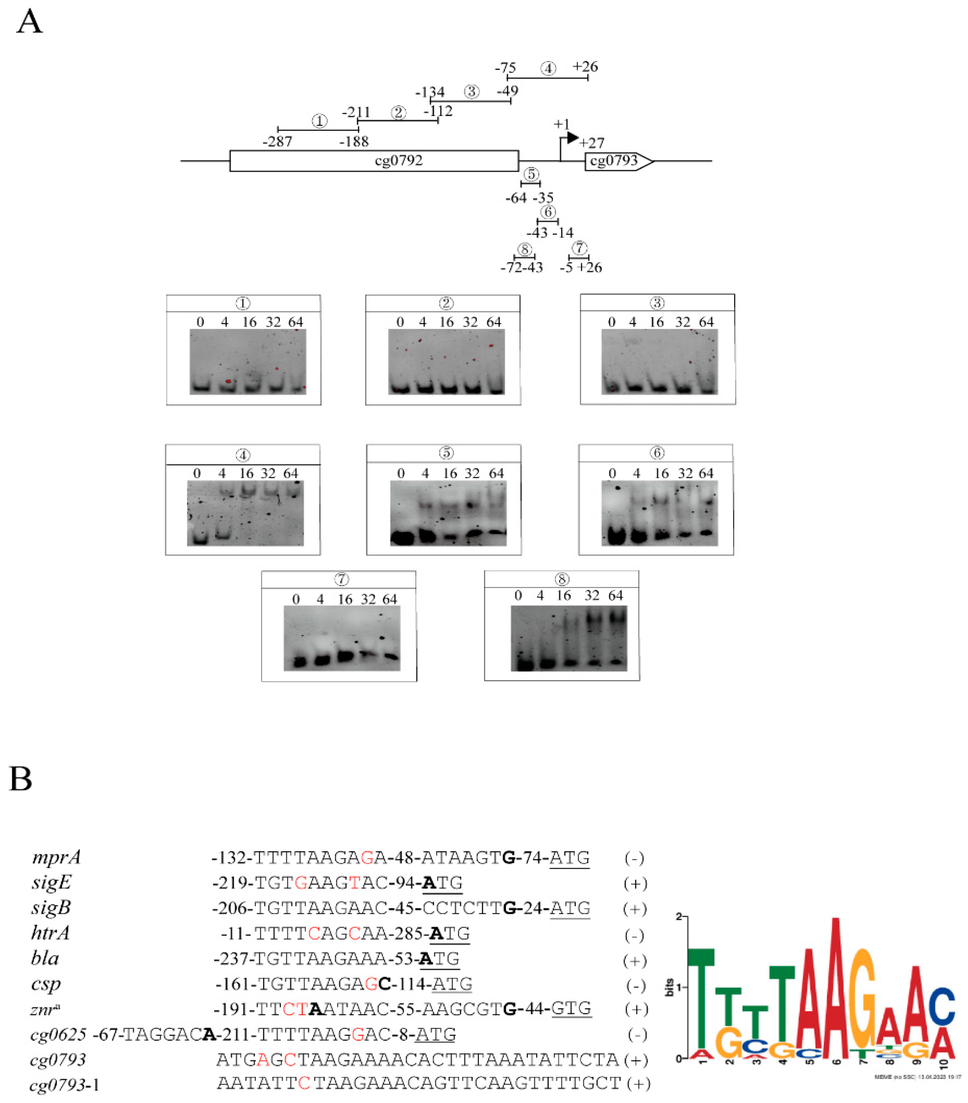
3.3. Identification of mprA-Binding Regions

To investigate the genes directly targeted by MprA, we conducted electrophoretic mobility shift assays (EMSAs). Purified phosphorylated MprA pre-incubated with the high-energy phosphate donor acetylphosphate and unphosphorylated MprA, phosphorylation-defective MprA (D51N), and phosphorylation-mimicking MprA (D51E) were individually used for experimentation (Figure 2A and Figure S2A,B). Surprisingly, concentration-dependent binding of both MprA variants to the promoter regions of mprA and cg0793 was observed (Figure 2). However, phosphorylated MprA showed significant band shifts at lower protein concentrations, suggesting that phosphorylation is not absolutely required for binding these promoter regions in vitro but can enhance its affinity for dsDNA. Furthermore, phosphorylated MprA binds to the upstream sequences of sigE and htrA, whereas unphosphorylated MprA fails to do so (Figure 2). This suggests that phosphorylation is likely indispensable for regulators to interact with dsDNA in vitro for certain genes. Likely, phosphorylation-defective MprA fails to bind to the promoter regions of sigE and htrA (Figure S2C). However, phosphorylation-mimicking MprA also binds to the promoter sequences of mprA, cg0793, sigE, and htrA (Figure S2C). The differential binding affinity of MprA protein toward these promoter sequences could be attributed to divergence in the conservation of their DNA motif sequences. Phosphorylated MprA also binds to the promoter regions of the cell envelope biosynthesis genes bla and csp and significantly binds to the upstream sequences of cg0625 (secreted proteins of unknown function), znr, and sigB (Figure 2). Moreover, phosphorylated and unphosphorylated MprA failed to bind to the upstream promoter regions of the potential target genes ycic and the negative control cysD (Figure 2). Altogether, MprAB directly regulates the stress response–related sigma factors sigE and sigB and a range of genes involved in cell envelope biosynthesis (bla, csp, and htrA), strongly emphasizing its importance in regulating cell wall synthesis.
To identify a common motif, truncated fragments (labels 1–8) of the promoter region of cg0793 were tested due to its high affinity for MprA. As shown in Figure 3A, a significant gel shift with 100 bp and 30 bp fragments was observed at a four-fold molar excess of the phosphorylated MprA protein (labels 4–6, 8), suggesting the presence of two MprA binding sites in the upstream sequence of cg0793 between −64 and −35 and between −43 and −14 (labels 5 and 6). The 10 MprA-targeted gene binding sites were analyzed via EMSA experiments using MEME 5.5.7 software. The results revealed a 10 bp common motif (consensus sequence: TKTTAAGAAM; K: G/T; M: A/C) that tolerated mismatches in conserved positions, indicating that the MprA-binding motif represents a loose sequence (Figure 3B).

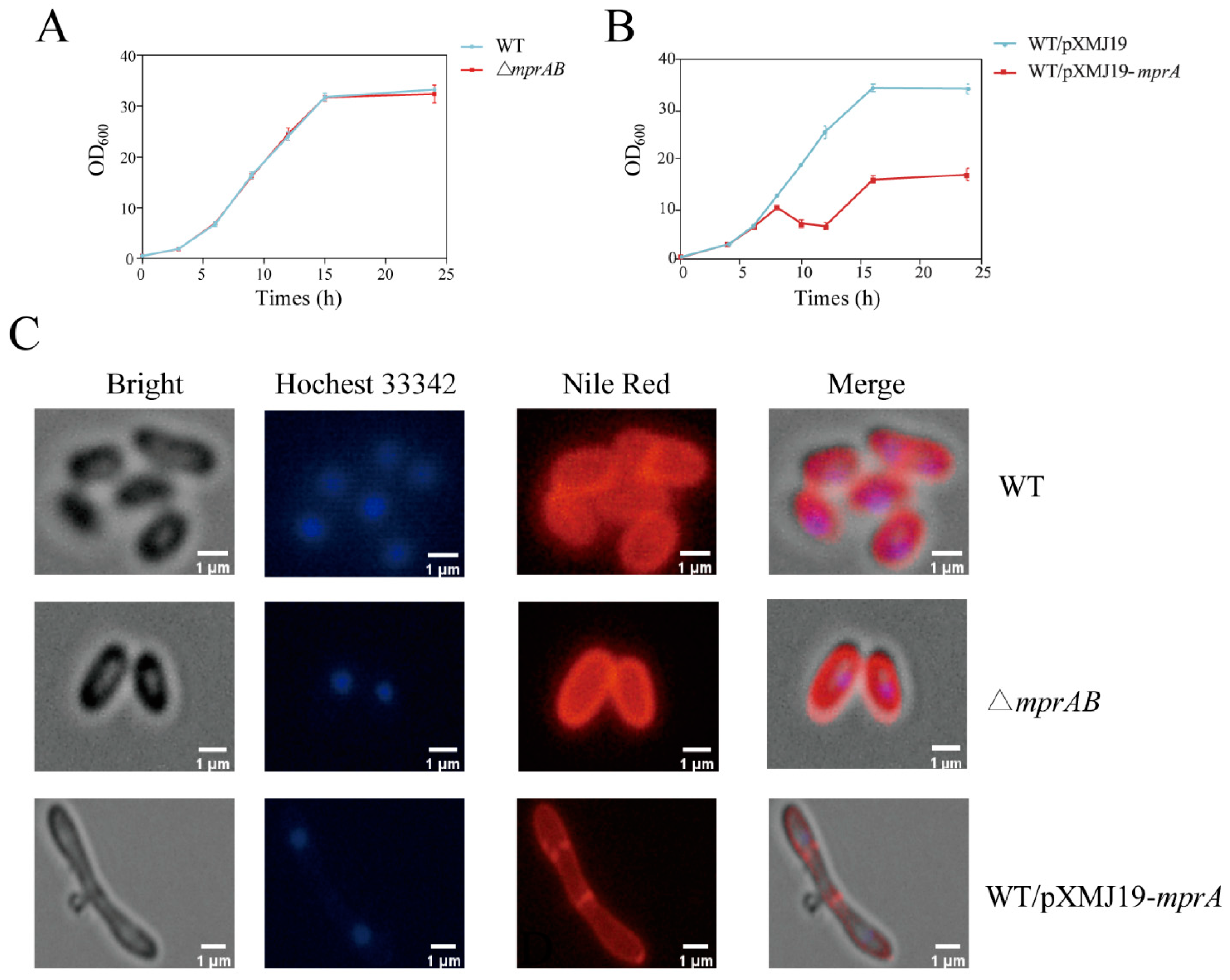
3.4. Overexpression of mprA Caused Cell Envelope Defects and Increased Alanine Titers in C. glutamicum

To characterize the physiological function of mprA in C. glutamicum, we compared the wild-type, △mprAB mutant, and WT/pXMJ19-mprA strains. The growth of the △mprAB mutant strain was similar to that of the wild-type strain in CGXII medium with 4% glucose at 30 °C (Figure 4A). However, mprA overexpression resulted in pronounced growth inhibition, and a much lower cell optical density was observed in the stationary phase (OD600, 33.29 ± 0.29 for WT/pXMJ19 strain versus 17.04 ± 0.60 for WT /pXMJ19-mprA strain) (Figure 4B).
Next, fluorescence microscopy analysis of cells stained with Nile Red (lipid components) and Hoechst 33342 (DNA) revealed that the mprA-overexpressing strain showed elongated cell length and a swollen cell shape, whereas the cell morphology of the △mprAB mutant strain was similar to the wild-type strain (Figure 4C). This morphological heterogeneity was analyzed by scanning electron microscopy (SEM); 1.89 ± 0.31 μm for the wild-type strain, 1.87 ± 0.34 μm for the WT △mprAB mutant strain, and 4.45 ± 1.69 μm for the WT/pXMJ19-mprA strain. This indicates that cell elongation was caused by mprA overexpression (Figure 5A,B). In addition, wrinkles were observed on the cell surface of the mprA-overexpressing strain, suggesting potential cell membrane damage, leading to increased cytoplasmic membrane permeability and the release of cell plasma [43]. These findings were further confirmed by propidium iodide (PI) staining and flow cytometric single-cell analysis (Figure S3). Moreover, TEM revealed a more atypical cell shape in the mprA-overexpressing strain (Figure 5B). Furthermore, the fur-like structure of the cell wall surface suggests that the outer layer of the cell envelope may be composed of mycolic acid or arabinogalactan, which is partially absent in the WT/pXMJ19-mprA strain (Figure 5B).
Previous studies have indicated that disruption of cell envelope biosynthesis affects the excretion of amino acids in Corynebacterium glutamicum [44]. The WT/pXMJ19 and WT/pXMJ19-mprA strains were investigated for glutamate and alanine production in batch fermentation. As expected, the WT/pXMJ19-mprA strain exhibited slower growth and glucose consumption rates compared to the wild-type strain (Figure 5C). Within 72 h of fermentation, the WT/pXMJ19 strain exhibited near-complete glucose utilization (97.3 g/L), accompanied by the production of 666.3 mg/L glutamate and 522.4 mg/L alanine, whereas the WT/pXMJ19-mprA strain retained 7.3 g/L residual glucose and showed altered amino acid profiles (320.1 mg/L glutamate, 805.6 mg/L alanine) (Figure 5D). It is noteworthy that, compared to the WT/pXMJ19 strain, the glutamate titers in the WT/pXMJ19-mprA strain gradually decreased after 24 h, while the alanine titers continued to increase over 72 h. The observed metabolic flux redirection toward alanine biosynthesis in the WT/pXMJ19-mprA strain is potentially attributable to transaminase-mediated nitrogen transfer, wherein glutamate serves as the amino group donor [45].

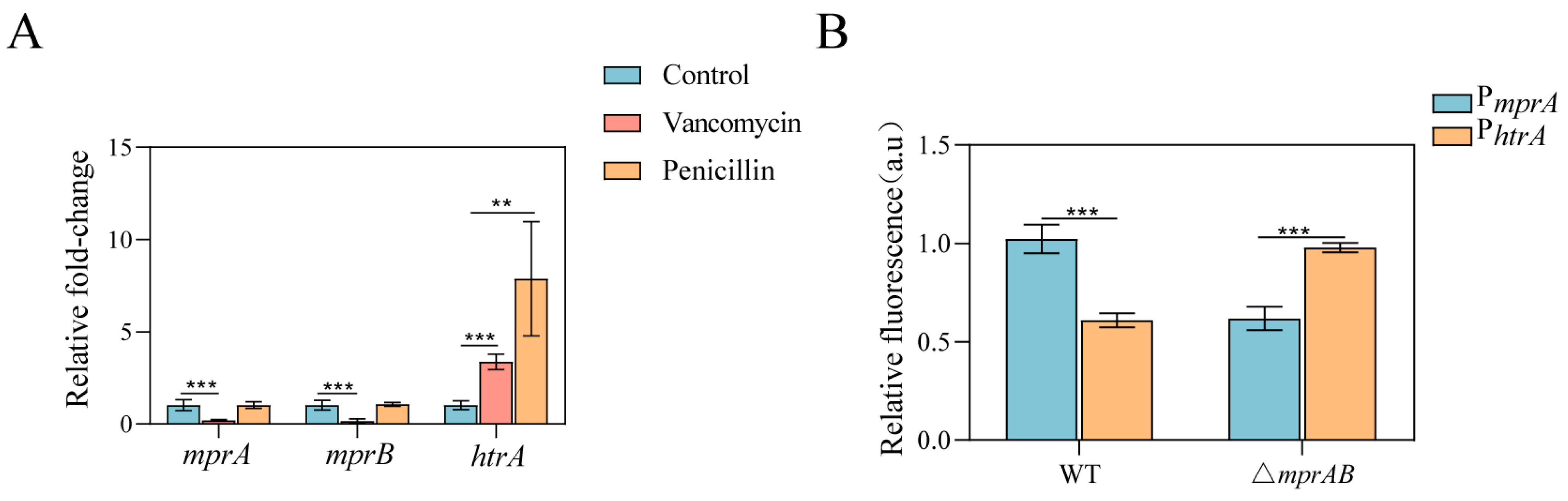
3.5. The MprAB Two-Component System and Its Direct Target Gene htrA Are Induced by Vancomycin

The MprAB system regulates several genes involved in cell wall biosynthesis, and overexpression of mprA causes cell wall defects in C. glutamicum. We tested whether cell wall–active antibiotics that impair cell envelope integrity induced MprAB expression. In the presence of vancomycin stimulation, the transcriptional levels of mprA and mprB were remarkably downregulated, as determined by qRT-PCR (Figure 6A). However, the transcription levels of mprA and mprB remained unaltered with the addition of penicillin. To investigate the activity of the Mpr regulon, a bicistronic plasmid containing the egfp gene under the control of the mprA promoter was constructed. To avoid the possible effect of plasmid copy number, the transcript level of red fluorescent protein (RFP) under the control of the constitutive promoter H36 on the same plasmid was determined by measuring the red fluorescence intensity [46]. The activities of the test promoters were easily and accurately evaluated using the GFP/RFP ratio. The promoter activity of the △mprAB mutant strain was decreased compared with that of the wild-type strain, which demonstrates positive autoregulation of expression of the MprAB system (Figure 6B).
Among the MprA target genes identified, htrA encodes a putative HtrA-like serine protease, which is involved in the response to protein secretion stress [47,48]. In M. tuberculosis, the HtrA homolog PepD plays a key role in the cell envelope stress response [49]. To determine whether cell envelope stress induces the MprA-regulated expression of htrA, we measured htrA transcript levels in the wild-type and △mprAB mutant strains in the presence or absence of vancomycin and penicillin by qRT-PCR. As shown in Figure 6A, treatment of wild-type cells with vancomycin and penicillin resulted in significantly increased expression of htrA relative to untreated cells. Moreover, the expression of htrA was increased in the WT △mprAB mutant strain (Figure 6B). These results indicate that the expression of MprAB TCS and its direct target gene, htrA, respond to vancomycin.

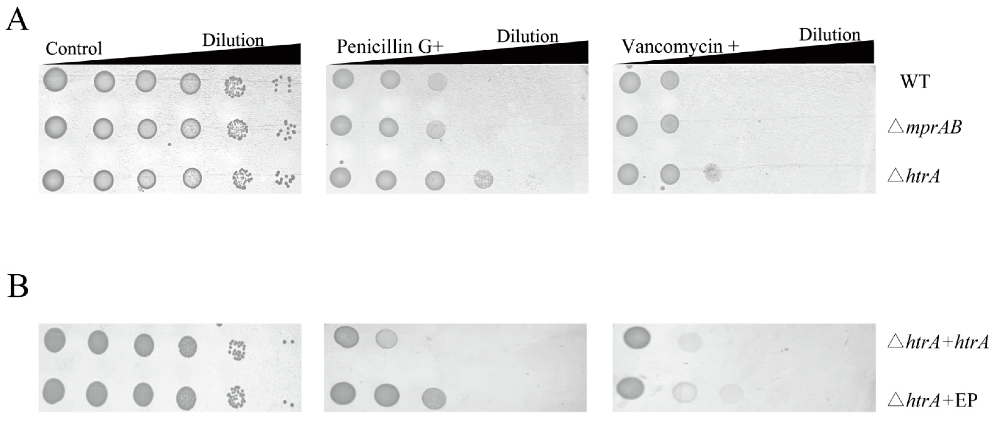
3.6. HtrA Mediates Vancomycin and Penicillin Resistance, but MprAB Does Not

M. tuberculosis MprAB and PepD have been reported to be required for resistance to cell envelope stress [14,49]. We therefore evaluated whether the MprAB TCS and HtrA are also essential for cell wall–active antibiotics resistance. The WT/pXMJ19-mprA strain was more sensitive to vancomycin and penicillin, and the WT/pXMJ19-htrA strain was more resistant to vancomycin, than the wild-type strain (Figure S4). However, there was no significant difference in the growth of wild-type and △mprAB mutant in penicillin and vancomycin resistance (Figure 7A). Thus, the MprAB TCS is responsive to, but does not mediate, vancomycin resistance in C. glutamicum. However, the △htrA mutant showed significantly better growth in the presence of penicillin and vancomycin compared to the wild-type strain (Figure 7A). Complementation by plasmid-encoded HtrA restored growth to wild-type levels (Figure 7B). Remarkably, the growth differences between the △htrA mutant and the △htrA/pXMJ19 mutant may be attributed to the presence of chloramphenicol. Thus, the MprAB TCS does not facilitate resistance to the cell envelope stressors vancomycin and penicillin, while its regulated gene htrA plays a critical role in mediating resistance to these stressors in C. glutamicum.

4. Discussion

The MprAB TCS is widely distributed in the genome of Actinobacteria, but its function and regulatory network have only been reported in mycobacteria. The cell envelope stress–responsive MprAB TCS, together with the alternative sigma factor SigE, forms a stress response network that facilitates the survival of tubercle bacilli inside host immune cells when they encounter nutrient and oxygen limitations and antibacterial mechanisms [16]. Characterization of the MprAB TCS in non-pathogenic Corynebacterium is critical for developing a complete knowledge of its conserved functions in Actinobacteria. In this study, we characterized the physiological functions and regulatory network of the MprAB TCS in C. glutamicum. Our results indicate that the MprAB TCS regulates a wide range of cellular pathways in response to impaired integrity of the cell envelope. RNA-seq analysis revealed only a slight perturbation of global transcript abundance in the △mprA mutant strain compared to the wild-type strain, suggesting that the MprAB TCS appears to be “off” under normal growth conditions. However, overexpression of mprA resulted in an intensely altered physiological status and transcript abundance in C. glutamicum. Several genes encoding secretory proteins (cg0793, cg0107, cg2069, psp2) and cell envelope biosynthesis proteins were significantly downregulated in the mprA-overexpressing strain (Table 1). One possible explanation is that these genes are involved in cell division, and a decrease in their expression may lead to morphological elongation. cg0793, encoding a cysteine-rich secretory protein with a CAP (cysteine-rich secretory protein) domain, is significantly regulated by MprA, as evidenced by a 3.45-fold increase upon mprA deletion and a significant decrease by 4.35-fold upon mprA overexpression. These protein families are most often involved in processes including extracellular matrix regulation and branching morphogenesis, potentially as either proteases or protease inhibitors. Cg0793 has been annotated as a hydrolase; however, its function is currently unclear. Considering the functionality of the MprAB TCS, we compared the growth of WT △cg0793 mutant and wild-type strains on agar plates supplemented with antibiotics affecting cell wall activity. However, no phenotypic differences were detected under these test conditions, and the function of cg0793 requires further investigation.
The purified MprA protein bound directly upstream of at least nine transcriptional units involved in multiple metabolic pathways, some with unknown functions. MEME analysis of the promoter regions of these regulatory sequences revealed an MprA-binding motif composed of a 10 bp flexible sequence (TKTTAAGAAM). In addition, the putative MprA-binding sites in the mprA promoter region, together with the PmprA-egfp reporter fusions, resulted in decreased relative activity in the mprAB deletion strain, suggesting positive autoregulation of the MprAB TCS. In mycobacteria, the MprAB TCS positively regulates sigE and sigB expression in response to cell envelope stress, and the absence of the regulator mprA results in downregulation of the expression of these genes [14]. In contrast, sigE and sigB expression in the △mprA mutant strain was comparable to that in the wild-type strain, whereas mprA overexpression directly repressed the transcript levels of sigE and sigB in C. glutamicum. Based on these findings, it is likely that an environmental signal is required for phosphorylation of the regulator MprA to control sigE and sigB expression in C. glutamicum. Thus, finding an appropriate extracellular stimulus to push the transition of MprAB TCS from the “off” to the “on” state may help to gain a more comprehensive understanding of the regulatory network between MprAB TCS and sigE and sigB in C. glutamicum.
Compared with C. glutamicum wild-type, an mprA-overexpressing mutant strain showed cell envelope defects and enhanced alanine titers. The enhanced alanine excretion could result from both impaired biosynthesis of cell envelope components, such as mycolic acids or peptidoglycan, and downregulation of alanine transporter Cg0524 expression. However, the loss of function of the MprAB TCS did not affect the growth of C. glutamicum under test conditions, including SDS, alkaline pH, and cell wall–active antibiotics. A different situation has been reported for M. tuberculosis and Mycobacterium smegmatis, which possess the MprAB TCS. In these cases, the mprAB TCS deletion strains were more sensitive to SDS than the wild-type strain. PepD not only is reported to be important in the stress response network mediated by MprAB in mycobacteria, but also its orthologous gene htrA can respond to periplasmic protein secretion stress in C. glutamicum, suggesting that HtrA family members maybe possess a conserved function in response to cell envelope stress in actinobacteria. Indeed, the promoter activity of htrA was MprAB-dependent, and htrA is responsive to vancomycin and penicillin. Compared to C. glutamicum wild-type, a △htrA mutant showed significantly increased growth on agar plates containing penicillin and vancomycin. These results clearly indicate that HtrA mediates penicillin and vancomycin resistance. In contrast, inactivation of pepD in M. smegmatis increases sensitivity to various cell wall stressors, but inactivation of pepD in M. tuberculosis is identical to wild-type with SDS-mediated stress [49].
Taken together, our results provide important novel insights into the transcriptional regulation and function of C. glutamicum MprAB TCS, indicating that it is clearly distinct from the M. tuberculosis MprAB TCS. (i) The MprAB TCS negatively affects the expression of genes that encode cell envelope functions and alternative sigma factors in C. glutamicum. (ii) Deletion of mprAB resulted a growth phenotype identical to C. glutamicum wild-type on agar plates containing cell envelope stressors. These functional differences may reflect C. glutamicum’s environmental adaptation strategy. The survival environments of non-pathogenic C. glutamicum fundamentally differ from those of pathogenic M. tuberculosis. C. glutamicum prioritizes the maintenance of cell envelope homeostasis to sustain rapid proliferation and high metabolic flux toward product synthesis. The MprAB TCS suppresses the expression of genes encoding cell envelope functions and alternative sigma factors, thereby preventing energy exhaustion caused by excessive cell envelope stress response. In contrast, M. tuberculosis requires continuous activation of cell envelope stress response mechanisms to survive harsh host environments. A future challenge is to elucidate how the activity of this system is controlled and how this TCS regulates the cell envelope process and function under diverse environmental stresses.

Supplementary Materials

The following supporting information can be downloaded at: https://www.mdpi.com/article/10.3390/microorganisms13051120/s1, Figure S1: Verification of RNA-seq results using qRT-PCR; Figure S2: EMSA analysis of MprAD-E and MprAD-N binding to the promoter of mprA, sigE, pepD, cg0793; Figure S3: Microscopic pictures and flow cytometric single-cell analysis of C. glutamicum wild type (ATCC 13032) and the overproduction of mprA in the wide type; Figure S4: Antibiotics sensitivity of C. glutamicum wide-type and overexpressing strains. Table S1: Bacterial strains and plasmids used in this study; Table S2: Primers used in this study.

Author Contributions

Formal analysis, Y.Y.; funding acquisition, Z.B.; methodology, Y.Z. and D.H.; project administration, Y.L.; resources, C.L.; supervision, Z.B.; validation, Y.Z.; writing—original draft, X.L.; writing—review & editing, X.L. All authors have read and agreed to the published version of the manuscript.

Funding

This work was supported by the National Natural Science Foundation of China (No. 22378167) and the Key Research and Development Program of Henan province (251111310300).

Institutional Review Board Statement

Not applicable.

Informed Consent Statement

Not applicable.

Data Availability Statement

The data used in this study can be accessed by request from the corresponding author.

Conflicts of Interest

The authors declare that they have no known competing financial interests or personal relationships that could have appeared to influence the work reported in this paper.

References

  1. McKitterick, A.C.; Bernhardt, T.G. Phage resistance profiling identifies new genes required for biogenesis and modification of the corynebacterial cell envelope. eLife 2017, 11, 79981–80005. [Google Scholar] [CrossRef] [PubMed]
  2. Zhou, X.; Rodriguez-Rivera, F.P.; Lim, H.C.; Bell, J.C.; Bernhardt, T.G.; Bertozzi, C.R.; Theriot, J.A. Sequential assembly of the septal cell envelope prior to V snapping in Corynebacterium glutamicum. Nat. Chem. Biol. 2019, 15, 221–231. [Google Scholar] [CrossRef] [PubMed]
  3. Lim, H.C.; Sher, J.W.; Rodriguez-Rivera, F.P.; Fumeaux, C.; Bertozzi, C.R.; Bernhardt, T.G. Identification of new components of the RipC-FtsEX cell separation pathway of Corynebacterineae. PLoS Genet. 2019, 15, e1008284. [Google Scholar] [CrossRef] [PubMed]
  4. Jacob-Dubuisson, F.; Mechaly, A.; Betton, J.M.; Antoine, R. Structural insights into the signalling mechanisms of two-component systems. Nat. Rev. Microbiol. 2018, 16, 585–593. [Google Scholar] [CrossRef]
  5. Wu, X.; Liu, J.; Liu, Z.; Gong, G.; Zha, J. Microbial cell surface engineering for high-level synthesis of bio-products. Biotechnol. Adv. 2022, 55, 107912. [Google Scholar] [CrossRef]
  6. Qin, X.; Zhang, K.; Nie, Y.; Wu, X.L. The roles of the two-component system, MtrAB, in response to diverse cell envelope stresses in Dietzia sp. DQ12-45-1b. Appl. Environ. Microbiol. 2022, 88, e0133722. [Google Scholar] [CrossRef]
  7. Mike, L.A.; Choby, J.E.; Brinkman, P.R.; Olive, L.Q.; Dutter, B.F.; Ivan, S.J.; Gibbs, C.M.; Sulikowski, G.A.; Stauff, D.L.; Skaar, E.P. Two-component system cross-regulation integrates Bacillus anthracis response to heme and cell envelope stress. PLoS Pathog. 2014, 10, e1004044. [Google Scholar] [CrossRef]
  8. Komazin, G.; Rizk, A.A.; Armbruster, K.M.; Bonnell, V.A.; Llinás, M.; Meredith, T.C. A copper-responsive two-component system governs lipoprotein remodeling in Listeria monocytogenes. J. Bacteriol. 2003, 205, e0039022. [Google Scholar] [CrossRef]
  9. Casino, P.; Rubio, V.; Marina, A. Structural insight into partner specificity and phosphoryl transfer in two-component signal transduction. Cell 2009, 139, 325–336. [Google Scholar] [CrossRef]
  10. Trajtenberg, F.; Albanesi, D.; Ruétalo, N.; Botti, H.; Mechaly, A.E.; Nieves, M.; Aguilar, P.S.; Cybulski, L.; Larrieux, N.; de Mendoza, D.; et al. Allosteric activation of bacterial response regulators: The role of the cognate histidine kinase beyond phosphorylation. mBio 2014, 5, e02105. [Google Scholar] [CrossRef]
  11. Schmidl, S.R.; Ekness, F.; Sofjan, K.; Daeffler, K.N.; Brink, K.R.; Landry, B.P.; Gerhardt, K.P.; Dyulgyarov, N.; Sheth, R.U.; Tabor, J.J. Rewiring bacterial two-component systems by modular DNA-binding domain swapping. Nat. Chem. Biol. 2019, 15, 690–698. [Google Scholar] [CrossRef] [PubMed]
  12. Parish, T. Two-component regulatory systems of Mycobacteria. Microbiol. Spectr. 2014, 2, MGM2-0010-2013. [Google Scholar] [CrossRef] [PubMed]
  13. Möker, N.; Brocker, M.; Schaffer, S.; Krämer, R.; Morbach, S.; Bott, M. Deletion of the genes encoding the MtrA-MtrB two-component system of Corynebacterium glutamicum has a strong influence on cell morphology, antibiotics susceptibility and expression of genes involved in osmoprotection. Mol. Microbiol. 2004, 54, 420–438. [Google Scholar] [CrossRef] [PubMed]
  14. He, H.; Hovey, R.; Kane, J.; Singh, V.; Zahrt, T.C. MprAB is a stress-responsive two-component system that directly regulates expression of sigma factors SigB and SigE in Mycobacterium tuberculosis. J. Bacteriol. 2006, 188, 2134–2143. [Google Scholar] [CrossRef]
  15. Sureka, K.; Dey, S.; Datta, P.; Singh, A.K.; Dasgupta, A.; Rodrigue, S.; Basu, J.; Kundu, M. Polyphosphate kinase is involved in stress-induced mprAB-sigE-rel signalling in mycobacteria. Mol. Microbiol. 2007, 65, 261–276. [Google Scholar] [CrossRef]
  16. Zahrt, T.C.; Deretic, V. Mycobacterium tuberculosis signal transduction system required for persistent infections. Proc. Natl. Acad. Sci. USA 2001, 98, 12706–12711. [Google Scholar] [CrossRef]
  17. Datta, P.; Ravi, J.; Guerrini, V.; Chauhan, R.; Neiditch, M.B.; Shell, S.S.; Fortune, S.M.; Hancioglu, B.; Igoshin, O.A.; Gennaro, M.L. The Psp system of Mycobacterium tuberculosis integrates envelope stress-sensing and envelope-preserving functions. Mol. Microbiol. 2015, 97, 408–422. [Google Scholar] [CrossRef]
  18. He, H.; Bretl, D.J.; Penoske, R.M.; Anderson, D.M.; Zahrt, T.C. Components of the Rv0081-Rv0088 locus, which encodes a predicted formate hydrogenlyase complex, are coregulated by Rv0081, MprA, and DosR in Mycobacterium tuberculosis. J. Bacteriol. 2011, 193, 5105–5118. [Google Scholar] [CrossRef]
  19. Bretl, D.J.; He, H.; Demetriadou, C.; White, M.J.; Penoske, R.M.; Salzman, N.H.; Zahrt, T.C. MprA and DosR coregulate a Mycobacterium tuberculosis virulence operon encoding Rv1813c and Rv1812c. Infect. Immun. 2012, 80, 3018–3033. [Google Scholar] [CrossRef]
  20. Pang, X.; Howard, S.T. Regulation of the alpha-crystallin gene acr2 by the MprAB two-component system of Mycobacterium tuberculosis. J. Bacteriol. 2007, 189, 6213–6221. [Google Scholar] [CrossRef]
  21. Pang, X.; Samten, B.; Cao, G.; Wang, X.; Tvinnereim, A.R.; Chen, X.L.; Howard, S.T. MprAB regulates the espA operon in Mycobacterium tuberculosis and modulates ESX-1 function and host cytokine response. J. Bacteriol. 2013, 195, 66–75. [Google Scholar] [CrossRef] [PubMed]
  22. He, H.; Zahrt, T.C. Identification and characterization of a regulatory sequence recognized by Mycobacterium tuberculosis persistence regulator MprA. J. Bacteriol. 2005, 187, 202–212. [Google Scholar] [CrossRef] [PubMed]
  23. Sureka, K.; Ghosh, B.; Dasgupta, A.; Basu, J.; Kundu, M.; Bose, I. Positive feedback and noise activate the stringent response regulator rel in mycobacteria. PLoS ONE 2008, 3, e1771. [Google Scholar] [CrossRef]
  24. Pang, X.; Cao, X.; Neuenschwander, P.F.; Haydel, S.E.; Hou, G.; Howard, S.T. The β-propeller gene Rv1057 of Mycobacterium tuberculosis has a complex promoter directly regulated by both the MprAB and TrcRS two-component systems. Tuberculosis 2011, 91, 142–149. [Google Scholar] [CrossRef]
  25. Pang, X.; Vu, P.; Byrd, T.F.; Ghanny, S.; Soteropoulos, P.; Mukamolova, G.V.; Wu, S.; Samten, B.; Howard, S.T. Evidence for complex interactions of stress-associated regulons in an mprAB deletion mutant of Mycobacterium tuberculosis. Microbiology 2007, 153, 1229–1242. [Google Scholar] [CrossRef]
  26. Bretl, D.J.; Bigley, T.M.; Terhune, S.S.; Zahrt, T.C. The MprB extracytoplasmic domain negatively regulates activation of the Mycobacterium tuberculosis MprAB two-component system. J. Bacteriol. 2014, 196, 391–406. [Google Scholar] [CrossRef]
  27. Wong, K.W. The Role of ESX-1 in Mycobacterium tuberculosis Pathogenesis. Microbiol. Spectr. 2017, 5. [Google Scholar] [CrossRef]
  28. Zahrt, T.C.; Wozniak, C.; Jones, D.; Trevett, A. Functional analysis of the Mycobacterium tuberculosis MprAB two-component signal transduction system. Infect. Immun. 2003, 71, 6962–6970. [Google Scholar] [CrossRef]
  29. Pan, L.; Chen, X.; Wang, K.; Mao, Z. Understanding high ε-poly-L-lysine production by Streptomyces albulus using pH shock strategy in the level of transcriptomic. J. Ind. Microbiol. Biotechnol. 2019, 46, 1781–1792. [Google Scholar] [CrossRef]
  30. Toyoda, K.; Inui, M. Regulons of global transcription factors in Corynebacterium glutamicum. Appl. Microbiol. Biotechnol. 2016, 100, 45–60. [Google Scholar] [CrossRef]
  31. Kleine, B.; Chattopadhyay, A.; Polen, T.; Pinto, D.; Mascher, T.; Bott, M.; Brocker, M.; Freudl, R. The three-component system EsrISR regulates a cell envelope stress response in Corynebacterium glutamicum. Mol. Microbiol. 2017, 106, 719–741. [Google Scholar] [CrossRef] [PubMed]
  32. Keilhauer, C.; Eggeling, L.; Sahm, H. Isoleucine synthesis in Corynebacterium glutamicum: Molecular analysis of the ilvB-ilvN-ilvC operon. J. Bacteriol. 1993, 175, 5595–5603. [Google Scholar] [CrossRef] [PubMed]
  33. Gao, G.R.; Wei, S.Y.; Ding, M.Z.; Hou, Z.J.; Wang, D.J.; Xu, Q.M.; Chen, Y.J. Enhancing fengycin production in the co-culture of Bacillus subtilis and Corynebacterium glutamicum by engineering proline transporter. Bioresour. Technol. 2003, 383, 129229. [Google Scholar] [CrossRef] [PubMed]
  34. SEQC/MAQC-III Consortium. A comprehensive assessment of RNA-seq accuracy, reproducibility and information content by the Sequencing Quality Control Consortium. Nat. Biotechnol. 2014, 32, 903–914. [Google Scholar] [CrossRef]
  35. Everaert, C.; Luypaert, M.; Maag, J.L.V.; Cheng, Q.X.; Dinger, M.E.; Hellemans, J.; Mestdagh, P. Benchmarking of RNA-sequencing analysis workflows using whole-transcriptome RT-qPCR expression data. Sci. Rep. 2017, 7, 1559–15570. [Google Scholar] [CrossRef]
  36. Baumgart, M.; Schubert, K.; Bramkamp, M.; Frunzke, J. Impact of LytR-CpsA-Psr proteins on cell wall biosynthesis in Corynebacterium glutamicum. J. Bacteriol. 2016, 198, 3045–3059. [Google Scholar] [CrossRef]
  37. Toyoda, K.; Inui, M. Extracytoplasmic function sigma factor σD confers resistance to environmental stress by enhancing mycolate synthesis and modifying peptidoglycan structures in Corynebacterium glutamicum. Mol. Microbiol. 2018, 107, 312–329. [Google Scholar] [CrossRef]
  38. Dietrich, C.; Li de la Sierra-Gallay, I.; Masi, M.; Girard, E.; Dautin, N.; Constantinesco-Becker, N.; Tropis, M.; Daffé, M.; van Tilbeurgh, H.; Bayan, N. The C-terminal domain of Corynebacterium glutamicum mycoloyltransferase A is composed of five repeated motifs involved in cell wall binding and stability. Mol. Microbiol. 2020, 114, 1–16. [Google Scholar] [CrossRef]
  39. Brand, S.; Niehaus, K.; Pühler, A.; Kalinowski, J. Identification and functional analysis of six mycolyltransferase genes of Corynebacterium glutamicum ATCC 13032: The genes cop1, cmt1, and cmt2 can replace each other in the synthesis of trehalose dicorynomycolate, a component of the mycolic acid layer of the cell envelope. Arch. Microbiol. 2003, 180, 33–44. [Google Scholar] [CrossRef]
  40. Teramoto, H.; Inui, M.; Yukawa, H. Corynebacterium glutamicum Zur acts as a zinc-sensing transcriptional repressor of both zinc-inducible and zinc-repressible genes involved in zinc homeostasis. FEBS J. 2012, 279, 4385–4397. [Google Scholar] [CrossRef]
  41. Pfeifer-Sancar, K.; Mentz, A.; Rückert, C.; Kalinowski, J. Comprehensive analysis of the Corynebacterium glutamicum transcriptome using an improved RNAseq technique. BMC Genom. 2013, 14, 888–911. [Google Scholar] [CrossRef] [PubMed]
  42. Schröder, J.; Jochmann, N.; Rodionov, D.A.; Tauch, A. The Zur regulon of Corynebacterium glutamicum ATCC 13032. BMC Genom. 2010, 7, 12. [Google Scholar] [CrossRef]
  43. Farkas, A.; Maróti, G.; Kereszt, A.; Kondorosi, É. Comparative Analysis of the Bacterial Membrane Disruption Effect of Two Natural Plant Antimicrobial Peptides. Front. Microbiol. 2017, 23, 51–63. [Google Scholar] [CrossRef] [PubMed]
  44. Li, H.; Xu, D.; Tan, X.; Huang, D.; Huang, Y.; Zhao, G.; Hu, X.; Wang, X. The role of trehalose biosynthesis on mycolate composition and L-glutamate production in Corynebacterium glutamicum. Microbiol. Res. 2023, 267, 127260. [Google Scholar] [CrossRef] [PubMed]
  45. Marienhagen, J.; Kennerknecht, N.; Sahm, H.; Eggeling, L. Functional analysis of all aminotransferase proteins inferred from the genome sequence of Corynebacterium glutamicum. J. Bacteriol. 2005, 187, 7639–7646. [Google Scholar] [CrossRef]
  46. Shimada, T.; Makinoshima, H.; Ogawa, Y.; Miki, T.; Maeda, M.; Ishihama, A. Classification and strength measurement of stationary-phase promoters by use of a newly developed promoter cloning vector. J. Bacteriol. 2004, 186, 7112–7122. [Google Scholar] [CrossRef]
  47. Jurischka, S.; Bida, A.; Dohmen-Olma, D.; Kleine, B.; Potzkei, J.; Binder, S.; Schaumann, G.; Bakkes, P.J.; Freudl, R. A secretion biosensor for monitoring Sec-dependent protein export in Corynebacterium glutamicum. Microb. Cell Fact. 2020, 19, 11. [Google Scholar] [CrossRef]
  48. Bakkes, P.J.; Lenz, P.; Müller, C.; Bida, A.; Dohmen-Olma, D.; Knapp, A.; Oldiges, M.; Jaeger, K.E.; Freudl, R. Biosensor-Based Optimization of Cutinase Secretion by Corynebacterium glutamicum. Front. Microbiol. 2021, 12, 750150–750167. [Google Scholar] [CrossRef]
  49. White, M.J.; He, H.; Penoske, R.M.; Twining, S.S.; Zahrt, T.C. PepD participates in the mycobacterial stress response mediated through MprAB and SigE. J. Bacteriol. 2010, 192, 1498–1510. [Google Scholar] [CrossRef]
Figure 1. The MprAB two-component system. (A) Genomic organization of the mprAB locus in Corynebacterium and Mycobacterium. Amino acid sequence identity with the C. glutamicum MprA and MprB ortholog is given in the upper panel. TSS: transcriptional start site. The genomic context of MprAB TCS was extracted from MicrobesOnline (http://microbesonline.org) (accessed on 1 October 2024). (B) Prediction of the protein domains of MprA and MprB, extracted from KEGG.
Microorganisms 13 01120 g001
Figure 2. Electrophoretic mobility shift assays (EMSAs) were performed to identify binding of MprA to promoter regions. (A) SDS-PAGE of the MprA protein. (B) DNA fragments about 300 bp upstream of the putative MprA target genes were incubated with or without a 4, 16, 32, or 64 molar excesses of phosphorylated purified MprA protein, as indicated below the respective lanes. The promoter region of the untargeted gene cysD was used as a negative control. The asterisk (*) indicates samples without prior phosphorylation of MprA by acetylphosphate. No DNA binding was observed without previous phosphorylation of MprA with acetylphosphate (htrA*, sigE*, and cysD*).
Microorganisms 13 01120 g002
Figure 3. Identification of the MprA-binding motif using MEME 5.5.7 software. (A) Search for MprA binding sites within the promoter region of cg0793. (B) MprA consensus binding motif predicted by MEME 5.5.7 software based on analysis of the upstream sequence of verified MprA-targeted genes (left panel). The TSSs are indicated in bold and were determined by 5′-end RNA-seq [41]. The start codons are underlined. Mismatched bases in the conserved motif are shown in red. The TSS of znr was derived from Schroder’s study [42]. Binding of MprA to the coding strand (+) or template strand (−) in the EMSA experiments.
Microorganisms 13 01120 g003
Figure 4. Characterization of wild-type (WT), △mprAB mutant, and mprA-overexpressing strains. Pre-cultured cells were cultivated in 3 mL of LBB medium in a 50 mL centrifuge tube at 30 °C overnight. (A) Growth curves of WT and △mprAB mutant strains in the CGXII medium containing 4% (w/v) glucose. (B) Growth curves of the WT strain carrying the empty plasmid pXMJ19 and pXMJ19-mprA in CGXII medium containing 4% glucose with 1 mM IPTG. (C) Fluorescence microscopy of the WT, △mprAB mutant, and mprA-overexpressing strains grown in CGXII medium containing 4% glucose for 24 h. The expression of mprA is induced by the addition of 1 mM IPTG. The lipid components of the cell membrane and DNA were marked using Nile red (red) and Hoechst 33342 (blue), respectively. The data are presented as mean ± SD of three independent biological replicates.
Microorganisms 13 01120 g004
Figure 5. Physiological characteristics of C. glutamicum cells. For microscopic analysis, cells were cultured in CGXII minimal medium containing 4% glucose for 24 h, and mprA expression was induced by adding 1 mM IPTG. (A) FSEM micrographs of WT, △mprAB mutant, and mprA-overexpressing strains. FSEM pictures were captured at 10,000× magnification. Size distribution of WT, △mprAB mutant, and mprA-overexpressing strains. The lengths of at least 100 individual cells were measured. Cells were fixed using 3% glutaraldehyde. (B) TEM images of wild-type and mprA-overexpressing strains at 5000×, 20,000×, and 40,000×. Insets show small portions of cell cross sections for each image. L-Glutamate and L-alanine fermentation by the WT/pXMJ19 (blue line) and WT/pXMJ19-mprA (cyan line) strains. OD600 and glucose consumption (C) and glutamate and alanine titers (D) were detected. The data are presented as mean ± SD of three independent biological replicates.
Microorganisms 13 01120 g005
Figure 6. The MprAB TCS and HtrA are induced by the cell wall–active antibiotics vancomycin or penicillin. Transcript levels were assayed before (blue) and 1 h after the addition of 4 μg/mL vancomycin or 2 U/mL penicillin in the exponential phase. (A) The levels of mprA, mprB, and htrA in the WT strains were determined using qRT-PCR. Transcript levels before induction in the control strain were taken as 1.0. (B) The WT and △mprAB mutant strains carrying PmprA-EGFP and PhtrA-EGFP were transformed with the reporter vector pEC-X1-PmprA and pEC-X1- PhtrA for 8 h. The relative fluorescence was evaluated by the GFP/RFP ratio. The data are presented as mean ± SD of three independent biological replicates. Statistical significance was determined by Student’s t test. **, p < 0.01, ***, p < 0.001.
Microorganisms 13 01120 g006
Figure 7. The wild-type strain and the indicated deletion mutants were grown, serially diluted, and spotted onto LBB plates containing either (A) 0.2 U/mL penicillin and 0.25 μg/mL vancomycin or (B) 0.2 U/mL penicillin, 0.25 μg/mL vancomycin, and 10 μg/mL chloramphenicol. EP: pXMJ19 empty plasmid.
Microorganisms 13 01120 g007
Table 1. Significantly regulated genes, as determined by RNA-seq or qRT-PCR.
Gene LocusGeneFunctionBinding Site amprA bp-Value cmprA/pXMJ19-mprA dp-Value c
cg0996mprAResponse regulator of two-component systemTTTTAAGAGA−3.65 ± 0.176.29 × 10−10111.11 ± 0.754.28 × 10−50
cg0997mprBHistidine kinase of two-component system −4.52 ± 0.173.91 × 10−148−0.82 ± 0.352.05 × 10−02
Cell envelope biosynthesis
cg0413cmt1Trehalose corynomycolyl transferase −0.03 ± 0.299.17 × 10−01−1.12 ± 0.083.22 × 10−45
cg0414wzzSaccharide synthesis 0.01 ± 0.139.51 × 10−01−1.37 ± 0.082.64 × 10−74
cg0650 L,D-transpeptidases 0.01 ± 0.139.51 × 10−01−1.62 ± 0.096.34 × 10−77
cg0905psp2Protein potentially involved in peptidoglycan biosynthesis 0.21 ± 0.416.10 × 10−01−3.07 ± 0.144.18 × 10−102
cg0998htrAHtrA-like serine proteaseTTTTCAGCAA1.00 ± 0.131.75 × 10−15−0.93 ±0.089.58 × 10−29
cg1108porAMycoloyltransferase 0.08 ± 0.124.81 × 10−01−1.45 ± 0.075.92 × 10−94
cg1109porBMycoloyltransferase 1.48 ± 0.119.33 × 10−43−0.74 ± 0.041.13 × 10−71
cg2069 Protein of LGFP repeat family 0.84 ± 0.198.46 × 10−06−2.67 ± 0.153.69 × 10−67
cg2398plsC1-Acyl-sn-glycerol-3-phosphate acetyltransferase 0.00 ± 0.129.85 × 10−01−1.16 ± 0.094.84 × 10−40
cg2478blaPenicillin-binding proteinTGTTAAGAAA0.21 ± 0.141.20 × 10−01−1.31 ± 0.105.11 × 10−38
cg3197cspProtein potentially involved in peptidoglycan biosynthesisTGTTAAGAGC0.00 ± 0.129.80 × 10−01−2.00 ± 0.082.76 × 10−144
Regulatory proteins
cg0317 ArsR family transcriptional regulator −1.11 ± 0.372.99 × 10−031.44 ± 0.493.54 × 10−03
cg1119 Putative stress-responsive transcriptional regulator 0.31 ± 0.211.37 × 10−01−1.38 ± 0.394.16 × 10−04
cg1271sigESigma factorTGTGAAGTAC−0.14 ± 0.15
(0.40 ± 0.10)
3.50 × 10−01−0.10 ± 0.10
(−1.24 ± 0.05) e
3.17 × 10−01
cg2102sigBSigma factorTGTTAAGAAC−0.29 ± 0.20
(0.35 ± 0.25)
1.42 × 10−01−0.11 ± 0.10
(−1.0 ± 0.07) e
2.96 × 10−01
cg2115sugRTranscriptional regulators of sugar metabolism −0.06 ± 0.136.52 × 10−01−1.05 ± 0.111.74 × 10−21
cg2500znrPutative transcriptional regulator −0.92 ± 0.369.44 × 10−030.64 ± 0.143.33 × 10−06
cg2648 Bacterial regulatory protein 3.83 ± 1.323.78 × 10−03−1.02 ± 1.303.80 × 10−02
Secreted
proteins
cg0085porH1PhoH-like ATPase 0.12 ± 0.143.99 × 10−01−1.36 ± 0.131.68 × 10−26
cg0625 Secreted proteinTTTTAAGGAC−0.42 ± 0.203.27 × 10−021.10 ± 0.097.50 × 10−33
cg0726 Secreted lipoprotein 0.22 ± 0.141.25 × 10−01−1.05 ± 0.141.72 × 10
cg0793 Secreted proteinAGCTAAGAAA
ATTCTAAGAA
3.45 ± 0.194.35 × 10−76−4.15 ± 0.292.30× 10−45
cg0918 Putative secreted protein −0.03 ± 0.107.29 × 10−01−1.31 ± 0.486.23 × 10−03
cg1247 Putative secreted protein 0.10 ± 0.588.67 × 10−01−1.10 ± 0.094.42 × 10−32
cg1514 Putative secreted protein −0.89 ± 0.253.17 × 10−041.02 ± 0.136.94 × 10−15
cg1936 Putative secreted protein −0.09 ± 0.287.44 × 10−01−1.16 ± 0.392.58 × 10−03
cg2061 Putative secreted protein −0.25 ± 0.212.26 × 10−01−2.93 ± 0.106.91 × 10−177
cg2518 Putative secreted protein 0.12 ± 0.174.69 × 10−01−2.60 ± 0.127.07 × 10−113
cg2566 Putative secreted protein −0.06 ± 0.217.80 × 10−01−2.07 ± 0.123.88 × 10−68
cg3197 Putative secreted protein 0.00 ± 0.129.80 × 10−01−2.00 ± 0.082.76 × 10−144
cg3343 Putative secreted protein 0.42 ± 0.259.70 × 10−02−2.90 ± 0.101.96 × 10−180
cg3394 Putative secreted protein 0.35 ± 0.271.98 × 10−01−1.70 ± 0.202.73 × 10−17
Other metabolism
cg0010 Hypothetical protein 0.06 ± 0.308.54 × 10−01−1.19 ± 0.192.70 × 10−10
cg0088 Citrate transporter −40 ± 0.322.15 × 10−011.00 ± 0.401.19 × 10−02
cg0096 Hypothetical protein −0.44 ± 0.473.56 × 10−011.16 ± 0.467.20 × 10−124
cg0107 Putative integral membrane transport protein −0.25 ± 0.606.78 × 10−01−5.07 ± 0.231.08 × 10−02
cg0108 Sirtuin-type KDAC homologues −0.08 ± 0.155.98 × 10−01−1.48 ± 0.274.42 × 10−08
cg0133abgTSecondary transporter of the AbgT family −0.08 ± 0.145.94 × 10−01−2.03 ± 0.081.43 × 10−137
cg0134abgBPeptidase −0.05 ± 0.167.75 × 10−01−2.01 ± 0.094.58 × 10−122
cg0135 Putative inner membrane protein 0.07 ± 0.207.28 × 10−011.13 ± 0.168.57 × 10−13
cg0182tagA2DNA-3-methyladenine glycosylase I −0.28 ± 0.303.63 × 10−01−0.65 ± 0.051.40 × 10−108
cg0253 Flavodoxin reductase 0.28 ± 0.0.242.45 × 10−01−1.04 ± 0.171.63 × 10−09
cg0254 Alanine symporter −0.98 ± 0.151.42 × 10−10−1.06 ± 0.085.51 × 10−37
cg0291 3,4-dioxygenase beta subunit 0.15 ± 0.101.10 × 10−011.16 ± 0.101.07 × 10−22
cg0391 UDP-glucose 4-epimerase −0.07 ± 0.125.75 × 10−01−1.97 ± 0.084.70 × 10−123
cg0415ptpA2Low molecular weight protein-tyrosine phosphatase 0.05 ± 0.126.65 × 10−01−1.11 ± 0.091.62 × 10−35
cg0437 Membrane protein 0.22 ± 0.141.03 × 10−01−1.19 ± 0.125.66 × 10−24
cg0441lpdDihydrolipoyl dehydrogenase 0.04 ± 0.147.70 × 10−01−1.31 ± 0.096.20 × 10−48
cg0623 Cobalt transport system −0.12 ± 0.195.28 × 10−011.10 ± 0.087.30 × 10−43
cg0646 IclR family proteins 0.12 ± 0.195.09 × 10−01−1.93 ± 0.157.33 × 10−38
cg0692 Transposase 1.37 ± 0.581.77 × 10−020.51 ± 0.412.13 × 10−01
cg0703guaAGMP synthase 0.21 ± 0.416.10 × 10−01−1.42 ± 0.632.49 × 10−02
cg0721crtB2Hytoene synthetase 0.2 ± 0.20 9.26 × 10−01−1.11 ± 0.211.82 × 10−07
cg0723crtEGeranylgeranyl diphosphate synthase 0.03 ± 0.188.69 × 10−01−1.06 ± 0167.74 × 10−11
cg0727 Nucleoside-diphosphate-sugar epimerase 0.16 ± 0.121.81 × 10−01−1.17 ± 0.111.76 × 10−24
cg0739 Putative integral membrane protein 0.06 ± 0.156.81 × 10−01−1.42 ± 0.161.51 × 10−18
cg0740 Membrane protein 0.13 ± 0.184.85 × 10−01−1.23 ± 0.131.53 × 10−21
cg0755metYO-Acetylhomoserine-lyase 0.03 ± 0.127.96 × 10−01−1.05 ± 0.131.53 × 10−21
cg0770 Fe3+-siderophores transport system −1.18 ± 0.121.32 × 10−22−1.57 ± 0.146.58 × 10−31
cg0794ycicCobalamin synthesis protein 3.27 ± 0.147.50 × 10−123−3.42 ± 0.113.91 × 10−195
cg0830 Membrane protein 0.58 ± 0.273.23 × 10−02−1.33 ± 0.453.05 × 10−03
cg0844 Type II restriction enzyme −0.59 ± 0.174.15 × 10−04−1.09 ± 0.099.28 × 10−35
cg0845 Superfamily II DNA/RNA helicase −0.17 ± 0.224.26 × 10−01−1.27 ± 0.132.73 × 10−21
cg0923 Membrane protein 0.15 ± 0.156.02 × 10−01−1.14 ± 0.222.44 × 10−07
cg0924 Fe3+-siderophores transport system −0.09 ± 0.097.50 × 10−01−1.04 ± 0.164.79 × 10−11
cg1016betPGlycine betaine transporter 0.09 ± 0.216.72 × 10−01−1.05 ± 0.111.62 × 10−11
cg1018recQATP-dependent DNA helicase −0.28 ± 0.168.29 × 10−021.20 ± 0.082.81 × 10−51
cg1019 Metal-dependent hydrolase 0.06 ± 0.197.77 × 10−011.21 ± 0.368.24 × 10−04
cg1055menG2-demethylmenaquinone methyltransferase 0.08 ± 0.196.71 × 10−011.31 ± 0.091.54 × 10−47
cg1061urtAUrea transport system substrate-binding protein 1.09 ± 0.471.96 × 10−02−0.52 ± 0.275.50 × 10−02
cg1077 Permease of the major facilitator superfamily 0.18 ± 0.152.30 × 10−01−1.22 ± 0.195.56 × 10−11
cg1088 ABC-type multidrug/protein/lipid transport system 0.22 ± 0.171.83 × 10−01−1.35 ± 0.081.67 × 10−60
cg1178 Transposase −2.43 ± 0.271.69 × 10−19−0.61 ± 0.214.00 × 10−03
cg1182 Putative membrane protein −1.97 ± 0.193.55 × 10−24−0.20 ± 0.162.20 × 10−01
cg1183 Predicted dinucleotide-utilizing enzyme −2.15 ± 0.211.78 × 10−25−0.13 ± 0.153.89 × 10−01
cg1184 Transposase −1.80 ± 0.208.97 × 10−20−0.13 ± 0.193.89 × 10−01
cg1244 Arsenate reductase or related protein 0.17 ± 0.396.55 × 10−01−1.37 ± 0.506.53 × 10−03
cg1305 Amino acid permease −0.09 ± 0.196.20 × 10−01−1.15 ± 0.216.84 × 10−08
cg1427 Extracellular deoxyribonuclease 0.43 ± 0.258.64 × 10−021.45 ± 0.123.40 × 10−35
cg1438 ATPase component 1.83 ± 0.934.98 × 10−02−0.27 ± 0.241.54 × 10−01
cg1493 D-alanine--d-alanine ligase A 0.00 ± 0.138.67 × 10−01−1.07 ± 0.102.12 × 10−29
cg1551uspA1Universal stress protein 0.12 ± 0.112.71 × 10−01−1.13 ± 0.081.18 × 10−50
cg1642 Siderophore-interacting protein −1.02 ± 0.493.80 × 10−021.33 ± 0.408.43 × 10−04
cg2043 Hypothetical protein −0.02 ± 0.138.98 × 10−01−1.01 ± 0.085.60 × 10−37
cg2181 ABC-type peptide transport system −0.59 ± 0.965.40 × 10−01−1.62 ± 0.265.46 × 10−10
cg2343 Decarboxylase 0.03 ± 0.279.24 × 10−01−1.05 ± 0.085.25 × 10−40
cg2358 Hypothetical protein −0.19 ± 0.406.25 × 10−01−1.33 ± 0.311.47 × 10−05
cg2359 Isoleucine-tRNA ligase-like protein −0.16 ± 0.131.96 × 10−01−1.21 ± 0.091.92 × 10−39
cg2386 Hypothetical protein −0.27 ± 0.283.36 × 10−01−1.01 ± 0.141.05 × 10−12
cg2397 Putative membrane protein 0.04 ± 0.117.07 × 10−01−1.49 ± 0.109.83 × 10−02
cg2565 Hypothetical protein −0.14 ± 0.133.03 × 10−01−2.45 ± 0.111.09 × 10−103
cg2651 Hypothetical protein 1.37 ± 0.261.96 × 10−07−3.71 ± 0.131.29 × 10−187
cg2662pepNAminopeptidase −0.19 ± 0.141.65 × 10−01−1.44 ± 0.098.29 × 10−53
cg2807tnp11aTransposase 0.31 ± 0.211.31 × 10−01−1.04 ± 0.152.78 × 10−12
cg2844pstAABC-type phosphate transport system −0.11 ± 0.266.65 × 10−01−1.12 ± 0.169.03 × 10−12
cg2845pstCABC-type phosphate transport system −0.28 ± 0.242.39 × 10−01−1.21 ± 0.122.31 × 10−19
cg2846pstSABC-type phosphate transport system −0.26 ± 0.242.80 × 10−01−1.36 ± 0.121.97 × 10−29
cg2870dctANa+/H+-dicarboxylate symporter 0.50 ± 0.201.16 × 10−02−1.47 ± 0.131.46 × 10−28
cg2895 Permease of the major facilitator superfamily −0.00 ± 0.132.80 × 10−01−2.43 ± 0.122.59 × 10−92
cg2896 Endoglucanase −0.05 ± 0.147.52 × 10−01−1.36 ± 0.121.97 × 10−29
cg3086 L,L-Cystathionine gamma-Lyase −0.21 ± 0.182.36 × 10−01−1.02 ± 0.201.93 × 10−07
cg3105 Hypothetical protein −0.30 ± 0.177.70 × 10−021.04 ± 0.131.97 × 10−29
cg3106 Hypothetical protein −0.13 ± 0.143.66 × 10−01−1.14 ± 0.096.64 × 10−37
cg3270 Hypothetical protein 0.34 ± 0.302.53 × 10−01−2.63 ± 0.407.68 × 10−05
cg3292 Copper chaperone −0.07 ± 0.166.73 × 10−01−1.19 ± 0.183.97 × 10−11
cg3395proPProline/betaine transporter 0.34 ± 0.197.13 × 10−02−2.37 ± 0.101.25 × 10−122
cg3403 Permease of the major facilitator superfamily 0.28 ± 0.303.60 × 10−01−1.32 ± 0.442.75 × 10−03
cg3404 ABC-type transport system −0.19 ± 0.305.26 × 10−01−1.15 ± 0.212.89 × 10−08
a The motif sequence of the MprA protein bound to these genes in the EMSA experiment. b The relative ratios of transcript level of genes of the C. glutamicummprA mutant strain vs. the C. glutamicum wild-type strain, as determined by RNA-seq, are shown as base 2 logarithm values. The data are presented as the mean ± SD of three independent biological replicates. c p-values were determined by Student’s t-test. d The relative ratios of transcript levels of genes in the mprA-overexpressing C. glutamicummprA mutant strain vs. the C. glutamicummprA mutant strain, as determined by RNA-seq and shown as base 2 logarithm values. The data are presented as the mean ± SD of three independent biological replicates. e The RNA-seq results were inconsistent with the qRT-PCR results, and the values in parentheses indicate the transcript levels measured by qRT-PCR. The data are presented as the mean ± SD of three independent biological replicates.
Disclaimer/Publisher’s Note: The statements, opinions and data contained in all publications are solely those of the individual author(s) and contributor(s) and not of MDPI and/or the editor(s). MDPI and/or the editor(s) disclaim responsibility for any injury to people or property resulting from any ideas, methods, instructions or products referred to in the content.

