Article

Widespread Detection of Fowl Adenovirus Serotype 2/11 Species D Among Cases of Inclusion Body Hepatitis–Hydropericardium Syndrome in Chickens in Egypt

1
Department of Poultry Diseases, Faculty of Veterinary Medicine, Beni-Suef University, Beni-Suef 62511, Egypt
2
Department of Animal and Veterinary Science, Clemson University, Clemson, SC 29634, USA
3
Clemson University School of Health Research (CUSHR), Clemson, SC 29634, USA
*
Author to whom correspondence should be addressed.
Microorganisms 2025, 13(5), 1107; https://doi.org/10.3390/microorganisms13051107
Submission received: 10 April 2025 / Revised: 3 May 2025 / Accepted: 6 May 2025 / Published: 12 May 2025
(This article belongs to the Special Issue Poultry Pathogens and Poultry Diseases, 2nd Edition)

Abstract

:
Fowl adenoviruses (FAdVs) are important emerging pathogens affecting the poultry industry in Egypt as they are the primary etiology of inclusion body hepatitis–hydropericardium syndrome (IBH-HPS) associated with severe economic losses. This study aims to identify the circulating FAdVs from cases of IBH-HPS in 5 Egyptian provinces during the period from October 2020 through September 2022. Out of the 210 examined flocks, liver samples from 66 flocks were positive for FAdVs (31.4%) using conventional polymerase chain reaction targeting loop 1 of the major hexon gene, with varying rates of mortality (1% to 14%). In the positive samples detected during the study, the histopathological examination revealed pathognomonic lesions of FAdVs, including basophilic and eosinophilic intra-nuclear inclusion bodies (INIBs). The percentage of FAdV positivity increased with the flock age; from samples collected at ages 1 to10, 11 to 20, 21 to 30, and >30 days of age, 10% (5/50), 25.6% (11/43), 34.3% (23/67), and 54% (27/50) were found positive for FAdVs, respectively. Notably, the positivity percentages among the flocks reared in cages were higher than for those reared in the deep litter system of the housing. The gene sequencing and phylogenetic analysis of 19 strains revealed clustering into FAdV species D serotype 2/11, demonstrating that serotype 2/11 is most prevalent in the targeted Egyptian provinces during the period of the study. Several point mutations in the sequenced region among different strains were reported. These findings underscore the prevalence of FAdV and provide a basis for further research on circulating strains to develop effective control strategies.

1. Introduction

Fowl adenoviruses (FAdVs) are widely spread in chicken flocks worldwide. FAdVs were initially considered as opportunistic pathogens that depend on the presence of synergism with other agents, such as immune-compromising factors including infectious bursal disease (IBD) virus, chicken anemia virus (CAV), and mycotoxins, to induce the inclusion body hepatitis–hydropericardium pericardium syndrome (IBH-HPS). Recently, the existence of FAdVs as primary pathogens seems to be increasing [1,2].
The virus is a member of the family Adenoviridae, genus Aviadenovirus. Based on the molecular structure of the hexon protein gene and serum neutralization profiles, they are classified into five different species (FAdV-A to FAdV-E) and 12 serotypes (FAdV-A serotype 1; FAdV-B serotype 5; FAdV-C serotypes 4 and 10; FAdV-D serotypes 2, 3, 9, and 11; and FAdV-E serotype 6, 7, 8a, and 8b). FAdVs have been isolated from cases of IBH-HPS and adenoviral gizzard erosion, which are associated with severe economic losses in poultry flocks [3].
IBH-HPS outbreaks have been reported in many countries globally, such as Australia, Japan, China, Korea, Iran, India, Pakistan, Malaysia, Spain, Greece, Saudi Arabia, United Arab Emirates, Morocco, and South Africa [4,5,6,7]. All serotypes of FAdV are implicated in IBH-HPS outbreaks; however, FAdVs from cases of IBH-HPS are commonly related to FAdV-2/11 (species D) and FAdV-8a and 8b (species E) [7].
IBH-HPS is associated with severe economic losses for many reasons; firstly, as a result of the mortality rates, which generally range between 2 and 10% of the flock, although in cases of co-infection with other immunosuppressive agents such as IBDV, CAV, and REO virus, the mortality rate may reach up to 70%. The infection of broiler flocks with virulent FAdVs, such as serotype-4, has been related to significantly increased (up to 70%) mortality rates, vaccination failure due to the immunosuppressive nature of FAdVs [6,8], and the negative effects of FAdVs on feed conversion and weight gain (growth retardation).
IBH-HPS usually occurs in broiler flocks aged 3–7 weeks but may also be detected in layer flocks up to 20 weeks of age with reduced egg production and hatchability. The affected birds may die with no prominent clinical signs or exhibit non-specific signs such as the adoption of a crouching position with ruffled feathers and decreased feed intake. However, in many flocks, the infected birds have lesions relevant to IBH, with the liver and heart being the most affected organs. The liver appears pale, enlarged, and friable, with focal areas of necrosis or congested with petechial hemorrhages. Heart lesions are also seen in the form of increased volumes of pericardial fluids, which are commonly clear, straw-colored, and “jelly-like”. Anemia with icteric skin was also noticed in some birds. The lesions in the kidneys were also reported as swollen, pale, and enlarged kidneys with petechial hemorrhages [9].
In Egypt, different serotypes within different species of FAdVs were isolated from IBH-HPS outbreaks [10]. During 2019–2020, FAdV species E serotype 8a was isolated from a 35-day-old commercial broiler flock in Behira province [11]. FAdV-D 2/11 was also isolated from IBH-HPS outbreaks in Sharkia province in 2020 [12]. During a molecular investigation across the Nile Delta provinces, the dominance of species D and E serotype 8a of FAdV in broiler flocks and the emergence of new FAdV serotypes 1, 3, and 8b were reported [13]. In 2021, the new highly virulent FAdV serotype 4 species C was isolated for the first time from IBH-HPS cases encountered in a 32 days-old broiler flock in Alexandria province that suffered from a high mortality rate [14]. In Assiut province, the primary pathogenicity of FAdV serotype 2 was assessed following its isolation from broiler flocks suffered from increased mortality rates and poor performance with the absence of any immunosuppressive agents [2].
This study aims to identify the different serotypes of FAdVs associated with IBH-HPS in five Egyptian provinces (Menia, Beni-Suef, Fayoum, Giza, and Behira) during the period from 2020 to 2022.

2. Materials and Methods
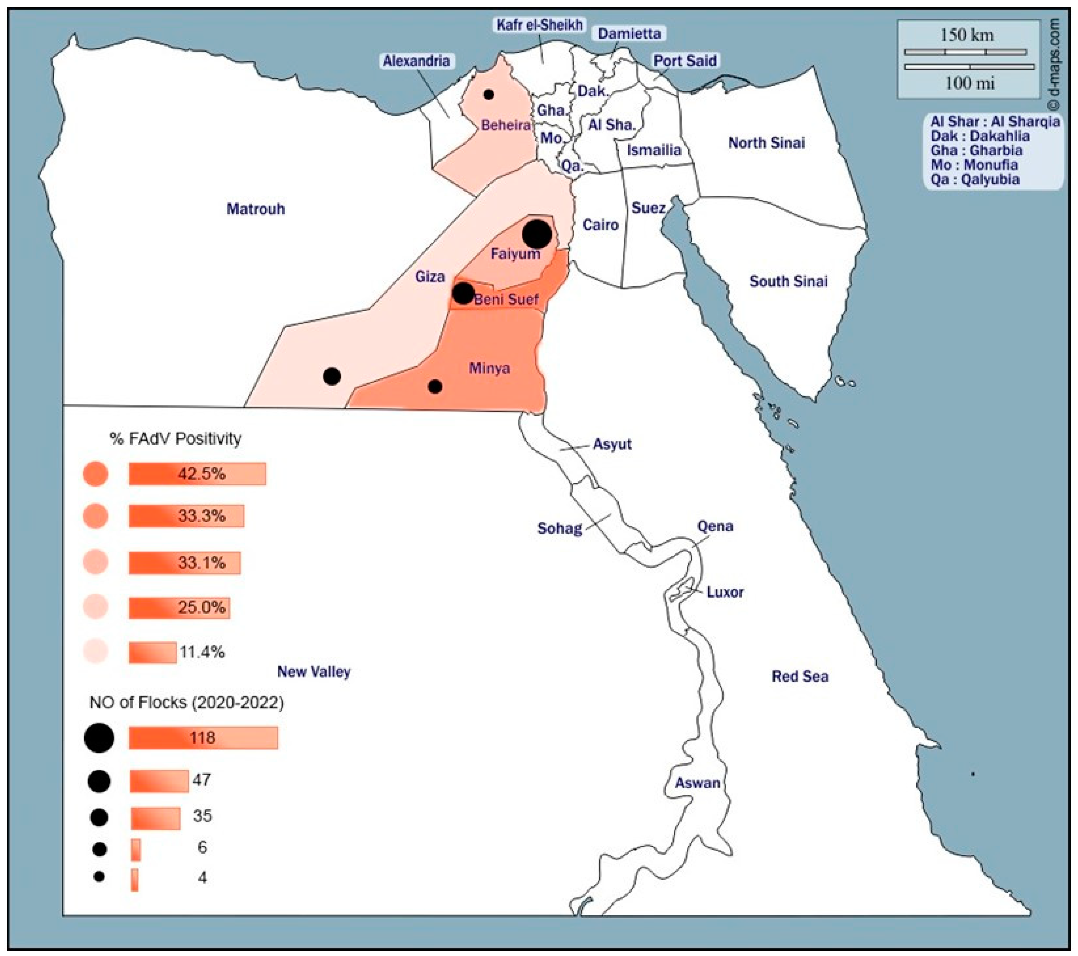
2.1. Study Area

The study was conducted in five Egyptian provinces (Menia, Beni-Suef, Fayoum, Giza, and Behira) from October 2020 through September 2022 (Figure 1).

2.2. Sample Collection and Preparation

Liver specimens (5–10 samples per flock) were collected from 208 broiler chicken farms (aged 1 day to 6 weeks), including freshly dead and euthanized moribund chickens, one native breed, and one commercial layer flock. The vaccination program of the broiler breeders was unavailable. The liver tissue samples were transferred to the laboratory on ice and stored at −80 °C for further processing. The collected liver specimens for each farm were pooled and homogenized using a sterile porcelain mortar and pistol. A sterile saline solution was used to prepare the 20% (w/v) suspensions.
Centrifugation of the suspensions was carried out for 10 min at 2000 rpm in a cooling centrifuge, and then the supernatant was collected and stored at −80 °C until further use [11]. Specimens from the liver (5 mm thickness) were also preserved in the Bouin solution for histopathological examination. The tissues were dehydrated, cleared, embedded in paraffin, and sectioned via microtome into 4 µm-thick sections. The histopathological sections were stained with hematoxylin and eosin stain and examined under a light microscope for the detection of inclusion bodies and other pathological changes [15].

2.3. Molecular Detection of FAdVs

Viral nucleic acid was extracted using a genomic DNA Mini kit (Geneaid, New Taipei City, Taiwan) according to the manufacturer’s instructions. The DNA extracts were kept at −20 °C for further analyses. A polymerase chain reaction (PCR) was performed using the sense hexon A (5′-CAARTTCAGRCAGACGGT-3′) and antisense hexon B (5′-TAGTGATGMCGSGACATCAT-3′) primers for the detection of FAdVs. The primers flank a region of 897 bp in positions 144–161 for primer hexon A and over positions 1040–1120 for primer hexon B [16]. DNA amplifications were carried out in a total volume of 50 μL containing 8 μL of viral DNA, 2 μL of a 20 pmol/µL concentration from each primer, 25 μL of ABT 2X PCR mix (Applied Biotechnology, New Cairo, Egypt), and 13 μL of PCR-grade water. The reactions were run in a Multigene Gradient TC9600-G-230V thermal cycler (Labnet International, Edison, NJ, USA) as follows: an initial denaturation step at 95 °C for 5 min; 35 cycles of amplification (each amplification cycle included secondary denaturation at 95 °C for 30 sec, annealing at 60 °C for 45 sec, and extension at 72 °C for 1.5 min), and a final extension step at 72 °C for 10 min. The amplified PCR products were analyzed via electrophoresis in a 1% agarose gel, stained with ethidium bromide 0.5 μg/mL [17], and visualized via UV transillumination.

2.4. Sequencing and Sequence Analysis of the L1 Region of the Hexon Gene

The amplified PCR products of size 897 bp were excised and purified using a GeneJet Gel Extraction Kit (GeneJet, Thermoscientific, Vilnius, Lithuania). The purified PCR products were shipped and subjected to sequencing reactions using a Big Dye Terminator v3.1 Cycle Sequencing Kit (Applied Biosystems, Foster City, CA, USA) according to the manufacturer’s specifications at Macrogen Korea. A multiple-nucleotide sequence alignment was assembled and analyzed with a representative reference to the FAdVs strains’ hexon gene sequences obtained from the GenBank database (http://www.ncbi.nlm.nih.gov/) using BioEdit software version 7.0 with the ClustalW alignment algorithm, with which the percentage identity matrices between different virus sequences were determined. Neighbor-joining phylogenetic trees were constructed using the distance-based method in MEGA software version 11.

3. Results

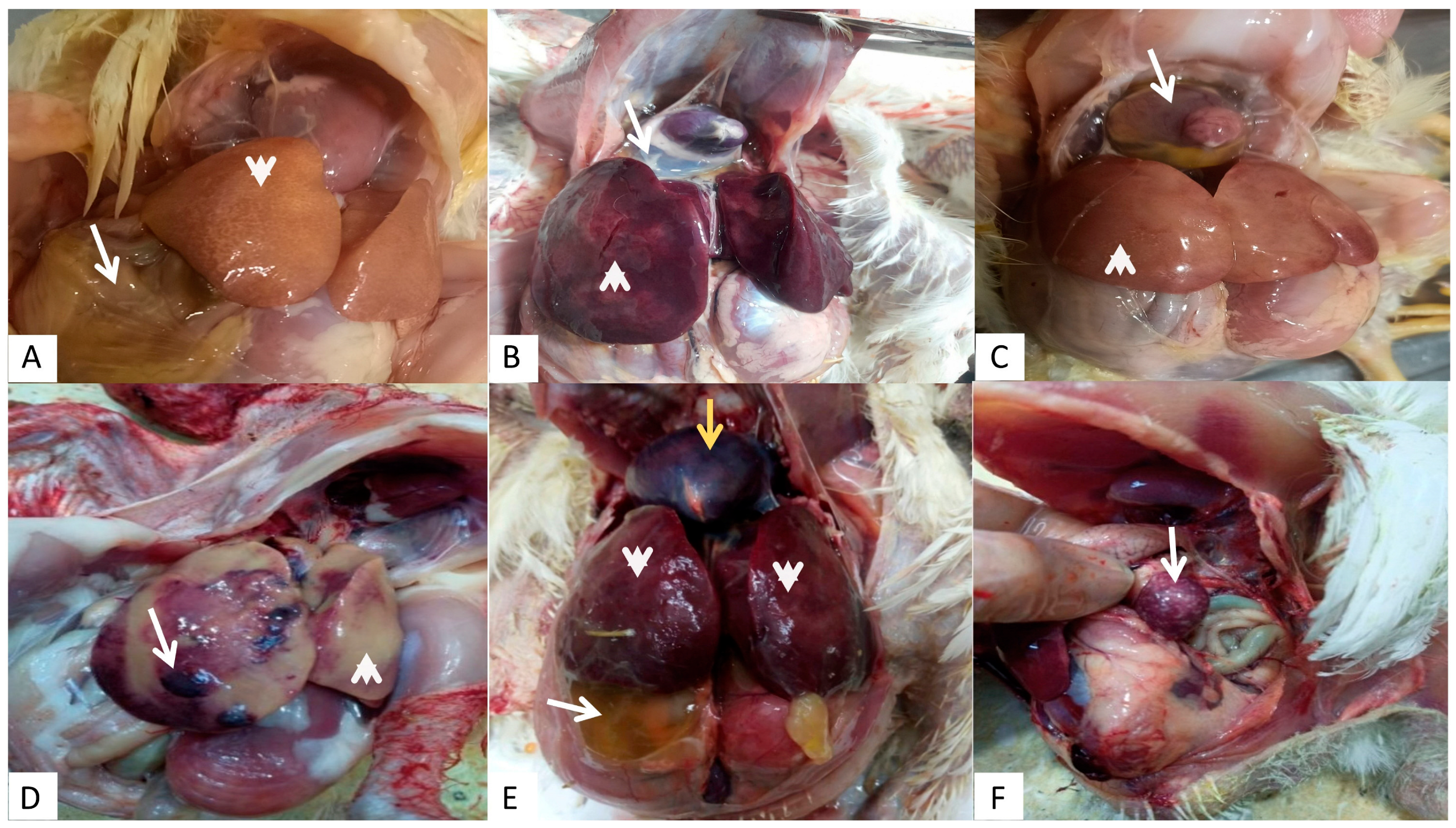
3.1. Clinical Cases and Gross Lesions

From October 2020 to September 2022, 210 flocks (208 broilers, one native breed, and one commercial layer flock) suffering from IBH or HPS in five Egyptian provinces were examined. The distribution and mortality rates among the different flocks are summarized in Supplementary Table S1. The most prominent gross lesions observed were enlarged hemorrhagic livers with necrotic foci and the accumulation of straw-colored serous fluid within the pericardial sac. Other associated lesions within other parenchymatous and lymphoid organs were observed, such as enlarged hemorrhagic kidneys and spleens with necrotic patches (Figure 2). The distribution and positivity percentages of IBH-HPS infection with different ages and systems of housing in the investigated chicken flocks are shown in Table 1 and Table 2.

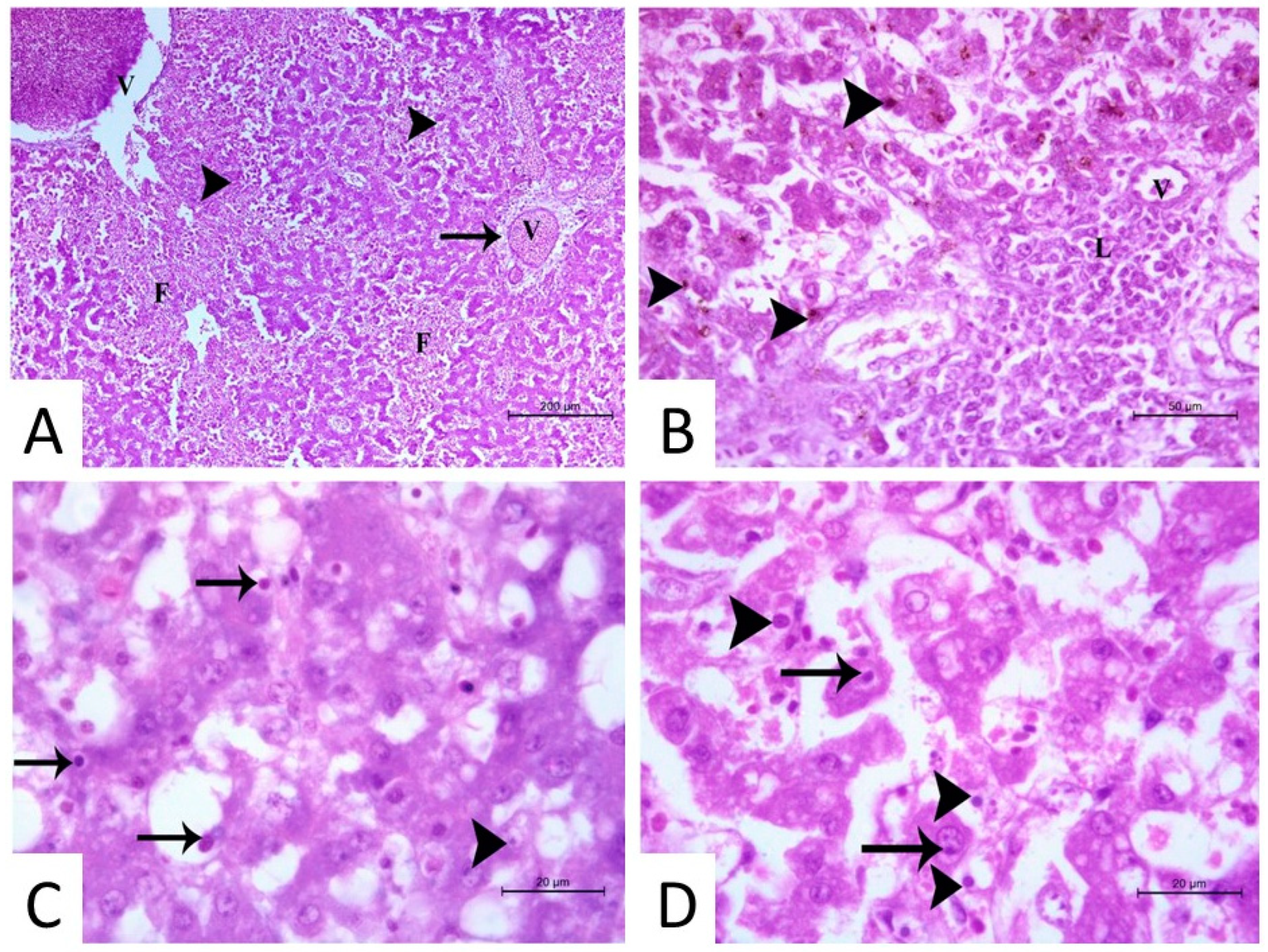
3.2. Histopathological Findings

The hepatic tissue appeared to be suffering from coagulative necrosis of the hepatocytes with prominent hemosiderin pigments, as well as hemorrhage; the infected cells appeared to be highly degenerated and vacuolated, and other hepatocytes appeared to be highly destructed, forming necrotic foci. Different forms and shapes of basophilic and acidophilic intranuclear inclusion bodies (INIBs) were prominent in the liver sections. The central veins and portal vessels were severely dilated and congested, with massive infiltration by lymphocytes and plasma cells around the portal area. Notably, massive fibrosis around the central vein and the portal area was evident (Figure 3).

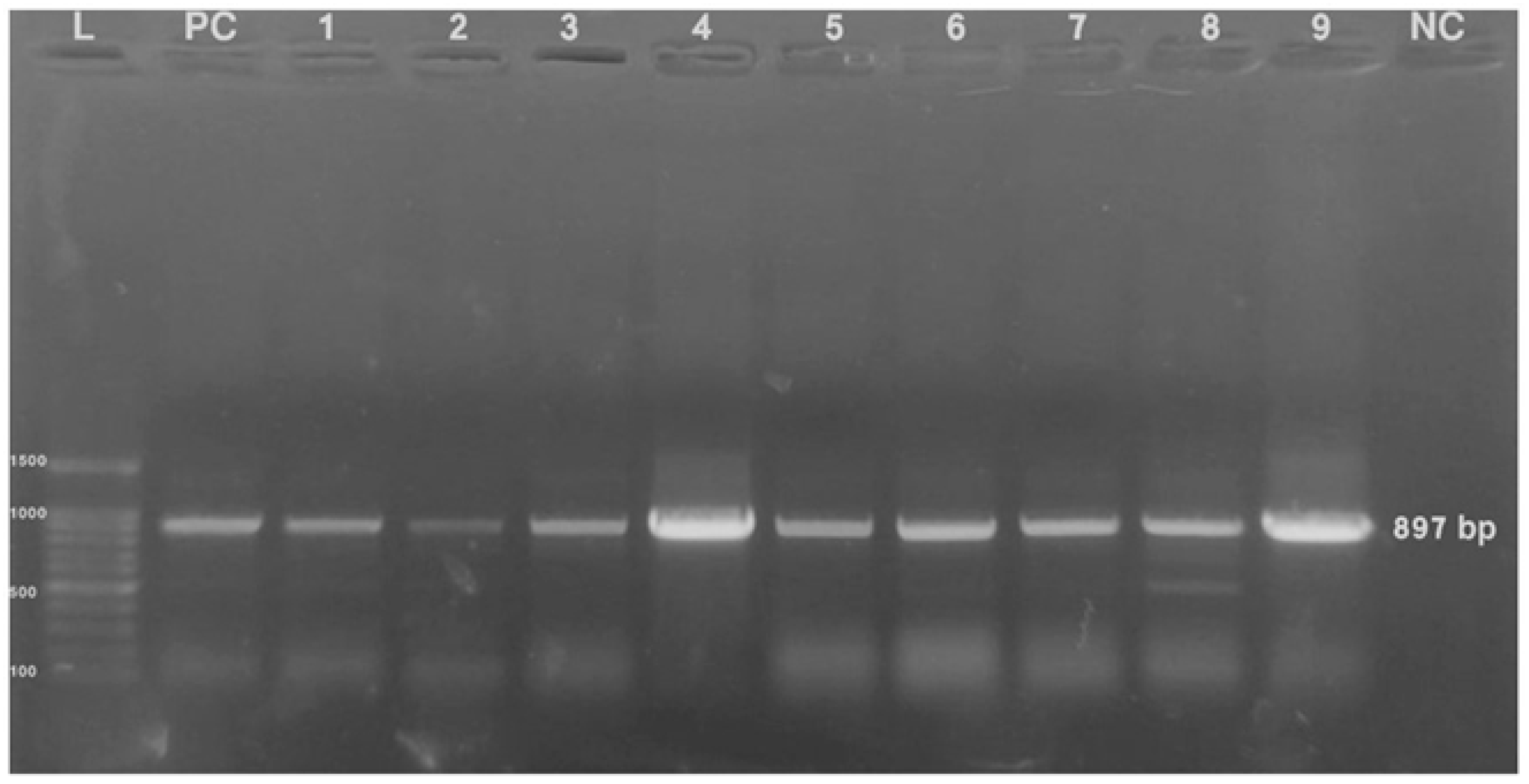
3.3. Virus Detection via PCR

A PCR was performed using fowl adenovirus hexon gene-specific primers targeting a PCR product of 897 bp (Figure 4) on the collected liver tissues. Of 210 examined flocks, 66 were positive for FAdVs (31.4%). The percentages of flocks found positive for FAdVs from samples collected at ages 1 to 10, 10 to 20, 21 to 30, and >30 days of age were 10% (5/50), 25.6% (11/43), 34.3% (23/67), and 54% (27/50), respectively. The percentages of FAdVs-positive flocks from samples collected from deep litter and cage system farms were 26.6% (46/173) and 54.1% (20/73), respectively.

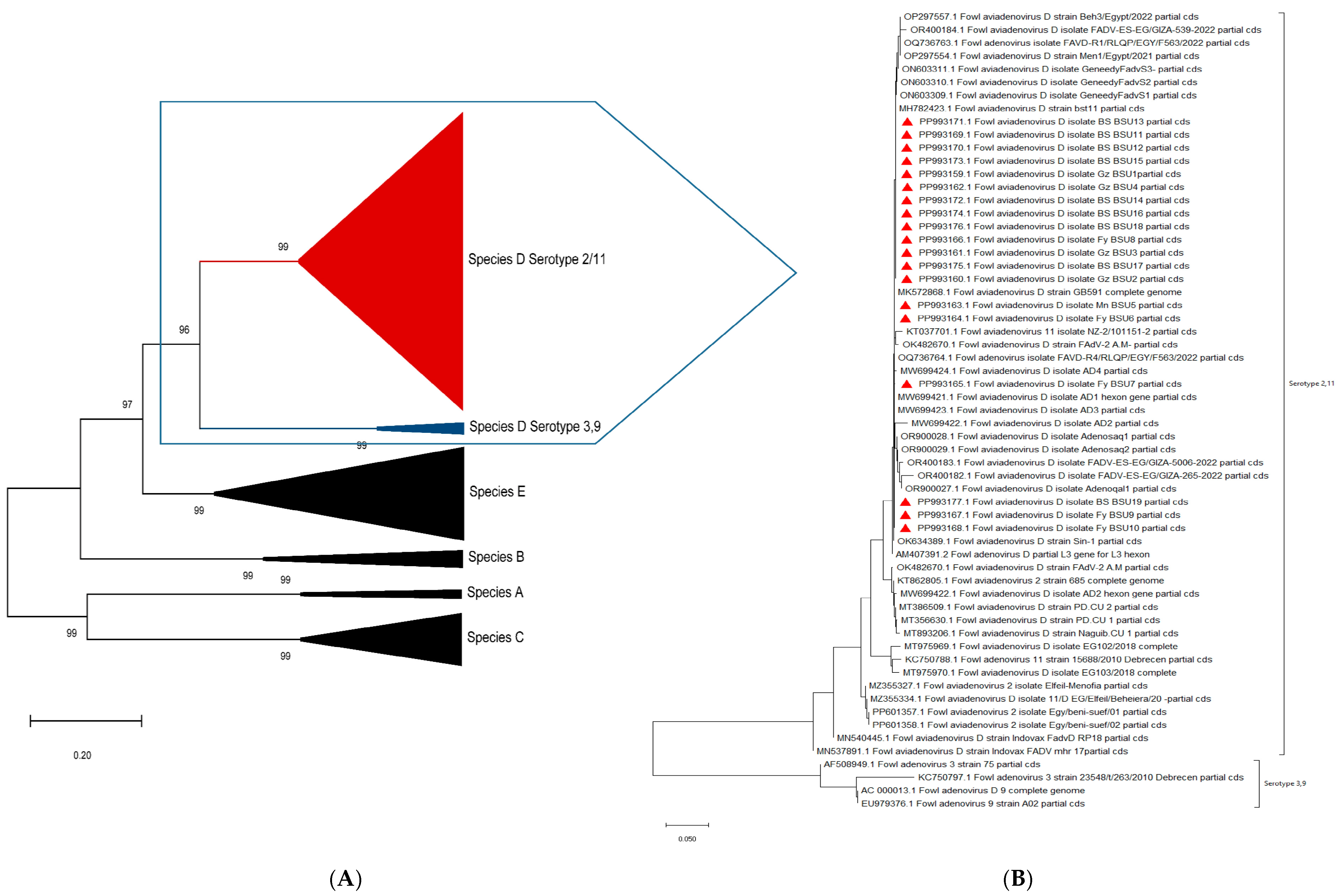
3.4. Gene Sequencing and Phylogenetic Analysis

The L1 regions of the hexon gene of the 19 selected FAdVs were sequenced, and the nucleotide sequence of each product was submitted to GenBank NCBI under various accession numbers (from PP993159 to PP993177). The 19 sequences obtained in the current study were aligned with other reference sequences of the different FAdV species (from A to E). The phylogenetic analyses and the pairwise identity matrix were created based on the alignment results. The phylogenetic analysis showed that the 19 sequenced FAdVs were clustered into fowl adenovirus species D serotype 2/11. A compressed phylogenetic tree was created based on our sequences, as well as the reference strain’s hexon gene and complete genome sequences from different countries that were available on the GenBank to confirm the clustering of the various FAdV species. Another phylogenetic tree expressing species D was constructed using our isolates and all of the Egyptian serotype 2/11 isolates until now (Figure 5). The genetic identity of the detected FAdV species D had a high homology (97.9%) to serotype 2 isolate accession number KT862805 from Egypt [18] and 94.9% to a serotype 2/11 isolate accession number MT975968 isolated from Poland [19] (Table 3).

3.5. Mutation Analysis of Amino Acid Residues

The amino acid sequences were aligned and the analysis showed several silent and non-silent mutations in the four HVRs (HVR1: n49–n243, aa17–aa81; HVR2: n244–n291, aa 82–aa97; HVR3: n337–n429, aa113–aa143; HVR4: n484–n501, aa162–aa167) [19] of the L1 region of the Egyptian and reference strains of 2/11 available on the GenBank NCBI and the sequences generated in this study (Table 4 and Supplementary Figure S1).

4. Discussion

IBH-HPS is considered an emerging vertically transmitted disease that impacts the broiler sector and leads to significant economic losses across the globe. While all serotypes of FAdV have been associated with significant IBH-HPS outbreaks, recent reports have documented that severe IBH-HPS outbreaks are primarily associated with serotypes D (FAdV-2 and FAdV-11) and E (FAdV-8a and 8b) [6].
Histopathology, a commonly used method for diagnosing numerous IBH-HPS outbreaks in various countries, including Canada and Iraq, remains an important diagnostic tool [20,21]. Other studies have also identified PCR as a reliable diagnostic tool [22]. In the current study, we utilized both histopathology and PCR targeting a 897 bp length of the hexon protein gene L1 region for the detection of FAdV in samples collected from chicken flocks [16]. The primers were used for partial hexon gene sequencing, as they flank a very important frame of the gene, which contains full-length 3HVR (2nd, 3rd, and 4th HVR) and a part of the 1st HVR of the hexon protein gene [19]. It has previously been reported that the virus persisted in the liver tissue for up to 35 days post-infection (dpi), with viral DNA detectable as early as three dpi, whereas the viral nucleic acid detection in cloacal swabs was intermittent [11]. In another study, viral DNA in cloacal swabs was detected at 3 and 6 dpi only, whereas in liver tissue, it was detectable at 3 dpi and persisted for up to 13 dpi [23,24].
It is well-known that internal organs, particularly the liver, are the primary sites for FAdV replication, with the viral load and viral DNA levels reported to be higher within liver tissue than in other organs, such as the kidney and spleen [25]. Therefore, in the current study, liver tissue specimens were targeted for FAdV DNA detection rather than cloacal swabs and internal organs other than the liver. An FAdV infection was considered positive when pathological alterations were noted in the hepatic tissues from FAdV PCR positive samples. Among the pathological changes are the presence of basophilic and eosinophilic INIB in the liver tissue, coagulative necrosis, a prominent deposition of hemosiderin pigments, and a large amount of fibrosis around the central vein and portal area [26,27]. The detection of viral-associated histologic lesions and viral nucleic acids among the tested flocks at different ages (from one day old to seven weeks old) throughout the study suggests a broad age susceptibility to FAdV infection [1,27,28].
Although FAdVs are associated with high mortality rates that may reach 40% in the case of IBH and up to 80% in the case of HPS, positive flocks exhibit non-specific clinical signs [29]. In this study, varying rates (1% to 14%) of mortality were noticed in FAdV-positive flocks. In line with these observations, significant variations in mortality rates (from 1% to 30%) were previously reported in different countries such as Japan, China, and Canada [30,31], indicating that several factors might aggravate the disease [1]. Grossly, the examined flocks had variable liver lesions, including hepatic enlargement with pale pinpointed foci, being congested with round edges, liver ecchymotic hemorrhage, and in some cases the liver appeared pale with hemorrhagic patches. Effusion of the pericardium with different amounts of straw-yellow-colored fluid from 1 mL up to 15 mL may explain the noticeable decrease in the size of the heart. Additionally, hemopericardium was seen in some cases. In addition, nephrosis and an enlarged spleen with necrotic patches were noticed, as in Figure 2. These lesions represent the typical gross features of IBH-HPS, as described in previous studies [9,32]. In this study, we noticed higher positivity rates of FAdV infection among flocks reared in cages than those reared in deep litter systems, and this could be attributed to the stressors (such as the higher stocking density and rate of metabolism encountered in cages) that play a role in exacerbating the multiplication and spread of FAdVs [33,34].
Notably, three of the investigated flocks (e.g., flocks #18, #19, and #20 in Supplementary Table S1) were positive for FAdVs at day one of age, highlighting the potential of FAdVs’ early transmission, either vertically from infected broiler breeders through eggs or horizontally post-hatching [26]. Moreover, two other flocks (#57 and #62) were negative for FAdVs at day one, and positive results were confirmed for the same flocks at an older age (at 21 and 33 days). This may be attributed to the low viral load in the samples collected earlier compared to those collected later for flock #57 or due to a horizontal infection at the farm during the rearing period for flock #62 (Supplementary Table S1). Additionally, an ascending pattern of FAdV-positive flocks was observed, beginning with 10% for young flocks (1–10 days old) and reaching 54% for flocks aged over 30 days [27]. Despite some studies indicated severe degenerative changes in FadV-infected chicks during the first week of life [35,36], the waning of the maternally derived antibodies (MDA) was reported to play an important role in the inactivation of virus replication in infected birds. If present, the MDA can delay FAdV’s replication and subsequent invasion of the virus into the internal organs, although it has no role in protecting against initial infection [37]. Further studies are needed to determine the role of vertically transmitted FAdVs in initiating severe outbreaks of disease.
Previous studies conducted in Egypt reported FAdV serotypes from various species, including serotype 8a [11,38] and 8b [13] species E and a new highly virulent FAdV serotype 4 species C [14], with two other different serotypes 1 and 3 belonging to different species A and B, respectively [13]. The current study only identified serotype 2/11, and it is worth mentioning that even though at least 110 sequences related to serotype 2/11 have been reported in Egypt, only one sequence of serotype 4, one sequence of serotype 1, two sequences of serotype 3, nine sequences of serotype 8a, and two sequences of serotype 8b from Egypt are available on the NCBI GenBank. These studies denote the widespread FAdV serotype 2/11 infection compared to other serotypes in Egypt [39].
Although the lengths of amino acid residues of the hexon protein are similar among FAdV-2/11 serotypes (950aa for each), they vary throughout other FAdV serotypes [40]. The primers used in the current study flank the most hypervariable region in the hexon gene, the L1 region, which is used to differentiate between the species of FAdVs, with seven hypervariable regions (HVRs 1–7) being recognized. Four HVRs are located in loop L1, two HVRs in loop L2, and the last one in loop L4; therefore, the L1 region is considered a type- or species-specific area, as it contains four HVRs, with characteristic nucleotide and amino acid sequence lengths for each FAdV species. For species 2/11, the lengths of the four HVR were as follows: HVR1: n49–n243, aa17–aa81; HVR2: n244–n291, aa 82–aa97; HVR3: n337–n429, aa113–aa143; HVR4: n484–n501, aa162–aa167 [19].
The major capsid proteins of FAdVs are efficient in triggering both cellular and humoral immune responses. The hexon protein, as it is the major structural component of the virus capsid and carries specific antigenic determinants on its surface, elicits the highest level of cellular or antibody response [41]. The L1 region represents one of the three variable loops (L1, L2, and L4) exposed on the outer surfaces between serotypes to form type-specific epitopes. It contains immunogenic HVRs on the surface of the hexon gene. HVRs from loops L1 and L2 code for type-specific antigenic determinants present on the hexon surface and are solely in charge of triggering type-specific neutralizing antibodies (NAbs). Furthermore, the L1 region’s HVRs load antigenic determinants unique to each serotype, especially the HVR1 [19]. The hexon molecule sizes differ according to the species and the serotype of FAdVs. A partial sequence analysis of the hexon gene of 19 samples in the current study indicated that all of them belong to FAdV-D serotype 2/11, which was also reported in many other countries, such as Canada [31], South Africa [35], China [32], Poland [19] Saudi Arabia [18], and Egypt [2,13,21,28,39].
The strains sequenced in our study shared from 99.5% upto 100% and from 98.0% upto 100% nucleotide and amino acids identity similarity between them, respectively. With the two reference strains, our sequences shared 94.2% to 94.9% and 94.7% to 95.9% nucleotide and amino acid identity similarity, respectively, with strain MT975968; and 96.7% to 97.9% and 96.2% to 97.4% nucleotide and amino acid identity similarity, respectively, with strain KT862805 (one of the only three isolates available with a full hexon gene sequence of Egyptian strain of FAdV 2/11) (Table 4). Differentiating serotype 2 from serotype 11 based solely on hexon gene sequencing is challenging, as FAdV-2 and FAdV-11 share a high degree of genetic identity and cross-neutralizing potential, and further studies are needed to address this issue. The percent identity similarity of FAdV species D detected in the present study was as high as 97.9% to serotype 2 isolate accession number KT862805 from Egypt [18] and 94.9% to a serotype 2/11 isolate accession number MT975968 isolated from Poland [19] (Table 3). Several studies were conducted to broaden the range of protection against FAdV infections in broiler flocks by the transferred MDA, a key factor in protecting the progeny until they develop age resistance to FAdV infections. These studies involved vaccines of broiler breeders containing either FAdV-11 mixed with FAdV-8a or -8b, which resulted in the development of antibodies against the related serotypes, as well as FAdV-2 and FAdV-7, as confirmed by a virus neutralization test (VNT) and challenge of the progeny [42]. In contrast, a single vaccination with live FAdV-8b raised antibodies against FAdV-8a and -8b but not against FAdV-2 or -11 [42,43]. It is, therefore, critical that FAdV be monitored continuously and that vaccine strains be evaluated and updated regularly to ensure sustained efficacy in disease prevention.
The deduced nucleotide and amino acid sequence analysis for the isolates detected in the current study with two reference strains (KT862805 and MT975968) retrieved from the GenBank NCBI revealed several silent and non-silent mutations in the four HVRs of LI of the hexon gene. Mutations in the sequenced region of the hexon protein gene were reported in all Egyptian strains available in the NCBI database and are tabulated in Table 4. The significance of the mutations reported in this study in the pathogenicity or immunogenicity of different FAdVs requires further studies.

5. Conclusions

Based on a sequencing analysis of loop 1 of the hexon gene, the most prevalent FAdV circulating in the targeted five Egyptian provinces was serotype 2/11 species D, which was associated with several outbreaks during the period 2020–2022. Further studies on the epidemiological situation, the role of vertical transmission in the spread, and the development of robust diagnostic tools and vaccines are essential for the establishment of better preventative measures for circulating FAdVs.

Supplementary Materials

The following supporting information can be downloaded at https://www.mdpi.com/article/10.3390/microorganisms13051107/s1: Figure S1. Nucleotide sequence (A) and deduced amino acid sequence (B) of strains of FAdV serotype 2/11 species D included in the current study. Table S1. Available data for fowl adenovirus-positive flocks detected from different investigated provinces.

Author Contributions

Conceptualization, D.M.A., A.A.E.-S., M.F.E. and S.A.S.S.; methodology, D.M.A., A.A., K.A. and S.A.S.S.; formal analysis, D.M.A., A.A., K.A. and S.A.S.S.; investigation, and data curation, D.M.A., A.A.E.-S., M.F.E., A.A. and S.A.S.S.; writing—original draft preparation, D.M.A., A.A., S.A.S.S., A.A.E.-S., M.F.E., K.A. and S.A.S.S. All authors have read and agreed to the published version of the manuscript.

Funding

This research received no external funding.

Institutional Review Board Statement

The authors declare that all procedures for chicken handling were carried out in compliance with the applicable legislation of the Institutional Animal Care and Use Committee (IACUC 022-444, 5 September 2023), Faculty of Veterinary Medicine, Beni-Suef University, Egypt.

Informed Consent Statement

Not applicable.

Data Availability Statement

The original contributions presented in this study are included in the article/supplementary materials. Further inquiries can be directed to the corresponding author.

Acknowledgments

The authors are grateful to the staff of the Poultry Diseases Department, Faculty of Veterinary Medicine, Beni-Suef University, Egypt, for their assistance and support; the Department of Histology, Beni-Suef University, Egypt, for their efforts with the histopathological techniques; and the Department of Parasitology, Beni-Suef University, Egypt, for granting permits for UV trans-illumination.

Conflicts of Interest

The authors declare that they have no known conflict or personal relationship that could have influenced the work reported in this paper.

References

  1. Hess, M. Commensal or pathogen—A challenge to fulfil Koch’s Postulates. Br. Poult. Sci. 2017, 58, 1–12. [Google Scholar] [CrossRef]
  2. Safwat, M.M.; Sayed, A.S.R.; Ali Elsayed, M.F.; Ibrahim, A. Genotyping and pathogenicity of fowl adenovirus isolated from broiler chickens in Egypt. BMC Vet. Res. 2022, 18, 325. [Google Scholar] [CrossRef] [PubMed]
  3. Franzo, G.; Faustini, G.; Tucciarone, C.M.; Pasotto, D.; Legnardi, M.; Cecchinato, M. Conflicting Evidence between Clinical Perception and Molecular Epidemiology: The Case of Fowl Adenovirus D. Animals 2023, 13, 3851. [Google Scholar] [CrossRef] [PubMed]
  4. El-Shall, N.A.; El-Hamid, H.S.A.; Elkady, M.F.; Ellakany, H.F.; Elbestawy, A.R.; Gado, A.R.; Geneedy, A.M.; Hasan, M.E.; Jaremko, M.; Selim, S.; et al. Epidemiology, pathology, prevention, and control strategies of inclusion body hepatitis and hepatitis-hydropericardium syndrome in poultry: A comprehensive review. Front. Vet. Sci. 2022, 9, 963199. [Google Scholar] [CrossRef]
  5. Ishag, H.Z.A.; Terab, A.M.A.; El Tigani-Asil, E.T.A.; Bensalah, O.K.; Khalil, N.A.H.; Khalafalla, A.I.; Al Hammadi, Z.; Shah, A.A.M.; Al Muhairi, S.S.M. Pathology and Molecular Epidemiology of Fowl Adenovirus Serotype 4 Outbreaks in Broiler Chicken in Abu Dhabi Emirate, UAE. Vet. Sci. 2022, 9, 154. [Google Scholar] [CrossRef]
  6. Schachner, A.; Matos, M.; Grafl, B.; Hess, M. Fowl adenovirus-induced diseases and strategies for their control—A review on the current global situation. Avian Pathol. 2018, 47, 111–126. [Google Scholar] [CrossRef]
  7. De Luca, C.; Hess, M. Vaccination strategies to protect chickens from fowl adenovirus (FAdV)-induced diseases: A comprehensive review. Vaccine 2025, 43 Pt 1, 126496. [Google Scholar] [CrossRef]
  8. Niu, Y.; Sun, Q.; Shi, Y.; Ding, Y.; Li, Z.; Sun, Y.; Li, M.; Liu, S. Immunosuppressive potential of fowl adenovirus serotype 4. Poult. Sci. 2019, 98, 3514–3522. [Google Scholar] [CrossRef]
  9. Franzo, G.; Prentza, Z.; Paparounis, T.; Tsiouris, V.; Centonze, G.; Legnardi, M.; Catelli, E.; Tucciarone, C.M.; Koutoulis, K.; Cecchinato, M. Molecular epidemiology of fowl adenoviruses in Greece. Poult. Sci. 2020, 99, 5983–5990. [Google Scholar] [CrossRef]
  10. Abdellatif, D.M.; El-Sawah, A.A.; Elkady, M.F.; Ali, A.; Shany, S.A.S. Inclusion Body Hepatitis: A Comprehensive Overview of Disease Impact and Control in Poultry. Egypt. J. Vet. Sci. 2025, 1–17. [Google Scholar] [CrossRef]
  11. Radwan, M.M.; El-Deeb, A.H.; Mousa, M.R.; El-Sanousi, A.A.; Shalaby, M.A. First report of fowl adenovirus 8a from commercial broiler chickens in Egypt: Molecular characterization and pathogenicity. Poult. Sci. 2019, 98, 97–104. [Google Scholar] [CrossRef] [PubMed]
  12. El-Basrey, Y.; Hamed, R.; Mohamed, M.; Lebdah, M. Detection of inclusion body hepatitis virus in broilers at Sharkia province, Egypt. J. Anim. Health Prod. 2020, 9, 84–89. [Google Scholar] [CrossRef]
  13. Adel, A.; Mohamed, A.A.E.; Samir, M.; Hagag, N.M.; Erfan, A.; Said, M.; Arafa, A.E.S.; Hassan, W.M.M.; El Zowalaty, M.E.; Shahien, M.A. Epidemiological and molecular analysis of circulating fowl adenoviruses and emerging of serotypes 1, 3, and 8b in Egypt. Heliyon 2021, 7, e08366. [Google Scholar] [CrossRef]
  14. Sultan, H.; Arafa, A.E.; Adel, A.; Selim, K.; Hossiny, M.; Talaat, S. Molecular Detection of a Novel Fowl Adenovirus Serotype-4 (FadV-4) from an Outbreak of Hepatitis Hydropericardium Syndrome in Commercial Broiler Chickens in Egypt. Avian Dis. 2021, 65, 385–390. [Google Scholar] [CrossRef]
  15. Suvarna, K.S.; Layton, C.; Bancroft, J.D. Bancroft’s Theory and Practice of Histological Techniques, 8th ed.; Elsevier: Amsterdam, The Netherlands, 2019; p. iii. [Google Scholar] [CrossRef]
  16. Meulemans, G.; Boschmans, M.; Berg, T.P.; Decaesstecker, M. Polymerase chain reaction combined with restriction enzyme analysis for detection and differentiation of fowl adenoviruses. Avian Pathol. 2001, 30, 655–660. [Google Scholar] [CrossRef]
  17. Lee, P.Y.; Costumbrado, J.; Hsu, C.Y.; Kim, Y.H. Agarose gel electrophoresis for the separation of DNA fragments. J. Vis. Exp. 2012, e3923. [Google Scholar] [CrossRef]
  18. Mohamed, M.H.A.; El-Sabagh, I.M.; Abdelaziz, A.M.; Al-Ali, A.M.; Alramadan, M.; Lebdah, M.A.; Ibrahim, A.M.; Al-Ankari, A.S. Molecular characterization of fowl aviadenoviruses species D and E associated with inclusion body hepatitis in chickens and falcons indicates possible cross-species transmission. Avian Pathol. 2018, 47, 384–390. [Google Scholar] [CrossRef]
  19. Niczyporuk, J.S. Deep analysis of Loop L1 HVRs1-4 region of the hexon gene of adenovirus field strains isolated in Poland. PLoS ONE 2018, 13, e0207668. [Google Scholar] [CrossRef]
  20. Dar, A.; Gomis, S.; Shirley, I.; Mutwiri, G.; Brownlie, R.; Potter, A.; Gerdts, V.; Tikoo, S.K. Pathotypic and molecular characterization of a fowl adenovirus associated with inclusion body hepatitis in Saskatchewan chickens. Avian Dis. 2012, 56, 73–81. [Google Scholar] [CrossRef]
  21. El-Tholoth, M.; Abou El-Azm, K.I. Molecular detection and characterization of fowl adenovirus associated with inclusion body hepatitis from broiler chickens in Egypt. Trop. Anim. Health Prod. 2019, 51, 1065–1071. [Google Scholar] [CrossRef]
  22. Gunes, A.; Marek, A.; Grafl, B.; Berger, E.; Hess, M. Real-time PCR assay for universal detection and quantitation of all five species of fowl adenoviruses (FAdV-A to FAdV-E). J. Virol. Methods 2012, 183, 147–153. [Google Scholar] [CrossRef]
  23. Abghour, S.; Mouahid, M.; Darkaoui, S.; Berrada, J.; Zro, K.; Kichou, F. Pathogenicity of field strain of fowl aviadenovirus serotype 11 isolated from chickens with inclusion body hepatitis in Morocco. PLoS ONE 2021, 16, e0261284. [Google Scholar] [CrossRef]
  24. Wang, T.; Meng, F.; Chen, C.; Shen, Y.; Li, P.; Xu, J.; Feng, Z.; Qu, X.; Wang, F.; Li, B.; et al. Pathogenicity and epidemiological survey of fowl adenovirus in Shandong Province from 2021 to 2022. Front. Microbiol. 2023, 14, 1166078. [Google Scholar] [CrossRef] [PubMed]
  25. Sun, J.; Zhang, Y.; Gao, S.; Yang, J.; Tang, Y.; Diao, Y. Pathogenicity of fowl adenovirus serotype 4 (FAdV-4) in chickens. Infect. Genet. Evol. 2019, 75, 104017. [Google Scholar] [CrossRef] [PubMed]
  26. Toro, H.; Gonzalez, O.; Escobar, C.; Cerda, L.; Morales, M.A.; Gonzalez, C. Vertical induction of the inclusion body hepatitis/hydropericardium syndrome with fowl adenovirus and chicken anemia virus. Avian Dis. 2001, 45, 215–222. [Google Scholar] [CrossRef] [PubMed]
  27. Chitradevi, S.; Sukumar, K.; Suresh, P.; Balasubramaniam, G.A.; Kannan, D. Molecular typing and pathogenicity assessment of fowl adenovirus associated with inclusion body hepatitis in chicken from India. Trop. Anim. Health Prod. 2021, 53, 412. [Google Scholar] [CrossRef]
  28. Elbestawy, A.R.; Ibrahim, M.; Hammam, H.; Noreldin, A.E.; Bahrawy, A.E.; Ellakany, H.F. Molecular characterization of fowl adenovirus D species in broiler chickens with inclusion body hepatitis in Egypt. Alex. J. Vet. Sci. 2020, 64, 110–117. [Google Scholar] [CrossRef]
  29. Rashid, F.; Xie, Z.; Wei, Y.; Xie, Z.; Xie, L.; Li, M.; Luo, S. Biological features of fowl adenovirus serotype-4. Front. Cell Infect. Microbiol. 2024, 14, 1370414. [Google Scholar] [CrossRef]
  30. Changjing, L.; Haiying, L.; Dongdong, W.; Jingjing, W.; Youming, W.; Shouchun, W.; Jida, L.; Ping, L.; Jianlin, W.; Shouzhen, X.; et al. Characterization of fowl adenoviruses isolated between 2007 and 2014 in China. Vet. Microbiol. 2016, 197, 62–67. [Google Scholar] [CrossRef]
  31. Ojkic, D.; Martin, E.; Swinton, J.; Vaillancourt, J.P.; Boulianne, M.; Gomis, S. Genotyping of Canadian isolates of fowl adenoviruses. Avian Pathol. 2008, 37, 95–100. [Google Scholar] [CrossRef]
  32. Zhang, J.; Xie, Z.; Pan, Y.; Chen, Z.; Huang, Y.; Li, L.; Dong, J.; Xiang, Y.; Zhai, Q.; Li, X.; et al. Prevalence, genomic characteristics, and pathogenicity of fowl adenovirus 2 in Southern China. Poult. Sci. 2024, 103, 103177. [Google Scholar] [CrossRef] [PubMed]
  33. Choi, K.S.; Kye, S.J.; Kim, J.Y.; Jeon, W.J.; Lee, E.K.; Park, K.Y.; Sung, H.W. Epidemiological investigation of outbreaks of fowl adenovirus infection in commercial chickens in Korea. Poult. Sci. 2012, 91, 2502–2506. [Google Scholar] [CrossRef]
  34. Matos, M.; Grafl, B.; Liebhart, D.; Hess, M. The outcome of experimentally induced inclusion body hepatitis (IBH) by fowl aviadenoviruses (FAdVs) is crucially influenced by the genetic background of the host. Vet. Res. 2016, 47, 69. [Google Scholar] [CrossRef]
  35. Maartens, L.H.; Joubert, H.W.; Aitchison, H.; Venter, E.H. Inclusion body hepatitis associated with an outbreak of fowl adenovirus type 2 and type 8b in broiler flocks in South Africa. J. S. Afr. Vet. Assoc. 2014, 85, e1–e5. [Google Scholar] [CrossRef]
  36. Lim, T.H.; Lee, H.J.; Lee, D.H.; Lee, Y.N.; Park, J.K.; Youn, H.N.; Kim, M.S.; Youn, H.S.; Lee, J.B.; Park, S.Y.; et al. Identification and virulence characterization of fowl adenoviruses in Korea. Avian Dis. 2011, 55, 554–560. [Google Scholar] [CrossRef] [PubMed]
  37. Hess, M. Aviadenovirus infections. In Diseases of Poultry; John Wiley & Sons, Inc.: Hoboken, NJ, USA, 2013; pp. 290–300. [Google Scholar] [CrossRef]
  38. Lebdah, M.; Alshaya, D.S.; Jalal, A.S.; Mousa, M.R.; Radwan, M.M.; Samir, M.; Adel, A.; Albaqami, N.M.; El-Saadony, M.T.; El-Tarabily, K.A.; et al. Molecular characterization of aviadenovirus serotypes and pathogenicity of the identified adenovirus in broiler chickens. Poult. Sci. 2022, 101, 101918. [Google Scholar] [CrossRef] [PubMed]
  39. Hossiny, M.; Abdelrazek, A.; Sultan, H. Molecular Characterization of Hydropericardium Hepatitis Syndrome in Broiler Chickens. J. Curr. Vet. Res. 2024, 6, 151–160. [Google Scholar] [CrossRef]
  40. Rashid, F.; Xie, Z.; Zhang, L.; Luan, Y.; Luo, S.; Deng, X.; Xie, L.; Xie, Z.; Fan, Q. Genetic characterization of fowl aviadenovirus 4 isolates from Guangxi, China, during 2017–2019. Poult. Sci. 2020, 99, 4166–4173. [Google Scholar] [CrossRef]
  41. Gupta, A. Pathogenesis and Control of Inclusion Body Hepatitis in Broiler Chickens. Ph.D. Thesis, University of Saskatchewan, Saskatoon, SK, Canada, 2018. [Google Scholar]
  42. Popowich, S.; Gupta, A.; Chow-Lockerbie, B.; Ayalew, L.; Ambrose, N.; Ojkic, D.; Gunawardana, T.; Kurukulasuriya, S.; Willson, P.; Tikoo, S.K.; et al. Broad spectrum protection of broiler chickens against inclusion body hepatitis by immunizing their broiler breeder parents with a bivalent live fowl adenovirus vaccine. Res. Vet. Sci. 2018, 118, 262–269. [Google Scholar] [CrossRef]
  43. Gupta, A.; Popowich, S.; Ojkic, D.; Kurukulasuriya, S.; Chow-Lockerbie, B.; Gunawardana, T.; Goonewardene, K.; Karunarathna, R.; Ayalew, L.E.; Ahmed, K.A.; et al. Inactivated and live bivalent fowl adenovirus (FAdV8b + FAdV11) breeder vaccines provide broad-spectrum protection in chicks against inclusion body hepatitis (IBH). Vaccine 2018, 36, 744–750. [Google Scholar] [CrossRef]
Figure 1. Distribution of fowl adenovirus serotype 2/11 in 5 Egyptian provinces, as well as the number of flocks collected from each province.
Microorganisms 13 01107 g001
Figure 2. Gross pathology of chickens naturally infected with FAdVS serotype 2/11 species D (HPS): (A) pale liver with severe necrosis (arrowhead) and beginning of ascites (arrow); (B) hydropericardium (arrow) with necrosis and enlargement of the liver (arrowhead); (C) severe hydropericardium, heart atrophy (arrow), and pale necrotic liver (arrowhead); (D) enlarged liver with areas of necrosis (arrowhead) and other areas of hemorrhages (arrow); (E) hemopericardium (yellow arrow), enlarged congested liver (arrowhead), and ascites (arrow); (F) mild to moderate enlargement and areas of necrosis in the spleen (arrow).
Microorganisms 13 01107 g002
Figure 3. Histopathology of livers from infected broiler chickens with fowl adenovirus species D serotype 2/11. (A) Congestion and dilation of central veins and portal vessels (V), degeneration of hepatocytes (arrowhead) forming necrotic foci (F), and massive fibrosis around the central vein and the portal area (arrow) (H & E staining, ×100) were observed. (B) The hepatic tissue appeared to be suffering from coagulative necrosis of hepatocytes (arrowhead) with prominent hemosiderin pigments. A massive infiltration by lymphocytes was detected around the portal area (L) around the portal vessels (V) (H & E staining, ×400). (C,D) Different forms (basophilic and acidophilic) and shapes of intra-nuclear inclusion bodies (arrow), lymphocytes, and plasma cells (arrowhead) were detected in the infected cells, which appeared to be highly degenerated and vacuolated (H & E staining, ×1000).
Microorganisms 13 01107 g003
Figure 4. Visualization of 897 bp PCR product of the hexon gene of fowl adenovirus via agarose gel electrophoresis (1%) after staining with ethidium bromide. Lane L: 100bp DNA ladder; lanes 1 to 9: positive samples; lane PC: positive control; lane NC: negative control.
Microorganisms 13 01107 g004
Figure 5. Compressed phylogenetic tree representing hexon gene all fowl adenovirus species (from A to E) (A) and detailed phylogenetic tree representing our sequenced isolates and other reference strains related to species D (B).
Microorganisms 13 01107 g005
Table 1. Distribution and positivity percentages of IBH-HPS infection at different ages in investigated chicken flocks.
ProvincePositivity at Different Ages (Positive/Total Tested (%))Totals
1–10 Days11–20 Days21–30 Days>30 Days
Menia0/01/11/30/22/6 (33.3%)
Beni-Suef4/173/48/165/1020/47 (42.5%)
^ Fayoum **1/227/2813/3618/3239/118 (33.1%)
Giza ***0/100/91/113/54/35 (11.4%)
Behera0/10/10/11/11/4 (25%)
Subtotal5/50 (10%)11/43 (25.6%)23/67 (34.3%)27/50 (54%)66/210 (31.4%)
^ Flocks included 117 broilers and one commercial layer. ** For samples collected from Fayoum and Giza provinces, one flock from each was negative early (at 1 and 4 days old, respectively) and positive later (at 21 and 33 days old, respectively). *** Flocks included 34 broilers and one native-breed chicken.
Table 2. Distribution and positivity percentages of IBH-HPS infection in different housing systems.
ProvinceSystem of Housing (Positive /Total Tested (%))Totals
Deep LitterCages
Menia2/60/02/6
Beni-Suef13/387/920/47 (42.5%)
Fayoum26/9013/2839/118 (33.1%)
Giza4/3504/35 (11.4%)
Behera¼01/4 (25%)
Subtotal46/173 (26.6%)20/37 (54.1%)66/210 (31.4%)
Table 3. Nucleotide and amino acids identities of identified FAdVs in the current study.
Strain123456789101112131415161718192021
Nucleotide idenity %
1. KT862805-FAdV 2 strain 685 UK 96.797.697.897.897.497.797.897.497.797.997.997.797.697.497.697.797.697.897.697.8
2. MT975968 FAdV D isolate EG101/201896.3 94.394.894.894.294.794.794.294.694.994.994.494.594.294.794.694.594.894.594.9
3. PP993159 FAdV D isolate Gz BSU196.595 10010099.999.999.999.710099.999.910010099.910010010010010099.9
4. PP993160 FAdV D isolate Gz BSU296.995.599.6 10099.999.999.999.610099.999.910010099.999.910010010010099.9
5. PP993161 FAdV D isolate Gz BSU396.995.599.699.7 99.999.999.999.610099.999.910010099.999.910010010010099.9
6. PP993162 FAdV D isolate Gz BSU496.69599.699.699.6 99.799.799.699.999.799.799.899.999.799.999.999.999.999.999.7
7. PP993163 FAdV D isolate Gz BSU597.395.899.699.699.699.6 10099.799.910010099.999.999.999.799.999.999.999.9100
8. PP993164 FAdV D isolate Gz BSU697.495.999.699.699.699.6100 99.799.910010099.999.999.799.999.999.999.999.9100
9. PP993165 FAdV D isolate Gz BSU796.594.999.298.898.899.299.299.2 99.699.799.799.699.699.599.699.699.699.699.699.7
10. PP993166 FAdV D isolate Gz BSU896.795.299.699.699.699.699.699.698.4 99.999.910010099.999.910010010010099.9
11. PP993167 FAdV D isolate Gz BSU997.295.799.299.399.399.210010099.299.3 10099.999.999.799.999.999.999.999.9100
12. PP993168 FAdV D isolate Gz BSU1097.295.899.299.399.399.210010099.299.399.7 99.999.999.799.999.999.999.999.9100
13. PP993169 FAdV D isolate Gz BSU1196.394.799.699.699.699.699.299.298.399.699.299.2 10010010010010010010099.9
14. PP993170 FAdV D isolate Gz BSU1296.594.999.699.699.699.699.299.298.399.699.299.299.6 10010010010010010099.9
15. PP993171 FAdV D isolate Gz BSU1396.294.799.299.399.399.299.698.99899.398.998.999.699.6 99.999.999.999.999.999.7
16. PP993172 FAdV D isolate Gz BSU1496.595.199.699.399.399.699.299.698.899.399.399.399.699.699.3 10010099.910099.9
17. PP993173 FAdV D isolate Gz BSU1597.195.610010010010099.699.698.899.699.699.699.699.699.3100 10010010099.9
18. PP993174 FAdV D isolate Gz BSU169795.510010010010099.699.698.810099.699.699.699.699.3100100 10010099.9
19. PP993175 FAdV D isolate Gz BSU1796.895.499.699.799.799.699.699.698.899.699.399.399.699.699.399.3100100 10099.9
20. PP993176 FAdV D isolate Gz BSU1896.795.299.699.699.699.699.699.698.599.699.399.399.699.699.399.699.610099.6 99.9
21. PP993177 FAdV D isolate Gz BSU1997.195.799.299.399.399.210010099.299.399.799.799.299.298.999.399.699.699.399.3
Amino acid identity %
Table 4. An amino acid mutation analysis of all Egyptian strains related to FAdV serotype 2/11 species D.
RegionAmino Acids PositionMajorityStrains in this Study (No. 19)Other Egyptian Strains (No. 91)
HVR1
(17–81)
58T--I (1/33), -- (N = 58)
59R--H (1/34), -- (N = 57)
60N--Y (1/34), -- (N = 57)
61VV (3/19), -- (N = 16)I (1/34), L (1/34), -- (N = 57)
62TT (5/19), S (1/19), -- (N = 13)A (1 /34), -- (N = 57)
63TT (11/19), -- (N = 8)A (1/34), -- (N = 57)
75PP (16/19), -- (N = 3)A (3/34), -- (N = 57)
78TT (17/19), -- (N = 2)-- (N = 57), H (1/34), N (1/34)
79DD (17/19), -- (N = 2)G (1 /34), -- (N = 57)
HVR2
(82–97)
84SS (18/19), G (1/19)S (33), -- (N = 58)
92NN (19/19)D (1/34), H (1/34), -- (N = 57)
HVR3
(113–143)
113DD (19/19)G (1/75), -- (N = 16)
114RR (19/19)G (1/75), S (1/75), -- (N = 16)
115GG (19/19)R (1/75), -- (N = 16)
116PP (19/19)S (1/75), -- (N = 16)
117SS (19/19)F (2/75), -- (N = 16)
118FF (19/19)S (1/76), P (1/76), N (1/76), -- (N = 15)
119KK (19/19)T (4/78), N (2/78), Q (1/87), -- (N = 13)
120PP (19/19)K (1/79), A (1/79), L (1/79), -- (N = 12)
121YY (19/19)S (1/83), -- (N = 8)
122GG (19/19)V (1/83), A (1/83), -- (N = 8)
123GG (19/19)E (1/84), -- (N = 7)
124TT (19/19)A (1/84), G (1/84), -- (N = 7)
125AA (19/19)T (1/90), V (1/90), D (1/90), G (1/90), -- (N = 1)
126YY (19/19)K (1/90), H (1/90), -- (N = 1)
135FF (19/19)L (1/91)
140II (19/19)V (10/91), L (3/91)
141DD (19/19)G (1/91), Q (1/91), E (1/91)
142TT (19/19)S (2/91), A (1/91)
143GG (19/19)E (11/91), D (2/91)
HVR4
(162–167)
162SS (19/19)G (7/91), A (2/91) T (1/91)
163AA (19/19)N (3/91)
164KK (19/19)N (2/91), Q (1/91)
165DD (19/19)T (3/91)
166KK (18/19), T (1/19)N (3/91), E (1/91), T (1/91)
Abbreviations: --: unavailable; A: alanine; E: glutamic acid, isoleucine; V: valine; R: arginine; Q: glutamine; L: leucine; P: proline; N: asparagine; G: glycine; K: lysine; S: serine; D: aspartic acid; H: histidine; F: phenylalanine; T: threonine; Y: tyrosine. These hypervariable regions (HVR1-4) were determined as previously described [19].
Disclaimer/Publisher’s Note: The statements, opinions and data contained in all publications are solely those of the individual author(s) and contributor(s) and not of MDPI and/or the editor(s). MDPI and/or the editor(s) disclaim responsibility for any injury to people or property resulting from any ideas, methods, instructions or products referred to in the content.

