Communication

Non-Secretor Status Due to FUT2 Stop Mutation Is Associated with Reduced Rotavirus Infections but Not with Other Enteric Pathogens in Rwandan Children

1
Department of Microbiology and Parasitology, School of Medicine and Pharmacy, University of Rwanda, Kigali P.O. Box 3286, Rwanda
2
Department of Infectious Diseases, Institute of Biomedicine, University of Gothenburg, 405 30 Gothenburg, Sweden
3
Department of Pathology, University Teaching Hospital of Kigali, Kigali P.O. Box 655, Rwanda
4
Department of Pediatrics, School of Medicine and Pharmacy, University of Rwanda, Kigali P.O. Box 3286, Rwanda
5
Department of Pediatrics, University Teaching Hospital of Kigali, Kigali P.O. Box 655, Rwanda
6
Department of Internal Medicine, Rwanda Military Referral and Teaching Hospital, Kigali P.O. Box 3377, Rwanda
7
Department of Internal Medicine, School of Medicine and Pharmacy, University of Rwanda, Kigali P.O. Box 3286, Rwanda
8
Department of Laboratory Medicine, Institute of Biomedicine, University of Gothenburg, 405 30 Gothenburg, Sweden
9
Department of Microbiology and Immunology, Institute of Biomedicine, University of Gothenburg, 405 30 Gothenburg, Sweden
*
Author to whom correspondence should be addressed.
Microorganisms 2025, 13(5), 1071; https://doi.org/10.3390/microorganisms13051071
Submission received: 20 March 2025 / Revised: 28 April 2025 / Accepted: 29 April 2025 / Published: 3 May 2025
(This article belongs to the Section Gut Microbiota)

Abstract

:
Enteric pathogens remain a health threat for children in low-income countries. A single nucleotide polymorphism (SNP) in the FUT2 gene that precludes the expression of fucosyltransferase 2 has been reported to influence the susceptibility to rotavirus and norovirus infections. The aim of this study was to investigate the association between G428A at rs601338 (stop codon variant) in the FUT2 gene and a range of enteric pathogens in children under 5 years of age. Rectal swab samples from 668 children (median age 13.6 months, 51% males, 93% rotavirus vaccinated, 468 with diarrhea) from Rwanda were analyzed via PCR for pathogen detection and SNP genotyping. A FUT2 stop codon (‘non-secretor’ status) was found in 19% of all children. Rotavirus was detected in 5.3% of non-secretors compared with in 13% of secretors (OR = 0.39, p = 0.019). Rotavirus P[8] was the predominant genotype and was found in 2.3% of non-secretors compared with 8.8% of secretors (p = 0.009). There was no association with any other pathogen, including noroviruses, of which 2 of 14 GII.4 infections were detected among non-secretors. Thus, the FUT2 stop codon variant was associated with rotavirus but not with any other pathogen.

1. Introduction

Enteric infections pose a significant threat to the health of children under five in low- and middle-income countries [1] where living conditions and lack of hygiene strongly contribute to the transmission of enteric pathogens [2,3]. Host genetic factors, represented by single nucleotide polymorphisms (SNPs), may influence an individual’s susceptibility to and clearance of infections, for example, hepatitis C, HIV, malaria and norovirus [4,5].
The FUT2 gene encodes a fucosyltransferase that is responsible for the presence of the so-called H antigen in secretions and mucosal tissues, including the gastrointestinal tract. People who are homozygotes for the A allele at the SNP rs601338 (known as the G428A stop codon variant) have a non-functional FUT2 gene and are referred to as non-secretors. They constitute approximately 20% of Caucasian and African populations and reportedly have a lower susceptibility for certain rota- and norovirus subtypes, such as the P[8] genotype of rotavirus and the GII.4 genotype of norovirus [6,7,8,9,10,11,12]. Both the circulating viral genotypes and FUT2 genotypes may vary geographically, and studies from additional regions are warranted to better understand the impact of secretor status on the susceptibility to rotavirus and norovirus infections. It is also of interest to investigate the possible relation between secretor status and other pathogens than those already studied. Therefore, we have investigated the potential impact of FUT2-related secretor status on a broad range of enteric infections in among children in Rwanda, where such data are lacking.

2. Methods

2.1. Study Design, Study Area and Characteristics of Participants

Rectal swab samples from children who had participated in two recent studies of enteric infections in Rwanda were analyzed by rs601338 SNP genotyping. One of them was cross-sectional and included 794 children with or without diarrhea between 6 September and 5 November 2021 [13]. The other was a study of 120 children who had a long-term follow-up after an episode of diarrhea and was conducted from 12 June to 15 December 2022. For the present study, we included the 668 children whose rectal swab samples contained enough human DNA to be genotyped for the rs601338 SNP. These included, from the first study, 362 samples from children with diarrhea and 198 without diarrhea, and from the second study, 108 samples collected at the enrollment when the children presented with diarrhea. Only one sample per child was included.
The median age of study participants was 13.6 months (age distribution is shown in Figure 1), 51% were boys and 93% were rotavirus vaccinated. Seventy percent had diarrhea at the time of sample collection.

2.2. PCR for Pathogen Detection

Real-time PCR targeting a broad range of pathogens was performed as previously described [13], with primers and probes described in Supplementary Table S1. Briefly, 250 µL of rectal swab sample were mixed with 2 mL of lysis buffer followed by total nucleic acid extraction in an EasyMag instrument (Biomerieux, Marcy l’Étoile, France). The nucleic acids were eluted in 110 μL volume, and 5 μL of this was used for 45 cycles of PCR in a QuantStudio 6 flex instrument (Applied Biosystems, Foster City, CA, USA) in 8 parallel amplifications using a two-step setting (15 s at 95 °C, 60 s at 56 °C).
A real-time PCR specific for norovirus GII.4 was applied on samples positive for norovirus GII in the multiplex PCR (forward primer, GATGGGTCCACAGCCAACCT; reverse primer, CCGCTACAGGTGCCGCAA; probe ACGGGCTCCAAAGCCATAACCTCATT) [14]. Rotavirus genotyping was performed by real-time PCR as previously described [15].

2.3. FUT2 SNP Genotyping

For FUT2 SNP genotyping at rs601338, a segment of the FUT2 gene was amplified by 45 cycles of PCR (15 s at 95 °C, 60 s at 60 °C) with primers FUT2F, GCAGAACTACCACCTGAACGACT, FUT2R, GTGGTCGTGCAGGGTGAAC and allele-specific probes FUT2-FAM-MGB_G, CTGCTCCTGGACCTT and FUT2-VIC-MGB_A, CTGCTCCTAGACCTT in a Taqman genotyping master mix (Applied Biosystems). The genotype (AA, GA or GG) was identified by the allele analysis setting in the QuantStudio 6 Pro instrument (Applied Biosystems).

2.4. Data Analysis

The IBM SPSS Statistics 28.0 software (IBM Corporation, New York, NY, USA) was used for data analyses. The relationship between FUT2 genotypes and pathogens was examined using a two-tailed Fisher exact test. Multiple logistic regression analysis was used with diarrhea as the outcome (dependent variable) and each pathogen and FUT2 status as independent variables.

2.5. Ethical Approval

The study was approved by the University of Rwanda’s ethics committee (226/CMHS IRB/2021) and the University Teaching Hospital of Kigali’s ethics council (EC/CHUK/110/2021). Informed consent to participate was obtained from the children’s parents or legal guardians.

3. Results

3.1. Pathogen Association with Diarrhea and FUT2 Genotype

Among all study participants, 19% were homozygous for the FUT2 rs601338 A allele and were thus defined as a non-secretor or FUT2, while 42% had a GA genotype and 39% a GG genotype and thus were defined as FUT2+. The proportion of FUT2 did not differ significantly between children with or without diarrhea (18% vs. 23%, p = 0.13). As shown in Table 1, only rotavirus was associated with the FUT2 genotype, with a significantly (p = 0.019) lower frequency among non-secretors (7/132, 5.3%) than among secretors (67/536, 13%). Since the impact of secretor status might be confined to symptomatic infection, we compared the pathogen detection rates for children with or without diarrhea in secretors and non-secretors. Several pathogens were associated with diarrhea with relatively high odds ratios, including rotavirus, Shigella, ETEC, Cryptosporidium and Campylobacter, and as shown in Table 2 this association was apparent for children with the FUT2+ genotype (secretors). If non-secretors had been protected from diarrhea, then the OR would be lower than for secretors. This was seen only for rotavirus (OR 1.35 vs. 3.84), whereas other pathogens associated with diarrhea had a similar elevation of OR in secretors and non-secretors (but with p above 0.05 in the FUT2 group, likely reflecting the low number of cases).
Multiple regression analysis showed that the association between the FUT2 genotype and rotavirus remained significant when age, sex, and rotavirus vaccination were considered. ETEC-eltB was strongly associated with symptomatic infection with an OR = 3.67 (p < 0.0001) for secretors and had a lower OR (1.95, p = 0.21) for non-secretors, suggesting that non-secretors might be protected against diarrhea, but the difference was not statistically significant by interaction term analysis. A complementary analysis of the potential impact on symptoms is shown in Table 3, comparing the risk of diarrhea by FUT2 status for children who were infected. Elevated odds ratios were seen for rotavirus, ETEC-eltB and Salmonella, suggesting that FUT2 children might have a lower risk of symptomatic infection by these pathogens, but the associations were not statistically significant (p values 0.10–0.20).

3.2. Rotavirus P Types and Secretor Status

Samples positive for rotavirus were further genotyped for virus variants. Rotavirus P[8] was the most prevalent P genotype and was found in 50 of the 61 (82%) rotavirus-positive samples that could be genotyped. The number of cases was too low to accurately analyze differences between the P genotypes. As shown in Table 4, the P[8] genotype of rotavirus was detected in 2.3% (3/132) of FUT2 compared with 8.8% (47/536) of FUT2+ children (p = 0.009), supporting that at least infection by this subtype is restricted by secretor status.

3.3. Norovirus GII and Secretor Status

Norovirus GII was detected in 52 of 536 secretors (9.7%) and in 17 of 132 non-secretors (13%), which contrasts with previously reported associations between non-secretor status and lower susceptibility to norovirus infection [6]. Since GII.4 has been the predominant genotype of norovirus GII in recent decades [16,17], we investigated the prevalence of this subtype with a specific real-time PCR [14]. This analysis identified GII.4 among 2/17 non-secretors (12%), compared with 12/52 secretors (23%, p = 0.49) with norovirus GII infection. The relatively small total number of GII.4 infections might, to some extent, be an underestimation due to the additional freeze–thawing prior to subtyping or to a lower sensitivity of the subtyping than with the initial PCR.

4. Discussion

The observed frequencies of FUT2 rs601338 SNP genotypes in this study in Rwanda, with 19% carrying the AA genotype (stop codon homozygote, ‘non-secretor’), are similar to previous findings in Burkina Faso and South Africa, where 21% and 17% were non-secretors [6,18]. Rotavirus infections were significantly less common among non-secretors (OR = 0.39, p = 0.019) also when age, sex and rotavirus vaccination were considered, which aligns with previous studies suggesting that FUT2 secretor status influences the susceptibility to rotavirus infection [9,12,19] by controlling the synthesis of glycans recognized by the virus [20]. In the first published studies, the non-secretor phenotype appeared to confer complete resistance against P[8] strains of rotavirus [10,19,21], but results from subsequent studies suggest that P[8] can also infect non-secretors [7]. We found that the P[8] genotype was less frequent in non-secretors than in secretors (2.3% vs. 8.8%, p = 0.009), supporting that non-secretors are significantly but not completely protected against P[8] infection. Likewise, a study from Tunisia found that 15% of the P[8]-infected subjects were non-secretors and proposed that this might reflect that preexisting conditions may influence susceptibility [22]. A more recent study from Tunisia, however, showed that the susceptibility was linked to the rotavirus P[8] subtype and that subtype P[8]-4 preferentially infects non-secretors while other P[8] strains, as well as P[4], preferentially infect secretors [23]. We found only one non-secretor among the 11 children infected with P[4], P[6] or P[9] rotavirus strains, suggesting possible protection (OR = 0.40), but the low number of patients precludes statistical evaluation (p = 0.70).
The lower rate of rotavirus infections among non-secretors that we observed reflects the situation among vaccinated children, who constituted 93% of the children in our study. Non-secretors might develop a poorer response to the vaccine [7], and it would be interesting to compare the impact of FUT2 status in vaccinated and unvaccinated children, but the latter group was too small to allow this analysis.
Norovirus has also been shown to bind to FUT2-dependent glycans in a strain-dependent manner [24,25]. Non-secretors are protected from infection by strains from GII.4 [26] and possibly also GII.3 strains [27], while other less common strains such as GII.2 [28] seem to infect in a FUT2-independent manner [29]. Our finding of norovirus GII in 13% of non-secretors compared with 9.7% of secretors does not support that non-secretor status provides protection against the norovirus GII strains that circulate in Rwanda. This observation was surprising and suggests that secretor-independent strains of norovirus GII were more common than secretor-dependent strains in Rwanda at the time of our study. This possibility was supported by our use of an additional GII.4-specific PCR, which detected GII.4 in only 14 of the 71 norovirus GII-positive samples. Two of these 14 GII.4 strains were found in non-secretors (12%), compared with 12 in secretors (23%). This difference was not statistically significant (p = 0.49) but suggests that non-secretors might be less susceptible to GII.4 infection compared with other GII types.
Previous studies have reported FUT2 activity to be associated with inflammatory bowel disease (IBD) [30] and Campylobacter infection [31], and secretors from Bangladesh have been reported to be less likely to have symptomatic enteric infections by certain ETEC strains [32,33]. We found that the odds ratios for diarrhea among secretors versus non-secretors were elevated for rotavirus, ETEC-eltB and Salmonella (however with p values 0.10–0.20), suggesting non-secretors might be protected from symptomatic infection by these pathogens. Further studies, including a higher number of non-secretors, and more detailed characterization of the pathogens (including subtyping of rotavirus and norovirus) are needed to clarify if and how secretor status might be associated with infection and diarrhea.
A strength of our study is that it explores possible associations between FUT2 inactivation and infection susceptibility for a broad range of enteric pathogens, but we did not find any previously unrecognized association. It should be pointed out though that we only investigated a marker for FUT2 activity, and that susceptibility for other infections might rather depend on FUT3 which determines the expression of the Lewis antigen on intestinal mucosa [10,24]. The relatively low number of norovirus infections is a limitation that precludes conclusions about the impact of FUT2 status on norovirus infection, and due to the cross-sectional design of the study potential influence by FUT2 status on infection duration or severity over time could not be investigated.
In summary, we found a significant association between FUT2 status and a lower frequency of rotavirus infection and specifically with rotavirus genotype P[8], but no association with infection with norovirus or other pathogens.

Supplementary Materials

The following supporting information can be downloaded at https://www.mdpi.com/article/10.3390/microorganisms13051071/s1. Table S1: Primers and probes for real-time PCR.

Author Contributions

Conceptualization, M.E.A. and M.L.; Methodology, M.E.A. and M.L.; Formal analysis, J.B.M., S.N., G.E.R., A.M., M.E.A. and M.L.; Investigation, J.B.M.; Data curation, J.B.M., S.N. and M.E.A.; Writing—original draft, J.B.M.; Writing—review and editing, S.N., G.E.R., A.M., M.E.A. and M.L.; Supervision, J.C.K., E.S., A.M., M.E.A. and M.L.; Project administration, J.C.K. and E.S.; Funding acquisition, M.L. All authors have read and agreed to the published version of the manuscript.

Funding

This study was supported by grants from the Swedish International Development Cooperation Agency (SIDA 2019/37:9).

Institutional Review Board Statement

The study was approved by the University of Rwanda’s ethics committee on 21 August 2021 (226/CMHS IRB/2021) and the University Teaching Hospital of Kigali’s ethics council on 23 September 2021 (EC/CHUK/110/2021).

Informed Consent Statement

Informed consent to participate was obtained from the children’s parents or legal guardians.

Data Availability Statement

The original contributions presented in this study are included in the article/Supplementary Material. Further inquiries can be directed to the corresponding author.

Conflicts of Interest

The authors report there are no competing interests to declare.

References

  1. Kotloff, K.L. The Burden and Etiology of Diarrheal Illness in Developing Countries. Pediatr. Clin. N. Am. 2017, 64, 799–814. [Google Scholar] [CrossRef] [PubMed]
  2. Vila-Guilera, J.; Parikh, P.; Chaturvedi, H.; Ciric, L.; Lakhanpaul, M. Towards transformative WASH: An integrated case study exploring environmental, sociocultural, economic and institutional risk factors contributing to infant enteric infections in rural tribal India. BMC Public Health 2021, 21, 1331. [Google Scholar] [CrossRef]
  3. Baker, K.K.; Mumma, J.A.O.; Simiyu, S.; Sewell, D.; Tsai, K.; Anderson, J.D.; MacDougall, A.; Dreibelbis, R.; Cumming, O. Environmental and behavioural exposure pathways associated with diarrhoea and enteric pathogen detection in 5-month-old, periurban Kenyan infants: A cross-sectional study. BMJ Open 2022, 12, e059878. [Google Scholar] [CrossRef] [PubMed]
  4. Kwok, A.J.; Mentzer, A.; Knight, J.C. Host genetics and infectious disease: New tools, insights and translational opportunities. Nat. Rev. Genet. 2021, 22, 137–153. [Google Scholar] [CrossRef]
  5. Hu, M.; Zhang, X.; Li, J.; Chen, L.; He, X.; Sui, T. Fucosyltransferase 2: A Genetic Risk Factor for Intestinal Diseases. Front. Microbiol. 2022, 13, 940196. [Google Scholar] [CrossRef] [PubMed]
  6. Nordgren, J.; Svensson, L. Genetic Susceptibility to Human Norovirus Infection: An Update. Viruses 2019, 11, 226. [Google Scholar] [CrossRef]
  7. Sharma, S.; Hagbom, M.; Svensson, L.; Nordgren, J. The Impact of Human Genetic Polymorphisms on Rotavirus Susceptibility, Epidemiology, and Vaccine Take. Viruses 2020, 12, 324. [Google Scholar] [CrossRef]
  8. Lopman, B.A.; Trivedi, T.; Vicuna, Y.; Costantini, V.; Collins, N.; Gregoricus, N.; Parashar, U.; Sandoval, C.; Broncano, N.; Vaca, M.; et al. Norovirus Infection and Disease in an Ecuadorian Birth Cohort: Association of Certain Norovirus Genotypes With Host FUT2 Secretor Status. J. Infect. Dis. 2015, 211, 1813–1821. [Google Scholar] [CrossRef]
  9. MacDonald, J.; Groome, M.J.; Mans, J.; Page, N. FUT2 Secretor Status Influences Susceptibility to VP4 Strain-Specific Rotavirus Infections in South African Children. Pathogens 2020, 9, 795. [Google Scholar] [CrossRef]
  10. Nordgren, J.; Sharma, S.; Bucardo, F.; Nasir, W.; Gunaydin, G.; Ouermi, D.; Nitiema, L.W.; Becker-Dreps, S.; Simpore, J.; Hammarstrom, L.; et al. Both Lewis and secretor status mediate susceptibility to rotavirus infections in a rotavirus genotype-dependent manner. Clin. Infect. Dis. 2014, 59, 1567–1573. [Google Scholar] [CrossRef]
  11. Bucardo, F.; Kindberg, E.; Paniagua, M.; Grahn, A.; Larson, G.; Vildevall, M.; Svensson, L. Genetic susceptibility to symptomatic norovirus infection in Nicaragua. J. Med. Virol. 2009, 81, 728–735. [Google Scholar] [CrossRef]
  12. Pena-Gil, N.; Santiso-Bellon, C.; Gozalbo-Rovira, R.; Buesa, J.; Monedero, V.; Rodriguez-Diaz, J. The Role of Host Glycobiology and Gut Microbiota in Rotavirus and Norovirus Infection, an Update. Int. J. Mol. Sci. 2021, 22, 13473. [Google Scholar] [CrossRef] [PubMed]
  13. Munyemana, J.B.; Kabayiza, J.C.; Nilsson, S.; Andersson, M.E.; Lindh, M. Shigella and Enterotoxigenic Escherichia coli Have Replaced Rotavirus as Main Causes of Childhood Diarrhea in Rwanda After 10 Years of Rotavirus Vaccination. J. Infect. Dis. 2024, 230, e1176–e1180. [Google Scholar] [CrossRef] [PubMed]
  14. Widström, J.; Andersson, M.E.; Westin, J.; Wahllöf, M.; Lindh, M.; Rydell, G.E. Complex norovirus transmission dynamics at hospital wards revealed by deep sequencing. J. Clin. Microbiol. 2023, 61, e0060823. [Google Scholar] [CrossRef]
  15. Andersson, M.; Lindh, M. Rotavirus genotype shifts among Swedish children and adults—Application of a real-time PCR genotyping. J. Clin. Virol. 2017, 96, 1–6. [Google Scholar] [CrossRef]
  16. Cannon, J.L.; Bonifacio, J.; Bucardo, F.; Buesa, J.; Bruggink, L.; Chan, M.C.; Fumian, T.M.; Giri, S.; Gonzalez, M.D.; Hewitt, J.; et al. Global Trends in Norovirus Genotype Distribution among Children with Acute Gastroenteritis. Emerg. Infect. Dis. 2021, 27, 1438–1445. [Google Scholar] [CrossRef] [PubMed]
  17. van Beek, J.; de Graaf, M.; Al-Hello, H.; Allen, D.J.; Ambert-Balay, K.; Botteldoorn, N.; Brytting, M.; Buesa, J.; Cabrerizo, M.; Chan, M.; et al. Molecular surveillance of norovirus, 2005–2016: An epidemiological analysis of data collected from the NoroNet network. Lancet Infect. Dis. 2018, 18, 545–553. [Google Scholar] [CrossRef]
  18. Rossouw, E.; Brauer, M.; Meyer, P.; du Plessis, N.M.; Avenant, T.; Mans, J. Virus Etiology, Diversity and Clinical Characteristics in South African Children Hospitalised with Gastroenteritis. Viruses 2021, 13, 215. [Google Scholar] [CrossRef]
  19. Imbert-Marcille, B.M.; Barbe, L.; Dupe, M.; Le Moullac-Vaidye, B.; Besse, B.; Peltier, C.; Ruvoen-Clouet, N.; Le Pendu, J. A FUT2 gene common polymorphism determines resistance to rotavirus A of the P[8] genotype. J. Infect. Dis. 2014, 209, 1227–1230. [Google Scholar] [CrossRef]
  20. Huang, P.; Xia, M.; Tan, M.; Zhong, W.; Wei, C.; Wang, L.; Morrow, A.; Jiang, X. Spike protein VP8* of human rotavirus recognizes histo-blood group antigens in a type-specific manner. J. Virol. 2012, 86, 4833–4843. [Google Scholar] [CrossRef]
  21. Van Trang, N.; Vu, H.T.; Le, N.T.; Huang, P.; Jiang, X.; Anh, D.D. Association between norovirus and rotavirus infection and histo-blood group antigen types in Vietnamese children. J. Clin. Microbiol. 2014, 52, 1366–1374. [Google Scholar] [CrossRef] [PubMed]
  22. Ayouni, S.; Sdiri-Loulizi, K.; de Rougemont, A.; Estienney, M.; Ambert-Balay, K.; Aho, S.; Hamami, S.; Aouni, M.; Neji-Guediche, M.; Pothier, P.; et al. Rotavirus P[8] Infections in Persons with Secretor and Nonsecretor Phenotypes, Tunisia. Emerg. Infect. Dis. 2015, 21, 2055–2058. [Google Scholar] [CrossRef]
  23. Khachou, A.; Le Moullac-Vaidye, B.; Peltier, C.; Breiman, A.; Imbert-Marcille, B.M.; Ruvoen-Clouet, N.; Aouni, M.; Mastouri, M.; Chouchane, S.; Le Pendu, J. Host-Range Shift Between Emerging P[8]-4 Rotavirus and Common P[8] and P[4] Strains. J. Infect. Dis. 2020, 222, 836–839. [Google Scholar] [CrossRef] [PubMed]
  24. Huang, P.; Farkas, T.; Marionneau, S.; Zhong, W.; Ruvoen-Clouet, N.; Morrow, A.L.; Altaye, M.; Pickering, L.K.; Newburg, D.S.; LePendu, J.; et al. Noroviruses bind to human ABO, Lewis, and secretor histo-blood group antigens: Identification of 4 distinct strain-specific patterns. J. Infect. Dis. 2003, 188, 19–31. [Google Scholar] [CrossRef] [PubMed]
  25. Marionneau, S.; Ruvoen, N.; Le Moullac-Vaidye, B.; Clement, M.; Cailleau-Thomas, A.; Ruiz-Palacois, G.; Huang, P.; Jiang, X.; Le Pendu, J. Norwalk virus binds to histo-blood group antigens present on gastroduodenal epithelial cells of secretor individuals. Gastroenterology 2002, 122, 1967–1977. [Google Scholar] [CrossRef]
  26. Thorven, M.; Grahn, A.; Hedlund, K.O.; Johansson, H.; Wahlfrid, C.; Larson, G.; Svensson, L. A homozygous nonsense mutation (428G-->A) in the human secretor (FUT2) gene provides resistance to symptomatic norovirus (GGII) infections. J. Virol. 2005, 79, 15351–15355. [Google Scholar] [CrossRef]
  27. Tan, M.; Jin, M.; Xie, H.; Duan, Z.; Jiang, X.; Fang, Z. Outbreak studies of a GII-3 and a GII-4 norovirus revealed an association between HBGA phenotypes and viral infection. J. Med. Virol. 2008, 80, 1296–1301. [Google Scholar] [CrossRef]
  28. Lindesmith, L.; Moe, C.; Lependu, J.; Frelinger, J.A.; Treanor, J.; Baric, R.S. Cellular and humoral immunity following Snow Mountain virus challenge. J. Virol. 2005, 79, 2900–2909. [Google Scholar] [CrossRef]
  29. Le Pendu, J.; Rydell, G.E.; Nasir, W.; Larson, G. Human norovirus receptors. In Viral Gastroenteritis: Molecular Epidemiology and Pathogenesis; Svensson, L., Desselberger, U., Greenberg, H.B., Estes, M.K., Eds.; Elsevier: Amsterdam, The Netherlands, 2016. [Google Scholar]
  30. Brazil, J.C.; Parkos, C.A. Finding the sweet spot: Glycosylation mediated regulation of intestinal inflammation. Mucosal Immunol. 2022, 15, 211–222. [Google Scholar] [CrossRef]
  31. Ruiz-Palacios, G.M.; Cervantes, L.E.; Ramos, P.; Chavez-Munguia, B.; Newburg, D.S. Campylobacter jejuni binds intestinal H(O) antigen (Fuc alpha 1, 2Gal beta 1, 4GlcNAc), and fucosyloligosaccharides of human milk inhibit its binding and infection. J. Biol. Chem. 2003, 278, 14112–14120. [Google Scholar] [CrossRef]
  32. Ahmed, T.; Lundgren, A.; Arifuzzaman, M.; Qadri, F.; Teneberg, S.; Svennerholm, A.M. Children with the Le(a+b−) blood group have increased susceptibility to diarrhea caused by enterotoxigenic Escherichia coli expressing colonization factor I group fimbriae. Infect. Immun. 2009, 77, 2059–2064. [Google Scholar] [CrossRef] [PubMed]
  33. Mottram, L.; Wiklund, G.; Larson, G.; Qadri, F.; Svennerholm, A.M. FUT2 non-secretor status is associated with altered susceptibility to symptomatic enterotoxigenic Escherichia coli infection in Bangladeshis. Sci. Rep. 2017, 7, 10649. [Google Scholar] [CrossRef] [PubMed]
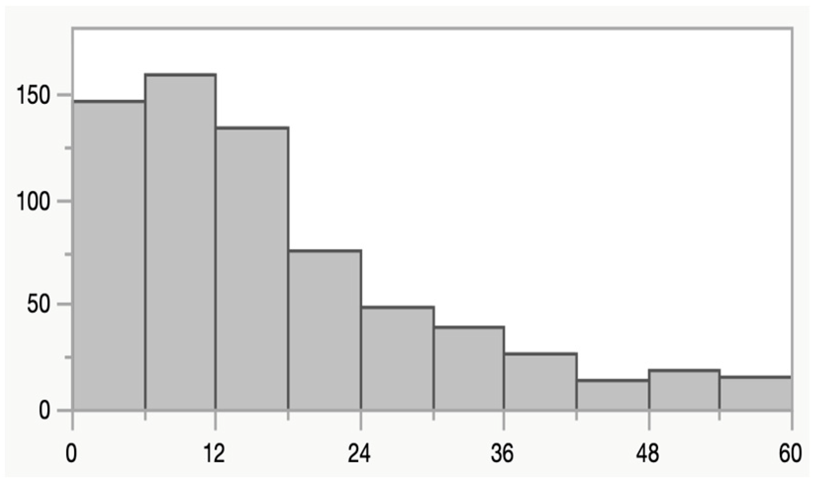
Figure 1. Age distribution (month of age on X axis) among the 668 children in the study.
Microorganisms 13 01071 g001
Table 1. Pathogen detection rates by FUT2 genotype.
FUT2+ (n = 536)FUT2 (n = 132)ORp
Adenovirus 40/41 (n = 52) 7.1% (38)11% (14)0.640.20
Astrovirus (n = 30)3.8% (25)4.7% (5)1.240.81
Norovirus GI (n = 24)0.6% (21)1.3% (3)1.750.45
Norovirus GII (n = 69)10% (52)13% (17)0.720.27
Rotavirus (n = 74)13% (67)5.3% (7)2.550.019
Sapovirus (n = 78)12% (62)12% (16)0.950.88
Campylobacter (n = 52)7.8% (42)7.6% (10)1.041.0
ETEC-eltB (n = 158)23% (125)25% (33)1.081.0
ETEC-estA (n = 81)12% (67)11% (14)1.200.66
Salmonella (n = 38)4.8% (26)9.1% (12)0.510.09
Shigella (n = 99)15% (83)12% (16)1.320.41
Cryptosporidium (n = 22)3.7% (20)1.5% (2)2.520.28
Table 2. Pathogen detection rates among children, stratified by FUT2 genotype and diarrhea.
FUT2  +FUT2  Pint.
DiarrheaNo
Diarrhea
ORpDiarrheaNo
Diarrhea
ORp
(n = 384)(n = 152) (n = 86)(n = 46)
Adenovirus 40/41 (n = 52)7.6% (29)5.9% (9)1.300.5810% (9)11% (5)0.961.00.67
Astrovirus (n = 30)5.2% (20)3.3% (5)1.620.504.6% (4)2.2% (1)2.200.660.80
Norovirus GI (n = 24)4.2% (16)3.3% (5)1.280.812.3% (2)2.2% (1)1.071.00.90
Norovirus GII (n = 69)11% (42)6.6% (10)1.740.1514% (12)11% (5)1.330.790.69
Rotavirus
(n = 74)
16% (60)4.6% (7)3.840.00035.8% (5)4.4% (2)1.351.00.30
Sapovirus
(n = 78)
12% (45)11% (17)1.051.013% (11)11% (5)1.201.00.84
Campylobacter (n = 52)9.6% (37)3.3% (5)3.130.01210% (9)2.2% (1)5.250.160.65
ETEC-eltB (n = 158)29% (110)10% (15)3.67<0.000129% (25)17% (8)1.950.210.25
ETEC-estA (n = 81)16% (67)4.6% (7)3.830.000314% (12)4.4% (2)3.570.140.94
Salmonella
(n = 38)
5.7% (22)2.6% (4)2.250.188.1% (7)11% (5)0.730.750.17
Shigella (n = 99)20% (75)5.3% (8)4.37<0.000116% (14)4.4% (2)4.280.0520.98
Cryptosporidium (n = 22)4.9% (19)0.7% (1)7.860.022.3% (2)0% (0) 0.540.62
Pint., p value for the potential impact by the interaction term (FUT2 status) on diarrhea.
Table 3. Odds ratios for pathogen-associated diarrhea among FUT2+ and FUT2 children.
FUT2+FUT2
Allwith DiarrheaAllwith DiarrheaORp
Adenovirus 40/41 (n = 52)3829 (76%)149 (64%)1.790.48
Astrovirus (n = 30)2520 (80%)54 (80%)1.001.00
Norovirus GI (n = 24)2116 (76%)32 (67%)1.601.00
Norovirus GII (n = 69)5242 (81%1712 (71%)1.750.50
Rotavirus (n = 74)6760 (90%)75 (71%)3.430.20
Sapovirus (n = 78)6245 (73%)1611 (69%)1.200.76
Campylobacter (n = 52)4237 (88%)109 (90%)0.821.00
ETEC-eltB (n = 158)125110 (88%)3325 (76%)2.350.10
ETEC-estA (n = 81)6760 (90%)1412 (86%)1.430.65
Salmonella (n = 38)2622 (85%)127 (58%)3.930.11
Shigella (n = 99)8375 (90%)1614 (88%)1.340.66
Cryptosporidium (n = 22)2019 (95%)22 (100%)0.001.00
Table 4. Presence of diarrhea among children with rotavirus P types stratified by FUT2 genotype.
FUT2+ (n = 536)FUT2 (n = 132)
nDiarrhea No Diarrhea Diarrhea No Diarrhea
Rotavirus7460752
P[8]5042521
P[4]7700 0
P[6]32010
P[9]11000
Disclaimer/Publisher’s Note: The statements, opinions and data contained in all publications are solely those of the individual author(s) and contributor(s) and not of MDPI and/or the editor(s). MDPI and/or the editor(s) disclaim responsibility for any injury to people or property resulting from any ideas, methods, instructions or products referred to in the content.