Article

Impacts of Tick Parasitism on the Rodent Gut Microbiome

1
Biology Department, University of Richmond, Richmond, VA 23173, USA
2
Biology Department, Old Dominion University, Norfolk, VA 23529, USA
3
School of Mathematics and Computer Science, University of KwaZulu-Natal, Durban 4041, South Africa
*
Author to whom correspondence should be addressed.
Microorganisms 2025, 13(4), 888; https://doi.org/10.3390/microorganisms13040888
Submission received: 5 February 2025 / Revised: 6 March 2025 / Accepted: 8 April 2025 / Published: 12 April 2025
(This article belongs to the Special Issue Ticks and Tick-Borne Diseases in Animals, 2nd Edition)

Abstract

:
Host microbiota may impact disease vector behavior and pathogen transmission, but little is known about associations between ectoparasites and microbial communities in wildlife reservoir species. We used Illumina metagenomic sequencing to explore the impacts of tick parasitism on the rodent fecal microbiome in both a field and laboratory setting. We found that tick parasitism on wild hosts was associated with variation in the fecal microbiota of both the white-footed deermouse, Peromyscus leucopus, and the southern cotton rat, Sigmodon hispidus. In a lab experiment, we detected significant changes to the fecal microbiome after experimental exposure to immature ticks in treated versus control BALB/c mice. Whereas there is variation in the fecal microbiome associated with each of the host species we tested, some of the same microbial taxa, notably members of the family Muribaculaceae, occurred at higher relative abundance in tick-parasitized hosts in both the field and laboratory studies, suggesting that there are consistent impacts of tick parasitism on the host gut microbiome. We recommend future studies to test the hypothesis that epithelial cell secretions, generated as part of the host’s immune response to tick parasitism, could provide resources that allow particular microbial lineages in the mammalian gut to flourish.

1. Introduction

Links between gut microbial flora and health outcomes in general [1], as well as specific effects of microbial species composition on development [2], neurological disease [3], and behavioral responses to stress [4], are becoming increasingly apparent, as are impacts on endocrine signaling [5], gastrointestinal dysfunction [6,7], depression [8], and obesity [9]. In non-human hosts, infections such as parvovirus can result in dysbiosis which, in turn, can lead to additional negative health outcomes such as intestinal inflammation, impaired bile acid metabolism, and development of chronic enteropathies [10]. Studies relating gut microbiota and acute infectious disease are fewer in number than those that explore impacts of the microbiome on chronic conditions, but are yielding novel insights into disease processes [11]. Round and Mazmanian [12] reviewed studies of mouse models and humans, and concluded that elements of the microbiome play a key role in protecting the host from potential pathogens either through immune system modulation or production of metabolites that reduce the likelihood of infection. More recent studies have tied changes in human microbiome composition to development of sequalae from emerging diseases like SARS-CoV-2 infection [13] and Lyme disease [14].
Whereas recent studies have demonstrated interactions between the host microbiome and vector-borne infections [15,16,17], little is known about how vectors themselves may interact with the host’s microbiome. The host microbiome may affect the likelihood of encountering or being parasitized by a vector through a number of potential mechanisms. Host-associated bacteria have been hypothesized to mediate volatile organic compounds, which may serve as important attractants to parasites such as flies, mosquitoes, and ticks [18,19,20]. Altered gut microbiome composition has also been implicated in inflammatory skin diseases [21,22,23]. Atopic dermatitis may be linked to an increase in abundance in the gut of Clostridium difficile or decrease in Bifidobacterium [21,22], while bacteria from the groups Clostridiales and Erysipelotrichales have been linked with changes in immune function, resulting in psoriasis [21]. As skin physiology and chemistry can affect attractiveness to ectoparasites [24], factors that affect the gut microbiome may influence ectoparasite burden.
Alternatively, the gut microbiome may affect, and be affected by, feeding and foraging behavior. Perry et al. [9] demonstrated that dysbiosed mice show different feeding behaviors than mice with unaltered microbiomes and, given that foraging behavior in mammals may be linked to tick exposure [25], the gut microbiome, through mediation of physiological condition, could indirectly affect ectoparasite exposure. Andre et al. [26] explored variation in the liver microbiome of Peromyscus leucopus, the principal reservoir of the Lyme disease pathogen Borrelia burgdorferi [27], but did not report any links between microbial community composition and infection with B. burgdorferi or another genus of vector-borne zoonoses, Bartonella. Whether parasitism impacts the host’s gut microbiome is similarly understudied. Internal parasites such as cestodes have been shown to affect the vertebrate gut microbiome, as well as expression of host immune genes [28], and it has been hypothesized that eradication of helminth parasites from humans has resulted in an altered gut microbiome and potentially led to increased incidence of autoimmune disease [29]. Whether an ectoparasite, through immune-mediated or other mechanisms, can im-pact the host microbiome is currently unknown.
Our goal was to assess links between variation in the host gut microbiome and exposure to ticks in both field- and lab-based studies to explore host–parasite interactions that may impact transmission of vector-transmitted pathogens. Specifically, we tested the hypotheses that (1) parasitism by ticks would be related to variation in the gut microbiome of wild rodents, and (2) experimental exposure to ticks would result in consistent and predictable changes in the host’s gut microbiome. In the field, we sampled the white-footed deermouse, Peromyscus leucopus, and the cotton rat, Sigmodon hispidus (a potential reservoir for the agent of Tidewater spotted fever, Rickettsia parkeri [30,31]), and compared gut microbiome composition among parasitized and unparasitized individuals. In the lab, we conducted an artificial tick infestation experiment to collect microbiome data from lab mice before and after experimentally induced tick parasitism.

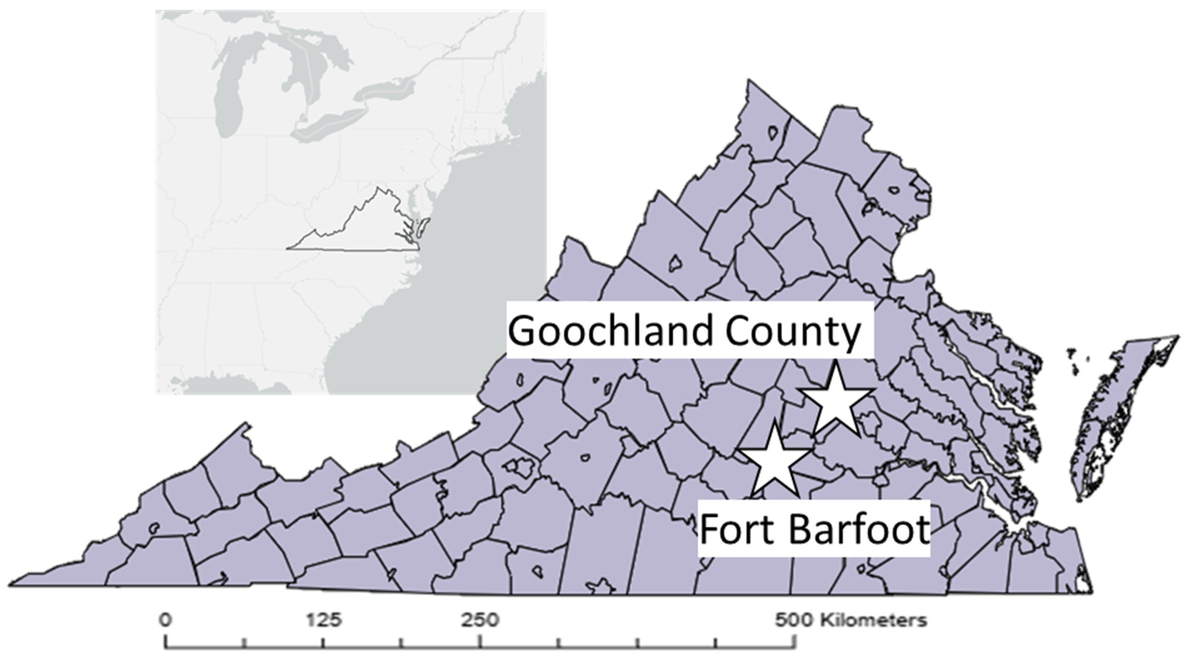
2. Materials and Methods

We sampled wild rodents for ongoing research on zoonotic pathogen transmission at two sites in central Virginia: a University of Richmond-owned property in eastern Goochland County; and Ft. Barfoot, which straddles Dinwiddie, Brunswick and Nottaway Counties (Figure 1).
We sampled early successional open-canopy habitats at each site, where we set eight transects of 10 Sherman live traps with 10 m spacing between traps and 150–1500 m between transects. Traps were baited with bird seed, and set for three consecutive nights, once every three weeks for four trapping sessions per site. Traps were checked by 10:30 each morning and left open, but not set, between sampling sessions. Captured rodents were marked using individually numbered ear tags, and data on species, sex, and weight were recorded prior to release. While rodents were in hand, we collected all visible ticks from the head, neck, ears, limbs, and axillae using fine pointed forceps; individuals were examined for 30–60 s following collection of the last tick, or in the event that no ticks were detected. We collected fecal samples directly into sterile 1.5 mL flip-cap microcentrifuge tubes that were kept on dry ice during multi-day sampling sessions before being frozen dry at −20 °C upon return to the lab. All animal work for the field project was reviewed and approved by the Institutional Animal Care and Use Committee of the University of Richmond (protocol 20-01-001).
In fall 2022, we ordered 10 female Balb/c mice of at least 7 weeks of age from Envigo (now Inotiv, Inc., Lafayette, IN, USA) for use in a parasitism experiment. Mice were held in individual mouse cages in the animal facility at the University of Richmond for approximately one week prior to the initiation of tick parasitism. Mice were assigned randomly to one of two experimental treatment groups for the parasitism trials, with initial fecal samples collected as described above prior to the first tick application. Tick applications were performed as follows: mice were held by the scruff and approximately 50–70 larval Ixodes scapularis, sourced from the Oklahoma State University tick colony, were applied to ears, head, ventrum, and dorsum of all individuals in the treatment group with a fine-bristled paintbrush. Individuals in the control group underwent a mock exposure identical to the tick application, but with no ticks on the paintbrush. Animals were then returned to clean cages with a wire mesh metabolic floor instead of shredded pine bedding for seven days. Cages were checked twice per day for engorged ticks, which were collected into sterile microcentrifuge tubes, and cleaned once per day. After the seven-day engorgement period, mice were moved to cages with shredded pine bedding for approximately five days prior to a second round of tick exposure, which was performed exactly like the first, except that animals were sacrificed at the end of the second tick exposure with a second fecal sample collected from each animal prior to euthanasia. All laboratory animal work was reviewed and approved by the Institutional Animal Care and Use Committee of the University of Richmond (protocol 20-06-002).

2.1. Molecular Methods

We extracted DNA from all individual fecal samples using commercial DNA isolation kits (Macherey-Nagel Blood and Tissue Kit, Macherey-Nagel, Inc., Bethlehem, PA, USA) using manufacturer’s protocols. We used different metagenomic sequencing protocols for the field and lab samples. For the 2020 field samples, we amplified a portion of the V4 hypervariable region of the 16S rRNA gene using conserved universal primers 515F (modified from [32]) and 806R (modified from [33]), and followed the Illumina 16S metagenomics library preparation guidelines [34] using an Illumina Nextera XT Index kit (Illumina, Inc., San Diego, CA, USA) through the library pooling step. Pooled libraries from up to twenty-five samples were loaded into an Illumina iSeq 100 i1 Reagent v2 (300-cycle) cartridge and run on an Illumina iSeq 100 instrument according to the manufacturer’s instructions, and with 5 µL of 100 pM PhiX positive control library added to 15 µL of the pooled library. Due to changes in resource availability during the project, sequencing of a portion of the V3-V4 hypervariable 16S region for fecal DNA extracts from the lab experiment was conducted on an Illumina MiSeq (2 × 250 reads) following Nextera library preparation by Novogene, Inc. (Novogene Corp., Sacramento, CA, USA). Sequencing of a portion of the V3-V4 hypervariable 16S region for fecal DNA extracts from the lab experiment was conducted on an Illumina MiSeq (2 × 250 reads) following Nextera library preparation by Novogene, Inc. (California, USA).

2.2. Informatic and Statistical Analyses

Sequence data were processed using Qiime2 [35] as follows: raw reads were imported, demultiplexed, truncated to remove loci with low read quality, and denoised/quality filtered with DADA2 [36], implemented in Qiime2 [35]; raw reads were truncated (but not trimmed) to 150 bases for the wild rodents and, for the lab mouse data, we truncated sequences (but did not trim them) at 225 and 222 bases for forward and reverse reads, respectively. We used weighted taxonomic classifiers [37] based on the Silva 138 rRNA reference database [38], and trained on the animal distal gut [39] to identify amplicon sequence variants (ASVs) for both the V4 and V3-V4 datasets. We assessed alpha and beta diversity outputs (e.g., unifrac analysis [40], rarefaction, evenness and diversity estimates) from Qiime2 and compared microbial community composition among metadata features (e.g., rodent species, sampling site, and presence/absence of ticks for the field data and treatment group and timepoint for the lab experiment) by perMANOVA using the R package vegan [41] implemented in Qiime2 [35]. We assessed relative over- or underrepresentation of taxa based on metadata features using ANCOM-BC [42] implemented in Qiime2 [35]. For these analyses, we implemented a prevalence cut-off of 0.5% and a significance threshold of 0.1.

3. Results

We collected fecal samples from 45 individual wild rodents sampled at two field sites and representing two species; one species (Sigmodon hispidus) was only detected at the Fort Barfoot site, whereas the other species (Peromyscus leucopus) was detected at both sites (Table 1).
Ticks were present on 15 individuals (Table 1), but these tick samples were lost before they could be identified or counted. Some individuals were captured more than once, but only the fecal sample from first collection was used for library preparation and analysis. Metagenomic libraries from these 45 samples generated a median read depth of 96,488 and a range of 24,996–312,172 reads per sample (Table 1). We identified 12,530 amplicon sequence variants (ASVs) that could be classified into 666 species-level-or-higher taxa. Taxonomic richness did not vary by rodent species (F = 082, p = 0.37), but did vary depending on whether the host was parasitized by ticks (F = 4.21, p = 0.047), with no significant interaction between host species and tick parasitism (F = 2.31, p = 0.14; Figure 2).
Microbial community composition, as characterized by weighted unifrac [40] distances, varied significantly by rodent species (F = 4.15, p = 0.007), sampling location (F = 3.0, p = 0.002), and tick parasitism (F = 1.78, p = 0.049; Figure 3).
In addition to main effects, there were significant interactions between rodent species and sampling location (F = 3.07, p = 0.006), and sampling location and presence of ticks (F = 2.14, p = 0.017), although these interactions are confounded by the lack of S. hispidus samples from the Goochland, Virginia site.
Particular microbial taxa varied in relative abundance for both P. leucopus and S. hispidus depending on whether the individual was parasitized by ticks. For S. hispidus, members of the genera Acinetobacter and Alistipes, as well as multiple taxa within the Muribaculaceae, were overrepresented in tick parasitized hosts, whereas one member of the Lachnospiraceae (GCA-900066575) was underrepresented (all q-values < 0.01; Figure 4A). For P. leucopus, the following taxa were overrepresented at the Goochland site: Rikenella, Paraeggerthelia, candidatus Saccharimonas, Clostridia UGC-014, and a members of the Muribacculaceae and family RF39 (all q-values < 0.05, Figure 4B). At the Ft. Barfoot site, only one taxon was overrepresented in tick-parasitized P. leucopus (Muribaculaceae, q = 0.044), whereas two (Acinetobacter (q = 0.02) and a member of the family Rs-E47 termite group of the Bacteriodales (q << 0.001) were underrepresented (Figure 4C).
For the lab experiment, the median number of reads per sample was 87,377 with a range of 63,716 to 101,481. The number of ASVs detected was 2807, and the number of taxa identified was 574. There was no effect of experimental grouping (i.e., control versus treatment; F = 0.005, p = 0.94), time (i.e., before versus after tick application, F = 0.06, p = 0.81), or tick parasitism (i.e., group-by-time interaction, F = 1.321, p = 0.27) on microbial taxonomic richness. There was, however, a significant effect of tick parasitism on fecal microbial community composition (i.e., a group-by-timepoint interaction; F = 3.35, p = 0.013), but there were no significant effects of either treatment group (F = 1.18, p = 0.25) or time (F = 0.90, p = 0.41) in isolation. As with the field data, there were significant effects of tick parasitism on the relative abundance of particular taxa. Tick-exposed mice showed significant overrepresentation of Muribaculaceae (q < 0.001), Lactococcus (q = 0.01), and Arthromitus (q = 0.02) relative to the pre-exposure timepoint, and significant underrepresentation of Tuzzerella (q = 0.03) (Figure 5A).
There were also significant changes in control mice before and after experimental infestation where Helicobacter (q = 0.03) was overrepresented in the second timepoint. and Tuzzerella (q = 0.02), Gastranaerophilales (q = 0.02), candidatus Saccharimonas (q = 0.03), NK4A214 group Oscillospiraceae (q = 0.002) were underrepresented in the second timepoint relative to the first (Figure 5B). We note that we recovered between seven and eighteen ticks per mouse per infestation (mean 12.8), but we note that it is likely that a number of ticks were undetected during daily cage cleaning; in a subsequent experiment (unpublished data), we found up to 71% more ticks during a second round of tick searching in each day’s cage remnants.

4. Discussion

We detected, in both a field study and laboratory experiment, that tick parasitism on rodents is associated with significant variation in the fecal microbiome. More specifically, experimental exposure to ticks resulted in a change in microbiome community composition in ways that were not seen in control mice, and with some changes paralleling differences between tick-parasitized and non-parasitized rodents in the field. The rodent gut microbiome is impacted by a wide variety of factors, and whereas certain lineages may be common to all members of a particular host species, heterogeneity among individuals and over time may mask other drivers of gut microbiome variation [43]. In our field study, the fecal microbiome varied by host species and sampling site, as well as tick parasitism, with substantial residual variation in microbiome composition unexplained by these variables; we did not test for variation by sex or age, and note that we did not have time-series data on tick parasitism for most of the field-sampled individuals. Given that the rodent species in our study have different ecological [44] and dietary characteristics [45,46], it is not surprising that their gut microbiota are different; prior studies have demonstrated that variation in habitat use, diet, and resource partitioning can impact the gut microbial communities in rodents [47,48,49], although variation among individuals may mask effects of diet and physiological state [43]. It is notable, however, that the same microbial taxon (Muribaculaceae) was associated with tick parasitism in both the field and lab studies, possibly suggesting at a mechanistic link between these two phenomena.
Recent research has highlighted links between the skin and gut microbiome, largely in reference to gut dysbiosis and effects on chronic skin conditions like psoriasis, atopic dermatitis and acne [22,50,51]. In humans, links between gut microbiome dysbiosis and skin pathologies have been described [52], and a weakened skin barrier to antigens can lead to immune-globulin E (IgE)-mediated food allergic response in the gut [53]. In another study, increased ultraviolet light exposure to the skin altered the fecal microbiome in ways that were consistent with oral vitamin D supplementation [54], further highlighting potential mechanisms by which impacts to the skin could manifest in changes to the gut microbiome. Mechanisms by which tick parasitism would elicit changes in the rodent fecal microbiome are unknown, and are beyond the scope of this study, but could involve immune response to tick salivary proteins or other impacts of tick feeding. Tick saliva is replete with proteins, lipids, and smaller peptides that are involved in mitigating pain/itch responses, promoting hemostasis, which can suppress the host’s immune response [55,56], and some of these host responses could have downstream impacts on the gut microbial community. For example, exposure to tick saliva, among other changes, promotes T helper 2 (Th2) cell response [57,58] and release of Th2-related cytokines [59], including interleukins 4 and 10, that are related with specific changes in the rodent gut microbiome [60].
One microbial taxon that was consistently represented by higher relative abundance in both the field and lab studies and across all three rodent taxa was the family Muribaculaceae (previously known as family S24-7) within the order Bacteroidales. Members of this family are common constituents of the rodent gut microbiome, representing as many as 83% of sequence reads assigned to the phylum Bacteriodota [61]. This family is characterized by metabolism of polysaccharides, including otherwise indigestible dietary fiber and mucin secreted by goblet cells in the gut [62,63]. Tick parasitism has been shown to elicit secretion of mucus from epithelial cells in the gut [64], which could result in increased relative abundance of microbial taxa that metabolize glycoproteins and other components of mucus. Related to disease and pathological processes, the high abundance of Muribaculaceae may be associated with better health outcomes because short-chain fatty acids, such as those produced by members of the Muribaculaceae, can have anti-inflammatory effects and lead to increased longevity in rodents [65]. In another study, two gut microbes, Akkermansia miciniphilia and a member of the Muribaculaceae (then known as family Bacteriodes S24-7), were associated with increased immunological response in mice [66]. With respect to parasitism, Kreisinger et al. [67] demonstrated that presence of tapeworms in the intestines of wild Apodemus flavicollis mice was associated with increased abundance of bacteria in the family S24-7 (now Muribaculaceae). We note that there were multiple sequence variants identified as members of the Muribaculaceae among our samples and studies, and we cannot be certain that all perform the same metabolic functions. Identification of any potential mechanisms by which tick parasitism would impact immune response in ways that could affect the gut microbiome, and specific taxa that may be affected by these immune responses, is worthy of future investigation.
With many hundreds of microbial taxa detected in a single fecal sample, it is prudent to interpret results of microbial community analyses with caution, particularly given the relatively small samples sizes in our field study, in particular where we were not able to control for effects of host sex, age, prior infection/parasitism status, genetics, or other aspects of individual-level variation. The amount of variation in wild rodent microbiota attributable to tick parasitism that we detected was far less than the variation between rodent species, and less than the variation attributable to sampling site, and we further cannot discount that potentially spurious or idiosyncratic changes in the microbiome were statistically attributed to tick parasitism. For example, Baxter et al. [43] reported that Peromyscus fecal microbiota varied substantially over time with samples taken from the same individual more than three days apart, showing more similarity to other individuals than to the initial sample. This result suggests that, at least for P. leucopus, gut microbiota change or turn over rapidly, and there is some risk of type I error when assessing the impacts of tick parasitism on the rodent gut microbiome, particularly in the field study. It is also possible that variation in the host microbiome affects parasitism by ticks, and some of the gut microbiome variation we describe in wild individuals with and without ticks may result from differences in likelihood of encountering or being parasitized by ticks. For example, elements of the skin microbiome can affect the attractiveness of certain host individuals to blood-feeding parasites [68], and infection with a microorganism may also influence blood-feeding by parasites, at least under some circumstances [69]. Similarly, the gut microbiome is linked to skin physiological function [21,22,23], and thus gut microbiome-mediated effects on skin could then impact parasitism by ticks. In cattle, differences in susceptibility to tick parasitism may be driven by variable immune system function in skin tissues [70]. Alternatively, gut microbiota can affect behavior [71], which can then lead to variation in likelihood of parasitism [25,72]. It is also possible that the composition of the internal microbiome could influence parasitism in other ways; Knutie [73] linked changes in the gut microbiome of eastern bluebirds following food supplementation to variation in parasitism by nest flies. Future studies where immune profiles are measured and quantified in response to tick parasitism and relative to changes in gut microbiome would help untangle the mechanistic relationship between tick parasitism and changes in the fecal microbiome.
In the lab experiment, we found that particular taxa in the microbiota of unparasitized mice changed significantly in relative abundance within a matter of several weeks, which is consistent with the findings of Baxter et al. [43]. We note that the characterization of tick parasitism for individuals in the field is based on a single point estimate, although tick parasitism status did not tend to change; for the subset of animals that were recaptured and checked for ticks on subsequent captures, there were two instances of change in tick parasitism status (both from parasitized to unparasitized, presumably because we removed the ticks on the previous occasion), and eight instances of the status remaining the same in subsequent captures (seven recaptures continued to be tick-free, whereas one individual was tick-parasitized on multiple occasions). This result is consistent with findings that (1) P. leucopus does not develop resistance to tick parasitism [74], and (2) that certain individuals in a population tend to have consistently higher tick burdens that others [74]. In other words, certain individuals may be more prone to tick parasitism than others [75,76]. It is also worth noting that interpretation of the field data, and comparisons to the lab experiment data, should account for the difference in the locus used to characterize microbiota. Use of different target loci within the 16S rRNA can affect the number of taxa identified in a sample [77,78], as can the taxonomic classification method [79]. Choice of sequencing platform (e.g., iSeq vs. MiSeq) can also affect detectable taxonomic resolution [80,81], but may be less impactful on overall microbiome characterization [82]. Whereas different sequence lengths between the field and lab samples preclude us from being able to directly assess similarity of taxa the species level, the iSeq is capable of characterizing taxa to genus and changes in abundance of particular taxa (e.g., Muribaculaceae) associated with tick parasitism should be fully comparable regardless of sequencing platform.
It is becoming increasingly apparent that the host and vector microbiome can influence transmission of vector-borne pathogens in a variety of ways, from facilitation or inhibition of pathogen acquisition to the probability of a vector feeding on a given host individual [16,20,21,22,23,24,25,83,84,85]. Our goal was to explore links between the host gut microbiome and tick parasitism by first describing variation and community composition of gut microbial communities in parasitized and unparasitized individuals of two wild rodent species, and second by tracking changes in the host microbiome following experimental exposure to ticks. Our results suggest that there may be particular gut microbial taxa that respond to physiological and/or immunological changes in hosts following tick parasitism, and that the overall gut microbial flora can be impacted by ectoparasite attachment and feeding; further studies that combine immunological assays with experimental tick parasitism and changes to the gut microbiome will elucidate such mechanisms. Whether these changes in gut microbial community composition impact host infection status to capacity to transmit vector-borne pathogens remains to be tested.

5. Conclusions

Tick parasitism on mammalian hosts, even in the absence of microbial pathogen transmission, results in an immune challenge that can affect physiological function [79]. Our data suggest that part of the host’s response to tick parasitism includes changes in the gut microbiome. Although the mechanisms by which the immunological response to ticks affect microbial community dynamics are unclear, it is reasonable to expect that immunologically mediated changes to epithelial cell function in the gut, possibly coordinated with responses by cells in the skin, would result in variation in resource availability for specific microbial taxa. We speculate that increased mucin production by goblet cells, stimulated by the host’s immune response to tick parasitism, facilitates reproduction of taxa in the Muribaculaceae and results in increased relative abundance of these lineages. We reach this conclusion based on the detection of consistent changes in abundance of this taxon in both observational and experimental studies of rodent fecal microbiota as part of broader changes in gut microbial community composition associated with parasitism by ticks.

Author Contributions

R.J.B. designed the study, with contributions from M.L. and J.P., as part of a larger-scale study of bacterial infections in Virginia rodents designed by R.J.B. and H.D.G.; R.J.B. and M.L. collected data and samples for the field study. R.J.B., M.L., J.P. and I.D.H. collected data for the lab study. R.J.B. and J.P. performed the bulk of the data analysis, with contributions from I.D.H. R.J.B., J.P. and I.D.H. wrote the manuscript and all authors edited the manuscript. All authors have read and agreed to the published version of the manuscript.

Funding

Support for this work was provided by award 5R01AI136035 from the National Institutes of Health to HDG (subaward to RJB), a Robert F. Smart Summer Fellowship in Biology to JP, and funding from the Department of Biology at the University of Richmond to RJB.

Institutional Review Board Statement

All vertebrate animal work described in this manuscript was approved by the University of Richmond Institutional Animal Care and Use Committee (IACUC) (protocol code #20-01-001, approved on 20 January 2020; and #20-06-002, approved on 20 June 2020).

Informed Consent Statement

Not applicable.

Data Availability Statement

The original contributions presented in this study are included in the article. Further inquiries can be directed to the corresponding author.

Acknowledgments

We would first like to thank B. Nelson for her support and for providing logistical assistance associated with field sampling at Ft. Barfoot. J. Gregorini assisted with field rodent sampling, and A. Jablonski, A. Johnson, and K. Waskas assisted with animal care and sample collection for the lab study.

Conflicts of Interest

The authors declare no conflicts of interest.

References

  1. Mackowiak, P.A. The normal microbial flora. N. Engl. J. Med. 1982, 307, 83–93. [Google Scholar] [CrossRef] [PubMed]
  2. Dominguez-Bello, M.G.; Godoy-Vitorino, F.; Knight, R.; Blaser, M.J. Role of the microbiome in human development. Gut 2019, 68, 1108–1114. [Google Scholar] [CrossRef] [PubMed]
  3. Tremlett, H.; Bauer, K.C.; Appel-Cresswell, S.; Finlay, B.B.; Waubant, E. The gut microbiome in human neurological disease: A review. Ann. Neurol. 2017, 81, 369–382. [Google Scholar] [CrossRef] [PubMed]
  4. Dinan, T.G.; Cryan, J.F. The microbiome-gut-brain axis in health and disease. Gastroenterol. Clin. 2017, 46, 77–89. [Google Scholar] [CrossRef]
  5. Flux, M.C.; Lowry, C.A. Finding intestinal fortitude: Integrating the microbiome into a holistic view of depression mechanisms, treatment, and resilience. Neurobiol. Dis. 2020, 135, 104578. [Google Scholar] [CrossRef]
  6. Wehkamp, J.; Frick, J.S. Microbiome and chronic inflammatory bowel diseases. J. Mol. Med. 2017, 95, 21–28. [Google Scholar] [CrossRef]
  7. Lleal, M.; Sarrabayrouse, G.; Willamil, J.; Santiago, A.; Pozuelo, M.; Manichanh, C. A single faecal microbiota transplantation modulates the microbiome and improves clinical manifestations in a rat model of colitis. eBioMedicine 2019, 48, 630–641. [Google Scholar] [CrossRef]
  8. Winter, G.; Hart, R.A.; Charlesworth, R.P.; Sharpley, C.F. Gut microbiome and depression: What we know and what we need to know. Rev. Neurosci. 2018, 29, 629–643. [Google Scholar] [CrossRef]
  9. Perry, R.J.; Peng, L.; Barry, N.A.; Cline, G.W.; Zhang, D.; Cardone, R.L.; Petersen, K.F.; Kibbey, R.G.; Goodman, A.L.; Shulman, G.I. Acetate mediates a microbiome–brain–β-cell axis to promote metabolic syndrome. Nature 2016, 534, 213–217. [Google Scholar] [CrossRef]
  10. Pilla, R.; Suchodolski, J.S. The role of the canine gut microbiome and metabolome in health and gastrointestinal disease. Front. Vet. Sci. 2020, 6, 498. [Google Scholar] [CrossRef]
  11. Libertucci, J.; Young, V.B. The role of the microbiota in infectious diseases. Nat. Microbiol. 2019, 4, 35–45. [Google Scholar] [CrossRef] [PubMed]
  12. Round, J.L.; Mazmanian, S.K. The gut microbiota shapes intestinal immune responses during health and disease. Nat. Rev. Immunol. 2009, 9, 313–323. [Google Scholar] [CrossRef]
  13. Yeoh, Y.K.; Zuo, T.; Lui, G.C.; Zhang, F.; Liu, Q.; Li, A.Y.; Chung, A.C.; Cheung, C.P.; Tso, E.Y.; Fung, K.S.; et al. Gut microbiota composition reflects disease severity and dysfunctional immune responses in patients with COVID-19. Gut 2021, 70, 698–706. [Google Scholar] [CrossRef]
  14. Morrissette, M.; Pitt, N.; González, A.; Strandwitz, P.; Caboni, M.; Rebman, A.W.; Knight, R.; D’onofrio, A.; Aucott, J.N.; Soloski, M.J.; et al. A distinct microbiome signature in posttreatment Lyme disease patients. mBio 2020, 11, e02310-20. [Google Scholar] [CrossRef]
  15. Bonnet, S.I.; Binetruy, F.; Hernández-Jarguín, A.M.; Duron, O. The tick microbiome: Why non-pathogenic microorganisms matter in tick biology and pathogen transmission. Front. Cell. Infect. Microbiol. 2017, 7, 236. [Google Scholar] [CrossRef] [PubMed]
  16. de la Fuente, J.; Villar, M.; Cabezas-Cruz, A.; Estrada-Peña, A.; Ayllon, N.; Alberdi, P. Tick–Host–Pathogen Interactions: Conflict and Cooperation. PLoS Pathog. 2016, 12, e1005488. [Google Scholar] [CrossRef] [PubMed]
  17. Cabezas-Cruz, A.; Estrada-Peña, A.; Rego, R.O.; De la Fuente, J. Tick-pathogen ensembles: Do molecular interactions lead ecological innovation? Front. Cell. Infect. Microbiol. 2017, 7, 74. [Google Scholar] [CrossRef] [PubMed]
  18. Faraone, N.; Light, M.; Scott, C.; MacPherson, S.; Hillier, N.K. Chemosensory and behavioural responses of Ixodes scapularis to natural products: Role of chemosensory organs in volatile detection. Insects 2020, 11, 502. [Google Scholar] [CrossRef] [PubMed]
  19. Lutz, H.L.; Gilbert, J.A.; Dick, C.W. Associations between Afrotropical bats, eukaryotic parasites, and microbial symbionts. Mol. Ecol. 2022, 31, 1939–1950. [Google Scholar] [CrossRef]
  20. Roodt, A.P.; Naudé, Y.; Stoltz, A.; Rohwer, E. Human skin volatiles: Passive sampling and GC× GC-ToFMS analysis as a tool to investigate the skin microbiome and interactions with anthropophilic mosquito disease vectors. J. Chromatogr. B 2018, 1097, 83–93. [Google Scholar] [CrossRef]
  21. Mann, E.A.; Bae, E.; Kostyuchek, D.; Chung, H.J.; McGee, J.S. The gut microbiome: Human health and inflammatory skin diseases. Ann. Dermatol. 2020, 32, 265. [Google Scholar] [CrossRef]
  22. Pessemier, B.D.; Grine, L.; Debaere, M.; Maes, A.; Paetzold, B.; Callewaert, C. Gut–skin axis: Current knowledge of the interrelationship between microbial dysbiosis and skin conditions. Microorganisms 2021, 9, 353. [Google Scholar] [CrossRef] [PubMed]
  23. Salem, I.; Ramser, A.; Isham, N.; Ghannoum, M.A. The gut microbiome as a major regulator of the gut-skin axis. Front. Microbiol. 2018, 9, 1459. [Google Scholar] [CrossRef] [PubMed]
  24. Frischknecht, F. The skin as interface in the transmission of arthropod-borne pathogens. Cell. Microbiol. 2007, 9, 1630–1640. [Google Scholar] [CrossRef]
  25. Fritzsche, A.; Allan, B.F. The ecology of fear: Host foraging behavior varies with the spatio-temporal abundance of a dominant ectoparasite. EcoHealth 2012, 9, 70–74. [Google Scholar] [CrossRef] [PubMed]
  26. André, A.; Mouton, A.; Millien, V.; Michaux, J. Liver microbiome of Peromyscus leucopus, a key reservoir host species for emerging infectious diseases in North America. Infect. Genet. Evol. 2017, 52, 10–18. [Google Scholar] [CrossRef]
  27. Hahn, M.A.; Piecyk, A.; Jorge, F.; Cerrato, R.; Kalbe, M.; Dheilly, N.M. Host phenotype and microbiome vary with infection status, parasite genotype, and parasite microbiome composition. Mol. Ecol. 2022, 31, 1577–1594. [Google Scholar] [CrossRef]
  28. Zaiss, M.M.; Harris, N.L. Interactions between the intestinal microbiome and helminth parasites. Parasite Immunol. 2016, 38, 5–11. [Google Scholar] [CrossRef]
  29. Bunikis, J.; Tsao, J.; Luke, C.J.; Luna, M.G.; Fish, D.; Barbour, A.G. Borrelia burgdorferi infection in a natural population of Peromyscus leucopus mice: A longitudinal study in an area where Lyme borreliosis is highly endemic. J. Infect. Dis. 2004, 189, 1515–1523. [Google Scholar] [CrossRef]
  30. Moraru, G.M.; Goddard, J.; Paddock, C.D.; Varela-Stokes, A. Experimental infection of cotton rats and bobwhite quail with Rickettsia parkeri. Parasites Vectors 2013, 6, 70. [Google Scholar] [CrossRef]
  31. Cumbie, A.N.; Espada, C.D.; Nadolny, R.M.; Rose, R.K.; Dueser, R.D.; Hynes, W.L.; Gaff, H.D. Survey of Rickettsia parkeri and Amblyomma maculatum associated with small mammals in southeastern Virginia. Ticks Tick-Borne Dis. 2020, 11, 101550. [Google Scholar] [CrossRef] [PubMed]
  32. Parada, A.E.; Needham, D.M.; Fuhrman, J.A. Every base matters: Assessing small subunit rRNA primers for marine microbiomes with mock communities, time series and global field samples. Environ. Microbiol. 2016, 18, 1403–1414. [Google Scholar] [CrossRef]
  33. Apprill, A.; McNally, S.; Parsons, R.; Weber, L. Minor revision to V4 region SSU rRNA 806R gene primer greatly increases detection of SAR11 bacterioplankton. Aquat. Microb. Ecol. 2015, 75, 129–137. [Google Scholar] [CrossRef]
  34. Illumina. 16s Metagenomic Sequencing Library Preparation; Illumina: San Diego, CA, USA, 2013; p. 21. [Google Scholar]
  35. Bolyen, E.; Rideout, J.R.; Dillon, M.R.; Bokulich, N.A.; Abnet, C.C.; Al-Ghalith, G.A.; Alexander, H.; Alm, E.J.; Arumugam, M.; Asnicar, F.; et al. Reproducible, interactive, scalable and extensible microbiome data science using QIIME 2. Nat. Biotechnol. 2019, 37, 852–857. [Google Scholar] [CrossRef]
  36. Callahan, B.J.; McMurdie, P.J.; Rosen, M.J.; Han, A.W.; Johnson, A.J.; Holmes, S.P. DADA2: High-resolution sample inference from Illumina amplicon data. Nat. Methods 2016, 13, 581–583. [Google Scholar] [CrossRef] [PubMed]
  37. Bokulich, N.A.; Kaehler, B.D.; Rideout, J.R.; Dillon, M.; Bolyen, E.; Knight, R.; Huttley, G.A.; Gregory Caporaso, J. Optimizing taxonomic classification of marker-gene amplicon sequences with QIIME 2′s q2-feature-classifier plugin. Microbiome 2018, 6, 90. [Google Scholar] [CrossRef]
  38. Quast, C.; Pruesse, E.; Yilmaz, P.; Gerken, J.; Schweer, T.; Yarza, P.; Peplies, J.; Glöckner, F.O. The SILVA ribosomal RNA gene database project: Improved data processing and web-based tools. Nucleic Acids Res. 2012, 41, D590–D596. [Google Scholar] [CrossRef] [PubMed]
  39. Kaehler, B.D.; Bokulich, N.A.; McDonald, D.; Knight, R.; Caporaso, J.G.; Huttley, G.A. Species abundance information improves sequence taxonomy classification accuracy. Nat. Commun. 2019, 10, 4643. [Google Scholar] [CrossRef] [PubMed]
  40. Lozupone, C.; Knight, R. UniFrac: A new phylogenetic method for comparing microbial communities. Appl. Environ. Microbiol. 2005, 71, 8228–8235. [Google Scholar] [CrossRef]
  41. Oksanen, J.; Blanchet, F.G.; Kindt, R.; Legendre, P.; Minchin, P.R.; O’hara, R.B.; Simpson, G.L.; Solymos, P.; Stevens, M.H.; Wagner, H.; et al. Package ‘vegan’, version 2.6-10; Community Ecology Package; 2013; Volume 2, pp. 1–295. Available online: https://CRAN.R-project.org/package=vegan (accessed on 7 April 2025).
  42. Lin, H.; Peddada, S.D. Analysis of compositions of microbiomes with bias correction. Nat. Commun. 2020, 11, 3514. [Google Scholar] [CrossRef]
  43. Baxter, N.T.; Wan, J.J.; Schubert, A.M.; Jenior, M.L.; Myers, P.; Schloss, P.D. Intra-and interindividual variations mask interspecies variation in the microbiota of sympatric Peromyscus populations. Appl. Environ. Microbiol. 2015, 81, 396–404. [Google Scholar] [CrossRef]
  44. Menzel, M.A.; Carter, T.C.; Houston, A.T.; Longe, R.L. Notes on the microhabitat associations of the hispid cotton rat (Sigmodon hispidus) and white-footed mouse (Peromyscus leucopus) in the piedmont physiographic province. Ga. J. Sci. 1999, 57, 180. [Google Scholar]
  45. Lackey, J.A.; Huckaby, D.G.; Ormiston, B.G. Peromyscus leucopus. Mamm. Species 1985, 247, 1. [Google Scholar] [CrossRef]
  46. Randolph, J.C.; Cameron, G.N.; Wrazen, J.A. Dietary choice of a generalist grassland herbivore, Sigmodon hispidus. J. Mammal. 1991, 72, 300–313. [Google Scholar] [CrossRef]
  47. Anders, J.L.; Moustafa, M.A.; Mohamed, W.M.; Hayakawa, T.; Nakao, R.; Koizumi, I. Comparing the gut microbiome along the gastrointestinal tract of three sympatric species of wild rodents. Sci. Rep. 2021, 11, 19929. [Google Scholar] [CrossRef]
  48. Bowerman, K.L.; Knowles, S.C.; Bradley, J.E.; Baltrūnaitė, L.; Lynch, M.D.; Jones, K.M.; Hugenholtz, P. Effects of laboratory domestication on the rodent gut microbiome. ISME Commun. 2021, 1, 49. [Google Scholar] [CrossRef]
  49. Kohl, K.D.; Dieppa-Colón, E.; Goyco-Blas, J.; Peralta-Martínez, K.; Scafidi, L.; Shah, S.; Zawacki, E.; Barts, N.; Ahn, Y.; Hedayati, S.; et al. Gut microbial ecology of five species of sympatric desert rodents in relation to herbivorous and insectivorous feeding strategies. Integr. Comp. Biol. 2022, 62, 237–251. [Google Scholar] [CrossRef] [PubMed]
  50. O’Neill, C.A.; Monteleone, G.; McLaughlin, J.T.; Paus, R. The gut-skin axis in health and disease: A paradigm with therapeutic implications. Bioessays 2016, 38, 1167–1176. [Google Scholar] [CrossRef]
  51. Sinha, S.; Lin, G.; Ferenczi, K. The skin microbiome and the gut-skin axis. Clin. Dermatol. 2021, 39, 829–839. [Google Scholar] [CrossRef]
  52. Lee, H.J.; Kim, M. Skin barrier function and the microbiome. Int. J. Mol. Sci. 2022, 23, 13071. [Google Scholar] [CrossRef]
  53. van Splunter, M.; Liu, L.; van Neerven, R.J.; Wichers, H.J.; Hettinga, K.A.; De Jong, N.W. Mechanisms underlying the skin-gut cross talk in the development of IgE-mediated food allergy. Nutrients 2020, 12, 3830. [Google Scholar] [CrossRef] [PubMed]
  54. Bosman, E.S.; Albert, A.Y.; Lui, H.; Dutz, J.P.; Vallance, B.A. Skin exposure to narrow band ultraviolet (UVB) light modulates the human intestinal microbiome. Front. Microbiol. 2019, 10, 477346. [Google Scholar] [CrossRef]
  55. Wikel, S.K.; Ramachandra, R.N.; Bergman, D.K. Tick-induced modulation of the host immune response. Int. J. Parasitol. 1994, 24, 59–66. [Google Scholar] [CrossRef]
  56. Brossard, M.; Wikel, S. Tick Immunobiology. In Ticks: Biology, Disease, and Control; Bowmand, A.S., Nuttall, P.A., Eds.; Cambridge University Press: Cambridge, UK, 2004; pp. 186–204. [Google Scholar]
  57. Skallová, A.; Iezzi, G.; Ampenberger, F.; Kopf, M.; Kopecký, J. Tick saliva inhibits dendritic cell migration, maturation, and function while promoting development of Th2 responses. J. Immunol. 2008, 180, 6186–6192. [Google Scholar] [CrossRef] [PubMed]
  58. Kotál, J.; Langhansová, H.; Lieskovská, J.; Andersen, J.F.; Francischetti, I.M.; Chavakis, T.; Kopecký, J.; Pedra, J.H.; Kotsyfakis, M.; Chmelař, J. Modulation of host immunity by tick saliva. J. Proteom. 2015, 128, 58–68. [Google Scholar] [CrossRef] [PubMed]
  59. Kovář, L.; Kopecký, J.; Říhová, B. Salivary gland extract from Ixodes ricinus tick modulates the host immune response towards the Th2 cytokine profile. Parasitol. Res. 2002, 88, 1066–1072. [Google Scholar] [CrossRef]
  60. Sang, H.; Xie, Y.; Su, X.; Zhang, M.; Zhang, Y.; Liu, K.; Wang, J. Mushroom Bulgaria inquinans modulates host immunological response and gut microbiota in mice. Front. Nutr. 2020, 7, 144. [Google Scholar] [CrossRef]
  61. Chung, Y.W.; Gwak, H.J.; Moon, S.; Rho, M.; Ryu, J.H. Functional dynamics of bacterial species in the mouse gut microbiome revealed by metagenomic and metatranscriptomic analyses. PLoS ONE 2020, 15, e0227886. [Google Scholar] [CrossRef]
  62. Zhu, Y.; Chen, B.; Zhang, X.; Akbar, M.T.; Wu, T.; Zhang, Y.; Zhi, L.; Shen, Q. Exploration of the Muribaculaceae family in the gut microbiota: Diversity, metabolism, and function. Nutrients 2024, 16, 2660. [Google Scholar] [CrossRef]
  63. Pereira, F.C.; Wasmund, K.; Cobankovic, I.; Jehmlich, N.; Herbold, C.W.; Lee, K.S.; Sziranyi, B.; Vesely, C.; Decker, T.; Stocker, R.; et al. Rational design of a microbial consortium of mucosal sugar utilizers reduces Clostridiodes difficile colonization. Nat. Commun. 2020, 11, 5104. [Google Scholar] [CrossRef]
  64. Dabbagh, K.; Takeyama, K.; Lee, H.M.; Ueki, I.F.; Lausier, J.A.; Nadel, J.A. IL-4 induces mucin gene expression and goblet cell metaplasia in vitro and in vivo. J. Immunol. 1999, 162, 6233–6237. [Google Scholar] [CrossRef] [PubMed]
  65. Smith, B.J.; Miller, R.A.; Ericsson, A.C.; Harrison, D.C.; Strong, R.; Schmidt, T.M. Changes in the gut microbiome and fermentation products concurrent with enhanced longevity in acarbose-treated mice. BMC Microbiol. 2019, 19, 130. [Google Scholar] [CrossRef]
  66. Ansaldo, E.; Slayden, L.C.; Ching, K.L.; Koch, M.A.; Wolf, N.K.; Plichta, D.R.; Brown, E.M.; Graham, D.B.; Xavier, R.J.; Moon, J.J.; et al. Akkermansia muciniphila induces intestinal adaptive immune responses during homeostasis. Science 2019, 364, 1179–1184. [Google Scholar] [CrossRef]
  67. Kreisinger, J.; Bastien, G.; Hauffe, H.C.; Marchesi, J.; Perkins, S.E. Interactions between multiple helminths and the gut microbiota in wild rodents. Philos. Trans. R. Soc. B Biol. Sci. 2015, 370, 20140295. [Google Scholar] [CrossRef] [PubMed]
  68. Ruiz-López, M.J. Mosquito behavior and vertebrate microbiota interaction: Implications for pathogen transmission. Front. Microbiol. 2020, 11, 573371. [Google Scholar] [CrossRef] [PubMed]
  69. Cozzarolo, C.S.; Glaizot, O.; Christe, P.; Pigeault, R. Enhanced attraction of arthropod vectors to infected vertebrates: A review of empirical evidence. Front. Ecol. Evol. 2020, 8, 296. [Google Scholar] [CrossRef]
  70. Franzin, A.M.; Maruyama, S.R.; Garcia, G.R.; Oliveira, R.P.; Ribeiro, J.M.; Bishop, R.; Maia, A.A.; Moré, D.D.; Ferreira, B.R.; de Miranda Santos, I.K. Immune and biochemical responses in skin differ between bovine hosts genetically susceptible and resistant to the cattle tick Rhipicephalus microplus. Parasites Vectors 2017, 10, 51. [Google Scholar] [CrossRef]
  71. Sherwin, E.; Bordenstein, S.R.; Quinn, J.L.; Dinan, T.G.; Cryan, J.F. Microbiota and the social brain. Science 2019, 366, eaar2016. [Google Scholar] [CrossRef]
  72. Sih, A.; Spiegel, O.; Godfrey, S.; Leu, S.; Bull, C.M. Integrating social networks, animal personalities, movement ecology and parasites: A framework with examples from a lizard. Anim. Behav. 2018, 136, 195–205. [Google Scholar] [CrossRef]
  73. Knutie, S.A. Food supplementation affects gut microbiota and immunological resistance to parasites in a wild bird species. J. Appl. Ecol. 2020, 57, 536–547. [Google Scholar] [CrossRef]
  74. Anderson, J.M.; Moore, I.N.; Nagata, B.M.; Ribeiro, J.M.; Valenzuela, J.G.; Sonenshine, D.E. Ticks, Ixodes scapularis, feed repeatedly on white-footed mice despite strong inflammatory response: An expanding paradigm for understanding tick–host interactions. Front. Immunol. 2017, 8, 1784. [Google Scholar] [CrossRef]
  75. Brunner, J.L.; Ostfeld, R.S. Multiple causes of variable tick burdens on small-mammal hosts. Ecology 2008, 89, 2259–2272. [Google Scholar] [CrossRef] [PubMed]
  76. Brehm, A.M.; Assis, V.R.; Martin, L.B.; Orrock, J.L. Individual variation underlies large-scale patterns: Host conditions and behavior affect parasitism. Ecology 2024, 106, e4478. [Google Scholar] [CrossRef] [PubMed]
  77. Lin, L.; Ju, F. Evaluation of different 16S rRNA gene hypervariable regions and reference databases for profiling engineered microbiota structure and functional guilds in a swine wastewater treatment plant. Interface Focus 2023, 13, 20230012. [Google Scholar] [CrossRef]
  78. López-Aladid, R.; Fernández-Barat, L.; Alcaraz-Serrano, V.; Bueno-Freire, L.; Vázquez, N.; Pastor-Ibáñez, R.; Palomeque, A.; Oscanoa, P.; Torres, A. Determining the most accurate 16S rRNA hypervariable region for taxonomic identification from respiratory samples. Sci. Rep. 2023, 13, 3974. [Google Scholar] [CrossRef]
  79. Clooney, A.G.; Fouhy, F.; Sleator, R.D.; O’Driscoll, A.; Stanton, C.; Cotter, P.D.; Claesson, M.J. Comparing apples and oranges?: Next generation sequencing and its impact on microbiome analysis. PLoS ONE 2016, 11, e0148028. [Google Scholar] [CrossRef] [PubMed]
  80. Salamon, D.; Zapała, B.; Krawczyk, A.; Potasiewicz, A.; Nikiforuk, A.; Stój, A.; Gosiewski, T. Comparison of iSeq and MiSeq as the two platforms for 16S rRNA sequencing in the study of the gut of rat microbiome. Appl. Microbiol. Biotechnol. 2022, 106, 7671–7681. [Google Scholar] [CrossRef]
  81. Kim, J.Y.; Yi, M.H.; Mahdi, A.A.; Yong, T.S. iSeq 100 for metagenomic pathogen screening in ticks. Parasites Vectors 2021, 14, 346. [Google Scholar] [CrossRef]
  82. Illumina. 16S Metagenomics Sequencing with the iSeq 100 System; Illumina: San Diego, CA, USA, 2018; p. 4. [Google Scholar]
  83. Jupatanakul, N.; Sim, S.; Dimopoulos, G. The insect microbiome modulates vector competence for arboviruses. Viruses 2014, 6, 4294–4313. [Google Scholar] [CrossRef]
  84. Ring, K.; Couper, L.I.; Sapiro, A.L.; Yarza, F.; Yang, X.F.; Clay, K.; Mateusiak, C.; Chou, S.; Swei, A. Host blood meal identity modifies vector gene expression and competency. Mol. Ecol. 2022, 31, 2698–2711. [Google Scholar] [CrossRef]
  85. Wikel, S.K. Tick-host interactions. Biol. Ticks 2014, 2, 88–128. [Google Scholar]
Figure 1. Locations of the field sites in central Virginia from which wild rodent fecal samples were collected.
Microorganisms 13 00888 g001
Figure 2. Microbial taxonomic richness detected in fecal samples from two wild rodent species and as a function of tick parasitism (Y indicates animals with ticks, N indicates animals without detected ticks). Numbers in each bar represent sample sizes and error bars represent standard error.
Microorganisms 13 00888 g002
Figure 3. Emperor principal coordinates analysis (PCoA) plots of weighted unifrac distances showing variation in microbial community assemblages for tick-parasitized (red) as well as un parasitized (blue) P. leucopus (panel A) and S. hispidus (panel B).
Microorganisms 13 00888 g003
Figure 4. Relative abundance of particular microbial taxa over- (blue) or underrepresented (orange) in tick parasitized (A) S. hispidus, (B) P. leucopus at the Fort Barfoot site, and (C) P. leucopus at the Goochland site. Taxa are listed by genus, where possible, with family or next highest taxonomic group in parenthesis. * q < 0.05, ** q < 0.005, *** q < 0.0005.
Microorganisms 13 00888 g004
Figure 5. Relative abundance of particular microbial taxa over- (blue) or underrepresented (orange) in laboratory mice at the end of the experimental treatment; panel (A) represents change following tick parasitism in the treatment group, and panel (B) shows change following mock parasitism in the control group. Taxa are listed by genus, where possible, with family or next highest taxonomic group in parenthesis. * q < 0.05, ** q < 0.005, *** q < 0.0005.
Microorganisms 13 00888 g005
Table 1. Individual rodents sampled at each of four field sites in Virginia, USA, as well as tick parasitism information.
Sampling LocationRodent SpeciesIndividuals SampledIndividuals with Ticks
GoochlandPeromyscus leucopus249
Fort BarfootPeromyscus leucopus143
Sigmodon hispidus73
Disclaimer/Publisher’s Note: The statements, opinions and data contained in all publications are solely those of the individual author(s) and contributor(s) and not of MDPI and/or the editor(s). MDPI and/or the editor(s) disclaim responsibility for any injury to people or property resulting from any ideas, methods, instructions or products referred to in the content.

