Article

Sulfurimonas microaerophilic sp. nov. and Sulfurimonas diazotrophicus sp. nov.: Two Novel Nitrogen-Fixing and Hydrogen- and Sulfur-Oxidizing Chemolithoautotrophs Within the Campylobacteria Isolated from Mangrove Sediments

1
Key Laboratory of Marine Genetic Resources, Third Institute of Oceanography, Ministry of Natural Resources of PR China, Xiamen 361005, China
2
Fujian Key Laboratory of Marine Genetic Resources, Xiamen 361005, China
3
School of Marine Biology, Xiamen Ocean Vocational College, Xiamen 361012, China
*
Authors to whom correspondence should be addressed.
These authors contributed equally to this work.
Microorganisms 2025, 13(4), 713; https://doi.org/10.3390/microorganisms13040713
Submission received: 7 February 2025 / Revised: 6 March 2025 / Accepted: 19 March 2025 / Published: 21 March 2025
(This article belongs to the Section Environmental Microbiology)

Abstract

:
Two novel marine hydrogen- and sulfur-oxidizing bacteria, designated HSL1-7T and HSL3-1T, were isolated from mangrove sediments from Fujian Province, China. Strain HSL1-7T exhibited Gram-negative, rod-shaped to slightly curved morphology with polar flagellum-driven motility, whereas strain HSL3-1T was Gram-negative, rod-shaped and non-motile. Strain HSL1-7T and HSL3-1T were obligate chemolithoautotrophs, capable of using molecular hydrogen and thiosulfate as an energy source, and molecular oxygen and elemental sulfur as the electron acceptors for growth. Cellular fatty acid profiles revealed similar predominant components (C16:1ω7c, C16:0, C18:1ω7c, and C14:0) in both strains. Strains HSL1-7T and HSL3-1T were strongly diazotrophic, as demonstrated by 15N2 fixation when a fixed nitrogen source was absent from the growth medium. The DNA G+C contents of strains HSL1-7T and HSL3-1T were determined to be 36.1% and 57.3%, respectively. Based on the 16S rRNA gene sequences, strains HSL1-7T and HSL3-1T exhibited the highest sequence similarities with Sulfurimonas marina B2T (98.5% and 94.45%, respectively). Notably, the 16S rRNA gene sequence similarity between strains HSL1-7T and HSL3-1T was 93.19%, indicating that they represent distinct species within the genus Sulfurimonas. Comparative genomic analyses revealed the presence of diverse metabolic profiles in strains HSL1-7T and HSL3-1T, including carbon fixation, hydrogen oxidation, sulfur oxidation, and nitrogen fixation. The combined phenotypic, chemotaxonomic, and phylogenetic evidence, including average nucleotide identity and in silico DNA-DNA hybridization values, shows that strains HSL1-7T and HSL3-1T represent two novel species of the genus Sulfurimonas for which the names Sulfurimonas microaerophilic sp. nov. and Sulfurimonas diazotrophicus sp. nov. are proposed, with the type strains HSL1-7T (=MCCC 1A18899T = KCTC 25640T) and HSL3-1T (=MCCC 1A18844T), respectively.

1. Introduction

Mangrove sediments are typically recognized as organic-rich but nitrogen-limited ecosystems [1,2]. As the rate-limiting step of nitrogen cycling, nitrogen fixation is particularly important to alleviate the nitrogen limitation of mangrove ecosystems. Our recent studies have demonstrated that chemolithoautotrophs, such as members of Campylobacterota, Proteobacteria, and Nitrospirota, are the dominant nitrogen fixers in mangrove sediments, rather than heterotrophs [3]. This study underscores the significance of chemolithoautotrophs in carbon-dominant ecosystems. Compared with heterotrophic nitrogen-fixing bacteria [4], fewer chemolithoautotrophic diazotrophs have been cultured so far. Therefore, isolating new related strains is conducive to deepening our understanding of the dark nitrogen fixation of this group in mangrove sediments.
The genus Sulfurimonas, belonging to the class Campylobacteria (formerly Epsilonproteobacteria) within the phylum of Campylobacterota (formerly Proteobacteria), was first proposed by Inagaki et al. in 2003 [5,6,7]. Members of the genus Sulfurimonas are globally distributed in sulfidic environments, including coastal sediments, pelagic redoxclines, deep-sea hydrothermal vents, and terrestrial soils [7,8]. They play an important role in the marine carbon, nitrogen, and sulfur cycles. The most widely shared feature of species of the genus Sulfurimonas is chemolithoautotrophic growth. A variety of electron donors and acceptors, such as molecular hydrogen (H2) and reduced sulfur compounds, are commonly utilized as the electron donors, and molecular oxygen (O2), nitrate (NO3), and elemental sulfur as the common electron acceptors. This may contribute to their widespread distribution [9,10,11]. The unique feature of sulfur-oxidizing coupled denitrification enables its application in wastewater denitrification [12], thereby facilitating the development of novel environmental remediation technologies.
At the time of writing, the genus Sulfurimonas comprises ten species with validly published names and three Candidatus species; these can be found in an up-to-date website (https://lpsn.dsmz.de/genus/sulfurimonas accessed on 1 November 2024). Among these, five species were isolated from deep-sea hydrothermal vents environments, including Sulfurimonas indica NW8NT, Sulfurimonas autotrophica OK10T, Sulfurimonas hydrogeniphila NW10T, Sulfurimonas paralvinellae GO25T, and Sulfurimonas sediminis S2-6T [7,13,14,15,16]. Four species were isolated from coastal marine sediments, including Sulfurimonas xiamenensis 1-1NT, Sulfurimonas denitrificans DSM 1251T, Sulfurimonas hongkongensis AST-10T, and Sulfurimonas lithotrophica GYSZ_1T [17,18,19]. Sulfurimonas marina B2T was isolated from a deep-sea sediment sample. Sulfurimonas crateris SN118T was isolated from a terrestrial mud volcano, and Sulfurimonas aquatica H1576T was isolated from a brackish lake, while Sulfurimonas gotlandica GD1T, Candidatus Sulfurimonas baltica GD2, and Candidatus Sulfurimonas marisnigri SoZ1 were isolated from the pelagic redoxcline of the Baltic Sea and Black Sea [20,21,22,23,24].
In this study, we report two novel strains, designated HSL1-7T and HSL3-1T, isolated from mangrove sediments sampled at the Jiulong River estuary, Fujian Province, China. Their physiological properties and an 16S rRNA gene sequence analysis indicate that strains HSL1-7T and HSL3-1T represent members of the genus Sulfurimonas. A comparative genomic analyses indicates that they exhibit diverse metabolic profiles, especially their unique nitrogen fixation capacity, which enables us to further elucidate their potential ecological functions within mangrove ecosystems.

2. Materials and Methods

2.1. Isolation of the Novel Strain and Cultivation

Sediment samples were collected from Zhangzhou mangroves in the Jiulong River estuary, Fujian Province, China (24°23’33” N, 117°54’11” E), and transported to the laboratory at 4 °C. For microbial enrichment, approximately 1 g of fresh sediment was aseptically inoculated into the MMJHS medium, followed by incubation at 28 °C for 7 days under a gas mixture of 76% H2/20% CO2/4% O2 (200 kPa) according to established protocols [25]. The MMJHS medium consisted of NaCl (30 g l−1), NH4Cl (0.25 g l−1), KCl (0.33 g l−1), CaCl2·2H2O (0.14 g l−1), MgCl2·6H2O (4.18 g l−1), K2HPO4 (0.14 g l−1), NaHCO3 (1 g l−1), Na2S2O3·5H2O (10 mM), Wolfe’s vitamins (1 mL l−1), and a trace element solution (10 mL l−1) [26]. After successful enrichment with the MMJHS medium, strains HSL1-7T and HSL3-1T were successfully isolated as axenic cultures using the dilution-to-extinction method [27]. The purity of the cultures was confirmed by microscopic examination coupled with a full-length 16S rRNA gene sequence analysis.

2.2. Phenotypic and Chemotaxonomic Analyses

Gram staining was performed following the manufacture’s protocol (Hangzhou Tianhe Microorganism Reagent, Hangzhou, China) using the standard Schaeffer-Fulton method with quality control strains included for validation. The cell morphology was observed using the cells grown in the MMJHS liquid medium at 32 °C for one day, using a transmission electron microscopy (TEM, JEM-1230, JEOL, Tokyo, Japan). The growth of strains HSL1-7T and HSL3-1T was measured via direct cell counting under a phase-contrast microscope using a hemocytometer. To determine the physiological characteristics of both strains, triplicate cultures grown in the MMJHS medium were analyzed under each experimental condition.
The growth temperature was determined by incubating the cultures in the MMJHS medium for 24 h across a temperature gradient from 4 to 55 °C (4, 10, 15, 20, 25, 28, 30, 35, 37, 40, 45, 50, and 55 °C). The salinity tolerance was assessed using NaCl concentrations ranging from 0 to 9.0% (w/v) in 0.5% increments. For pH profiling, cultures in the MMJHS medium were incubated for 24 h across pH 4.0–9.0 (0.5 pH unit intervals) with specific buffer systems: acetate/acetic acid (pH 4.0–5.0), MES (pH 5.0–6.0), PIPES (pH 6.0–7.0), HEPES (pH 7.0–7.5), and Tris/CAPSO (pH 8.0 and above) [28]. Oxygen requirements were evaluated under controlled atmospheres with O2 concentrations from 0 to 20% (0, 1, 2, 4, 6, 8, 10, 12, 15, and 20% at 200 kPa). In the case of oxygen absence, 10 mM nitrate was added as a potential electron acceptor.
The utilization of alternative electron donors was evaluated by substituting sulfite (5 mM), thiocyanate (5 mM), tetrathionate (5 mM), elemental sulfur (1% w/v), or sodium sulfide (50–100 μM), for thiosulfate in the MMJS medium with a gas phase of 76% N2/20% CO2/4% O2 (200 kPa). The molecular hydrogen was also examined in the MMJH medium in the absence of thiosulfate under a gas phase of 76% H2/20% CO2/4% O2 (200 kPa). For the electron acceptor analysis, nitrate (10 mM), nitrite (5 mM), ferric citrate (20 mM), ferrihydrite (20 mM), and manganese (IV) oxide (200 μM) were tested with thiosulfate as the sole electron donor under 80% N2/20% CO2 (200 kPa).
The metabolic capability of inorganic nitrogen sources was evaluated by supplementing the nitrogen-deprived MMJHS medium (lacking all nitrogen sources) with ammonium chloride (5 mM), sodium nitrate (5 mM), or sodium nitrite (5 mM). Cultures were incubated under a gas phase of 76% H2/20% CO2/4% O2 (200 kPa). Utilization of N2 was also tested under a 76% N2/20% CO2/4% O2 (200 kPa) gas phase. Potential nutrients required for growth, such as selenite, tungstate, and vitamins, were examined with the MMJHS medium with, and without, the specified nutrients.
The heterotrophic growth capability was investigated in the MMJHS medium by substituting NaHCO3 with various organic carbon sources under a gas phase of 96% N2/4% O2 (200 kPa). To determine whether there was culture after 24 h of incubation, direct cell counting using a hemocytometer was carried out. These organic carbon sources included: 0.1% (w/v) peptone, starch, tryptone, casein, yeast extract, and casamino acids; 5 mM acetate, citrate, formate, succinate, propionate, tartrate and pyruvate; 5 mM of each of 20 amino acids; and 0.02% (w/v) sucrose, galactose, glucose, fructose, lactose, maltose, and trehalose. To systematically identify the alternative energy sources, these organic compounds were used as an energy source in the MMHJS medium to replace thiosulfate under a gas phase of 76% N2/20% CO2/4% O2 (200 kPa).
The whole-cell fatty acid profiles were analyzed using the MIDI Sherlock Microbial Identification System (v 6.0B). Cultures grown in the MMJHS medium (32 °C, 24 h) underwent sequential saponification, extraction, and methylation. Fatty acid methyl esters (FAMEs) were separated via an Agilent 6850 gas chromatograph and identified against the TSBA 6.0 reference database [29].

2.3. Phylogeny Analysis Based on the 16S rRNA Gene Sequences

The genomic DNA isolation from strains HSL1-7T and HSL3-1T was performed with a commercial bacterial DNA extraction kit (SBS Genetech Co., Ltd., Shanghai, China) following the manufacturer’s protocol. The DNA purity and concentrations were determined using a NanoDrop 2000 spectrophotometer (Thermo Scientific, Waltham, MA, USA). The amplification of the full-length 16S rRNA genes was achieved with the universal primer pair 27F (5′-AGAGTTTGATCMTGGCTCAG-3′) and 1492R (5′-GGTTACCTTGTTACGACTT-3′), as previously described [30]. The 16S rRNA genes were sequenced via the Sanger method (Applied BiosystemsTM 3730XL, Waltham, MA, USA) by Sangon Biotech (Shanghai) Co., Ltd., Shanghai, China. The full-length 16S rRNA gene sequencing was performed using the EzTaxon-e server [31]. The sequences of related taxa were obtained from the GenBank database. The phylogenetic reconstruction was conducted in MEGA 7.0 [32] employing three distinct algorithmic approaches: neighbor-joining [33], maximum-likelihood [34], and minimum evolution [35]. The multiple alignment was first performed using CLUSTAL_W, followed by an evolutionary distance calculation through Kimura’s two-parameter model [36]; the bootstrap values were determined based on 1000 replications [37].

2.4. Genome Analysis and Functional Annotation

The complete genome of strains HSL1-7T and HSL3-1T was sequenced by Shanghai Majorbio Bio-Pharm Technology Co., Ltd. (Shanghai, China) on a Pacific Biosciences (PacBio, Menlo Park, CA, USA) sequencing platform using a single-molecule real-time (SMRT) technology. The sequenced reads were filtered and the high-quality paired-end reads were assembled to reconstruct the circulating genome using SOAPdenovo2 software (v 2.0) [38]. The genomic DNA G+C content was determined from the complete genome sequences. To identify tRNA and rRNA within the genome, tRNAscan-SE and rRNAmmer were utilized [39]. The complete 16S rRNA gene sequence was extracted from the whole genome using RNAmmer [40]. The gene prediction was performed using Glimmer (v 3.02) [41]. The genome annotation was carried out using RAST server [42], METABOLIC (v 4.0) [43] and KofamtKOALA [44] against the KEGG database. An orthoANI algorithm [45] was implemented to calculate the average nucleotide identity (ANI) values between the paired genomes. The digital DNA-DNA hybridization (dDDH) values were estimated through the Genome-to-Genome Distance Calculator (GGDC) platform (https://ggdc.dsmz.de/, accessed on 5 December 2024). The phylogenomic tree was reconstructed with the Up-to-date Bacterial Core Gene (UBCG) pipeline (v 3.0) [46]. The phylogenomic tree was visualized and decorated using ChiPlot (https://www.chiplot.online/, accessed on 25 December 2024) [47].

2.5. Characterization of N2 Fixation

The nitrogen-fixing ability of isolates in this study were determined using the 15N2 assimilation method, as previously described [3]. Cells were grown microaerobically in a 60 mL serum bottle containing 10 mL of the nitrogen-free MMJHS medium, under a gas phase of 70% H2/20% CO2/4% O2 (200 kPa). 10 mL of 15N2 (99% atomic 15N; Isotec) was added to the growth medium. The nitrogen fixation ability was also tested by the addition of inorganic nitrogen compounds including 1 mM or 40 mM NH4Cl. In addition, the strain Sulfurimonas xiamenensis 1-1NT, isolated from the coastal sediments of Xiamen Island, Fujian Province, China, was used as the control; this lacked nitrogen fixation gene clusters. All the cultures were incubated at 32 °C in the dark for 24 h, and subsequently harvested by centrifugation (10,000× g, 4 °C, 20 min) when visible turbidity was observed. The cells were then washed twice with ice-cold 20 mM Tris buffer prepared in artificial seawater, followed by freeze-drying overnight [48]. The 15N atomic percentage of freeze-dried cells was determined by a CarloErba elemental analyzer (Model NA 1500, Fisons Instruments, San Carlos, CA, USA) couple with a Finnegan MAT (ThermoQuest, Temecula, CA, USA) Delta S isotope ratio mass spectrometer [49].

3. Results and Discussion

3.1. Morphological and Phenotypic Characteristics

The cells of the strain HSL1-7T were Gram-stain-negative, rods to slightly curved, with dimensions of approximately 1.8–2.3 μm in length and 0.4–0.6 μm in width and exhibited motility via a single polar flagellum (Figure S1a). The cells of strain HSL3-1T were Gram-stain-negative, rod-shaped, with dimensions of approximately 0.7–2.5 μm in length and 0.2–0.5 μm in width, and non-motile with no flagellum observed in the MMJHS medium (Figure S1b). Strain HSL1-7T exhibited growth over a temperature range of 4 °C to 40 °C (optimum 32 °C). It also grew in NaCl concentrations from 1.0 to 4.0% (w/v) (optimum 3.0%). The pH range for growth was 5.0 to 8.5 (optimum pH 7.0). Additionally, growth was observed in oxygen concentrations ranging from 1% to 10% (optimum 4% O2). No growth was observed without oxygen, or with over 15% in the headspace gas (Table 1). Growth of strain HSL3-1T was observed at temperatures ranging from 4 °C to 45 °C (optimum 37 °C), at NaCl concentrations ranging from 2.0–4.0% (w/v) (optimum 3.0%), at a pH range of 5.0–8.5 (optimum pH 7.0), and at an O2 range of 0–20% (optimum 6% O2). Strains HSL1-7T and HSL3-1T both exhibit morphological and phenotypic similarities with the other species in the Sulfurimonas genus, including growth temperature range, pH range, and maximum O2 concentration. However, there were some differential features between strains HSL1-7T and HSL3-1T and the other reference strains, as shown in Table 1.
Physiological characterization through the chemoautotrophic cultivation experiments revealed that strains HSL1-7T and HSL3-1T could grow with molecular hydrogen and thiosulfate as the energy sources, while utilizing molecular oxygen and elemental sulfur as electron acceptors. Nitrate and nitrite could not be utilized as electron acceptors. The results of the inorganic nitrogen source tests indicated that strains HSL1-7T and HSL3-1T could grow with ammonium and molecular nitrogen as the nitrogen source. Nitrite, tungsten, selenium, and vitamin supplementation were not required for growth. The results of the heterotrophic growth tests indicated that none of the organic compounds sustained the growth of these two strains as the carbon or energy source. These results indicate that strains HSL1-7T and HSL3-1T are obligate chemolithoautotrophs.

3.2. Chemotaxonomy

To analyze the cellular fatty acid, strains HSL1-7T and HSL3-1T, and the closely related species, were cultivated in the MMJHS medium at 32 °C for 24 h. The fatty acid compositions of strains HSL1-7T and HSL3-1T and the closely related type species of the genus Sulfurimonas are listed in Table 2. The major cellular fatty acids of strains HSL1-7T and HSL3-1T were C16:1ω7c, C16:0, C18:1ω7c, and C14:0, which are similar to those of S. marina B2T, S. lithotrophica GYSZ_1T, and S. xiamenensis 1-1NT. However, differences were observed in the relative abundance of some fatty acids between strains HSL1-7T and S. marina B2T. For example, the fatty acid C16:1ω5c was observed at 0.6% in HSL1-7T but was not found in S. marina B2T. The fatty acid C16:1ω7c was observed at 28.7% in HSL1-7T, while it was observed at 37.7% in S. marina B2T. Some differences in fatty acids profiles were also observed between strains HSL3-1T and S. marina B2T; the fatty acid C12:0 was observed at 4.0% in HSL3-1T, while it was only observed at 1.3% in S. marina B2T. The fatty acid C16:1ω5c was observed at 0.4% in HSL3-1T but not found in S. marina B2T (Table 2). In addition, there were some differences between strains HSL1-7T and HSL3-1T. The fatty acid C12:0 was observed at 0.3% in HSL1-7T, while it was observed at 4.0% in HSL3-1T. The fatty acid of C18:0 was observed at 2.6% in HSL3-1T but was observed at 0.7% in HSL1-7T (Table 2).

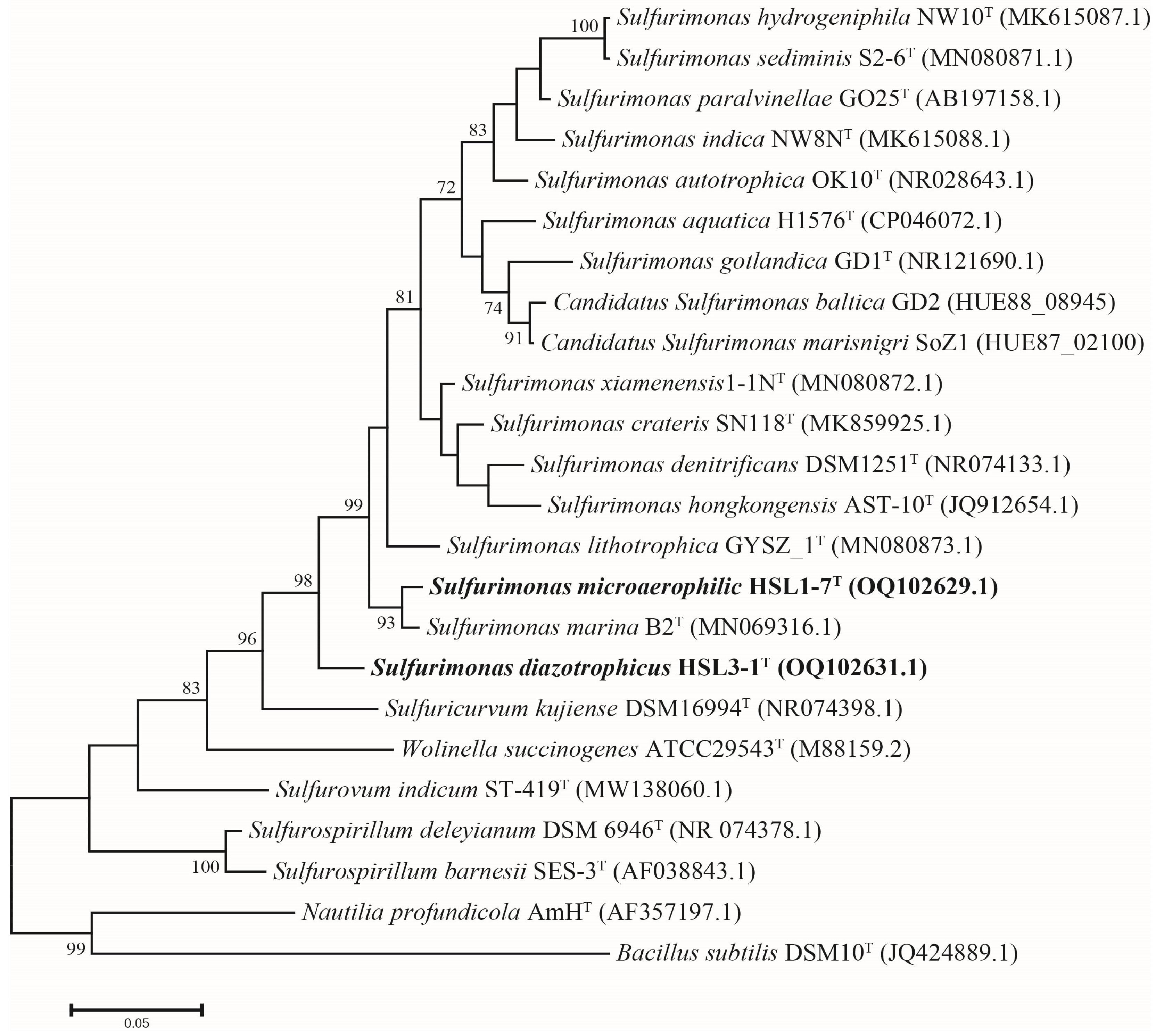
3.3. Phylogeny of 16S rRNA Gene Sequences

The comparative analysis of the full-length 16S rRNA gene sequences (1450bp) revealed that strain HSL1-7T shares the closest phylogenetic affiliation with S. marina B2T, exhibiting a 98.45% sequence identity; the other strains shared sequence similarities below 95.5%. Strain HSL3-1T was also most closely related to S. marina B2T with a 94.45% sequence identity; the other strains shared sequence similarities below 94.0%. These sequence similarities were lower than the threshold of 98.65% for species delineation [50]. The phylogenetic analysis using the maximum-likelihood method revealed that strain HSL1-7T was clustered with S. marina B2T, and strain HSL3-1T was clustered with the other reference strains of Sulfurimonas (Figure 1). The phylogenetic topology was further supported by neighbor-joining and minimum-evolution analyses, which indicates that strains HSL1-7T and HSL3-1T represent members of the genus Sulfurimonas (Figures S2 and S3).

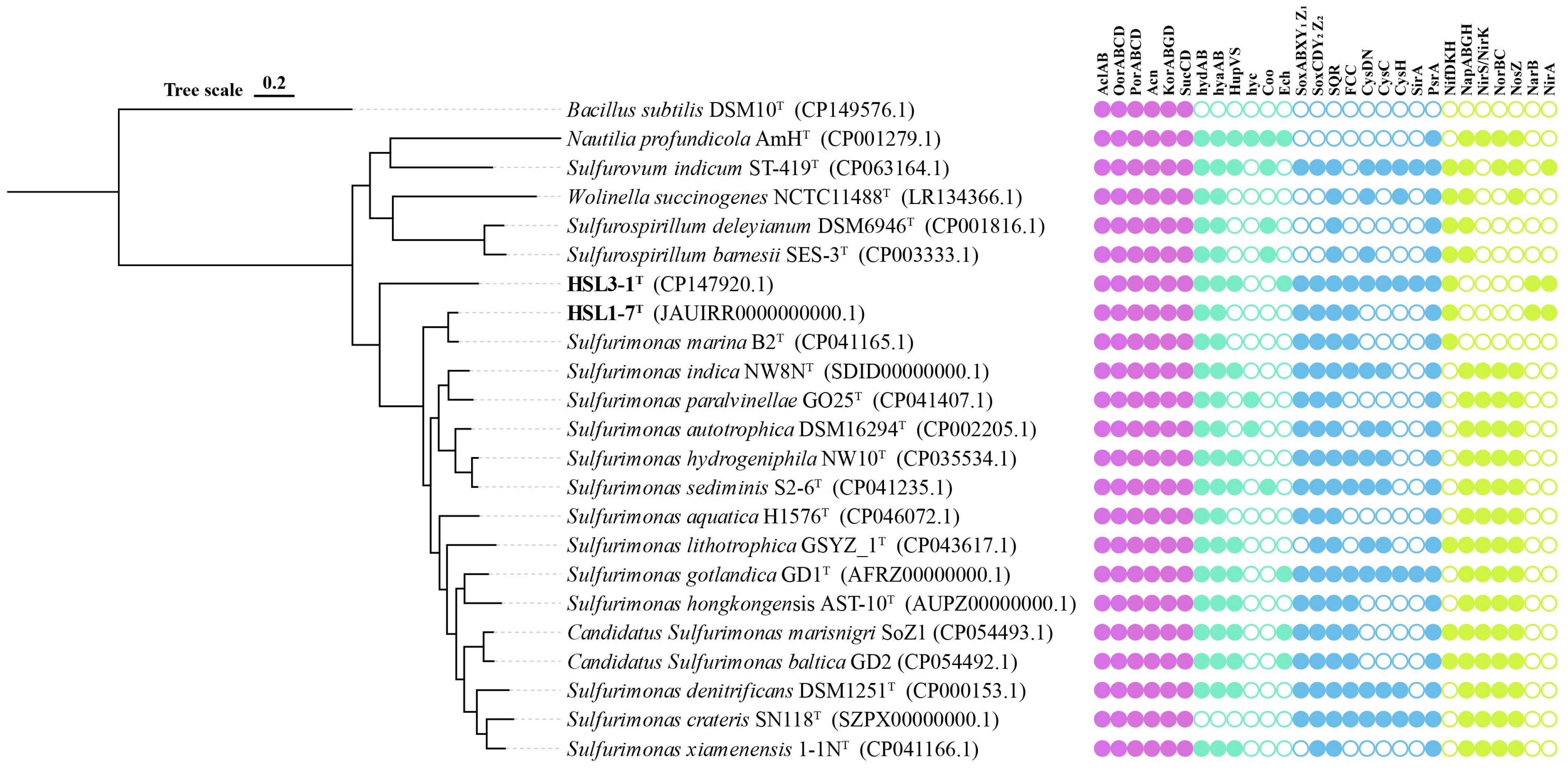
3.4. Genomic Properties

The genome size of strains HSL1-7T and HSL3-1T were 2.28 Mb and 2.49 Mb, respectively. The G+C contents of the genomic DNA of strains HSL1-7T and HSL3-1T were determined to be 36.1% and 57.3%, respectively (Table 1). The pairwise ANI values between HSL1-7T and the closest related type strain S. marina B2T was 89.2%, and the dDDH values between them were 36.9% (Table S1). The pairwise ANI values between strain HSL3-1T and the closest related type strain S. marina B2T were 67.41%, and the dDDH values between them were 23.7% (Table S2). All these values fell well below the established thresholds for prokaryotic species delineation (95–96% ANI and 70% dDDH) [51,52]. These data indicate that strains HSL1-7T and HSL3-1T represent two novel species of the genus Sulfurimonas.
The phylogenetic tree based on the 92 core gene sequences revealed that strain HSL1-7T formed a distinct branch with S. marina B2T, while HSL3-1T clustered together with other reference strains of Sulfurimonas (Figure 2). This result corroborated phylogenetic analyses of the 16S rRNA gene sequences, which supports that strains HSL1-7T and HSL3-1T represent members of the genus Sulfurimonas. Therefore, the genome-based phylogeny clearly indicates that strains HSL1-7T and HSL3-1T represent two novel species of the genus Sulfurimonas.

3.5. Genomic Functional Analysis

3.5.1. Carbon Metabolism

The genome analysis revealed that strains HSL1-7T and HSL3-1T possess the complete set of enzymes required for carbon fixation via the reductive tricarboxylic acid (rTCA) cycle, including the key enzymes encoding ATP citrate lyase (AclAB), 2-oxoglutarate: ferredoxin oxidoreductase (OorABCD), and pyruvate:ferredoxin oxidoreductase (PorABCD) (Figure 2). This genome characteristic was accordant with the metabolic features of chemoautotrophic growth in strains HSL1-7T and HSL3-1T, which was also conserved in the Sulfurimonas. In addition, the genomes of strains HSL1-7T and HSL3-1T contained enzymes for the intact tricarboxylic acid (TCA) cycle, including aconitehydratase (Acn), 2-oxoglutarateoxidoreductase (KorABGD), and succinyl-CoAsynthetase (SucCD) (Figure 2). Emerging evidence reveals the TCA cycle operates bidirectionally as a metabolic orchestrator, not only fueling oxidative phosphorylation but dynamically supplying core metabolic nodes (α-ketoglutarate, succinyl-CoA, and oxaloacetate) which coordinate anabolic flux partitioning through moonlighting enzyme-mediated substrate channeling [53].

3.5.2. Hydrogen Metabolism

The genomic analysis revealed that strain HSL1-7T contained one type of [Ni-Fe]-hydrogenase (Group I), while strain HSL3-1T contained three types of [Ni-Fe]-hydrogenases (Group I, Group II, and Group IV) (Figure 2). Group I hydrogenases are membrane-bound enzymes that catalyze the respiratory oxidation of hydrogen, coupled to the reduction of electron acceptors [54]. Like most members of the genus Sulfurimonas, strains HSL1-7T and HSL3-1T both harbor two copies of Group I [Ni-Fe]-hydrogenase. This suggests that Group I hydrogenases might be essential for growth, like the genus Sulfurimonas. Group II hydrogenases are possibly involved in hydrogen sensing or energy conversion at low hydrogen concentrations [30]; this was only found in strain HSL3-1T and some isolates from the seawater and sediments of the genus Sulfurimonas. Moreover, only strain HSL3-1T contained Group IV hydrogenases Ech. However, neither strain HSL1-7T nor HSL3-1T contained Group IV hydrogenases Hyc and Coo (Figure 2).

3.5.3. Sulfur Metabolism

The genomic analysis revealed that strains HSL1-7T and HSL3-1T harbor two distinct Sox gene clusters (soxABXY1Z1 and soxCDY2Z2), which collectively constitute a complete Sox multienzyme system (Figure 2). While in the Sulfurimonas, only strains Sulfurimonas xiamenensis and Sulfurimonas lithotrophica had the soxCDY2Z2 gene cluster [55]. Thus, the complete Sox gene clusters confer the capability of the isolated strains to oxidize thiosulfate to sulfate. Moreover, the genomes of strains HSL1-7T and HSL3-1T also contained flavocytochrome c sulfide dehydrogenase (Fcc) and sulfide:quinone oxidizing reductase (Sqr) for sulfide oxidation (Figure 2), which are also found in all members of genus Sulfurimonas. Only strain HSL3-1T contained the complete enzymes required for the assimilatory sulfate reduction pathway, including the key enzymes sulfate adenylyltransferase (CysDN), adenylylsulfate kinase (CysC), phosphoadenosine phosphosulfate reductase (CysH), and ferredoxin-sulfite reductase (SirA), which are capable of reducing sulfate to organic sulfur compounds. Further, the genomes of strains HSL1-7T and HSL3-1T both encoded polysulfide reductase (Psr) (Figure 2), which could reduce elemental sulfur to sulfide.

3.5.4. Nitrogen Metabolism

The comparative genome analysis revealed that strains HSL1-7T and HSL3-1T contain the complete nitrogen fixation pathway catalyzed by the nitrogenase encoded NifH (nitrogenase iron protein), NifD (nitrogenase Mo-iron alpha chain), and NifK (nitrogenase Mo-iron beta chain) (Figure 2). While in genus Sulfurimonas, only strains S. marisnigri SoZ1, S. lithotrophica GYSZ_1, S. baltica GD2, and S. marina B2 possessed the nitrogen fixation pathways [56]. Mangrove sediments are established as nitrogen-limited ecosystems due to the unbalance of carbon and nitrogen caused by the burying of plant litter or animal corpses. Nitrogen-fixing bacteria are considered to play a key role in nitrogen amendment [3]. Thus, the strains that contained nitrogenase genes in this study may have a competitive advantage in mangrove sediments. Moreover, strains HSL1-7T and HSL3-1T lack the genes encoding all the components required for the complete reduction of nitrate to nitrogen gas. Specifically, the genes for nitrate reductases (NapABGH), nitrite reductases (NirKS), nitric oxide reductases (NorBC), and nitrous oxide reductases (NosZ) were not detected in these strains (Figure 2), suggesting that they are unable to utilize nitrate as a terminal electron acceptor. In addition, strains HSL1-7T and HSL3-1T possessed the complete assimilatory nitrate reduction pathway, including the key enzyme encoding ferredoxin-nitrate reductase (NarAB) and ferredoxin-nitrite reductase (NirA).

3.6. Nitrogen Fixation Activity

The 15N2 isotope labelling experiments demonstrated that both strains, HSL1-7T and HSL3-1T, possessed the capability to fix 15N2 in the absence of an available nitrogen source in the medium, and that they exhibited similar nitrogen-fixing rates (Figure 3A). Whereas the reference strain Sulfurimonas xiamenensis 1-1NT from the coastal sediments, which lacks nitrogen fixation gene clusters, could not perform nitrogen fixation. Moreover, 15N2 fixation in both strains were completely inhibited by the addition of ammonia, regardless of whether the concentration was low or high (1 mM and 40 mM of NH4Cl) (Figure 3A). This observation aligns with the fact that nitrogen fixation is often repressed when exogenous nitrogen sources are readily available in their environment [57]. While in Sulfurimonas, only strains S. marina B2T, S. lithotrophica GYSZ_1T, and S. baltica GD2 harbored the complete nitrogenase gene clusters, suggesting that they have the potential for nitrogen fixation (Figure 3B), but this has not been verified so far. These results suggest an important role for nitrogen fixation of these isolates in the nitrogen-limited mangrove ecosystems in which they may be present.

3.7. Taxonomic Conclusion

Based on these results, strains HSL1-7T and HSL3-1T represent two novel species of the genus Sulfurimonas, for which the names Sulfurimonas microaerophilic sp. nov. and Sulfurimonas diazotrophicus sp. nov. are proposed.

3.7.1. Description of Sulfurimonas microaerophilic sp. nov.

Sulfurimonas microaerophilic (mi.cro.ae.ro’phi.lus. Gr. adj. mikros small, little; Gr. n. aer-aeros air; N.L. masc. adj. philus (from Gr. masc. adj. philos) loving; N.L. masc. adj. microaerophilus low-air loving, where the type strain was isolated).
The bacterial cells are Gram-stain-negative, rod-shaped to slightly curved (about 1.8–2.3 µm length × 0.4–0.6 µm in width), exhibiting motility via a single polar flagellum. The isolate was an obligate chemolithoautotroph capable of growing with molecular hydrogen and thiosulfate as an energy source, and molecular oxygen and elemental sulfur as the electron acceptors. Sodium bicarbonate and carbon dioxide can be used as inorganic carbon sources, whereas the growth of the strain was not observed under organic carbon source conditions such as peptone, yeast paste, and starch. The strain exhibited facultative microaerophilic physiology (growth up to 0–10% O2 in the gas phase; optimum growth in the presence of 4% O2). Growth parameters were established with temperature optima at 32 °C (range 4–40 °C), a salinity preference at 3.0% NaCl (w/v) (range 1.0–4.0%), and a pH optima at 7.0 (range 5.0–8.5). The major cellular fatty acids are C16:0 (31.6%), C16:1ω7c (28.7%), C18:1ω7c (19.5%), and C14:0 (10.5%).
The type strain, HSL1-7T (=MCCC 1A18899T = KCTC 25640T), was isolated from mangrove sediments of the Jiulong River tributaries in Zhangzhou, Fujian province, China. The genomic DNA G+C content of the type strain is 36.1%. The GenBank accession number for the 16S rRNA gene sequence is OQ102629, and the genome accession number is PRJNA989520, respectively.

3.7.2. Description of Sulfurimonas diazotrophicus sp. nov.

Sulfurimonas diazotrophicus (di.a.zo.trophi.cus. Gr. pref. di two, double; N. L. n. azotum nitrogen; Gr. adj. trophikos tending or feeding; N. L. masc. adj. diazotrophicus, one that feeds on dinitrogen).
The bacterial cells are Gram-stain-negative, displaying rod-shaped morphology (approximately 0.7–2.5 µm length × 0.2–0.5 µm in width) with non-motile characteristics. As a strict chemolithoautotroph, the isolate utilizes molecular hydrogen and thiosulfate as an energy source, while employing molecular oxygen and elemental sulfur as terminal electron acceptors. Nitrogen assimilation occurs through ammonium ions, sodium nitrate, and dinitrogen fixation, whereas growth showed no requirement for selenium, tungsten, or vitamin supplementation. Nitrogen fixation is completely inhibited by NH4Cl. Growth occurs at 4–45 °C with optimum growth at 37 °C, and at pH 5.0–8.5 with an optimum at pH 7.0. Growth is observed in the presence of 2.0–4.0% (w/v) NaCl with an optimum at 3.0% NaCl (w/v). The major cellular fatty acids are C16:1ω7c (35.0%), C16:0 (29.6%), C18:1ω7c (16.7%), and C14:0 (11.3%).
The type strain, HSL3-1T (=MCCC 1A18844T), was isolated from mangrove sediments of the Jiulong River tributaries in Zhangzhou, Fujian province, China. The G+C content of DNA was 57.3%. The GenBank accession number for the 16S rRNA gene sequence is OQ102631 and the genome accession number is PRJNA1085846, respectively.

4. Conclusions

The phylogenetic analysis based on the 16S rRNA gene sequences and whole genome, revealed that strains HSL1-7T and HSL3-1T represent two novel members of the genus Sulfurimonas. The 16S rRNA gene sequence similarity, and the ANI and dDDH values of strains HSL1-7T and HSL3-1T with closely related species, were lower than the thresholds for the delineation of a novel species. These results indicate that strains HSL1-7T and HSL3-1T do not represent any previously described species and should be considered to represent two novel species.
In addition, the physiological and biochemical characteristics of strains HSL1-7T and HSL3-1T differed from those of other species of the genus Sulfurimonas in terms of morphology, major fatty acid fractions, and patterns of electron donor-acceptor utilization. The comparative genomic analysis revealed that strains HSL1-7T and HSL3-1T possessed diverse metabolic profiles, including carbon fixation, hydrogen oxidation, sulfur oxidation, and nitrogen fixation. These unique metabolic capabilities suggests their potential role in driving the biogeochemical cycling of carbon, nitrogen, and sulfur in mangrove ecosystems. Notably, unlike most members of the genus Sulfurimonas, neither strain HSL1-7T or HSL3-1T genomes contained a complete denitrification pathway, but both harbored key genes for nitrogen fixation (nifHDK). Further, the 15N2 isotope labeling experiments demonstrated that they both exhibited strong nitrogen-fixing abilities. This characteristic implies that they may play an important role in the dark nitrogen fixation process in the mangrove ecosystem. The combined phenotypic, chemotaxonomic and phylogenetic evidence indicates that strains HSL1-7T and HSL3-1T represents two novel species in the genus Sulfurimonas. The findings of our study expand the understanding of Sulfurimonas species and their adaptation to mangrove environments.

Supplementary Materials

The following supporting information can be downloaded at: https://www.mdpi.com/article/10.3390/microorganisms13040713/s1, Figure S1: Transmission electron micrographs of cells of Sulfurimonas microaerophilic HSL1-7T (a) and Sulfurimonas diazotrophicus HSL3-1T (b); Figure S2: Neighbor joining phylogenetic tree based on 16S rRNA gene sequences showing the relationship between strains HSL1-7T and HSL3-1T with other members within the genus Sulfurimonas; Figure S3: Minimum-evolution phylogenetic tree based on 16S rRNA gene sequences showing the relationship between strains HSL1-7T and HSL3-1T with other members within the genus Sulfurimonas; Table S1: The average nucleotide identity (ANI) and in silico DNA-DNA hybridization values (dDDH) between strain HSL1-7T and related strains of the genus Sulfurimonas; Table S2: The average nucleotide identity (ANI) and in silico DNA-DNA hybridization values (dDDH) between strain HSL3-1T and related strains of the genus Sulfurimonas.

Author Contributions

Y.Z. and Y.L.: methodology, software, formal analysis, investigation, data curation, writing-original draft preparation, and visualization. Z.W.: software. L.C.: formal analysis. S.L.: investigation and data curation. H.Z. and Q.Y.: investigation and data curation. Q.L.: resources. S.W. and L.J.: conceptualization, resources, writing-review and editing, supervision, project administration, and funding acquisition. All authors have read and agreed to the published version of the manuscript.

Funding

This work was financially supported by the National Natural Science Foundation of China (No. 42176134 and 42030412) and Scientific Research Foundation of Third Institute of Oceanography (No. 2024002).

Institutional Review Board Statement

Not applicable.

Informed Consent Statement

Not applicable.

Data Availability Statement

The GenBank accession number for the 16S rRNA gene sequence and complete genome sequence of Sulfurimonas microaerophilic HSL1-7T are OQ102629 and PRJNA989520, respectively. The GenBank accession number for the 16S rRNA gene sequence and complete genome sequence of Sulfurimonas diazotrophicus HSL3-1T are OQ102631 and PRJNA1085846, respectively. Sulfurimonas microaerophilic HSL1-7: The 16S rRNA gene sequence can be accessed in GenBank under accession number OQ102629 via the following link: https://www.ncbi.nlm.nih.gov/search/all/?term=OQ102629 accessed on 20 October 2024; The complete genome sequence can be accessed in GenBank under accession number PRJNA989520 via the following link: https://www.ncbi.nlm.nih.gov/search/all/?term=PRJNA989520 accessed on 20 October 2024; Sulfurimonas diazotrophicus HSL3-1: The 16S rRNA gene sequence can be accessed in GenBank under accession number OQ102631 via the following link: https://www.ncbi.nlm.nih.gov/search/all/?term=OQ102631 accessed on 20 October 2024; The complete genome sequence can be accessed in GenBank under accession number PRJNA1085846 via the following link: https://www.ncbi.nlm.nih.gov/search/all/?term=PRJNA1085846 accessed on 20 October 2024.

Acknowledgments

We thank all the participants of the Marine Culture Collection of China for their kind help during the experiment.

Conflicts of Interest

The authors declare that there are no conflicts of interest.

References

  1. Duke, N.C.; Meynecke, J.O.; Dittmann, S.; Ellison, A.M.; Anger, K.; Berger, U.; Cannicci, S.; Diele, K.; Ewel, K.C.; Field, C.D.; et al. A world without mangroves? Science 2007, 317, 41–42. [Google Scholar] [CrossRef]
  2. Holguin, G.; Vazquez, P.; Bashan, Y. The role of sediment microorganisms in the productivity, conservation, and rehabilitation of mangrove ecosystems: An overview. Biol. Fertil. Soils 2001, 33, 265–278. [Google Scholar] [CrossRef]
  3. Wang, S.; Jiang, L.; Zhao, Z.; Chen, Z.; Wang, J.; Alain, K.; Cui, L.; Zhong, Y.; Peng, Y.; Lai, Q.; et al. Chemolithoautotrophic diazotrophs dominate dark nitrogen fixation in mangrove sediments. ISME J. 2024, 18, wrae119. [Google Scholar] [CrossRef]
  4. Sengupta, A.; Chaudhuri, S. Ecology of heterotrophic dinitrogen fixation in the rhizosphere of mangrove plant community at the Ganges river estuary in India. Oecologia 1991, 87, 560–564. [Google Scholar] [CrossRef]
  5. Biderre-Petit, C.; Courtine, D.; Hennequin, C.; Galand, P.E.; Bertilsson, S.; Debroas, D.; Monjot, A.; Lepère, C.; Divne, A.-M.; Hochart, C. A pan-genomic approach reveals novel Sulfurimonas clade in the ferruginous meromictic Lake Pavin. Mol. Ecol. Resour. 2024, 24, e13923. [Google Scholar] [CrossRef]
  6. Inagaki, F.; Takai, K.; Kobayashi, H.; Nealson, K.H.; Horikoshi, K. Sulfurimonas autotrophica gen. nov., sp. nov., a novel sulfur-oxidizing epsilon-proteobacterium isolated from hydrothermal sediments in the Mid-Okinawa Trough. Int. J. Syst. Evol. Microbiol. 2003, 53, 1801–1805. [Google Scholar] [CrossRef]
  7. Waite, D.W.; Vanwonterghem, I.; Rinke, C.; Parks, D.H.; Zhang, Y.; Takai, K.; Sievert, S.M.; Simon, J.; Campbell, B.J.; Hanson, T.E.; et al. Comparative Genomic Analysis of the Class Epsilonproteobacteria and Proposed Reclassification to Epsilonbacteraeota (phyl. nov.). Front. Microbiol. 2017, 8, 682, Erratum in Front. Microbiol. 2018, 9, 772. [Google Scholar] [CrossRef]
  8. Campbell, B.J.; Engel, A.S.; Porter, M.L.; Takai, K. The versatile epsilon-proteobacteria: Key players in sulphidic habitats. Nat. Rev. Microbiol. 2006, 4, 458–468. [Google Scholar] [CrossRef]
  9. Han, Y.; Perner, M. The globally widespread genus Sulfurimonas: Versatile energy metabolisms and adaptations to redox clines. Front. Microbiol. 2015, 6, 989. [Google Scholar] [CrossRef]
  10. Sievert, S.M.; Scott, K.M.; Klotz, M.G.; Chain, P.S.; Hauser, L.J.; Hemp, J.; Hügler, M.; Land, M.; Lapidus, A.; Larimer, F.W.; et al. Genome of the epsilonproteobacterial chemolithoautotroph Sulfurimonas denitrificans. Appl. Environ. Microbiol. 2008, 74, 1145–1156. [Google Scholar] [CrossRef]
  11. Watanabe, T.; Kubo, K.; Kamei, Y.; Kojima, H.; Fukui, M. Dissimilatory microbial sulfur and methane metabolism in the water column of a shallow meromictic lake. Syst. Appl. Microbiol. 2022, 45, 126320. [Google Scholar] [CrossRef]
  12. Zhang, D.; Cheng, L.; Zhang, S.; Zheng, J. Denitrification performance and microbial community analysis of sulfur autotrophic denitrification filter for low-temperature treatment of landfill leachate. J. Environ. Chem. Eng. 2023, 11, 109314. [Google Scholar] [CrossRef]
  13. Hu, Q.; Wang, S.; Lai, Q.; Shao, Z.; Jiang, L. Sulfurimonas indica sp. nov., a hydrogen- and sulfur-oxidizing chemolithoautotroph isolated from a hydrothermal sulfide chimney in the Northwest Indian Ocean. Int. J. Syst. Evol. Microbiol. 2021, 71, 004575. [Google Scholar] [CrossRef]
  14. Takai, K.; Suzuki, M.; Nakagawa, S.; Miyazaki, M.; Suzuki, Y.; Inagaki, F.; Horikoshi, K. Sulfurimonas paralvinellae sp. nov., a novel mesophilic, hydrogen- and sulfur-oxidizing chemolithoautotroph within the Epsilonproteobacteria isolated from a deep-sea hydrothermal vent polychaete nest, reclassification of Thiomicrospira denitrificans as Sulfurimonas denitrificans comb. nov. and emended description of the genus Sulfurimonas. Int. J. Syst. Evol. Microbiol. 2006, 56, 1725–1733. [Google Scholar] [CrossRef]
  15. Wang, S.; Jiang, L.; Hu, Q.; Liu, X.; Yang, S.; Shao, Z. Elemental sulfur reduction by a deep-sea hydrothermal vent Campylobacterium Sulfurimonas sp. NW10. Environ. Microbiol. 2021, 23, 965–979. [Google Scholar] [CrossRef]
  16. Wang, S.; Shao, Z.; Lai, Q.; Liu, X.; Xie, S.; Jiang, L.; Yang, S. Sulfurimonas sediminis sp. nov., a novel hydrogen- and sulfur-oxidizing chemolithoautotroph isolated from a hydrothermal vent at the Longqi system, southwestern Indian ocean. Antonie Van Leeuwenhoek 2021, 114, 813–822. [Google Scholar] [CrossRef]
  17. Cai, L.; Shao, M.F.; Zhang, T. Non-contiguous finished genome sequence and description of Sulfurimonas hongkongensis sp. nov., a strictly anaerobic denitrifying, hydrogen- and sulfur-oxidizing chemolithoautotroph isolated from marine sediment. Stand. Genom. Sci. 2014, 9, 1302–1310. [Google Scholar] [CrossRef]
  18. Hoor, A.T. Energetic aspects of the metabolism of reduced sulphur compounds in Thiobacillus dentrificans. Antonie Van Leeuwenhoek 1976, 42, 483–492. [Google Scholar] [CrossRef]
  19. Wang, S.; Jiang, L.; Liu, X.; Yang, S.; Shao, Z. Sulfurimonas xiamenensis sp. nov. and Sulfurimonas lithotrophica sp. nov., hydrogen- and sulfur-oxidizing chemolithoautotrophs within the Epsilonproteobacteria isolated from coastal sediments, and an emended description of the genus Sulfurimonas. Int. J. Syst. Evol. Microbiol. 2020, 70, 2657–2663. [Google Scholar] [CrossRef]
  20. Henkel, J.V.; Vogts, A.; Werner, J.; Neu, T.R.; Spröer, C.; Bunk, B.; Schulz-Vogt, H.N. Candidatus Sulfurimonas marisnigri sp. nov. and Candidatus Sulfurimonas baltica sp. nov., thiotrophic manganese oxide reducing chemolithoautotrophs of the class Campylobacteria isolated from the pelagic redoxclines of the Black Sea and the Baltic Sea. Syst. Appl. Microbiol. 2021, 44, 126155. [Google Scholar] [CrossRef]
  21. Kojima, H.; Kato, Y.; Watanabe, T.; Fukui, M. Sulfurimonas aquatica sp. nov., a sulfur-oxidizing bacterium isolated from water of a brackish lake. Arch. Microbiol. 2022, 204, 559. [Google Scholar] [CrossRef]
  22. Labrenz, M.; Grote, J.; Mammitzsch, K.; Boschker, H.T.S.; Laue, M.; Jost, G.; Glaubitz, S.; Jürgens, K. Sulfurimonas gotlandica sp. nov., a chemoautotrophic and psychrotolerant epsilonproteobacterium isolated from a pelagic redoxcline, and an emended description of the genus Sulfurimonas. Int. J. Syst. Evol. Microbiol. 2013, 63, 4141–4148. [Google Scholar] [CrossRef]
  23. Ratnikova, N.M.; Slobodkin, A.I.; Merkel, A.Y.; Kopitsyn, D.S.; Kevbrin, V.V.; Bonch-Osmolovskaya, E.A.; Slobodkina, G.B. Sulfurimonas crateris sp. nov., a facultative anaerobic sulfur-oxidizing chemolithoautotrophic bacterium isolated from a terrestrial mud volcano. Int. J. Syst. Evol. Microbiol. 2020, 70, 487–492. [Google Scholar] [CrossRef]
  24. Wang, Z.; Wang, S.; Lai, Q.; Wei, S.; Jiang, L.; Shao, Z. Sulfurimonas marina sp. nov., an obligately chemolithoautotrophic, sulphur-oxidizing bacterium isolated from a deep-sea sediment sample from the South China Sea. Int. J. Syst. Evol. Microbiol. 2022, 72, 005582. [Google Scholar] [CrossRef]
  25. Jiang, L.; Lyu, J.; Shao, Z. Sulfur Metabolism of Hydrogenovibrio thermophilus Strain S5 and Its Adaptations to Deep-Sea Hydrothermal Vent Environment. Front. Microbiol. 2017, 8, 2513. [Google Scholar] [CrossRef]
  26. Liu, X.; Jiang, L.; Hu, Q.; Lyu, J.; Shao, Z. Thiomicrorhabdus indica sp. nov., an obligately chemolithoautotrophic, sulfur-oxidizing bacterium isolated from a deep-sea hydrothermal vent environment. Int. J. Syst. Evol. Microbiol. 2020, 70, 234–239. [Google Scholar] [CrossRef]
  27. Takai, K.; Horikoshi, K. Thermosipho japonicus sp. nov., an extremely thermophilic bacterium isolated from a deep-sea hydrothermal vent in Japan. Extremophiles 2000, 4, 9–17. [Google Scholar] [CrossRef]
  28. Li, Y.; Wang, S.; Ji, B.; Yuan, Q.; Wei, S.; Lai, Q.; Wu, K.; Jiang, L.; Shao, Z. Sulfurovum mangrovi sp. nov., an obligately chemolithoautotrophic, hydrogen-oxidizing bacterium isolated from coastal marine sediments. Int. J. Syst. Evol. Microbiol. 2023, 73, 006142. [Google Scholar] [CrossRef]
  29. Adiguzel, A.; Ozkan, H.; Baris, O.; Inan, K.; Gulluce, M.; Sahin, F. Identification and characterization of thermophilic bacteria isolated from hot springs in Turkey. J. Microbiol. Methods 2009, 79, 321–328. [Google Scholar] [CrossRef]
  30. Jiang, L.; Zheng, Y.; Peng, X.; Zhou, H.; Zhang, C.; Xiao, X.; Wang, F. Vertical distribution and diversity of sulfate-reducing prokaryotes in the Pearl River estuarine sediments, Southern China. FEMS Microbiol. Ecol. 2009, 70, 93–106. [Google Scholar] [CrossRef]
  31. Yoon, S.H.; Ha, S.M.; Kwon, S.; Lim, J.; Kim, Y.; Seo, H.; Chun, J. Introducing EzBioCloud: A taxonomically united database of 16S rRNA gene sequences and whole-genome assemblies. Int. J. Syst. Evol. Microbiol. 2017, 67, 1613–1617. [Google Scholar] [CrossRef]
  32. Kumar, S.; Stecher, G.; Tamura, K. MEGA7: Molecular Evolutionary Genetics Analysis Version 7.0 for Bigger Datasets. Mol. Biol. Evol. 2016, 33, 1870–1874. [Google Scholar] [CrossRef]
  33. Saitou, N.; Nei, M. The neighbor-joining method: A new method for reconstructing phylogenetic trees. Mol. Biol. Evol. 1987, 4, 406–425. [Google Scholar] [CrossRef]
  34. Felsenstein, J. Evolutionary trees from DNA sequences: A maximum likelihood approach. J. Mol. Evol. 1981, 17, 368–376. [Google Scholar] [CrossRef]
  35. Rzhetsky, A.; Nei, M. Statistical properties of the ordinary least-squares, generalized least-squares, and minimum-evolution methods of phylogenetic inference. J. Mol. Evol. 1992, 35, 367–375. [Google Scholar] [CrossRef]
  36. Kimura, M. A simple method for estimating evolutionary rates of base substitutions through comparative studies of nucleotide sequences. J. Mol. Evol. 1980, 16, 111–120. [Google Scholar] [CrossRef]
  37. Felsenstein, J. Confidence limits on phylogenies: An approach using the bootstrap. Evolution 1985, 39, 783–791. [Google Scholar] [CrossRef]
  38. Prjibelski, A.; Antipov, D.; Meleshko, D.; Lapidus, A.; Korobeynikov, A. Using SPAdes De Novo Assembler. Curr. Protoc. Bioinformatics 2020, 70, e102. [Google Scholar] [CrossRef]
  39. Lowe, T.M.; Chan, P.P. tRNAscan-SE On-line: Integrating search and context for analysis of transfer RNA genes. Nucleic Acids Res. 2016, 44, W54–W57. [Google Scholar] [CrossRef]
  40. Lagesen, K.; Hallin, P.; Rødland, E.A.; Staerfeldt, H.H.; Rognes, T.; Ussery, D.W. RNAmmer: Consistent and rapid annotation of ribosomal RNA genes. Nucleic Acids Res. 2007, 35, 3100–3108. [Google Scholar] [CrossRef]
  41. Delcher, A.L.; Harmon, D.; Kasif, S.; White, O.; Salzberg, S.L. Improved microbial gene identification with GLIMMER. Nucleic Acids Res. 1999, 27, 4636–4641. [Google Scholar] [CrossRef] [PubMed]
  42. Aziz, R.K.; Bartels, D.; Best, A.A.; DeJongh, M.; Disz, T.; Edwards, R.A.; Formsma, K.; Gerdes, S.; Glass, E.M.; Kubal, M.; et al. The RAST Server: Rapid annotations using subsystems technology. BMC Genom. 2008, 9, 75. [Google Scholar] [CrossRef] [PubMed]
  43. Zhou, Z.; Tran, P.Q.; Breister, A.M.; Liu, Y.; Kieft, K.; Cowley, E.S.; Karaoz, U.; Anantharaman, K. METABOLIC: High-throughput profiling of microbial genomes for functional traits, metabolism, biogeochemistry, and community-scale functional networks. Microbiome 2022, 10, 33. [Google Scholar] [CrossRef] [PubMed]
  44. Aramaki, T.; Blanc-Mathieu, R.; Endo, H.; Ohkubo, K.; Kanehisa, M.; Goto, S.; Ogata, H. KofamKOALA: KEGG Ortholog assignment based on profile HMM and adaptive score threshold. Bioinformatics 2020, 36, 2251–2252. [Google Scholar] [CrossRef]
  45. Medlar, A.J.; Törönen, P.; Holm, L. AAI-profiler: Fast proteome-wide exploratory analysis reveals taxonomic identity, misclassification and contamination. Nucleic Acids Res. 2018, 46, W479–W485. [Google Scholar] [CrossRef]
  46. Na, S.I.; Kim, Y.O.; Yoon, S.H.; Ha, S.M.; Baek, I.; Chun, J. UBCG: Up-to-date bacterial core gene set and pipeline for phylogenomic tree reconstruction. J. Microbiol. 2018, 56, 280–285. [Google Scholar] [CrossRef]
  47. Xie, J.; Chen, Y.; Cai, G.; Cai, R.; Hu, Z.; Wang, H. Tree Visualization By One Table (tvBOT): A web application for visualizing, modifying and annotating phylogenetic trees. Nucleic Acids Res. 2023, 51, W587–W592. [Google Scholar] [CrossRef]
  48. Bazylinski, D.A.; Morillo, V.; Lefèvre, C.T.; Viloria, N.; Dubbels, B.L.; Williams, T.J. Endothiovibrio diazotrophicus gen. nov., sp. nov., a novel nitrogen-fixing, sulfur-oxidizing gammaproteobacterium isolated from a salt marsh. Int. J. Syst. Evol. Microbiol. 2017, 67, 1491–1498. [Google Scholar] [CrossRef]
  49. Zheng, H.; Dietrich, C.; Radek, R.; Brune, A. Endomicrobium proavitum, the first isolate of Endomicrobia class. nov. (phylum Elusimicrobia)—An ultramicrobacterium with an unusual cell cycle that fixes nitrogen with a Group IV nitrogenase. Environ. Microbiol. 2016, 18, 191–204. [Google Scholar] [CrossRef]
  50. Kim, M.; Oh, H.-S.; Park, S.-C.; Chun, J. Towards a taxonomic coherence between average nucleotide identity and 16S rRNA gene sequence similarity for species demarcation of prokaryotes. Int. J. Syst. Evol. Microbiol. 2014, 64, 346–351. [Google Scholar] [CrossRef]
  51. Auch, A.F.; von Jan, M.; Klenk, H.-P.; Göker, M. Digital DNA-DNA hybridization for microbial species delineation by means of genome-to-genome sequence comparison. Stand. Genom. Sci. 2010, 2, 117–134. [Google Scholar] [CrossRef] [PubMed]
  52. Richter, M.; Rosselló-Móra, R. Shifting the genomic gold standard for the prokaryotic species definition. Proc. Natl. Acad. Sci. USA 2009, 106, 19126–19131. [Google Scholar] [CrossRef]
  53. Pitson, S.M.; Mendz, G.L.; Srinivasan, S.; Hazell, S.L. The tricarboxylic acid cycle of Helicobacter pylori. Eur. J. Biochem. 1999, 260, 258–267. [Google Scholar] [CrossRef] [PubMed]
  54. Dong, X.; Zhang, C.; Peng, Y.; Zhang, H.X.; Shi, L.D.; Wei, G.; Hubert, C.R.J.; Wang, Y.; Greening, C. Phylogenetically and catabolically diverse diazotrophs reside in deep-sea cold seep sediments. Nat. Commun. 2022, 13, 4885. [Google Scholar] [CrossRef]
  55. Wang, S.; Jiang, L.; Hu, Q.; Cui, L.; Zhu, B.; Fu, X.; Lai, Q.; Shao, Z.; Yang, S. Characterization of Sulfurimonas hydrogeniphila sp. nov., a Novel Bacterium Predominant in Deep-Sea Hydrothermal Vents and Comparative Genomic Analyses of the Genus Sulfurimonas. Front. Microbiol. 2021, 12, 626705. [Google Scholar] [CrossRef] [PubMed]
  56. Yuan, Q.; Wang, S.; Wang, S.; Zhong, Y.; Jiang, L. Genome sequence of Sulfurimonas sp. C5, a potential chemolithoautotrophic, sulfur-oxidizing bacterium isolated from a mangrove sediment. Microbiol. Resour. Announc. 2024, 13, e0047424. [Google Scholar] [CrossRef]
  57. König, S.; Gros, O.; Heiden, S.E.; Hinzke, T.; Thürmer, A.; Poehlein, A.; Meyer, S.; Vatin, M.; Mbéguié-A-Mbéguié, D.; Tocny, J.; et al. Nitrogen fixation in a chemoautotrophic lucinid symbiosis. Nat. Microbiol. 2016, 2, 16193. [Google Scholar] [CrossRef]
Figure 1. Maximum-likelihood phylogenetic tree based on the 16S rRNA gene sequences showing the relationship between strains HSL1-7T and HSL3-1T with other members within the genus Sulfurimonas. Species names highlighted in bold represent novel taxa discovered in this work. Bootstrap numbers (>70%) were shown with 1000 calculations. Bar, 0.05 substitutions per nucleotide position.
Microorganisms 13 00713 g001
Figure 2. Phylogenetic reconstruction using 92 core genes was performed for strains HSL1-7T and HSL3-1T, along with their functional genes involved in carbon, hydrogen, sulfur, and nitrogen metabolism, in comparison with closely related species. Bootstrap values (>70%) are indicated at nodes based on 1000 replicates. Species names highlighted in bold represent novel taxa discovered in this work. Bar, 0.2 substitutions per nucleotide position. Column descriptions are as follows: Carbon pathway: ATP citrate lyase alpha and beta subunits (AclAB), 2- oxoglutarate:ferredoxin oxidoreductase (OorABCD), and pyruvate:ferredoxin oxidoreductase (PorABCD), aconite hydratase (Acn), 2-oxoglutarate oxidoreductase alpha-, beta-, gamma-, and delta- subunits (KorABGD), succinyl-CoA synthetase alpha and beta subunits (SucCD). Hydrogen pathways: [NiFe]-Hydrogenases Group I (HydAB and HyaAB), [NiFe]-Hydrogenases Group II (HupVS), [NiFe]-Hydrogenases Group IV (Hyc, Coo and Ech). Sulfur pathways: thiosulfate oxidating Sox proteins (SoxABXY1Z1, SoxY2Z2CD), sulfide:quinone oxidoreductase (Sqr), flavocytochrome c sulfide dehydrogenase (Fcc), sulfate adenylyltransferase (CysDN), adenylylsulfate kinase (CysC), phosphoadenosine phosphosulfate reductase (CysH), ferredoxin-sulfite reductase (SirA), polysulfide reductase chain A (PsrA). Nitrogen pathways: nitrogenase molybdenum-iron (alpha- and beta-chains) and iron protein (NifDKH), periplasmic nitrate reductase components (NapABGH), nitrite reductase (NirK/NirS), nitric oxide reductase subunit A/B (NorBC), nitrous-oxide reductase (NosZ), ferredoxin-nitrate reductase (NarAB), ferredoxin-nitrite reductase (NirA).
Microorganisms 13 00713 g002
Figure 3. Determination of nitrogen fixation activity of strains HSL1-7T and HSL3-1T and analysis of nitrogen fixation gene clusters. (A) 15N abundance based on the 15N incorporation assay, the incubation conditions were H2 as the sole electron donor and O2 as the electron acceptor, Sulfurimonas xiamenensis 1-1NT which lacked nif gene cluster served as a control. (B) Green represents nitrogen fixation genes (nifHDK), red represents nitrogenase metal cofactor biosynthesis genes (nifBSZVENUWOQXTN), purple represents transcriptional regulator genes (P-II, nifA, araC), pink represent electron transfer genes (fdx), and yellow represents transporter genes (ABC).
Microorganisms 13 00713 g003
Table 1. Comparison of phenotypic characteristics of Sulfurimonas microaerophilic HSL1-7T sp. nov. and Sulfurimonas diazotrophicus HSL3-1T sp. nov. with related type strains within the genus Sulfurimonas.
Characteristic1234567
ShapeRods to slightly curvedRodsRodsRods to slightly curvedRods to slightly curvedRodsRods
Motility+++
Doubling time under optimal (h)NDND3.681212ND
Temperature range (Optimal T) (°C)10–40 (32)4–45 (37)10–45 (35)4–45 (33)10–45 (30)ND15–35 (30)
pH range
(Optimal pH)
5.0–8.5
(7.0)
5.0–8.5 (7.0)4.5–9.0 (7.0)5.0–8.5 (6.5)5.5–8.0
(7.0)
ND (7.0)6.5–8.5 (7.0–7.5)
NaCl requirement+++++
Maximum O2 concentration (%)10201520200.50
Electron donorH2, S2O32−H2, S2O32−HS, S0, S2O32−H2, S0, S2O32−, HSH2, S0, S2O32−, S4O62−, HSS2O32−, HSH2, S2O32−, HS
Organic electron donorsFormate, fumarate, yeast extract alcohol mix
Electron acceptorS0, O2S0, O2O2S0, NO3, O2S0, NO3, O2NO3, O2, NO2NO3
DNA G+C content (%)36.157.336.033.234.53634.9
+, positive; −, negative; ND, not determined. Strains: 1, HSL1-7T; 2, HSL3-1T; 3, S. marina B2T [24]; 4, S. lithotrophica GYSZ_1T [19]; 5, S. xiamenensis 1-1NT [19]; 6, S. denitrificans DSM 1251T [14]; 7, S. hongkongensis AST-10T [17].
Table 2. Cellular fatty acids compositions (%) of Sulfurimonas microaerophilic HSL1-7T sp. nov. and Sulfurimonas diazotrophicus HSL3-1T sp. nov. with related type strains within the genus Sulfurimonas.
Fatty acid (%)1234567
C12:00.34.01.32.6---
C14:010.511.39.13.21.10.44.8
C14:0 3-OH2.44.06.64.9---
C16:031.629.625.618.923.415.332.8
C16:1ω7c28.735.037.750.531.867.9-
C16:1ω5c0.60.4---2.0-
C18:02.60.71.11.310.0-16.9
C18:1ω7c19.516.713.212.118.712.1-
Fatty acids present at levels exceeding 10% were highlighted by bold type. -, not detected. Strains: 1, HSL1-7T; 2, HSL3-1T; 3, S. marina B2T; 4, S. lithotrophica GYSZ_1T; 5, S. xiamenensis 1-1NT; 6, S. denitrificans DSM 1251T; 7, S. hongkongensis AST-10T.
Disclaimer/Publisher’s Note: The statements, opinions and data contained in all publications are solely those of the individual author(s) and contributor(s) and not of MDPI and/or the editor(s). MDPI and/or the editor(s) disclaim responsibility for any injury to people or property resulting from any ideas, methods, instructions or products referred to in the content.

