Article

From Isolation to Application: Designing a Multi-Target Phage Cocktail for Bivalve Safety

1
CESAM, Department of Biology, University of Aveiro, Campus Universitário de Santiago, 3810-193 Aveiro, Portugal
2
CRETUS, Department of Microbiology and Parasitology, CIBUS—Faculty of Biology, University of Santiago de Compostela, 15782 Santiago de Compostela, Spain
*
Authors to whom correspondence should be addressed.
Microorganisms 2025, 13(12), 2708; https://doi.org/10.3390/microorganisms13122708
Submission received: 24 October 2025 / Revised: 20 November 2025 / Accepted: 24 November 2025 / Published: 27 November 2025
(This article belongs to the Collection Feature Papers in Antimicrobial Agents and Resistance)

Abstract

Narrow host specificity and bacterial resistance often limit single-phage treatments. Phage cocktails address these challenges by expanding the host range, reducing resistance, and enhancing bacterial inactivation. This study aimed to develop an optimised phage cocktail targeting Escherichia coli, Salmonella enterica serovar Typhimurium, Salmonella enterica serovar Enteritidis, and Aeromonas hydrophila, key pathogens in bivalve consumption. Twelve phages were isolated, purified, and screened for bacterial inactivation using resazurin-based viability assays. Host range analysis showed that all phages infected at least one additional bacterial species, with four (phEc4, phSE1, phAh2, phAh4) targeting three of the four bacteria. Cocktail formulation aimed to maximise bacterial reduction while balancing host range expansion with factors such as the risks of resistance development and inter-phage competition. Among the tested combinations, the most effective cocktail consisted of E. coli phage phEc3, S. Typhimurium phage phST1, S. Enteritidis phage phSE1, and A. hydrophila phage phAh2. Future studies should evaluate the cocktail’s efficacy in vitro and assess both safety and performance in vivo in bivalve depuration systems.

1. Introduction

Bacteriophages (phages) are viruses that specifically infect bacteria [1] and offer a promising alternative to conventional antibiotics by selectively targeting harmful bacteria while preserving native microbiota [2,3,4,5]. Since their discovery over a century ago, phages have been explored as antibacterial agents in both therapeutic and environmental contexts, with renewed interest driven by the global rise in antimicrobial resistance [6,7,8].
In food safety, phage-based biocontrol has been explored to mitigate contamination risks in various products [9], including bivalve shellfish, which pose significant public health risks due to pathogens such as Escherichia coli, Salmonella enterica (serovars Typhimurium and Enteritidis), and Aeromonas hydrophila [10]. Industrial depuration is widely used to reduce microbial loads in live shellfish before consumption, but its effectiveness against bacterial pathogens varies [10]. Integrating phage biocontrol into depuration could improve bacterial removal efficiency while maintaining sustainability and consumer safety [10]. However, practical implementation faces challenges such as phages’ narrow host range [11,12,13] and the emergence of phage-resistant bacteria [14], both of which limit the success of single-phage applications [1,12,15,16,17,18]. To address these limitations, phage cocktails—combinations of multiple phages—are often employed to expand host range and suppress resistance [19]. These benefits are often described as enhancing a cocktail’s breadth (host range) or depth (resistance suppression) of activity [19].
Several studies have called for cocktails targeting different genera and serovars of bacteria transmitted via bivalves, such as S. Enteritidis and S. Typhimurium [20,21]. However, the careful selection of appropriate phages is key to success [22]. Host range and bacterial inactivation efficiency are essential criteria during the early stages of phage cocktail development [10]. This is particularly important given that the efficacy of phage cocktails can vary considerably: while some studies show synergistic or additive effects, others report reduced activity—often due to phage competition for the same bacterial receptors [22]. Thus, formulating an effective phage cocktail requires selecting potent candidates and carefully considering their potential interactions [22].
Bacteria employ various resistance mechanisms against phages, including blocking adsorption, degrading or preventing phage nucleic acid entry through restriction-modification systems and the CRISPR/Cas system, and using abortive infection systems to inhibit closely related phages, thereby limiting competitor phage propagation [22,23]. These defences may limit phage propagation and reduce overall cocktail effectiveness [22]. Combining phages that rely on the same receptor can result in antagonistic effects, as a single mutation may render multiple phages ineffective [24].
In contrast, some studies suggest that phage cocktails achieve greater bacterial inactivation than single-phage treatments [25,26], whether targeting the same bacterium [25,27], different strains of a species [28] or multiple bacterial species [26]. Thus, optimising a cocktail requires balancing bacterial reduction, host range, and stability—key challenges in developing effective phage biocontrol strategies.
Several strategies have been developed to counteract bacterial resistance to phages, including phage training, genetic engineering, and cocktails [29,30,31]. In a well-balanced mixture, bacteria resistant to one phage may remain susceptible to others [32], enhancing reliability and reducing treatment failure [33], a concept that could also extend to cocktails targeting multiple bacterial species [26]. However, randomly assembled cocktails can be inconsistent due to negative phage interactions [11].
An alternative strategy is to use fewer phages if individual phages can infect multiple bacterial species [34,35]—commonly referred to as polyvalence [36,37]. However, the chances of isolating effective polyvalent phages decrease with broader bacterial diversity, especially across Gram classifications [19]. For this reason, cocktails combining phages that target distinct receptors are preferred [38], though identifying these phages requires significant time, labour, and screening [24].
Since phages recognise their bacterial hosts through specific surface receptors, cocktail design often requires a degree of personalisation [13]. Given these complexities, several studies advocate for an empirical approach—testing diverse phage combinations with overlapping host ranges to identify the most effective mixes without the need for full receptor characterization [22].
Conventional methods for assessing phage–bacteria dynamics, such as colony counting and optical density (OD) measurements, are either too time-consuming or lack precision for large-scale screening [39]. To overcome these limitations, we employed the resazurin cell viability assay—a high-throughput method that simultaneously evaluates multiple bacteria–phage interactions [39]. This assay relies on the reduction of blue, non-fluorescent resazurin to the pink, fluorescent compound resorufin by metabolically active cells, allowing bacterial viability to be quantified by fluorescence or absorbance as an indirect measure of bacterial inactivation [39]. This approach enabled us to identify cocktails with broader host ranges, designed to delay resistance and maintain or improve upon single-phage efficacy.
In this study, we isolated phages targeting E. coli, S. enterica serovars Typhimurium and Enteritidis, and A. hydrophila. These were formulated into cocktails, and their antibacterial activity was evaluated in vitro using a viability-based approach. Our aim was to select reliable candidates for further in vitro and in vivo application in bivalve depuration systems.

2. Materials and Methods

2.1. Bacterial Strains and Growth Conditions

The bacterial strains used in this study as phage hosts included E. coli (ATCC 25922), S. Typhimurium (ATCC 13311), S. Enteritidis (CVB) and A. hydrophila (ATCC 7966). A. hydrophila (ATCC 7966), E. coli (ATCC 25922) and S. Typhimurium (ATCC 13311) were purchased from the ATCC collection. S. Enteritidis (CVB) is a food sample isolate generously provided by Controlvet Laboratory.
Fresh bacterial cultures were preserved on Tryptic Soy Agar (TSA; Liofilchem, Roseto degli Abruzzi, Italy) at 4 °C. Before each assay, an isolated colony was aseptically transferred to Tryptic Soy Broth (TSB; Liofilchem, Roseto degli Abruzzi, Italy) and incubated overnight at 25 °C. One hundred microliters of this culture were aseptically transferred to fresh TSB and incubated overnight at 25 °C until an optical density (OD600 of 0.8), corresponding to approximately 109 cells per mL, was reached.

2.2. Phage Isolation, Host Range and Purification

All phages were isolated from the Aveiro sewage network (station EEIS9 of the Multi-Sanitation System of Ria de Aveiro—SIMRIA). Sewage water samples were filtered through 0.45 µm pore size polycarbonate membranes (Millipore, Bedford, MA, USA). The resulting filtrate was added to a double-strength TSB medium, along with 1 mL of fresh cultures of each target host bacterium: E. coli (ATCC 25922), S. Typhimurium (ATCC 13311), S. Enteritidis (CVB), and A. hydrophila (ATCC 7966). These mixtures were incubated at 25 °C with shaking at 80 rpm for 18 h to promote phage–bacteria interactions, thereby accelerating the lytic cycle and enhancing phage amplification. Following incubation, samples were filtered through 0.2 µm membranes to remove bacterial cells. Phage presence was assessed using the double-layer agar method [40]. Briefly, 100 µL of phage lysate was mixed with 300 µL of an overnight bacterial culture (~108 colony-forming units per millilitre [CFU/mL]) and added to 5 mL of molten soft agar (0.6% w/v, pre-warmed to 50 °C), which was then overlaid onto TSA plates (1.5% w/v). Plates were incubated at 25 °C and examined for lytic plaques after 12 h.
Each unpurified phage stock was tested against the other target bacteria to identify phages capable of infecting multiple bacterial hosts. Phages with overlapping host infections were identified, and 22 phages were isolated. Individual plaques were picked and transferred to a TSB medium containing fresh cultures of the corresponding host bacteria. These mixtures were incubated at 25 °C with shaking at 80 rpm for 18 h, as previously described, to allow phage propagation. After incubation, the cultures were centrifuged at 10,000× g for 10 min at room temperature to pellet bacterial cells, and the resulting supernatant was used for subsequent rounds of phage isolation. Three successive single-plaque isolation cycles were performed to obtain pure phage stocks. Lysates were centrifuged at 10,000× g for 10 min at 4 °C to remove bacterial debris, and the purified phage stocks were stored at 4 °C. Of the 22 isolated phages, 12 were successfully purified. Phage titres were determined using the double-layer agar method [40], as previously described. Plates were incubated at 25 °C for 8 h, and the number of lytic plaques was counted. The results were expressed as plaque-forming units per millilitre (PFU/mL). Phages phEc1, phEc2, phEc3, phST1, phST2, and phST3 were previously described in a separate study [39]; however, all phages reported here, including these, were originally isolated using the protocol detailed above.

2.3. Bacterial Killing Curves

Bacterial inactivation was assessed using E. coli (ATCC 25922), S. Typhimurium (ATCC 13311), S. Enteritidis (CVB), and A. hydrophila (ATCC 7966) in TSB at 25 °C, using a multiplicity of infection (MOI) of 10, based on each phage’s original host bacterium, to achieve ≥99.99% infection efficiency in phage–bacteria systems [41]. Each assay was initiated with approximately 1 × 10⁷ CFU/mL of bacteria and 1 × 10⁸ PFU/mL of phages. For phage cocktails, each phage was added at an equal concentration relative to its primary host, ensuring consistent MOI and minimizing potential dominance or suppression effects. Only two-phage cocktail combinations were tested in this study. While the twelve isolated phages allowed for possible three- and four-phage formulations, testing all permutations would have required a prohibitively large and complex matrix of experimental conditions. This complexity, combined with the need for replication and continuous viability monitoring over 24 h, led us to focus on two-phage combinations as a tractable and informative first step. A bacterial control (no phage added) was included for each assay, and all samples—control and test—were incubated under identical conditions.
Bacterial viability was assessed at each timepoint (0, 2, 4, 6, 8, 10, and 24 h) using a resazurin-based fluorescence assay, as previously described [39]. At each timepoint, 100 µL of each test or control sample was transferred into black, opaque 96-well plates, followed by the addition of 20 µL of resazurin solution (0.15 mg/mL; Fluorochem Ltd., Glossop, UK). Blank wells containing only sterile media and resazurin were included to correct for background fluorescence; their relative fluorescent unit (RFU) values were subtracted from the sample readings.
To facilitate resazurin reduction by metabolically active bacteria, plates were incubated at 37 °C in the dark for 2 h without shaking. At the 24 h timepoint, only a 10-min incubation was required due to elevated metabolic activity. Although the main phage inactivation assays were conducted at 25 °C, this brief temperature shift during the assay is unlikely to have influenced bacterial growth [42,43,44]. Furthermore, since all samples—including controls—underwent the same treatment, comparative viability assessments remain valid. All experiments were performed in triplicate.

2.4. Statistical Analysis

Statistical analysis was conducted using GraphPad Prism software 9 (San Diego, CA, USA). The normality of the data was assessed with the Kolmogorov–Smirnov test, while Levene’s test was employed to confirm the homogeneity of variance. Two-way ANOVA with repeated measures, followed by Tukey’s multiple comparison post-hoc test, was used to evaluate significant differences between RFU values (see Section 3.2). A p-value of less than 0.05 was considered statistically significant.

3. Results

3.1. Phage Isolation and Purification

Phages phEc1, phEc2, phEc3 [39], and phEc4 were isolated using E. coli as host; phages phST1, phST2, and phST3 were isolated using S. Typhimurium as host [39]; phages phSE1, phSE2, and phSE3 were isolated using S. Enteritidis as host; and phages phAh2 and phAh4 were isolated using A. hydrophila as host. High titre suspensions (>109 PFU/mL) were achieved for all phages. Host range revealed that E. coli, S. Typhimurium, S. Enteritidis and A. hydrophila displayed lysis plates on 11, 11, 4 and 2 of the 12 phages tested, respectively (Table 1).

3.2. Phage Screening Assays

Since phages can infect and inactivate bacteria other than their original hosts [26], screening assays were performed to further evaluate the effectiveness of individual phage suspensions and various phage combinations in bacterial inactivation. As such, we employed the resazurin cell viability assay [39] to assess the inactivation profiles of each phage and different phage combinations to identify the most effective phages for inclusion in the previously delineated optimal cocktail.

3.2.1. E. coli Inactivation Screening

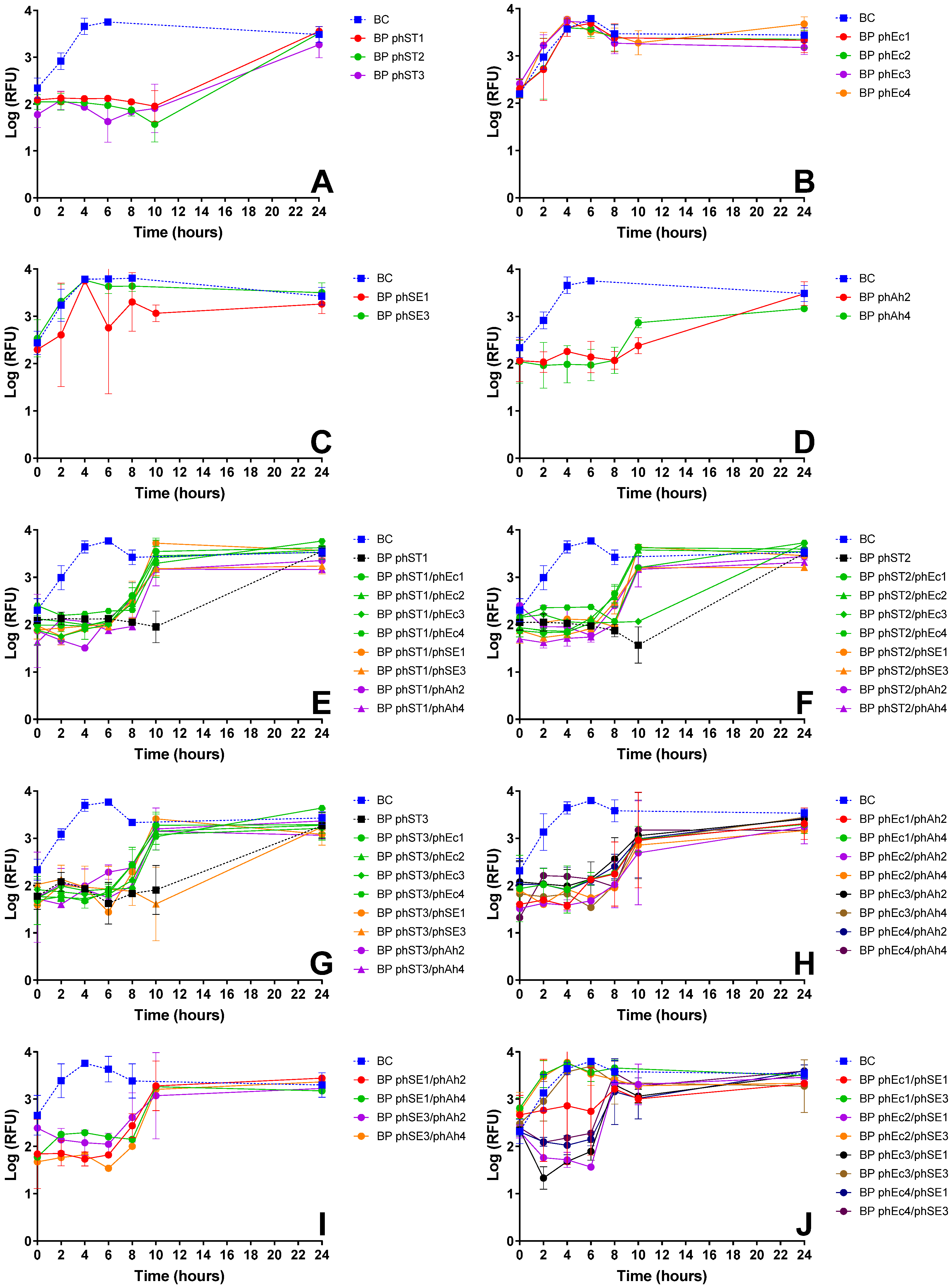
Screening assays were performed with all E. coli-infecting phages and cocktails combining two phages from different bacterial hosts (Figure 1, Table A1). Single-phage suspensions reduced E. coli viability for 6 to 24 h (Figure 1A–D), with phages phEc2, phEc3 (Figure 1A), phST1, phST2 (Figure 1B), phSE1 (Figure 1C), and phAh2 (Figure 1D) demonstrating the strongest inactivation, reducing bacterial viability by approximately 1 to 2.5 log RFU relative to the control (ANOVA, p < 0.05).
Cocktails combining E. coli phages with S. Typhimurium (Figure 1E–H, green) or S. Enteritidis phages (Figure 1E–H, orange) generally showed no improvement compared with each E. coli phage alone (Figure 1E–G, black), except for the cocktail phEc1/phSE1 (Figure 1E), which enhanced inactivation by ~1 log RFU (ANOVA, p < 0.05). In contrast, combinations such as phEc3/phST2 (Figure 1G), phEc2 with S. Typhimurium phages (Figure 1F, green) and phEc4 with S. Typhimurium phages (Figure 1H, green) showed reduced efficacy. Adding A. hydrophila phages enhanced inactivation by ~1 to 2 log RFU (ANOVA, p < 0.05), particularly phages phAh2 (Figure 1E–G) and phAh4 (Figure 1E), or maintained similar profiles (Figure 1H, purple).
Cocktails without E. coli phages, including combinations of S. Typhimurium, S. Enteritidis, and A. hydrophila phages, frequently showed increased bacterial viability after 10 h (Figure 1I,J). However, combinations such as phST1/phSE1 (Figure 1I), phST2/phAh4 (Figure 1J), and phSE1/phAh2 (Figure 1K) exhibited 1.5–3 log RFU reductions compared to controls (ANOVA, p < 0.05).

3.2.2. S. Typhimurium Inactivation Screening

Screening assays were performed with all S. Typhimurium-infecting phages and cocktails combining two phages from different bacterial hosts (Figure 2, Table A2). Single-phage suspensions of S. Typhimurium and A. hydrophila phages produced ~1.5 log RFU reductions over 24 h (Figure 2A,D, ANOVA, p < 0.05), while E. coli and S. Enteritidis phages showed no effect (Figure 2B,C).
Cocktails combining S. Typhimurium phages with phages targeting other bacteria generally showed an earlier rebound in bacterial viability after 10 h, compared to single-phage treatments (Figure 2E–G). However, certain combinations such as phST2/phEc3 (Figure 2F) and phST3/phSE3 (Figure 2G) maintained inactivation comparable to that of S. Typhimurium phages alone (~1.5 log RFU, ANOVA, p < 0.05), indicating no loss of efficacy.
For cocktails without S. Typhimurium phages, combinations of A. hydrophila phages with E. coli or S. Enteritidis phages produced similar inactivation profiles with increased bacterial viability after 10 h (Figure 2H,I). The combination phEc3/phSE1 exhibited higher early inactivation (~2 log RFU, ANOVA, p < 0.05), whereas other E. coli-S. Enteritidis phage cocktails, such as phEc1/phSE3, phEc2/phSE3, and phEc3/phSE3, showed no additional effect on bacterial viability (Figure 2J).

3.2.3. S. Enteritidis Inactivation Screening

Screening assays were performed with all S. Enteritidis-infecting phages and cocktails combining both (Figure 3, Table A3). Single-phage suspensions of S. Enteritidis phages effectively reduced bacterial viability by ~1.5–2 log RFU (ANOVA, p < 0.05), except phSE3, which showed minimal effect, with RFU values similar to the control after 6 h (Figure 3A). While phEc4 could infect S. Enteritidis, it did not impact bacterial viability (Figure 3A).
In combination assays, phEc4 either extended the effectiveness of S. Enteritidis phages, as seen with phSE3/phEc4 after 6 h, or reduced their effectiveness by ~1 log RFU, as observed with phSE2/phEc4 after 10 h (Figure 3B, ANOVA, p < 0.05).

4. Discussion

Phage cocktails are a promising approach to overcoming the limitations of single-phage treatments [11], particularly regarding narrow host specificity [11,12,13] and bacterial resistance [14]. Additionally, they can achieve greater bacterial inactivation than single-phage treatments [25,26]. However, their success depends on selecting complementary phages [38]. This study aimed to develop an optimised phage cocktail against E. coli, S. Typhimurium, S. Enteritidis, and A. hydrophila—key pathogens in bivalve consumption. Using resazurin-based viability assays, we identified a cocktail that matched or exceeded single-phage performance, targeted multiple bacteria, and minimised resistance development.
To identify phages capable of infecting more than one target bacterium, we used an isolation workflow that allowed us to recover candidates exhibiting activity beyond their original host (Section 2.2). Host range analysis (Table 1) showed that all phages infected at least one additional bacterial species, with some capable of targeting three of the four bacteria, which is expected for closely related enteric bacteria such as E. coli and S. Typhimurium due to genomic and ecological similarities [45]. This approach provided a practical means to narrow down and identify viable candidates for cocktail formulation [26] and subsequent testing and optimization. Similar strategies to identify broad-host-range phages from environmental samples have been reported [46,47], though with varying methodologies and goals. However, relying on phages with broader host ranges, like phAh2, poses important challenges. Broad-spectrum infectivity does not guarantee uniform efficacy across hosts, as discussed below; phages may exhibit reduced adsorption efficiency or lower burst size on secondary hosts [35]. Furthermore, such phages may exert selective pressure that promotes resistance in unintended bacterial populations [48,49]. Thus, while broad-host-range phages can simplify cocktail design, their inclusion must be supported by robust, context-specific efficacy data—an aspect we explore in the following sections.
To generate these efficacy data, after isolation and purification, each phage was then tested for its ability to inactivate target bacteria. In most cases, phages effectively reduced the viability of non-host bacteria (Figure 1A–D and Figure 2A,D). Similar findings were reported by Costa et al. (2019) [26], where an E. coli phage inactivated S. Typhimurium, and a S. Typhimurium phage inactivated E. coli. However, some phages unexpectedly formed plaques in host range assays but failed to reduce bacterial viability in liquid cultures. Notably, E. coli and S. Enteritidis phages showed no effect on S. Typhimurium viability (Figure 2B,C), and phEc4 failed to inactivate S. Enteritidis (Figure 3A) despite all forming plaques in host range assays (Table 1). This discrepancy may be attributed to a low efficiency of plating (EOP), as supported by follow-up testing of the selected phages, which showed low or undetectable EOP on infectable but non-host bacteria [50]. In such cases, phage replication and subsequent inactivation may occur at such low levels that any effect is masked by rapid bacterial regrowth at later assay stages [51,52]. To explore this, we conducted a preliminary, CFU-based inactivation assay using phSE3 against S. Typhimurium. The initial PFU count was very low, and CFU reduction was only observed at a single time point, with no differences from the untreated control at any other time. Although not conclusive, this supports the hypothesis that low EOP may hinder detectable inactivation in RFU-based assays, especially when bacterial regrowth dominates. While resazurin provides a rapid, high-throughput screening tool, it primarily measures metabolic activity, which may not always correlate directly with viable cell counts [39]. This can result in false positives (e.g., metabolically active but non-replicating cells), false negatives (e.g., low metabolic activity despite viability) [53], or early phage effect at the beginning of the assays on bacterial metabolic activity during the incubation period [39], as seen in some instances (Figure 1C and Figure 2H). Such limitations become especially relevant when phage replication is inefficient, reinforcing the importance of complementary quantification methods. Although CFU enumeration has been applied to the selected phages [50], further work using additional confirmatory assays—such as adsorption rate measurements, one-step growth curves, and time-kill assays—will be necessary to validate findings across the entire phage panel.
Building on these single-phage assessments, we next examined how phages performed in combination. Phage cocktails sometimes provided enhanced bacterial suppression relative to individual phages, but this benefit depended strongly on the compatibility of specific phage partners (Figure 2J and Figure 3). Similar observations have been reported by Costa et al. (2019) [26], who also noted improved bacterial control when certain phages were combined. Such enhanced performance is often attributed to synergistic interactions, in which one phage facilitates or amplifies the activity of another through complementary infection strategies [13,54]. For example, Schmerer et al. (2014) [54] described synergy arising from enzymatic modification of the bacterial surface that increased access for a partner phage, while Regeimbal et al. (2016) [13] showed that selective pressure imposed by one phage can generate variants that become more susceptible to another. Mechanisms of this kind may underline the improved suppression observed in some combinations in this study. However, because synergy is highly context-dependent and can emerge from multiple interacting biological processes, future work will be needed to determine the specific factors contributing to these effects.
Despite some promising results, most phage combinations either produced inactivation profiles similar to the most effective single-phage treatment (Figure 1E–H, Figure 2E–G and Figure 3B), showed reduced efficacy compared to single-phage treatments—as observed for the phEc3/phST2 combination (Figure 1G), highlighting potential inter-phage antagonism within multicomponent mixtures—or led to earlier bacterial regrowth (Figure 1F–J, Figure 2E–I and Figure 3B). The latter was supported by visible turbidity accompanying the late-stage fluorescence rise, confirming biomass recovery rather than a resazurin artifact, which we interpreted as an indirect indicator of resistance development [55]. These findings are consistent with prior reports that randomly assembled cocktails often display variable effectiveness due to interactions among component phages [11,22] and reinforce the importance of an empirical screening workflow, since phage–phage interactions frequently diverged from expectations based solely on host-range profiles or individual efficacy.
One likely explanation is competition between phages targeting the same bacterial surface receptor, which can render cocktails ineffective if bacteria mutate that receptor [24]. Additional resistance mechanisms may also contribute, including prophage-encoded defences, mobile genetic elements, intercellular communication, quorum sensing, and superinfection exclusion [22]. Given the overlapping host ranges of the phages, quorum sensing and superinfection exclusion are particularly relevant. Quorum sensing allows bacteria to modulate susceptibility based on population density, downregulating phage receptors and upregulating defence systems such as CRISPR–Cas in response to phage detection [22]. Superinfection exclusion, a prophage-encoded mechanism, prevents entry of closely related phages once one has already infected the host, inhibiting secondary infections and suppressing competing phages [22]. Although direct phenotypic or genetic confirmation of resistance was beyond the scope of this study—including receptor profiling or assays to monitor resistance emergence—the earlier or more pronounced regrowth observed in some cocktails suggests that inter-phage interactions and these resistance mechanisms likely influenced treatment outcomes. Future work will aim to isolate and characterize resistant strains to better understand these dynamics and assess how bacterial exposure to the phage cocktail affects virulence, growth dynamics, surface receptors, and potential genomic or functional changes in the phages themselves. Nonetheless, mitigation strategies such as sequential phage application, selection of geographically diverse phages, or inclusion of naturally co-evolved “guard” phages may offer promising avenues to minimize resistance [22].
As summarized in Table 2, optimizing a phage cocktail requires balancing bacterial inactivation, host range [19], and resistance management [22] while minimising unnecessary complexity [56]. Several highly effective combinations were identified, but not all were suitable for inclusion in the final formulation. Selection was guided by three main considerations: (1) maximizing bacterial suppression, (2) targeting multiple pathogens, and (3) minimizing phage interactions that could reduce efficacy. For E. coli, phEc3 consistently appeared in top-performing cocktails, demonstrating strong inactivation both alone and in combination with other phages. Although phEc4 exhibited a broad host range, it was ultimately excluded because it did not enhance bacterial suppression and, in some cases, reduced inactivation when combined with other phages. For S. Typhimurium, several phages performed well, but no single candidate stood out across all conditions. Given its frequent presence in effective E. coli-targeting cocktails, phST1 was selected to ensure strong suppression while maintaining synergy with other phages. For S. Enteritidis, phSE2 showed slightly higher efficacy in single-phage treatments; however, phSE1 was chosen due to its broader host range, maximizing overall cocktail coverage. For A. hydrophila, both phAh2 and phAh4 showed promise. Although phAh4 exhibited slightly better E. coli inactivation when combined with S. Typhimurium phages, phAh2 was selected for its broader efficacy across target bacteria. Comparative analysis confirmed that phAh2 provided similar inactivation without adding complexity. The final cocktail—comprising phEc3 (E. coli), phST1 (S. Typhimurium), phSE1 (S. Enteritidis), and phAh2 (A. hydrophila)—represents a carefully balanced combination of effectiveness, host coverage, and resistance mitigation.
Following selection of the four candidate phages, subsequent work focused on validating their suitability for application. This study represents the preliminary screening phase of a broader project aimed at developing an effective phage cocktail for bivalve depuration. The selected phages then underwent comprehensive morphological, genomic, and physiological characterization, including genome sequencing, annotation, and taxonomic classification [50]. The genomes were screened to confirm the absence of genes associated with lysogeny, virulence, or antimicrobial resistance. The phages—renamed PCEc3, PCST1, PCSE1, and PCAh2—were subsequently tested as a cocktail in targeted in vitro assays using CFU enumeration and evaluated under relevant environmental conditions, including seawater stability, pH tolerance, and thermal resilience [57]. Although incomplete lysis was observed under the controlled, nutrient-rich conditions of the in vitro assay, this does not necessarily indicate reduced efficacy in practical settings, where bacterial densities, nutrient availability, the presence of non-target microorganisms, and environmental conditions differ markedly [58,59,60]. Indeed, the cocktail’s efficacy and persistence were confirmed in in vivo bivalve depuration trials [57], indicating that regrowth observed in vitro reflects optimal laboratory conditions and does not compromise its applied potential. Together, these findings lay the groundwork for a robust and practical approach to phage cocktail development for food safety applications.

5. Conclusions

This study presents a systematic framework for phage cocktail design that integrates host-range screening, cross-species efficacy testing, and assessment of phage–phage interactions. Our findings show that empirical cocktail screening can reveal both synergistic and antagonistic phage interactions that are not predictable from host-range data alone. These results demonstrate that effective cocktail formulation requires balancing bacterial suppression, breadth of activity, and resistance management through rational trade-offs—maximizing efficacy and coverage while minimizing complexity and antagonism. The proposed workflow provides a transferable template for designing multi-target phage cocktails for food safety and environmental biocontrol applications. Future work should validate these findings under realistic depuration conditions, integrating genomic and environmental stability analyses to confirm safety and performance. Ultimately, this approach advances the practical implementation of phage-based strategies to enhance microbial safety in aquaculture and shellfish production.

Author Contributions

Conceptualization, P.C., C.P., J.L.R. and A.A.; methodology, P.C.; validation, P.C., C.P., J.L.R. and A.A.; formal analysis, P.C.; investigation, P.C.; resources, A.A.; data curation, P.C.; writing—original draft preparation, P.C.; writing—review and editing, P.C., C.P., J.L.R. and A.A.; visualization, P.C., C.P., J.L.R. and A.A.; supervision, C.P., J.L.R. and A.A.; funding acquisition, A.A. All authors have read and agreed to the published version of the manuscript.

Funding

This research was funded by national funds through FCT—Fundação para a Ciência e a Tecnologia I.P., under the project CESAM-Centro de Estudos do Ambiente e do Mar, references UID/50017/2025 (doi.org/10.54499/UID/50017/2025) and LA/P/0094/2020 (doi.org/10.54499/LA/P/0094/2020).

Institutional Review Board Statement

Not applicable.

Informed Consent Statement

Not applicable.

Data Availability Statement

The original contributions presented in this study are included in the article. Further inquiries can be directed to the corresponding authors.

Acknowledgments

The authors express their gratitude to the Department of Biology and the University of Aveiro, where this research was carried out. The authors are also grateful to CESAM and its funding sources. Pedro Costa was supported in the form of a PhD grant (PD/BD/150360/2019) and Carla Pereira by a Junior Research contract (DOI: 10.54499/CEECIND/03974/2017/CP1459/CT0022), financed by national funds through the FCT—Foundation for Science and Technology, I.P. During the preparation of this manuscript, the authors used OpenAI’s ChatGPT (version 5.1) to check grammar and improve readability and language. The authors have reviewed and edited the output and take full responsibility for the content of this publication.

Conflicts of Interest

The authors declare no conflicts of interest.

Abbreviations

The following abbreviations are used in this manuscript:
CFUColony-Forming Unit
EOPEfficiency of Plating
MOIMultiplicity Of Infection
ODOptical Density
PFUPlaque-Forming Unit
RFURelative Fluorescent Unit
TSATryptic Soy Agar
TSBTryptic Soy Broth

Appendix A

Table A1. Host infectivity * of phages against E. coli.
PhagesphEc1phEc2phEc3phEc4phST1phST2phST3phSE1phSE2phAh2phAh4
phEc110----------
phEc2ND10---------
phEc3NDND10--------
phEc4NDNDND10-------
phST11010101010------
phST210101010ND10-----
phST310101010NDND8----
phSE1101010101081010---
phSE2101010108810ND10--
phAh210101010886101010-
phAh41010101081081010ND4
* Host infectivity is measured in hours of hold time, representing the duration of phage suppression on bacterial viability in liquid culture. Microorganisms 13 02708 i001 Hold Time (Hours).
Table A2. Host infectivity * of phages against S. Typhimurium.
PhagesphST1phST2phST3phEc1phEc2phEc3phEc4phSE1phSE3phAh2phAh4
phST110----------
phST2ND10---------
phST3NDND10--------
phEc18880-------
phEc2888ND0------
phEc38108NDND0-----
phEc4888NDNDND0----
phSE188866662---
phSE388100606ND0--
phAh288888888810-
phAh4888888888ND10
* Host infectivity is measured in hours of hold time, representing the duration of phage suppression on bacterial viability in liquid culture. Microorganisms 13 02708 i001 Hold Time (Hours).
Table A3. Host infectivity * of phages against S. Enteritidis.
PhagesphSE1phSE2phSE3phEc4
phSE110---
phSE2ND10--
phSE3NDND4-
phEc410106ND
* Host infectivity is measured in hours of hold time, representing the duration of phage suppression on bacterial viability in liquid culture. Microorganisms 13 02708 i001 Hold Time (Hours).

References

  1. Chan, B.K.; Abedon, S.T.; Loc-Carrillo, C. Phage cocktails and the future of phage therapy. Future Microbiol. 2013, 8, 769–783. [Google Scholar] [CrossRef] [PubMed]
  2. Curtright, A.J.; Abedon, S.T. Phage therapy: Emergent property pharmacology. J. Bioanal. Biomed. 2012, s6, 2. [Google Scholar] [CrossRef]
  3. Abedon, S.; Thomas-Abedon, C. Phage therapy pharmacology. Curr. Pharm. Biotechnol. 2010, 11, 28–47. [Google Scholar] [CrossRef] [PubMed]
  4. Loc-Carrillo, C.; Abedon, S. Pros and cons of phage therapy. Bacteriophage 2011, 1, 111–114. [Google Scholar] [CrossRef]
  5. Olszowska-Zaremba, N.; Borysowski, J.; Dąbrowska, K.; Górski, A. Phage translocation, safety and immunomodulation. In Bacteriophages in Health and Disease; Hyman, P., Abedon, S.T., Eds.; CABI: Wallingford, UK, 2012; pp. 168–184. [Google Scholar]
  6. Sulakvelidze, A.; Alavidze, Z.; Morris, J.G. Bacteriophage Therapy. Antimicrob. Agents Chemother. 2001, 45, 649–659. [Google Scholar] [CrossRef]
  7. Abedon, S.; García, P.; Mullany, P.; Aminov, R. Editorial: Phage therapy: Past, present and future. Front. Microbiol. 2017, 8, 981. [Google Scholar] [CrossRef]
  8. Lin, D.M.; Koskella, B.; Lin, H.C. Phage therapy: An alternative to antibiotics in the age of multi-drug resistance. World J. Gastrointest. Pharmacol. Ther. 2017, 8, 162. [Google Scholar] [CrossRef] [PubMed]
  9. Moye, Z.D.; Woolston, J.; Sulakvelidze, A. Bacteriophage applications for food production and processing. Viruses 2018, 10, 205. [Google Scholar] [CrossRef]
  10. Pereira, C.; Costa, P.; Duarte, J.; Balcão, V.M.; Almeida, A. Phage therapy as a potential approach in the biocontrol of pathogenic bacteria associated with shellfish consumption. Int. J. Food Microbiol. 2021, 338, 108995. [Google Scholar] [CrossRef] [PubMed]
  11. Chen, L.; Yuan, S.; Liu, Q.; Mai, G.; Yang, J.; Deng, D.; Zhang, B.; Liu, C.; Ma, Y. In vitro design and evaluation of phage cocktails against Aeromonas salmonicida. Front. Microbiol. 2018, 9, 1476. [Google Scholar] [CrossRef]
  12. Hyman, P.; Abedon, S. Bacteriophage host range and bacterial resistance. Adv. Appl. Microbiol. 2010, 70, 217–248. [Google Scholar] [CrossRef]
  13. Regeimbal, J.M.; Jacobs, A.C.; Corey, B.W.; Henry, M.S.; Thompson, M.G.; Pavlicek, R.L.; Quinones, J.; Hannah, R.M.; Ghebremedhin, M.; Crane, N.J.; et al. Personalized therapeutic cocktail of wild environmental phages rescues mice from Acinetobacter baumannii wound infections. Antimicrob. Agents Chemother. 2016, 60, 5806–5816. [Google Scholar] [CrossRef]
  14. Valappil, S.K.; Shetty, P.; Deim, Z.; Terhes, G.; Urbán, E.; Váczi, S.; Patai, R.; Polgár, T.; Pertics, B.Z.; Schneider, G.; et al. Survival comes at a cost: A coevolution of phage and its host leads to phage resistance and antibiotic sensitivity of Pseudomonas aeruginosa multidrug resistant strains. Front. Microbiol. 2021, 12, 783722. [Google Scholar] [CrossRef]
  15. Pirnay, J.-P.; De Vos, D.; Verbeken, G.; Merabishvili, M.; Chanishvili, N.; Vaneechoutte, M.; Zizi, M.; Laire, G.; Lavigne, R.; Huys, I.; et al. The phage therapy paradigm: Prêt-à-Porter or Sur-mesure? Pharm. Res. 2011, 28, 934–937. [Google Scholar] [CrossRef]
  16. Castillo, D.; Christiansen, R.H.; Dalsgaard, I.; Madsen, L.; Middelboe, M. Bacteriophage resistance mechanisms in the fish pathogen Flavobacterium psychrophilum: Linking genomic mutations to changes in bacterial virulence factors. Appl. Environ. Microbiol. 2015, 81, 1157–1167. [Google Scholar] [CrossRef] [PubMed]
  17. Melo, L.D.R.; Veiga, P.; Cerca, N.; Kropinski, A.M.; Almeida, C.; Azeredo, J.; Sillankorva, S. Development of a phage cocktail to control Proteus mirabilis catheter-associated urinary tract infections. Front. Microbiol. 2016, 7, 1024. [Google Scholar] [CrossRef] [PubMed]
  18. Yen, M.; Cairns, L.S.; Camilli, A. A cocktail of three virulent bacteriophages prevents Vibrio cholerae infection in animal models. Nat. Commun. 2017, 8, 14187. [Google Scholar] [CrossRef]
  19. Abedon, S.T.; Danis-Wlodarczyk, K.M.; Wozniak, D.J. Phage cocktail development for bacteriophage therapy: Toward improving spectrum of activity breadth and depth. Pharmaceuticals 2021, 14, 1019. [Google Scholar] [CrossRef] [PubMed]
  20. Butt, A.A.; Aldridge, K.E.; Sanders, C. V Infections related to the ingestion of seafood Part I: Viral and bacterial infections. Lancet Infect. Dis. 2004, 4, 201–212. [Google Scholar] [CrossRef]
  21. Feldhusen, F. The role of seafood in bacterialfoodborne diseases. Microbes Infect. 2000, 2, 1651–1660. [Google Scholar] [CrossRef]
  22. Costa, P.; Pereira, C.; Romalde, J.L.; Almeida, A. A game of resistance: War between bacteria and phages and how phage cocktails can be the solution. Virology 2024, 599, 110209. [Google Scholar] [CrossRef]
  23. Labrie, S.J.; Samson, J.E.; Moineau, S. Bacteriophage resistance mechanisms. Nat. Rev. Microbiol. 2010, 8, 317–327. [Google Scholar] [CrossRef]
  24. Yoo, S.; Lee, K.-M.; Kim, N.; Vu, T.N.; Abadie, R.; Yong, D. Designing phage cocktails to combat the emergence of bacteriophage-resistant mutants in multidrug-resistant Klebsiella pneumoniae. Microbiol. Spectr. 2024, 12, e0125823. [Google Scholar] [CrossRef]
  25. Mateus, L.; Costa, L.; Silva, Y.J.; Pereira, C.; Cunha, A.; Almeida, A. Efficiency of phage cocktails in the inactivation of Vibrio in aquaculture. Aquaculture 2014, 424–425, 167–173. [Google Scholar] [CrossRef]
  26. Costa, P.; Pereira, C.; Gomes, A.T.P.C.; Almeida, A. Efficiency of single phage suspensions and phage cocktail in the inactivation of Escherichia coli and Salmonella Typhimurium: An in vitro preliminary study. Microorganisms 2019, 7, 94. [Google Scholar] [CrossRef] [PubMed]
  27. Duarte, J.; Pereira, C.; Moreirinha, C.; Salvio, R.; Lopes, A.; Wang, D.; Almeida, A. New insights on phage efficacy to control Aeromonas salmonicida in aquaculture systems: An in vitro preliminary study. Aquaculture 2018, 495, 970–982. [Google Scholar] [CrossRef]
  28. Chen, H.; Liu, H.; Gong, Y.; Dunstan, R.A.; Ma, Z.; Zhou, C.; Zhao, D.; Tang, M.; Lithgow, T.; Zhou, T. A Klebsiella-phage cocktail to broaden the host range and delay bacteriophage resistance both in vitro and in vivo. NPJ Biofilms Microbiomes 2024, 10, 127. [Google Scholar] [CrossRef] [PubMed]
  29. Burrowes, B.H.; Molineux, I.J.; Fralick, J.A. Directed in vitro evolution of therapeutic bacteriophages: The appelmans protocol. Viruses 2019, 11, 241. [Google Scholar] [CrossRef] [PubMed]
  30. Chen, Y.; Batra, H.; Dong, J.; Chen, C.; Rao, V.B.; Tao, P. Genetic engineering of bacteriophages against infectious diseases. Front. Microbiol. 2019, 10, 954. [Google Scholar] [CrossRef]
  31. Merabishvili, M.; Pirnay, J.-P.; De Vos, D. Guidelines to Compose an Ideal Bacteriophage Cocktail. Methods Mol. Biol. 2018, 1693, 99–110. [Google Scholar]
  32. Haines, M.E.K.; Hodges, F.E.; Nale, J.Y.; Mahony, J.; van Sinderen, D.; Kaczorowska, J.; Alrashid, B.; Akter, M.; Brown, N.; Sauvageau, D.; et al. Analysis of selection methods to develop novel phage therapy cocktails against antimicrobial resistant clinical isolates of bacteria. Front. Microbiol. 2021, 12, 613529. [Google Scholar] [CrossRef]
  33. Merril, C.R.; Scholl, D.; Adhya, S.L. The prospect for bacteriophage therapy in Western medicine. Nat. Rev. Drug Discov. 2003, 2, 489–497. [Google Scholar] [CrossRef]
  34. Koskella, B.; Meaden, S. Understanding bacteriophage specificity in natural microbial communities. Viruses 2013, 5, 806–823. [Google Scholar] [CrossRef] [PubMed]
  35. de Jonge, P.A.; Nobrega, F.L.; Brouns, S.J.J.; Dutilh, B.E. Molecular and evolutionary determinants of bacteriophage host range. Trends Microbiol. 2019, 27, 51–63. [Google Scholar] [CrossRef]
  36. Ross, A.; Ward, S.; Hyman, P. More is better: Selecting for broad host range bacteriophages. Front. Microbiol. 2016, 7, 1352. [Google Scholar] [CrossRef]
  37. Hyman, P. Phages for phage therapy: Isolation, characterization, and host range breadth. Pharmaceuticals 2019, 12, 35. [Google Scholar] [CrossRef]
  38. Yang, Y.; Shen, W.; Zhong, Q.; Chen, Q.; He, X.; Baker, J.L.; Xiong, K.; Jin, X.; Wang, J.; Hu, F.; et al. Development of a bacteriophage cocktail to constrain the emergence of phage-resistant Pseudomonas aeruginosa. Front. Microbiol. 2020, 11, 327. [Google Scholar] [CrossRef]
  39. Costa, P.; Gomes, A.T.P.C.; Braz, M.; Pereira, C.; Almeida, A. Application of the resazurin cell viability assay to monitor Escherichia coli and Salmonella Typhimurium inactivation mediated by phages. Antibiotics 2021, 10, 974. [Google Scholar] [CrossRef] [PubMed]
  40. Adams, M. Bacteriophages; John Wiley and Sons Inc.: New York, NY, USA, 1959. [Google Scholar]
  41. Abedon, S.T. Kinetics of phage-mediated biocontrol of bacteria. Foodborne Pathog. Dis. 2009, 6, 807–815. [Google Scholar] [CrossRef]
  42. Janda, J.M.; Abbott, S.L. The genus Aeromonas: Taxonomy, pathogenicity, and infection. Clin. Microbiol. Rev. 2010, 23, 35–73. [Google Scholar] [CrossRef] [PubMed]
  43. Tuttle, A.R.; Trahan, N.D.; Son, M.S. Growth and maintenance of Escherichia coli laboratory strains. Curr. Protoc. 2021, 1, e20. [Google Scholar] [CrossRef]
  44. Futoma-Kołoch, B.; Małaszczuk, M.; Korzekwa, K.; Steczkiewicz, M.; Gamian, A.; Bugla-Płoskońska, G. The prolonged treatment of Salmonella enterica strains with human serum effects in phenotype related to virulence. Int. J. Mol. Sci. 2023, 24, 883. [Google Scholar] [CrossRef] [PubMed]
  45. Meysman, P.; Sánchez-Rodríguez, A.; Fu, Q.; Marchal, K.; Engelen, K. Expression divergence between Escherichia coli and Salmonella enterica serovar Typhimurium reflects their lifestyles. Mol. Biol. Evol. 2013, 30, 1302–1314. [Google Scholar] [CrossRef]
  46. Jensen, E.C.; Schrader, H.S.; Rieland, B.; Thompson, T.L.; Lee, K.W.; Nickerson, K.W.; Kokjohn, T.A. Prevalence of broad-host-range lytic bacteriophages of Sphaerotilus natans, Escherichia coli, and Pseudomonas aeruginosa. Appl. Environ. Microbiol. 1998, 64, 575–580. [Google Scholar] [CrossRef]
  47. Yu, P.; Mathieu, J.; Li, M.; Dai, Z.; Alvarez, P.J.J. Isolation of polyvalent bacteriophages by sequential multiple-host approaches. Appl. Environ. Microbiol. 2016, 82, 808–815. [Google Scholar] [CrossRef]
  48. Burrowes, B.; Harper, D.R.; Anderson, J.; McConville, M.; Enright, M.C. Bacteriophage therapy: Potential uses in the control of antibiotic-resistant pathogens. Expert Rev. Anti. Infect. Ther. 2011, 9, 775–785. [Google Scholar] [CrossRef]
  49. Wandro, S.; Oliver, A.; Gallagher, T.; Weihe, C.; England, W.; Martiny, J.B.H.; Whiteson, K. Predictable molecular adaptation of coevolving Enterococcus faecium and lytic phage EfV12-phi1. Front. Microbiol. 2019, 9, 3192. [Google Scholar] [CrossRef] [PubMed]
  50. Costa, P.; Pereira, C.; Oliveira, V.; Gomes, N.C.M.; Romalde, J.L.; Almeida, A. Characterising phages for the control of pathogenic bacteria associated with bivalve consumption. Int. J. Food Microbiol. 2025, 432, 111096. [Google Scholar] [CrossRef]
  51. Shkoporov, A.N.; Khokhlova, E.V.; Fitzgerald, C.B.; Stockdale, S.R.; Draper, L.A.; Ross, R.P.; Hill, C. ΦCrAss001 represents the most abundant bacteriophage family in the human gut and infects Bacteroides intestinalis. Nat. Commun. 2018, 9, 4781. [Google Scholar] [CrossRef]
  52. Mulla, Y.; Müller, J.; Trimcev, D.; Bollenbach, T. Extreme diversity of phage amplification rates and phage–antibiotic interactions revealed by PHORCE. PLoS Biol. 2025, 23, e3003065. [Google Scholar] [CrossRef] [PubMed]
  53. Vieira-da-Silva, B.; Castanho, M.A.R.B. Resazurin reduction-based assays revisited: Guidelines for accurate reporting of relative differences on metabolic status. Molecules 2023, 28, 2283. [Google Scholar] [CrossRef]
  54. Schmerer, M.; Molineux, I.J.; Bull, J.J. Synergy as a rationale for phage therapy using phage cocktails. PeerJ 2014, 2, e590. [Google Scholar] [CrossRef]
  55. Oechslin, F. Resistance development to bacteriophages occurring during bacteriophage therapy. Viruses 2018, 10, 351. [Google Scholar] [CrossRef]
  56. Molina, F.; Simancas, A.; Ramírez, M.; Tabla, R.; Roa, I.; Rebollo, J.E. A new pipeline for designing phage cocktails based on phage-bacteria infection networks. Front. Microbiol. 2021, 12, 564532. [Google Scholar] [CrossRef]
  57. Costa, P.; Pereira, C.; Barja, J.L.; Romalde, J.L.; Almeida, A. Enhancing bivalve depuration using a phage cocktail: An in vitro and in vivo study. Food Control 2025, 177, 111442. [Google Scholar] [CrossRef]
  58. Attrill, E.L.; Łapińska, U.; Westra, E.R.; Harding, S.V.; Pagliara, S. Slow growing bacteria survive bacteriophage in isolation. ISME Commun. 2023, 3, 95. [Google Scholar] [CrossRef] [PubMed]
  59. Fister, S.; Robben, C.; Witte, A.K.; Schoder, D.; Wagner, M.; Rossmanith, P. Influence of environmental factors on phage–bacteria interaction and on the efficacy and infectivity of phage P100. Front. Microbiol. 2016, 7, 1152. [Google Scholar] [CrossRef] [PubMed]
  60. Šivec, K.; Podgornik, A. Determination of bacteriophage growth parameters under cultivating conditions. Appl. Microbiol. Biotechnol. 2020, 104, 8949–8960. [Google Scholar] [CrossRef] [PubMed]
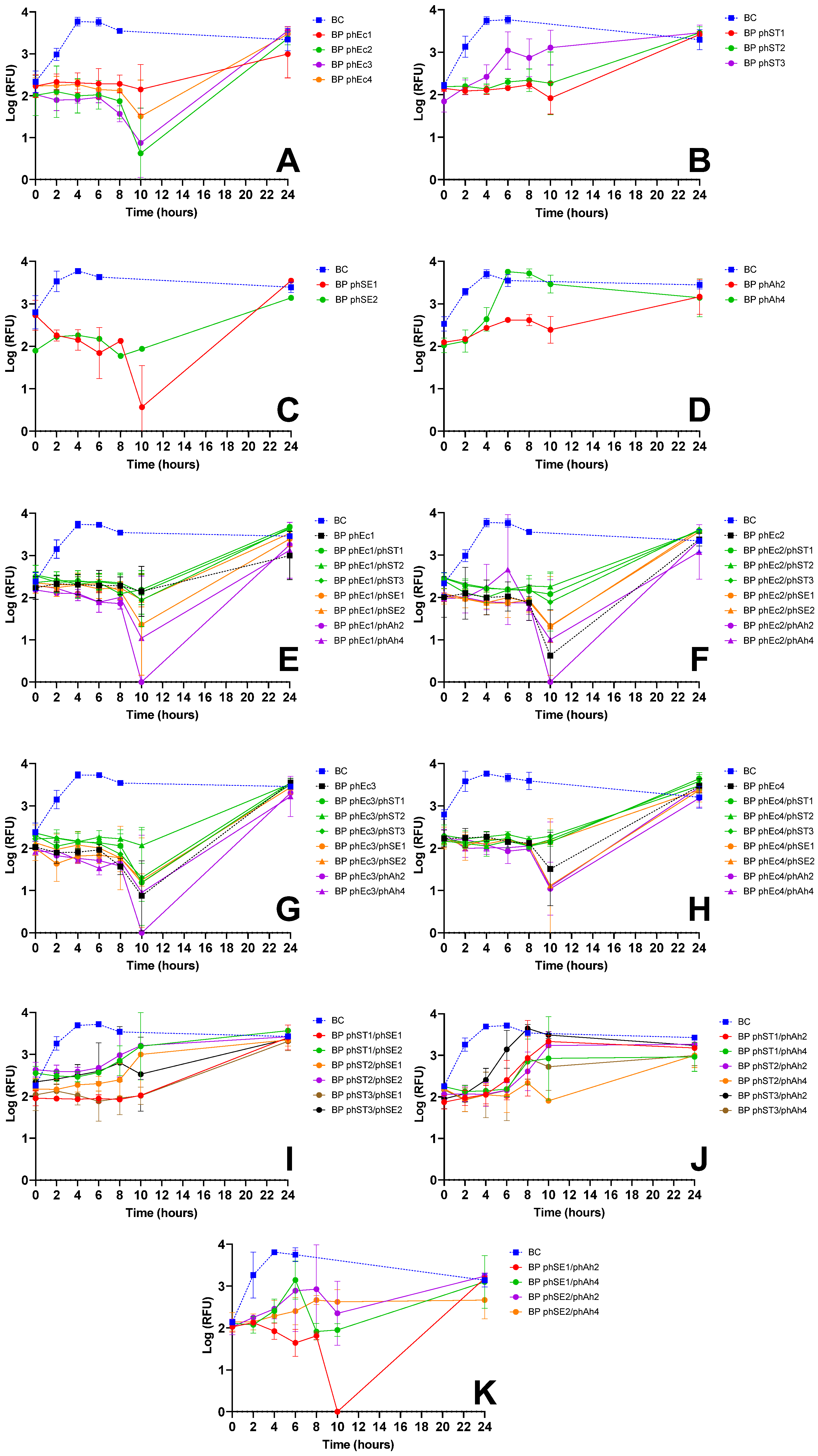
Figure 1. Inactivation of E. coli over 24 h using single-phage suspensions and two-phage cocktails at an MOI of 10, evaluated via bacterial viability through resazurin-based cell viability assay. Panels show the effect of (A) E. coli phages (phEc1, phEc2, phEc3, phEc4); (B) S. Typhimurium phages (phST1, phST2, phST3); (C) S. Enteritidis phages (phSE1, phSE2, phSE3); and (D) A. hydrophila phages (phAh2, phAh4); E. coli phages (black) and cocktails combining each E. coli phage, phEc1 (E), phEc2 (F), phEc3 (G), and phEc4 (H) with other S. Typhimurium (green), S. Enteritidis (orange) and A. hydrophila (purple) phages; (I) cocktails combining S. Typhimurium phages with S. Enteritidis phages; (J) cocktails combining S. Typhimurium phages with A. hydrophila phages; and (K) cocktails combining S. Enteritidis phages with A. hydrophila phages. RFU: relative fluorescence unit, an indicator of metabolic activity. BC: bacteria control; BP: bacteria plus phage. Values represent the mean of three independent assays; error bars represent the standard deviation.
Microorganisms 13 02708 g001
Figure 2. Inactivation of S. Typhimurium over 24 h using single-phage suspensions and two-phage cocktails at an MOI of 10, evaluated via bacterial viability through resazurin-based cell viability assay. Panels show the effect of (A) S. Typhimurium phages (phST1, phST2, phST3); (B) E. coli phages (phEc1, phEc2, phEc3, phEc4); (C) S. Enteritidis phages (phSE1, phSE2, phSE3); and (D) A. hydrophila phages (phAh2, phAh4); S. Typhimurium phages (black) and cocktails combining each S. Typhimurium phage, phST1 (E), phST2 (F), and phST3 (G), with other E. coli (green), S. Enteritidis (orange) and A. hydrophila (purple) phages; (H) cocktails combining E. coli phages with A. hydrophila phages; (I) cocktails combining S. Enteritidis phages with A. hydrophila phages; and (J) cocktails combining E. coli phages with S. Enteritidis phages. RFU: relative fluorescence unit, an indicator of metabolic activity. BC: bacteria control; BP: bacteria plus phage. Values represent the mean of three independent assays; error bars represent the standard deviation.
Microorganisms 13 02708 g002
Figure 3. Inactivation of S. Enteritidis over 24 h using single-phage suspensions and two-phage cocktails at an MOI of 10, evaluated via bacterial viability through resazurin-based cell viability assay. Panels show the effect of: (A) S. Enteritidis phages (phSE1, phSE2, phSE3) and E. coli phage phEc4; and (B) cocktails combining each S. Enteritidis phage (phSE1, phSE2, phSE3) with the E. coli phage phEc4. RFU: relative fluorescence unit, an indicator of metabolic activity. BC: bacteria control; BP: bacteria plus phage. Values represent the mean of three independent assays; error bars represent the standard deviation.
Microorganisms 13 02708 g003
Table 1. Phage host ranges.
PhagesE. coliS. TyphimuriumS. EnteritidisA. hydrophila
phEc1HXX
phEc2HXX
phEc3HXX
phEc4HX
phST1HXX
phST2HXX
phST3HXX
phSE1HX
phSE2XHX
phSE3XHX
phAh2XH
phAh4XH
H—Isolation host. —Lysis plaques. X—No lysis plaques.
Table 2. Phage Selection Based on Efficacy vs. Host Range Trade-Offs.
Target BacteriumSelected PhageEfficacy HighlightsHost Range JustificationReason for Selection over Alternatives
E. coliphEc3Strong inactivation alone (Figure 1A) and in cocktails (e.g., phEc3/phAh2); no antagonism observedGood specificity without cross-host antagonismMore effective than other E. coli phages; synergetic in cocktails
S. TyphimuriumphST1Robust suppression; maintained efficacy in multiple cocktails (e.g., phST1/phSE1); did not induce early regrowthFunctional in both single and combination treatmentsPreferred over phST2, which showed antagonism with phEc3 (Figure 1G)
S. EnteritidisphSE1Effective in single and combo assays (Figure 1C,E,K); e.g., phEc1/phSE1 and phSE1/phAh2 showed enhanced activityBroader host range than phSE2 or phSE3, aligning with multi-pathogen coverage goalBetter synergy and coverage than more potent but narrower phSE2
A. hydrophilaphAh2Strong enhancement in cocktails with phEc3 and phSE1 (Figure 1E–G); consistent inactivation across targetsBroader efficacy across host bacteria compared to phAh4Chosen over phAh4 for broader effectiveness with fewer phage–phage
antagonisms
Disclaimer/Publisher’s Note: The statements, opinions and data contained in all publications are solely those of the individual author(s) and contributor(s) and not of MDPI and/or the editor(s). MDPI and/or the editor(s) disclaim responsibility for any injury to people or property resulting from any ideas, methods, instructions or products referred to in the content.