Review

Advances in Pseudomonas aeruginosa-Induced Programmed Cell Death and Potential Targeted Treatment Strategies

1
Division of Pulmonary and Critical Care Medicine, The First Affiliated Hospital of Sun Yat-sen University, Guangzhou 510080, China
2
Institute of Respiratory Diseases, Sun Yat-sen University, Guangzhou 510080, China
3
Department of Emergency Medicine, The First Affiliated Hospital of Sun Yat-sen University, Guangzhou 510080, China
*
Author to whom correspondence should be addressed.
Microorganisms 2025, 13(11), 2560; https://doi.org/10.3390/microorganisms13112560
Submission received: 15 September 2025 / Revised: 31 October 2025 / Accepted: 4 November 2025 / Published: 10 November 2025
(This article belongs to the Section Molecular Microbiology and Immunology)

Abstract

Pseudomonas aeruginosa (P. aeruginosa) is responsible for the high prevalence of various nosocomial infections, and it is challenging to completely eradicate P. aeruginosa infection in clinics. One of P. aeruginosa’s main pathogenic mechanisms is to trigger multiple forms of programmed cell death (PCD), such as apoptosis, pyroptosis, necroptosis, ferroptosis, and PANoptosis, among which PANoptosis is a newly discovered PCD pathway mediated by PANoptosome complexes and their key upstream regulators. Compared with other well-studied PCD pathways, advances in P. aeruginosa-induced PANoptosis have yet to be thoroughly reviewed. This review highlights research advances in this pathway, providing mechanistic insights by summarizing the upregulation and/or activation of PANoptosome sensor proteins and their key upstream regulators during P. aeruginosa infection. We also offer perspectives on the mechanistic links between P. aeruginosa cytotoxicity and other forms of PCD. Additionally, pharmacological compounds that may be used to target P. aeruginosa-induced PCD, particularly PANoptosis, are discussed, and future research and therapeutic directions are proposed. Our work helps bridge the knowledge gap, paving the way for further understanding of P. aeruginosa-induced PCD and the development of novel therapeutics against P. aeruginosa infection.

1. Introduction

Pseudomonas aeruginosa (P. aeruginosa) is a highly virulent and versatile pathogen that causes various nosocomial infections [1,2]. Among Gram-negative bacteria, a high prevalence of P. aeruginosa has been reported in a wide range of nosocomial infections such as pneumonia [1,3], bloodstream infections [4,5], urinary tract infection [6], intra-abdominal infections [7], and sepsis [8]. Of note, as an opportunistic pathogen, critically ill and immunocompromised patients are more susceptible to P. aeruginosa-associated infections [9]. Moreover, drug-resistant strains even contribute to increased disease severity [10]. P. aeruginosa is one of the leading bacterial pathogens, accounting for more than 500,000 deaths in 2019, with a high age-standardized mortality rate of 7.4% [11]. Although new-generation antibiotics have been put into clinical use, it still remains challenging to completely eradicate P. aeruginosa infection due to its high level of antibiotic resistance [12,13]. To explore alternative and effective therapeutics against this infection, it is crucial to further investigate the pathogenicity of P. aeruginosa.
One of the main pathogenic mechanisms of P. aeruginosa is to induce multiple forms of cell death [14]. Previously published research has demonstrated the cytotoxicity of P. aeruginosa since multiple forms of programmed cell death (PCD) can be induced by P. aeruginosa virulence factors. For example, exotoxins secreted from bacterial type III secretion system (T3SS) cause cell apoptosis [15]. The lipopolysaccharide (LPS) of P. aeruginosa is responsible for triggering cell pyroptosis [16]. Furthermore, P. aeruginosa exolysin leads to the activation of necroptosis [17]. It is noteworthy that PANoptosis, a new form of PCD that shares key molecular characteristics of apoptosis, pyroptosis, and necroptosis, has been demonstrated to occur in various infectious diseases [18,19,20]. Pathogen-induced PANoptosis (e.g., by SARS-CoV-2 or F. novicida) has been shown to drive cell death and disease pathology [19,20], and, importantly, preventing PANoptosis significantly reduces mortality in preclinical models of infection [19]. Crucially, works from Kanneganti’s team [21] and Liu and colleagues [22] provided evidence that P. aeruginosa triggers PANoptosis in target cells. As a Gram-negative opportunistic pathogen similar to F. novicida, P. aeruginosa-induced PANoptosis is proposed to contribute to disease progression. However, compared with other well-studied PCD pathways (e.g., apoptosis and pyroptosis), advances in P. aeruginosa-induced PANoptosis have yet to be comprehensively reviewed. In addition, progress in other forms of P. aeruginosa-induced PCD needs to be examined.
In this review, we focus on providing up-to-date, integrated, and critical perspectives on the close association between P. aeruginosa cytotoxicity and the induction of multiple PCD pathways. Moreover, potential treatment strategies targeting P. aeruginosa-induced PCD, particularly PANoptosis, are discussed. Gaining new insights into P. aeruginosa-induced PCD may enhance the understanding of P. aeruginosa’s pathogenicity, thereby boosting the discovery of novel therapeutics against P. aeruginosa infection.

2. P. aeruginosa-Induced PCD

2.1. P. aeruginosa-Induced PANoptosis

As described in the Introduction, P. aeruginosa is able to induce multiple forms of PCD such as apoptosis [15], pyroptosis [16], and necroptosis [23], which are essential biological processes that regulate immune response to pathogen invasion. As these PCD pathways are collectively involved in host immune defense, potential cross-talk may exist among them. In 2019, a new form of PCD that shares important molecular features of apoptosis, pyroptosis, and necroptosis was discovered and termed PANoptosis [24], highlighting the intricate interplay among these PCD pathways (Figure 1). It is critical to point out that individual inhibition of key molecular components of apoptosis, pyroptosis, or necroptosis cannot prevent PANoptosis, and only the combined deletion of PANoptotic components can effectively abrogate the PANoptosis process. PANoptosis has been reported to occur in various infectious diseases [20,25,26], including Gram-negative bacteria infection [18,20], and is regulated by the PANoptosome complexes and their key upstream regulators, as discussed below.

2.1.1. Composition of PANoptosome Complexes

The process of PANoptosis is sophisticatedly regulated by the PANoptosome, a multiprotein complex comprising pivotal molecular components of apoptosis, pyroptosis, and necroptosis [25]. Although multiple signal pathways are involved in the formation of PANoptosome complexes and the composition of each complex may vary under different stimuli [20,24,25], the PANoptosome is primarily composed of the following proteins.
Sensor proteins: (1) Z-DNA binding protein 1 (ZBP-1). ZBP-1 is heavily engaged in innate immunity and cell death through the sensing of aberrant nucleic acids [27,28]. Notably, apart from nucleic acid recognition, ZBP-1 functions as an essential pathogen sensor to regulate cell death and inflammatory responses [29]. Activated ZBP-1 promotes the assembly of the PANoptosome by mediating the recruitment and interaction of other molecular components of PANoptosis, such as caspase-8 and receptor-interacting serine/threonine-protein kinase 3 (RIPK3), in the context of Gram-negative bacteria infection [24,25]. Importantly, ZBP-1 deficiency or mutation leads to attenuated levels of PANoptosis [20]. (2) Absent in melanoma 2 (AIM2). This well-known DNA sensor is essential for host immune defense against Gram-negative bacteria infection [30,31]. The work from Lee and colleagues reveals that AIM2 controls PANoptosome formation by cooperating with ZBP-1 and Pyrin [20]. In cells with AIM2 deficiency, PANoptosis is totally abrogated, accompanied by reduced expression levels of ZBP-1 and Pyrin [20]. Furthermore, complete inactivation of PANoptotic molecules (e.g., caspase-8, -3, -7, -1, Gasdermin D, and RIPK3) is found in AIM2-deficient cells [20]. These results suggest the important role of AIM2 in PANoptosis. (3) Receptor-interacting serine/threonine-protein kinase 1 (RIPK1). In addition to ZBP-1 and AIM2, Gram-negative bacteria infection induces the assembly of the RIPK1-PANoptosome, which is crucial for regulating cell death and host antimicrobial defense [18]. Importantly, loss of RIPK1 leads to a reduction in PANoptotic molecules (e.g., caspase-8, -3, -7, -1, and Gasdermin D), along with decreased levels of pyroptosis and apoptosis [18]. Interestingly, necroptosis is activated upon loss of RIPK1 [18], and it might act as a compensatory mechanism to preserve immune homeostasis in response to bacterial infection. (4) NLR family pyrin domain containing 12 (NLRP12). Recently, the NLRP12-PANoptosome has been discovered upon heme plus pathogen-associated molecular pattern (PAMP) stimulation [32], implicating its regulatory role in infectious and inflammatory diseases.
Adaptor proteins: (1) Apoptosis-associated speck-like protein containing CARD (ASC). This functions as an adaptor that bridges the interaction between upstream signaling and downstream effector proteins [33,34]. The interactions of ASC with molecular components of PANoptosis (e.g., ZBP-1, AIM2, Pyrin, RIPK1, caspase-1, and RIPK3) are observed following infection, and ASC deletion leads to decreased expression of ZBP-1, Pyrin, and caspase-1 [20]. (2) FAS-associated via death domain (FADD). This serves as an adaptor for caspase enzymes (e.g., caspase-8) and is engaged in the regulation of inflammation and cell death [35,36]. Notably, loss of FADD diminishes both the PANoptotic molecule activation and cell death [19].
Effector proteins: (1) Caspase-1. This is a crucial effector of PCD, and it can specifically cleave Gasdermin D (GSDMD) to form pores in the plasma membrane [37]. (2) RIPK3. This phosphorylates downstream protein mixed-lineage kinase domain-like protein (MLKL), and activated MLKL traffics to the cell membrane for pore formation [38]. In the context of PANoptosis, RIPK3 is co-expressed with other molecular components of PANoptosis, such as ZBP1, RIPK1, and ASC [25], suggesting that these proteins assemble into the PANoptosome complex. (3) Caspase-8. This is a PANoptotic caspase that mediates the cleavage and activation of downstream caspase-3 and -7, as well as GSDMD, Gasdermin E (GSDME), and RIPK3 [39]. Active caspase-3 and -7 drive membrane pore generation via the cleavage of GSDME [39,40]. Together, these effector proteins jointly contribute to pore formation in the plasma membrane, ultimately driving PANoptosis in a synergistic manner. Figure 2 presents the molecular processes of PANoptosis and four types of PANoptosome complexes, and highlights apoptosis-, pyroptosis-, and necroptosis-associated molecules involved in PANoptosis.

2.1.2. Key Upstream Regulators of PANoptosome Complexes

Although the sensor, adaptor, and effector proteins discussed above together constitute the PANoptosome, it is essential to identify upstream molecules mediating PANoptosome formation. Interferon regulatory factor 1 (IRF1) has been discovered to serve as an upstream regulator in the PANoptosis pathway [19]. IRF1-deficient cells are protected from PANoptosis in response to tumor necrosis factor-α and IFN-γ co-treatment, along with decreased activation of both PANoptotic molecules (e.g., caspase-3, -7, and -8) and pore-forming molecules (e.g., GSDME and MLKL) [19]. More importantly, prior studies have confirmed the key role of IRF1 in regulating the expression of PANoptosome sensor proteins. For example, IRF1 was identified as a transcriptional modulator of ZBP1 because the level of ZBP1 is diminished in cells with IRF1 deficiency during infection [41]. IRF1 has also been confirmed to contribute to the formation and activation of ZBP1-PANoptosome [42]. Similar to ZBP-1, results from another study show that both AIM2 activation and host survival require IRF1 during F. novicida infection [31], a Gram-negative bacterium demonstrated to induce PANoptosis [20]. In addition to these, loss of IRF1 leads to impaired activation of RIPK1 and PANoptotic molecules, as well as reduced RIPK1-mediated PANoptosis [42], revealing that IRF1 promotes the formation of RIPK1-PANoptosome. Recently, the regulatory role of IRF1 in mediating NLRP12 expression and NLRP12-dependent PANoptosis has been recognized, as IRF1-deficient cells have significantly lower NLRP12 expression and cell death [32]. All the evidence mentioned above proves that IRF1 acts as a key upstream regulator for orchestrating PANoptosome assembly and PANoptosis.
Apart from IRF1, growing evidence has indicated the importance of caspase-8 in regulating the emergence of PANoptosis. For instance, work from Karki and colleagues unveils that caspase-8 is a core molecule involved in triggering PANoptosis because cells with ablation of caspase-8 and RIPK3 are completely resistant to PANoptosis [19]. Interestingly, loss of RIPK3 alone does not protect cells against PANoptosis [19]. (Cells from mice with caspase-8 deficiency alone are not available, due to embryonic lethality [43]). Furthermore, substantially declined activation of PANoptosis-associated caspases (e.g., caspase-3 and -7) and downstream pore-forming proteins are found upon deletion of caspase-8 and RIPK3 [19]. Results from another study show that caspase-8 functions as a pivotal molecular switch that intricately controls the process of apoptosis, pyroptosis, and necroptosis [44]. Additionally, caspase-8 was demonstrated to closely interact with AIM2-ASC complex upon F. novicida infection [45]. Aside from caspase-8, caspase-6 associates with RIPK3 to facilitate RIPK3-ZBP1 interaction, promoting PANoptosis and innate immunity activation [46]. Activation levels of PANoptosis-associated caspases and downstream effector proteins are attenuated upon caspase-6 deficiency [46]. These findings demonstrate that caspase-8 and -6 act as core upstream regulators of PANoptosome complexes, and they are capable of interacting with PANoptosome components to modulate the process of PANoptosis.

2.1.3. Progress in P. aeruginosa-Induced PANoptosis

As a leading Gram-negative pathogen responsible for various nosocomial infections, growing evidence has revealed that the severe cytotoxicity of P. aeruginosa drives PANoptosis. A study from Kanneganti’s team first discovered that the P. aeruginosa PAO1 strain triggers PANoptosis in mouse bone marrow-derived macrophages, and cells with concomitant loss of caspase-8, RIPK3, and caspase-1/11 are fully protected from PANoptosis [21]. Nevertheless, this research did not include in vivo data, and the potential influences of host microenvironment (e.g., pH and metabolites) and immune cell interactions on PANoptosis should not be overlooked. Another study identified that a low dose of P. aeruginosa causes PANoptosis in tumor cells, as evidenced by concurrent activation of apoptotic and necroptotic cell death pathways [47]. Notably, a recently published study shows that the PAO1 strain with rhl (a subsystem of the bacterial quorum sensing (QS) network) deficiency induces PANoptosis in mouse bone marrow-derived macrophages [22]. More importantly, mice are resistant to infection caused by the PAO1 strain with dual deletion of rhl and pqs (another subsystem of QS) [22], demonstrating that P. aeruginosa-induced PANoptosis is intricately regulated by the bacterial QS system. All these findings confirm that P. aeruginosa infection leads to PANoptosis. However, the underlying mechanisms of P. aeruginosa-induced PANoptosis are still largely unknown. Interestingly and importantly, accumulating evidence has demonstrated that P. aeruginosa infection promotes the upregulation and/or activation of PANoptosome sensor proteins as well as key upstream regulators of PANoptosome complexes, providing mechanistic evidence for the emergence of PANoptosis in the context of P. aeruginosa infection, which are summarized below.
Upregulation and/or Activation of PANoptosome Sensor Proteins Upon P. aeruginosa Infection
As previously summarized, ZBP-1, AIM2, RIPK1, and NLRP12 are crucial sensor proteins of the PANoptosome. As an important pathogen sensor orchestrating cell death and inflammatory responses, enhanced expression of ZBP-1 is detected in neutrophils following P. aeruginosa infection, along with augmented levels of neutrophil cell death [23]. This result implies that P. aeruginosa infection is closely associated with potential neutrophil PANoptosis. Furthermore, prior research has shown that ZBP-1 interacts with other components of PANoptosis, including RIPK3, RIPK1, and caspase-8, to intricately regulate cell death and immune response [48]. More importantly, as previously described, ZBP-1 deficiency or mutation attenuates PANoptosis [20]. These findings strongly support the ability of P. aeruginosa-induced ZBP-1 expression to trigger PANoptosis because of ZBP-1′s pivotal role in promoting PANoptosome assembly and the interaction between the PANoptosome components.
Results from a prior study show that AIM2 expression (another important PANoptosome sensor protein) is elevated in both in vivo and in vitro models of pulmonary P. aeruginosa infection [49]. Considering the key role of AIM2 in PANoptosome formation as discussed earlier, P. aeruginosa-induced AIM2 upregulation appears to contribute to the arise of PANoptosis. It is worth noting that inhibition of AIM2 activation alleviates the level of pulmonary inflammation and severity of P. aeruginosa pneumonia [49], indicating a close link between AIM2-mediated PANoptosis and the pathogenesis of pulmonary P. aeruginosa infection.
It is important to note that works from Sundaram et al. provide evidence that the phosphorylation level of RIPK1 is upregulated in a cell model of P. aeruginosa infection, confirming the activation of RIPK1 [21]. Intriguingly, activation of caspase-8 and -1 is also observed in this model [21], both of which are important components of RIPK1-PANoptosome. Moreover, P. aeruginosa-induced PANoptosis is totally abrogated in cells with combined deficiency of caspase-8, RIPK3, and caspase-1/11 [21]. Collectively, these results reveal that RIPK1 is strongly engaged in PANoptosome assembly and PANoptosis through interaction with caspase-1, caspase-8, and RIPK3 in the context of P. aeruginosa infection.
Regarding NLRP12, it is involved in host defense against P. aeruginosa infection as NLRP12-deficient mice are more susceptible to this pathogen [50]. Nevertheless, to date, NLRP12 has only been shown to control PANoptosome formation following heme and PAMP stimulation [32]. Together, activated RIPK1 has been shown to contribute to PANoptosome formation, and future studies are needed to deeply elucidate the underlying mechanistic roles of other PANoptosome sensor proteins (ZBP-1, AIM2, and NLRP12) in PANoptosome assembly and PANoptosis during P. aeruginosa infection.
Upregulation and/or Activation of Key Upstream Regulators of PANoptosome Complexes Upon P. aeruginosa Infection
As previously summarized, IRF1 is a key upstream regulator in PANoptosome assembly and PANoptosis. Interestingly, increased expression of IRF1 is observed in bladder epithelial cells upon P. aeruginosa infection [51]. Moreover, P. aeruginosa infection results in the upregulation of IRF1 in alveolar macrophages and lung tissue [52]. Given the pivotal role of IRF1 in PANoptosome assembly, these important findings suggest a connection between P. aeruginosa-induced IRF1 upregulation and PANoptosis. In another study, the N-(3-oxododecanoyl) homoserine lactone (3-oxo-C12-HSL) derived from P. aeruginosa causes caspase-8 activation [53], which plays a core role in mediating PANoptosis. Taken together, all the aforementioned findings clearly demonstrate that P. aeruginosa infection triggers the upregulation and/or activation of key upstream regulators and sensor proteins of PANoptosome complexes.

2.1.4. Outstanding Questions in P. aeruginosa-Induced PANoptosis

Although prior studies have demonstrated that P. aeruginosa triggers PANoptosis and activates PANoptosome sensors and upstream regulators, precise functions of individual PANoptosome components are not well understood. Their mechanistic roles in PANoptosome assembly and PANoptosis should be studied in conditional knockout conditions, and their direct interplay should be revealed through high-resolution structures of PANoptosome complexes.
As previously mentioned, the QS system of P. aeruginosa is mechanistically involved in the occurrence of PANoptosis. As the P. aeruginosa QS system controls the production and activation of diverse virulence factors [54], such as toxins, pyocyanin, elastase, and proteases, future studies need to thoroughly investigate whether and how each of these virulence factor contributes to the emergence of PANoptosis, which has been extensively demonstrated in other well-studied PCD pathways. A special focus should be given to the virulence factors that have been proven to simultaneously induce several types of PCD, including apoptosis, pyroptosis, and necroptosis.
Additionally, different strains of P. aeruginosa may vary in their capacity to induce PANoptosis. In the aforementioned studies, researchers investigated P. aeruginosa-induced PANoptosis using the PAO1 strain. Although it is commonly used in the research, the PAO1 strain and clinical P. aeruginosa isolates may differ in their QS systems and virulence factors. Accordingly, to discover potential targeted therapeutic strategies against P. aeruginosa infection in clinics, there is an urgent need to thoroughly explore the precise roles and mechanisms of clinical P. aeruginosa isolates, particularly the multidrug-resistant (MDR) isolates, in PANoptosome assembly and PANoptosis.
Furthermore, despite confirming that P. aeruginosa triggers PANoptosis in macrophages, it still remains unknown whether P. aeruginosa triggers PANoptosis in other immune cells that play crucial roles in pathogen clearance (e.g., neutrophils and T cells). Given the strong cytotoxicity and pathogenicity of P. aeruginosa, this pathogen might induce PANoptosis in both innate and adaptive pathogen-clearing cells, thereby impeding both innate and adaptive immunity, a direction that strongly requires exploration.

2.2. P. aeruginosa-Induced Apoptosis

Apoptosis is characterized by nuclear fragmentation, chromatin condensation, DNA fragmentation, cellular shrinking and blebbing, and apoptosome formation [55,56]. Apoptosis is a fundamental biological process essential for maintaining hemostasis, and it also plays a vital role in regulating host immune defense in response to pathogen invasion [57]. At the molecular level, apoptosis is driven by apoptotic initiator caspases such as caspase-8, -9, and -10 [58,59]. Dimerization of initiator caspases facilitates their activation, and they subsequently promote the cleavage and activation of apoptotic effector caspases (e.g., caspase-3 and -7), which carry out the execution of apoptosis [58].
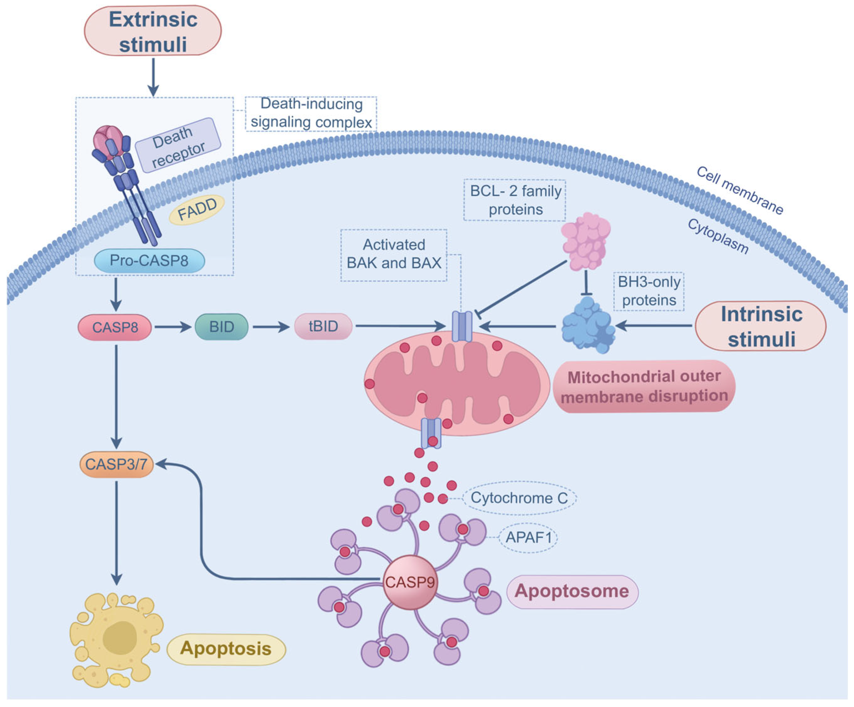
Apoptosis can be classified into intrinsic and extrinsic pathways. In intrinsic apoptosis, BH3-only proteins (e.g., PUMA) are overexpressed and activated by diverse factors such as hypoxia and DNA damage [60]. When the activation of BH3-only proteins exceeds a specific threshold, the suppressive effect of anti-apoptotic B-cell lymphoma-2 (BCL-2) proteins (e.g., MCL1 and BCL-2) is counteracted, promoting the assembly and oligomerization of BAK and BAX in the outer membranes of mitochondria [61,62]. This allows cytochrome c to be released from the mitochondria into cytosol, where it interacts with the apoptotic initiator caspase-9 and apoptotic protease-activating factor-1 [63], ultimately contributing to apoptosome formation and apoptosis. The extrinsic pathway occurs upon ligands (extracellular stimuli) binding to death receptors (DRs) on the cell membrane. The DRs belong to the tumor necrosis factor (TNF) superfamily, such as TNFR1, Fas, DR4, and DR5, and their ligands are TNF-α, FasL, and TRAIL, respectively [64]. Activation of DRs is able to recruit the adaptor proteins (e.g., FADD), and the DRs, FADD, and procaspase-8 assemble into the death-inducing signaling complex, which facilitates the activation of procaspase-8 [62]. Once activated, caspase-8 provokes apoptosis by activating the effector caspases [62]. In some conditions, active caspase-8 is capable of cleaving Bid (a member of BH3-only proteins) into tBId [65], enabling the interaction between extrinsic and intrinsic apoptotic signaling. Apart from this, caspase-8 is a crucial molecular switch orchestrating the interplay between apoptosis and other PCD pathways such as necroptosis and pyroptosis [44]. The molecular mechanisms of apoptosis are shown in Figure 3.

Progress and Outstanding Questions in P. aeruginosa-Induced Apoptosis

Prior research has provided evidence that P. aeruginosa can induce apoptosis through various virulence factors. For instance, T3SS-secreted ExoS leads to intrinsic apoptosis via activation of the apoptotic initiator caspase-9 and effector caspase-3 [15], and the FADD/caspase-8-dependent extrinsic pathway is also found in response to ExoS treatment [66]. The GTPase-activating protein domain of T3SS-secreted ExoT exhibits the ability to induce caspase-9-dependent intrinsic apoptosis [67]. Moreover, characteristic features of apoptosis are observed following P. aeruginosa ExoY exposure [68]. Aside from the T3SS-secreted toxins, the porin from P. aeruginosa is reported to initiate the apoptotic pathway through inhibiting BCL-2 expression [69]. Similarly, LPS, another virulence factor of P. aeruginosa, is recognized for its ability to drive apoptosis [70]. Pyocyanin, a potent cytotoxic pigment produced by P. aeruginosa, contributes to mitochondrial injury and cytochrome c release [71], suggesting induction of the intrinsic apoptotic pathway. Furthermore, the 3-oxo-C12-HSL derived from P. aeruginosa drives spontaneous trimerization of TNFR1, promoting caspase-8 and -3-mediated extrinsic apoptosis [72]. Intrinsic apoptosis is also detected after 3-oxo-C12-HSL exposure, but is completely abolished by BCL-2 overexpression [73]. In addition to the virulence factors discussed above, the bacterial ExoA mediates intrinsic apoptosis since ExoA treatment causes MCL1 degradation and BAK activation [74]. Besides that, ExoA facilitates extrinsic apoptosis via activating caspase-8 and -3 [75]. It is notable that pyocyanin-induced neutrophil apoptosis impairs host bacterial clearance and inflammatory response [76]. Nevertheless, results from another study reveal that P. aeruginosa-induced apoptosis is important for lung bacterial clearance, and defective apoptosis in human bronchial epithelial cells is likely to contribute to the pathogenesis of persistent P. aeruginosa colonization in cystic fibrosis [77]. Based on these aforementioned findings, it is important to note that, although P. aeruginosa is well known to trigger apoptosis, it still remains controversial whether P. aeruginosa-induced apoptosis is beneficial or detrimental for bacterial eradication and infection resolution. In our perspective, underlying mechanisms of this question may be complex and multifaceted. Future studies should thoroughly investigate this question by comprehensively considering factors such as strain diversity (e.g., differences in virulence factor expression between strains), organ and tissue specificity, target cells, and complex bacteria-host interactions.

2.3. P. aeruginosa-Induced Pyroptosis

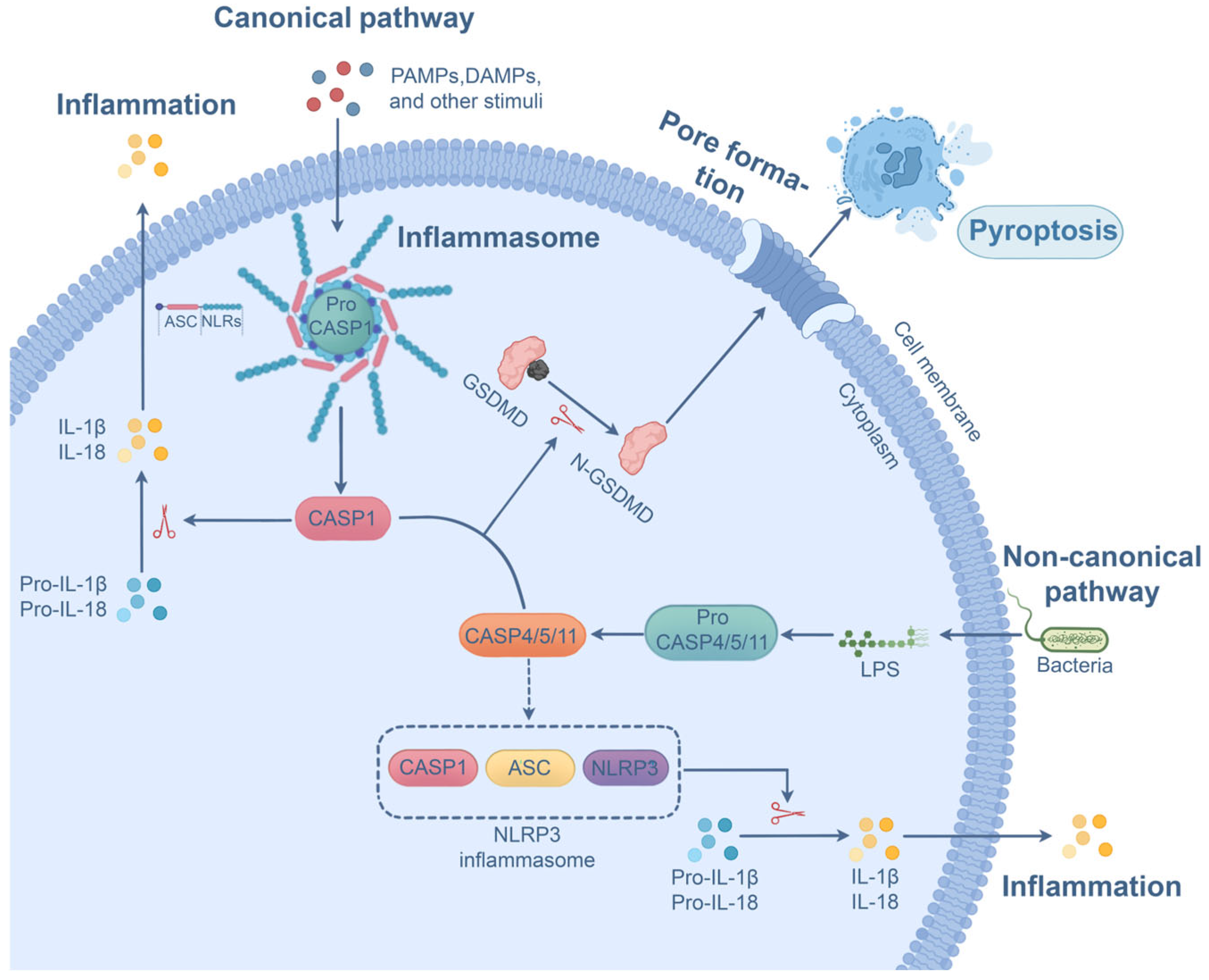
Contrary to apoptosis, pyroptosis is a highly pro-inflammatory PCD pathway with distinct morphological changes such as plasma membrane rupture, membrane blebbing, and cytoplasm flattening [78]. Pyroptosis can be induced by either the canonical or non-canonical pathway. The canonical pyroptotic pathway is mediated by inflammasomes (e.g., NLRP3 inflammasome or NLRC4 inflammasome), a multiprotein complex comprising cytoplasmic sensor NOD-like receptors (NLRs) or AIM2/Pyrin, procaspase-1, and the adaptor ASC [79]. The assembly of inflammasomes is initiated upon detection of PAMPs and danger-associated molecular patterns (DAMPs) by NLRs [79,80]. The oligomerization and activation of NLRs engage ASC, an adaptor bridging the sensors (NLRs or AIM2/Pyrin) and the procaspase-1 [80]. Procaspase-1 is subsequently recruited, followed by self-cleavage and activation [81]. The activated form of caspase-1 mediates the cleavage of GSDMD and the generation of N-terminal domain (N-GSDMD), forming pores on the cell membrane and promoting pyroptosis [82]. Meanwhile, active caspase-1 drives IL-1β and IL-18 maturation through cleavage of their inactive precursors (proIL-1β and proIL-18), after which they are released through pores created by N-GSDMD [79,83]. In non-canonical pyroptosis, upon direct binding to LPS, caspase-4, -5, and -11 undergo oligomerization and activation [84]. Active forms of these caspases cleave GSDMD to produce N-GSDMD, resulting in plasma membrane pore formation and pyroptosis. In addition, these caspases indirectly contribute to the maturation of IL-18 and IL-1β in an NLRP3 inflammasome-dependent manner in certain cells [85]. It is notable that other Gasdermin family proteins, such as Gasdermin B and GSDME, are found to execute pyroptosis through their pore-forming activity after being cleaved [40,86]. Figure 4 depicts the molecular pathways of pyroptosis.

Progress and Outstanding Questions in P. aeruginosa-Induced Pyroptosis

Previously published studies have provided evidence that diverse virulence factors of P. aeruginosa can cause pyroptosis. For instance, flagellin, a pathogenic protein from P. aeruginosa, causes pyroptotic cell death through NLRC4 inflammasome activation [87,88]. In the presence of ExoS ADP ribosyl transferase, neutrophils undergo pyroptosis in an NLRP3-dependent manner [89]. Intriguingly, caspase-11-dependent non-canonical pyroptosis also occurs during T3SS-negative P. aeruginosa infection [90]. The exolysin secreted by P. aeruginosa was identified to provoke pyroptotic cell death through activation of the NLRP3 inflammasome and downstream caspase-1 [17]. It is noteworthy that, contrary to its apoptosis-inducing capacity, pyocyanin suppresses inflammasome activation and caspase-1-dependent canonical pyroptosis, which enables bacteria to evade immune surveillance [91]. Aside from the aforementioned virulence factors, deficiency in oprC (one of the P. aeruginosa porins) reduces GSDMD cleavage and IL-1β release, implicating the role of oprC in pyroptosis induction [92]. Augmented levels of active caspase-1, IL-18, and IL-1β are also detected following P. aeruginosa biofilm stimulation [93], indicating that the bacterial biofilm mediates pyroptotic cell death. The pyroptosis induced by bacterial virulence factors is usually considered a host defense mechanism to restrict infection through activation of the pro-inflammatory immune response and, ultimately, sacrifice of infected cells. In line with this, in an animal model of P. aeruginosa-induced keratitis, mice deficient in caspase-1 and caspase-1/11 exhibit higher bacterial load and more severe corneal disease [89]. Nevertheless, NLRC4 inflammasome activation and the resulting IL-18 generation are reported to suppress host antimicrobial response against the P. aeruginosa strain with functional T3SS, leading to reduced bacterial clearance and an increased level of lung injury [94]. Similarly, findings from Cohen and colleagues unveil that inhibition of caspase-1 and IL-18/IL-1β improves the clearance of P. aeruginosa and alleviates the severity of P. aeruginosa pulmonary infection [95]. Accordingly, these findings suggest that the precise role of pyroptosis in host defense to P. aeruginosa invasion is not well determined. Given the variations in virulence factor expression among different strains, the pathophysiological effect of P. aeruginosa-induced pyroptosis may significantly vary. Importantly, when a P. aeruginosa strain harbors both pyroptosis-inducing (e.g., T3SS) and pyroptosis-inhibiting (e.g., pyocyanin) virulence factors, their multifaceted impact on pyroptosis remains uncertain. Furthermore, the complexity of bacteria-host interactions may be another determinant for P. aeruginosa-induced pyroptosis. For example, different organs and tissues exhibit a distinct microenvironment and composition of resident cells. Consequently, the target cells undergoing P. aeruginosa-induced pyroptosis may differ, and the role of pyroptosis in bacterial eradication and disease resolution may alter.

2.4. P. aeruginosa-Induced Necroptosis

Necroptosis is a form of pro-inflammatory PCD, and it is morphologically characterized by cellular and organelle edema, cell membrane damage, and cell content release [78,96]. Necroptosis acts as a complementary pathway for cell death if caspase-8-mediated apoptosis is disrupted [97]. Necroptosis is initiated following the activation of TNFRs [98,99,100], TLR3/4 [101,102], Fas [98,103], TRAILR [104], and NLR [105], as well as upon stimulation by various factors such as IFNs [106], hypoxia [107], and bacterial infection [108]. Subsequently, the interaction and phosphorylation of RIPK1 and RIPK3 activate pronecrotic kinase activity [109,110] and promote the formation of the necroptosis signaling complex called the necrosome [111]. Activated RIPK3 facilitates the recruitment and phosphorylation of the necroptosis effector MLKL [38,112]. After phosphorylation, MLKL undergoes translocation and oligomerization in the plasma membrane [113], perforating the membrane to form necroptotic pores. In the meantime, necroptotic cells release cell contents and DAMPs that elicit substantial inflammatory responses through permeabilized membranes [114]. Besides RIPK1, RIPK3 can also interact with either TRIF or ZBP-1 for the induction of necroptosis [115,116]. The necroptosis signaling pathway is illustrated in Figure 5.

Progress and Outstanding Questions in P. aeruginosa-Induced Necroptosis

It has been confirmed that P. aeruginosa virulence factors are critically involved in the process of necroptosis. The exolysin secreted by P. aeruginosa was demonstrated to mediate necroptosis as the use of RIPK1 and RIPK3 inhibitors diminishes exolysin-induced cytotoxicity [17]. The bacterial QS system also plays a role in regulating host necroptosis during infection [22]. P. aeruginosa LPS interrupts the binding of RIPK3 to histone and facilitates subsequent RIPK3 translocation [117], implying that LPS is involved in the regulation of necroptosis signaling. In contrast to apoptosis and pyroptosis, pyocyanin is not involved in necroptosis signaling, because pyocyanin-induced cytotoxicity is not reversed by the necroptosis inhibitor [118]. It is worth mentioning that a prior study unveils that P. aeruginosa-mediated necroptosis contributes to disease pathology as the pharmacological inhibition of necroptosis signaling mitigates the severity of acute lung injury [119]. Conversely, results from another study show that the necroptosis initiator RIPK3 functions as an attenuator of pulmonary inflammation caused by P. aeruginosa after its translocation from the nucleus to the cytoplasm, although the activation of necroptosis signaling was not assessed in this research [117]. Thus, similar to aforementioned PCD pathways, the aforementioned findings highlight the need to clarify whether P. aeruginosa-induced necroptosis is host-beneficial or -detrimental. This question should be explored through comprehensive consideration of multiple factors such as strain diversity, organ and tissue specificity, and host immune status.

2.5. P. aeruginosa-Induced Ferroptosis

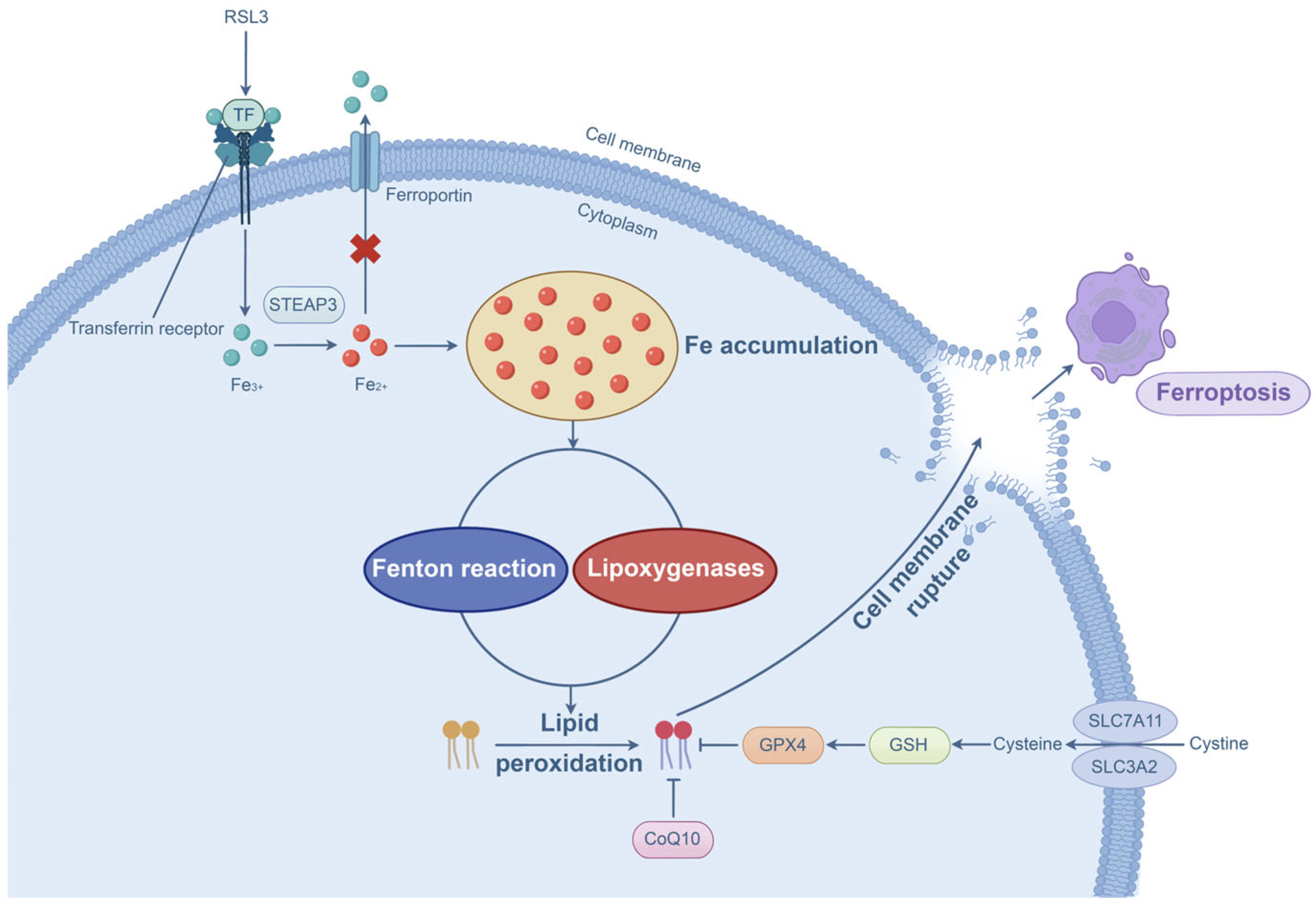
Ferroptosis is a form of PCD with distinct morphological features such as shrunken mitochondria, loss of mitochondrial cristae, and disruption of outer mitochondrial membrane integrity [120]. Notably, unlike other types of PCD described earlier, a normal nuclear size, lack of chromatin condensation, and intact plasma membrane are observed in cells undergoing ferroptosis [120]. Ferroptosis primarily occurs through iron accumulation and lipid peroxidation. The ferroptosis inducer RSL3 can induce intracellular iron accumulation [121]. Moreover, enhanced transferrin expression and declined ferroportin expression lead to iron accumulation [122]. Accumulated Fe3+ can be converted into Fe2+ by the STEAP3 metalloreductase, resulting in the excessive production of reactive oxygen species through the Fenton reaction [123]. More importantly, this reaction mediates the non-enzymatic peroxidation of polyunsaturated fatty acids [124,125], a key pathway for ferroptosis initiation. Compared with the non-enzymatic pathway, enzymatic lipid peroxidation is controlled by lipoxygenases, which modulate the peroxidation of polyunsaturated fatty acids for ferroptosis induction [126,127]. Furthermore, inactivation and depletion of GPX4 and its crucial cofactor GSH elicit ferroptotic cell death because GPX4 is responsible for detoxification of the hydroperoxides [126]. In addition to GPX4, the coenzyme Q10 functions as another important antioxidant that halts lipid peroxidation [128]. Figure 6 presents the molecular processes of ferroptosis.

Progress in P. aeruginosa-Induced Ferroptosis

It has been evidenced that bacterial infections trigger ferroptosis [129], including Gram-negative bacteria [130,131]. Among them, P. aeruginosa was demonstrated to generate lipoxygenase (pLoxA), which promotes host lipid peroxidation (from arachidonic acid-phosphatidylethanolamines to 15-hydroperoxy-arachidonic acid-phosphatidylethanolamines) and ferroptosis [132]. Another study reveals that P. aeruginosa is able to degrade GPX4 activity [133]. Similarly, inhibition of the anti-ferroptotic pathway (GSH/GPX4) and activation of the pro-ferroptotic signal have been found during P. aeruginosa infection, and the use of the P. aeruginosa lipoxygenase inhibitor mitigates the ferroptotic signal and disease severity [134]. Importantly, a previous study discovered that ferroptosis inhibitor liproxstatin-1 is capable of blocking PANoptosis by suppressing the cleavage of PANoptosome upstream regulators (caspase-8 and -6) [135]. Results from another study indicate that there exists an association and cross-talk between ferroptosis and PANoptosis under hypoxic conditions [136]. Given that the proliferation and pathogenicity of P. aeruginosa are closely associated with anaerobic conditions [137], new treatment strategies for P. aeruginosa-induced PANoptosis, particularly caused by MDR strains, warrant further exploration by considering the interplay between ferroptosis and PANoptosis as well as the potential use of ferroptosis inhibitors to target PANoptosis.

3. Potential Targeted Treatment Strategies for P. aeruginosa-Induced PCD

P. aeruginosa infection poses a serious risk to patients due to its antibiotic resistance capacity. As one of the major pathogenic mechanisms of P. aeruginosa is to induce multiple forms of PCD, targeting them may provide alternative therapeutic options for resolving P. aeruginosa infection, particularly for infection caused by MDR strains. As potential treatments for well-studied PCD pathways (e.g., apoptosis, pyroptosis, and necroptosis) have been discussed by previous studies, we herein focus on discussing pharmacological inhibitors and compounds targeting the recently discovered P. aeruginosa-induced PANoptosis.

3.1. Targeting Key Upstream Regulators

As previously discussed, P. aeruginosa triggers PANoptosis, and previous studies have confirmed that P. aeruginosa infection results in the upregulation of IRF1. Intriguingly, genetic depletion or pharmacological inhibition of IRF1 attenuates IRF1-mediated inflammatory cell death [19,138] and reduces the cleavage of caspase-1 and GSDMD [138]. These results suggest that targeted disruption of IRF1 using specific inhibitors (e.g., I-2 and I-19) may be a potential therapeutic approach for P. aeruginosa-induced PANoptosis. Importantly, no obvious multiorgan side effects have been observed in mice treated with I-2 or I-19 [138], indicating that they may be suitable for clinical translation after human trials. In a prior study, treatment with necrostatin-1 (an RIPK1 inhibitor) reverses the activation of caspase-8 and alleviates the cell death resulting from stimulation of P. aeruginosa-secreted 3-oxo-C12-HSL [53]. Another study reveals that miR-29a-3p exhibits the capacity of suppressing caspase-8 activation and PANoptosis in alveolar epithelial cells [139]. Furthermore, administration of the caspase-8 inhibitor Z-IETD-FMK enhances neutrophil antimicrobial activity in the absence of increased cell death [140]. These findings indicate that blockade of caspase-8 activation using pharmacological compounds or small molecules offers a possible treatment strategy for P. aeruginosa-induced PANoptosis. Another investigation shows that caspase-8 activation occurs downstream of caspase-6, another core upstream regulator of PANoptosis, and the use of a selective caspase-6 inhibitor Z-VEID-FMK prevents apoptotic cell death and facilitates cell survival [141]. This underscores the important role of caspase-6 in controlling cell death, implicating the therapeutic potential of applying caspase-6 inhibitors against P. aeruginosa-induced PANoptosis. Nevertheless, necrostatin-1 and the aforementioned caspase inhibitors have not yet entered clinical trials, and their efficacy and safety for the treatment of P. aeruginosa-induced PANoptosis still need further investigation and optimization.

3.2. Targeting Sensor Proteins

As discussed earlier, ZBP-1 is a pivotal sensor protein that composes the PANoptosome and modulates the process of PANoptosis. It is reported that ADAR1 can limit the activation of ZBP-1, preventing ZBP-1-mediated cell death and autoinflammation [142]. ADAR1 has been involved in clinical investigations in the field of cancer therapy efficacy (NCT07108348 and NCT05482451), and its clinical feasibility in treating P. aeruginosa-induced PANoptosis warrants further assessment. The miR-29a-3p treatment also diminishes the levels of ZBP-1 and PANoptosis [139]. Furthermore, a prior study shows that CDK1 can bind with ZBP-1 PANoptosome, and the administration of cucurbitacin E (a CDK1 inhibitor) is able to limit tumor cell growth by regulating PANoptosis [143]. With regard to other PANoptosome sensor proteins, IFI16-β represses AIM2 activation since IFI16-β can prevent the AIM2 recognition of cytosolic dsDNA [144]. In addition, IFI16-β impairs the assembly of AIM2-ASC complex [144]. J114 also displays a potent inhibitory effect on AIM2-dependent cell death as it disturbs AIM2-ASC interaction and dampens AIM2 activation [145]. 4-Sulfonic calix[6]arene was identified as another AIM2 inhibitor because it blocks the DNA-binding HIN domain of AIM2 [146]. In this study, researchers have confirmed that suramin, a clinically available polysulfonated drug exhibiting similar properties to 4-Sulfonic calix[6]arene, effectively inhibits AIM2 inflammasome activation [146], suggesting the feasibility for clinical use. In comparison with ZBP-1 and AIM2, many potent and highly specific RIPK1 inhibitors have been discovered over the past decade. 6E11, a highly selective human RIPK1 inhibitor, is shown to restrict TNF-α (one of the PANoptosis inducers [19])-induced, RIPK1-driven cell death while exhibiting low cytotoxicity toward normal cells [147]. This high-affinity, non-ATP competitive inhibitor provides more potent protection for cell survival compared with necrostatin-1, a well-studied RIPK1 inhibitor [147]. Further animal and clinical studies are needed to evaluate its clinical potential. ZB-R-55 was found to be another highly effective RIPK1 inhibitor with desirable kinase selectivity and therapeutic effects in sepsis induced by LPS (a major virulence factor of P. aeruginosa) [148]. Importantly, ZB-R-55 shows approximately tenfold greater potency compared with GSK2982772 [148], the first RIPK1 inhibitor that entered clinical investigations. Moreover, treatment with RIPA-56 efficiently protects mice from TNF-α-induced mortality, systemic inflammation, and multiorgan damage, proving its high effectiveness, selectivity, and metabolic stability for RIPK1 inhibition [149]. Given the high selectivity, efficacy, and metabolic stability of these specific RIPK1 inhibitors in preclinical models, their therapeutic potential and adverse effects require further investigation in clinical settings. As for NLRP12, there are currently no direct NLRP12 inhibitors available for human or animal studies. Screening, optimization, and application of NLRP12 inhibitors are strongly encouraged to evaluate their specificity and potency in limiting NLRP12-mediated PCD, especially in the context of P. aeruginosa infection.

3.3. Targeting Other Molecular Components of PANoptosome Complexes or Downstream Executioners

ASC, an important adaptor protein bridging the upstream signaling and downstream effectors for PANoptosome assembly and activation, can be regarded as a potential therapeutic target. Spirodalesol analog 8A was confirmed to be a desirable ASC inhibitor as it directly interacts with ASC to prevent ASC speck formation [150]. Treatment of 8A also attenuates the severity of endotoxemia induced by LPS [150]. As previously reviewed, pharmacological inhibition of AIM2-ASC interaction and ASC oligomerization by J114 protects cells from AIM2-dependent cell death [145]. Since AIM2 and ASC play pivotal roles in modulating PANoptosis, the potential of applying these selective ASC inhibitors against P. aeruginosa-induced PANoptosis is promising. Besides ASC, caspase-1 is another important component of the PANoptosome. Tetracycline is evidenced to mitigate caspase-1-dependent cell death and lung inflammation [151]. Notably, tetracycline dose-dependently reduces IL-18 and IL-1β production by alveolar leukocytes from patients with acute lung inflammation [151]. This finding strengthens the feasibility of applying tetracycline to target caspase-1 in clinical settings. The unsaturated ester derivative-compound 9 blocks caspase-1 activity and prevents caspase-1-driven cell death in a time- and concentration-dependent manner [152]. However, although compound 9 shows strong protective efficacy against caspase-1-driven cell death, its high cytotoxicity needs further evaluation in animal studies. A prior study reports that compound 42 (an analog of TAK-632) attenuates RIPK3-dependent cell death through inhibiting the phosphorylation of RIPK3 [153], another cell death mediator. Furthermore, compound 42 exhibits over 60-fold selectivity for RIPK3 over RIPK1 [153]. AZD5423 can also directly target RIPK3 to repress its kinase activity, impeding RIPK3 activation and alleviating RIPK3-dependent cell damage [154]. It is notable that these RIPK3 inhibitors are not yet clinically available. However, as an important component of PANoptosome complexes, RIPK3 still remains a promising therapeutic target for P. aeruginosa-induced PANoptosis.
Aside from the PANoptosome components described before, GSDMD and MLKL (downstream executioners) may be targetable molecules for mitigating cell death. For instance, necrosulfonamide (NSA) is able to reduce GSDMD-mediated cell death and ameliorate LPS-induced sepsis through directly binding to the Cys191 of GSDMD [155]. Through the blockade of GSDMD’s pore-forming activity, disulfiram exerts an inhibitory effect on pyroptotic cell death and LPS-induced sepsis in mice [156]. As mentioned earlier, LPS serves as a major pathogenic factor of P. aeruginosa, implicating the possibility of employing NSA or disulfiram against P. aeruginosa-induced PANoptosis. Surprisingly, cell death triggered by the AIM2 inflammasome is also impaired by disulfiram in a dose-dependent manner [156], suggesting that disulfiram can target both AIM2 and GSDMD. As an FDA-approved anti-alcoholism agent, this dual-target drug shows great promise against P. aeruginosa-induced PANoptosis, provided that its dosage, pharmacodynamics, and oral bioavailability are carefully assessed in clinical trials. With regard to MLKL, treatment with NSA can downregulate its phosphorylation and activation [157]. Mechanistically, the Cys86 of human MLKL was identified as the covalent binding and modification site for NSA [38]. Thus, NSA acts as a dual-target inhibitor of GSDMD and MLKL. Nevertheless, unlike disulfiram, NSA is not yet available for clinical use, and thus clinical investigations are needed to study the efficacy and safety of this promising dual-target drug. Saracatinib, a drug with encouraging preclinical and clinical results for multiple diseases [158], suppresses TNF-induced cell death by disrupting phosphorylation, translocation, and oligomerization of MLKL [159]. All aforementioned pharmacological compounds and small molecules are summarized in Table 1. Recently, it has been reported that Imipenem-Relebactam treatment shows a high clinical success rate (75.9%) and reduced 30-day mortality (24.1%) in patients with difficult-to-treat P. aeruginosa infection [160]. This highlights the potential of combining the aforementioned pharmacological compounds or small molecules with antibiotics to provide more favorable efficacy against P. aeruginosa infection.

4. Conclusions and Future Directions

P. aeruginosa is responsible for the onset of many hospital-acquired infections, and it still remains challenging to fully eradicate this pathogen in clinics. It has been demonstrated that it triggers multiple types of PCD, such as apoptosis, pyroptosis, necroptosis, ferroptosis, and PANoptosis, the latter of which is intricately regulated by PANoptosome complexes and their key upstream regulators. Progress in our understanding of these molecules, as well as their upregulation and/or activation in response to P. aeruginosa infection, offers up-to-date and comprehensive insights into the close mechanistic association between P. aeruginosa infection and PANoptosis. In addition to PANoptosis, P. aeruginosa virulence factors induce apoptosis, pyroptosis, necroptosis, and ferroptosis, and they are closely associated with the host’s immune reaction in response to P. aeruginosa invasion. Targeting P. aeruginosa-induced PCD using pharmacological compounds or small molecules may be an alternative treatment strategy for P. aeruginosa infection, especially for infection caused by MDR strains. Our efforts help bridge the knowledge gap and pave the way for future research to further understand the pathogenicity of P. aeruginosa.
However, although PANoptosome complexes and their key upstream regulators have been identified as PANoptosis mediators, little is known about the precise mechanistic role of each PANoptosome component in PANoptosis. To elucidate their functions and potential functional redundancies, conditional knockout experiments for each molecule in cells or animal models are needed. In vivo studies are preferable since host microenvironmental factors (e.g., pH and metabolites) and interactions among innate and adaptive immune cells may also influence the PANoptosis process. Furthermore, it is of great value to investigate how individual virulence factors of P. aeruginosa mechanistically contribute to PANoptosome assembly and PANoptosis. Besides laboratory P. aeruginosa strains, clinical MDR isolates should be used for research. To precisely define the cytotoxicity of a specific virulence factor, loss-of-function mutant strains can be generated and utilized. Moreover, it is crucial to clarify the precise roles of P. aeruginosa-induced PCD and identify when it plays a beneficial role (e.g., restricting infection or activating immune response) or detrimental role (e.g., mediating organ and tissue damage) at different phases of infection. Lastly, the development of clinically effective medications targeting P. aeruginosa-induced PCD is highly anticipated. Considering recent advances in antibiotic treatment against P. aeruginosa infection, combining P. aeruginosa-induced PCD-targeting agents with antibiotics may achieve more favorable efficacy for P. aeruginosa infection. Unveiling the high-resolution structures of PANoptosome complexes is of great significance, as it will substantially facilitate structure-based drug design. Thereafter, the translational feasibility of drugs, such as selectivity, metabolic stability, and oral bioavailability, can be further evaluated and optimized in preclinical models. Finally, clinical studies are indispensable for validating therapeutic effectiveness and assessing potential side effects. Taken together, gaining new knowledge and insights into P. aeruginosa-induced PCD will open new avenues for developing effective targeted therapeutics against P. aeruginosa infection.

Author Contributions

Conceptualization, C.T. and Y.L.; investigation, C.T. and Y.L.; writing—original draft preparation, C.T.; writing—review and editing, Y.L.; visualization, C.T.; supervision, Y.L.; project administration, Y.L. All authors have read and agreed to the published version of the manuscript.

Funding

This research received no external funding.

Institutional Review Board Statement

Not applicable.

Informed Consent Statement

Not applicable.

Data Availability Statement

No new data were created or analyzed in this study. Data sharing is not applicable to this article.

Conflicts of Interest

The authors declare no conflicts of interest.

References

  1. Wunderink, R.G.; Matsunaga, Y.; Ariyasu, M.; Clevenbergh, P.; Echols, R.; Kaye, K.S.; Kollef, M.; Menon, A.; Pogue, J.M.; Shorr, A.F.; et al. Cefiderocol versus high-dose, extended-infusion meropenem for the treatment of Gram-negative nosocomial pneumonia (APEKS-NP): A randomised, double-blind, phase 3, non-inferiority trial. Lancet Infect. Dis. 2021, 21, 213–225. [Google Scholar] [CrossRef]
  2. Edelstein, M.V.; Skleenova, E.N.; Shevchenko, O.V.; D’SOuza, J.W.; Tapalski, D.V.; Azizov, I.S.; Sukhorukova, M.V.; Pavlukov, R.A.; Kozlov, R.S.; Toleman, M.A.; et al. Spread of extensively resistant VIM-2-positive ST235 Pseudomonas aeruginosa in Belarus, Kazakhstan, and Russia: A longitudinal epidemiological and clinical study. Lancet Infect. Dis. 2013, 13, 867–876. [Google Scholar] [CrossRef] [PubMed]
  3. Ferrer, M.; Difrancesco, L.F.; Liapikou, A.; Rinaudo, M.; Carbonara, M.; Li Bassi, G.; Gabarrus, A.; Torres, A. Polymicrobial intensive care unit-acquired pneumonia: Prevalence, microbiology and outcome. Crit. Care 2015, 19, 450. [Google Scholar] [CrossRef]
  4. Bauer, K.A.; Puzniak, L.A.; Yu, K.C.; Finelli, L.; Moise, P.; Ai, C.; Watts, J.A.; Gupta, V. Epidemiology and outcomes of culture-positive bloodstream pathogens prior to and during the SARS-CoV-2 pandemic: A multicenter evaluation. BMC Infect. Dis. 2022, 22, 841. [Google Scholar] [CrossRef]
  5. Karaba, S.M.; Cosgrove, S.E.; Lee, J.H.; Fiawoo, S.; Heil, E.L.; Quartuccio, K.S.; Shihadeh, K.C.; Tamma, P.D. Extended-Infusion beta-Lactam Therapy, Mortality, and Subsequent Antibiotic Resistance Among Hospitalized Adults With Gram-Negative Bloodstream Infections. JAMA Netw. Open 2024, 7, e2418234. [Google Scholar] [CrossRef] [PubMed]
  6. Weiner-Lastinger, L.M.; Abner, S.; Edwards, J.R.; Kallen, A.J.; Karlsson, M.; Magill, S.S.; Pollock, D.; See, I.; Soe, M.M.; Walters, M.S.; et al. Antimicrobial-resistant pathogens associated with adult healthcare-associated infections: Summary of data reported to the National Healthcare Safety Network, 2015–2017. Infect. Control Hosp. Epidemiol. 2020, 41, 1–18. [Google Scholar] [CrossRef] [PubMed]
  7. Sartelli, M.; Catena, F.; Ansaloni, L.; Coccolini, F.; Corbella, D.; Moore, E.E.; Malangoni, M.; Velmahos, G.; Coimbra, R.; Koike, K.; et al. Complicated intra-abdominal infections worldwide: The definitive data of the CIAOW Study. World J. Emerg. Surg. 2014, 9, 37. [Google Scholar] [CrossRef]
  8. Rhee, C.; Chen, T.; Kadri, S.S.; Lawandi, A.; Yek, C.; Walker, M.; Warner, S.; Fram, D.; Chen, H.C.; Shappell, C.N.; et al. Trends in Empiric Broad-Spectrum Antibiotic Use for Suspected Community-Onset Sepsis in US Hospitals. JAMA Netw. Open 2024, 7, e2418923. [Google Scholar] [CrossRef]
  9. Johnson, L.E.; D’Agata, E.M.; Paterson, D.L.; Clarke, L.; Qureshi, Z.A.; Potoski, B.A.; Peleg, A.Y. Pseudomonas aeruginosa bacteremia over a 10-year period: Multidrug resistance and outcomes in transplant recipients. Transpl. Infect. Dis. 2009, 11, 227–234. [Google Scholar] [CrossRef]
  10. Montero, M.; Sala, M.; Riu, M.; Belvis, F.; Salvado, M.; Grau, S.; Horcajada, J.P.; Alvarez-Lerma, F.; Terradas, R.; Orozco-Levi, M.; et al. Risk factors for multidrug-resistant Pseudomonas aeruginosa acquisition. Impact of antibiotic use in a double case-control study. Eur. J. Clin. Microbiol. Infect. Dis. 2010, 29, 335–339. [Google Scholar] [CrossRef]
  11. Ikuta, K.S.; Swetschinski, L.R.; Aguilar, G.R.; Sharara, F.; Mestrovic, T.; Gray, A.P.; Weaver, N.D.; Wool, E.E.; Han, C.; Hayoon, A.G.; et al. Global mortality associated with 33 bacterial pathogens in 2019: A systematic analysis for the Global Burden of Disease Study 2019. Lancet 2022, 400, 2221–2248. [Google Scholar] [CrossRef]
  12. Yayan, J.; Ghebremedhin, B.; Rasche, K. Antibiotic Resistance of Pseudomonas aeruginosa in Pneumonia at a Single University Hospital Center in Germany over a 10-Year Period. PLoS ONE 2015, 10, e0139836. [Google Scholar] [CrossRef] [PubMed]
  13. Reyes, J.; Komarow, L.; Chen, L.; Ge, L.; Hanson, B.M.; Cober, E.; Herc, E.; Alenazi, T.; Kaye, K.S.; Garcia-Diaz, J.; et al. Global epidemiology and clinical outcomes of carbapenem-resistant Pseudomonas aeruginosa and associated carbapenemases (POP): A prospective cohort study. Lancet Microbe 2023, 4, e159–e170. [Google Scholar] [CrossRef]
  14. Wood, S.J.; Goldufsky, J.W.; Seu, M.Y.; Dorafshar, A.H.; Shafikhani, S.H. Pseudomonas aeruginosa Cytotoxins: Mechanisms of Cytotoxicity and Impact on Inflammatory Responses. Cells 2023, 12, 195. [Google Scholar] [CrossRef] [PubMed]
  15. Kaminski, A.; Gupta, K.H.; Goldufsky, J.W.; Lee, H.W.; Gupta, V.; Shafikhani, S.H. Pseudomonas aeruginosa ExoS Induces Intrinsic Apoptosis in Target Host Cells in a Manner That is Dependent on its GAP Domain Activity. Sci. Rep. 2018, 8, 14047. [Google Scholar] [CrossRef] [PubMed]
  16. Chu, L.H.; Indramohan, M.; Ratsimandresy, R.A.; Gangopadhyay, A.; Morris, E.P.; Monack, D.M.; Dorfleutner, A.; Stehlik, C. The oxidized phospholipid oxPAPC protects from septic shock by targeting the non-canonical inflammasome in macrophages. Nat. Commun. 2018, 9, 996. [Google Scholar] [CrossRef]
  17. Basso, P.; Wallet, P.; Elsen, S.; Soleilhac, E.; Henry, T.; Faudry, E.; Attree, I. Multiple Pseudomonas species secrete exolysin-like toxins and provoke Caspase-1-dependent macrophage death. Environ. Microbiol. 2017, 19, 4045–4064. [Google Scholar] [CrossRef]
  18. Malireddi, R.K.S.; Kesavardhana, S.; Karki, R.; Kancharana, B.; Burton, A.R.; Kanneganti, T.D. RIPK1 Distinctly Regulates Yersinia-Induced Inflammatory Cell Death, PANoptosis. Immunohorizons 2020, 4, 789–796. [Google Scholar] [CrossRef]
  19. Karki, R.; Sharma, B.R.; Tuladhar, S.; Williams, E.P.; Zalduondo, L.; Samir, P.; Zheng, M.; Sundaram, B.; Banoth, B.; Malireddi, R.K.S.; et al. Synergism of TNF-alpha and IFN-gamma Triggers Inflammatory Cell Death, Tissue Damage, and Mortality in SARS-CoV-2 Infection and Cytokine Shock Syndromes. Cell 2021, 184, 149–168.e17. [Google Scholar] [CrossRef]
  20. Lee, S.; Karki, R.; Wang, Y.; Nguyen, L.N.; Kalathur, R.C.; Kanneganti, T.D. AIM2 forms a complex with pyrin and ZBP1 to drive PANoptosis and host defence. Nature 2021, 597, 415–419. [Google Scholar] [CrossRef]
  21. Sundaram, B.; Karki, R.; Kanneganti, T.D. NLRC4 Deficiency Leads to Enhanced Phosphorylation of MLKL and Necroptosis. Immunohorizons 2022, 6, 243–252. [Google Scholar] [CrossRef] [PubMed]
  22. Liu, Z.; Sun, L.; Li, L.; Miao, E.A.; Amer, A.O.; Wozniak, D.J.; Wen, H. Pseudomonas aeruginosa Mediates Host Necroptosis through Rhl-Pqs Quorum Sensing Interaction. Immunohorizons 2024, 8, 721–728. [Google Scholar] [CrossRef]
  23. Yue, L.; Cao, H.; Qi, J.; Yuan, J.; Wang, X.; Wang, Y.; Shan, B.; Ke, H.; Li, H.; Luan, N.; et al. Pretreatment with 3-methyladenine ameliorated Pseudomonas aeruginosa-induced acute pneumonia by inhibiting cell death of neutrophils in a mouse infection model. Int. J. Med. Microbiol. 2023, 313, 151574. [Google Scholar] [CrossRef] [PubMed]
  24. Malireddi, R.K.S.; Kesavardhana, S.; Kanneganti, T.D. ZBP1 and TAK1: Master Regulators of NLRP3 Inflammasome/Pyroptosis, Apoptosis, and Necroptosis (PAN-optosis). Front. Cell Infect. Microbiol. 2019, 9, 406. [Google Scholar] [CrossRef] [PubMed]
  25. Christgen, S.; Zheng, M.; Kesavardhana, S.; Karki, R.; Malireddi, R.K.S.; Banoth, B.; Place, D.E.; Briard, B.; Sharma, B.R.; Tuladhar, S.; et al. Identification of the PANoptosome: A Molecular Platform Triggering Pyroptosis, Apoptosis, and Necroptosis (PANoptosis). Front. Cell Infect. Microbiol. 2020, 10, 237. [Google Scholar] [CrossRef]
  26. Karki, R.; Lee, S.; Mall, R.; Pandian, N.; Wang, Y.; Sharma, B.R.; Malireddi, R.S.; Yang, D.; Trifkovic, S.; Steele, J.A.; et al. ZBP1-dependent inflammatory cell death, PANoptosis, and cytokine storm disrupt IFN therapeutic efficacy during coronavirus infection. Sci. Immunol. 2022, 7, eabo6294. [Google Scholar] [CrossRef]
  27. Takaoka, A.; Wang, Z.; Choi, M.K.; Yanai, H.; Negishi, H.; Ban, T.; Lu, Y.; Miyagishi, M.; Kodama, T.; Honda, K.; et al. DAI (DLM-1/ZBP1) is a cytosolic DNA sensor and an activator of innate immune response. Nature 2007, 448, 501–505. [Google Scholar] [CrossRef]
  28. Maelfait, J.; Liverpool, L.; Bridgeman, A.; Ragan, K.B.; Upton, J.W.; Rehwinkel, J. Sensing of viral and endogenous RNA by ZBP1/DAI induces necroptosis. EMBO J. 2017, 36, 2529–2543. [Google Scholar] [CrossRef]
  29. Kuriakose, T.; Kanneganti, T.D. ZBP1: Innate Sensor Regulating Cell Death and Inflammation. Trends Immunol. 2018, 39, 123–134. [Google Scholar] [CrossRef]
  30. Hornung, V.; Ablasser, A.; Charrel-Dennis, M.; Bauernfeind, F.; Horvath, G.; Caffrey, D.R.; Latz, E.; Fitzgerald, K.A. AIM2 recognizes cytosolic dsDNA and forms a caspase-1-activating inflammasome with ASC. Nature 2009, 458, 514–518. [Google Scholar] [CrossRef]
  31. Man, S.M.; Karki, R.; Malireddi, R.K.; Neale, G.; Vogel, P.; Yamamoto, M.; Lamkanfi, M.; Kanneganti, T.D. The transcription factor IRF1 and guanylate-binding proteins target activation of the AIM2 inflammasome by Francisella infection. Nat. Immunol. 2015, 16, 467–475. [Google Scholar] [CrossRef] [PubMed]
  32. Sundaram, B.; Pandian, N.; Mall, R.; Wang, Y.; Sarkar, R.; Kim, H.J.; Malireddi, R.K.S.; Karki, R.; Janke, L.J.; Vogel, P.; et al. NLRP12-PANoptosome activates PANoptosis and pathology in response to heme and PAMPs. Cell 2023, 186, 2783–2801.e20. [Google Scholar] [CrossRef]
  33. Broz, P.; Dixit, V.M. Inflammasomes: Mechanism of assembly, regulation and signalling. Nat. Rev. Immunol. 2016, 16, 407–420. [Google Scholar] [CrossRef] [PubMed]
  34. Samir, P.; Malireddi, R.K.S.; Kanneganti, T.D. The PANoptosome: A Deadly Protein Complex Driving Pyroptosis, Apoptosis, and Necroptosis (PANoptosis). Front. Cell Infect. Microbiol. 2020, 10, 238. [Google Scholar] [CrossRef]
  35. Tummers, B.; Mari, L.; Guy, C.S.; Heckmann, B.L.; Rodriguez, D.A.; Ruhl, S.; Moretti, J.; Crawford, J.C.; Fitzgerald, P.; Kanneganti, T.D.; et al. Caspase-8-Dependent Inflammatory Responses Are Controlled by Its Adaptor, FADD, and Necroptosis. Immunity 2020, 52, 994–1006.e8. [Google Scholar] [CrossRef]
  36. Schwarzer, R.; Jiao, H.; Wachsmuth, L.; Tresch, A.; Pasparakis, M. FADD and Caspase-8 Regulate Gut Homeostasis and Inflammation by Controlling MLKL- and GSDMD-Mediated Death of Intestinal Epithelial Cells. Immunity 2020, 52, 978–993.e6. [Google Scholar] [CrossRef] [PubMed]
  37. Shi, J.; Zhao, Y.; Wang, K.; Shi, X.; Wang, Y.; Huang, H.; Zhuang, Y.; Cai, T.; Wang, F.; Shao, F. Cleavage of GSDMD by inflammatory caspases determines pyroptotic cell death. Nature 2015, 526, 660–665. [Google Scholar] [CrossRef]
  38. Sun, L.; Wang, H.; Wang, Z.; He, S.; Chen, S.; Liao, D.; Wang, L.; Yan, J.; Liu, W.; Lei, X.; et al. Mixed lineage kinase domain-like protein mediates necrosis signaling downstream of RIP3 kinase. Cell 2012, 148, 213–227. [Google Scholar] [CrossRef]
  39. Pandian, N.; Kanneganti, T.D. PANoptosis: A Unique Innate Immune Inflammatory Cell Death Modality. J. Immunol. 2022, 209, 1625–1633. [Google Scholar] [CrossRef]
  40. Wang, Y.; Gao, W.; Shi, X.; Ding, J.; Liu, W.; He, H.; Wang, K.; Shao, F. Chemotherapy drugs induce pyroptosis through caspase-3 cleavage of a gasdermin. Nature 2017, 547, 99–103. [Google Scholar] [CrossRef]
  41. Kuriakose, T.; Zheng, M.; Neale, G.; Kanneganti, T.D. IRF1 Is a Transcriptional Regulator of ZBP1 Promoting NLRP3 Inflammasome Activation and Cell Death during Influenza Virus Infection. J. Immunol. 2018, 200, 1489–1495. [Google Scholar] [CrossRef]
  42. Sharma, B.R.; Karki, R.; Rajesh, Y.; Kanneganti, T.D. Immune regulator IRF1 contributes to ZBP1-, AIM2-, RIPK1-, and NLRP12-PANoptosome activation and inflammatory cell death (PANoptosis). J. Biol. Chem. 2023, 299, 105141. [Google Scholar] [CrossRef]
  43. Varfolomeev, E.E.; Schuchmann, M.; Luria, V.; Chiannilkulchai, N.; Beckmann, J.S.; Mett, I.L.; Rebrikov, D.; Brodianski, V.M.; Kemper, O.C.; Kollet, O.; et al. Targeted disruption of the mouse Caspase 8 gene ablates cell death induction by the TNF receptors, Fas/Apo1, and DR3 and is lethal prenatally. Immunity 1998, 9, 267–276. [Google Scholar] [CrossRef] [PubMed]
  44. Fritsch, M.; Gunther, S.D.; Schwarzer, R.; Albert, M.C.; Schorn, F.; Werthenbach, J.P.; Schiffmann, L.M.; Stair, N.; Stocks, H.; Seeger, J.M.; et al. Caspase-8 is the molecular switch for apoptosis, necroptosis and pyroptosis. Nature 2019, 575, 683–687. [Google Scholar] [CrossRef]
  45. Pierini, R.; Juruj, C.; Perret, M.; Jones, C.L.; Mangeot, P.; Weiss, D.S.; Henry, T. AIM2/ASC triggers caspase-8-dependent apoptosis in Francisella-infected caspase-1-deficient macrophages. Cell Death Differ. 2012, 19, 1709–1721. [Google Scholar] [CrossRef]
  46. Zheng, M.; Karki, R.; Vogel, P.; Kanneganti, T.D. Caspase-6 Is a Key Regulator of Innate Immunity, Inflammasome Activation, and Host Defense. Cell 2020, 181, 674–687.e13. [Google Scholar] [CrossRef] [PubMed]
  47. Qi, J.L.; He, J.R.; Jin, S.M.; Yang, X.; Bai, H.M.; Liu, C.B.; Ma, Y.B. P. aeruginosa Mediated Necroptosis in Mouse Tumor Cells Induces Long-Lasting Systemic Antitumor Immunity. Front. Oncol. 2020, 10, 610651. [Google Scholar] [CrossRef] [PubMed]
  48. Kuriakose, T.; Man, S.M.; Malireddi, R.K.; Karki, R.; Kesavardhana, S.; Place, D.E.; Neale, G.; Vogel, P.; Kanneganti, T.D. ZBP1/DAI is an innate sensor of influenza virus triggering the NLRP3 inflammasome and programmed cell death pathways. Sci. Immunol. 2016, 1, aag2045. [Google Scholar] [CrossRef]
  49. Han, Y.; Ge, C.; Ye, J.; Li, R.; Zhang, Y. Demethyleneberberine alleviates Pseudomonas aeruginosa-induced acute pneumonia by inhibiting the AIM2 inflammasome and oxidative stress. Pulm. Pharmacol. Ther. 2023, 83, 102259. [Google Scholar] [CrossRef]
  50. Ulland, T.K.; Jain, N.; Hornick, E.E.; Elliott, E.I.; Clay, G.M.; Sadler, J.J.; Mills, K.A.; Janowski, A.M.; Volk, A.P.; Wang, K.; et al. Nlrp12 mutation causes C57BL/6J strain-specific defect in neutrophil recruitment. Nat. Commun. 2016, 7, 13180. [Google Scholar] [CrossRef]
  51. Penaranda, C.; Chumbler, N.M.; Hung, D.T. Dual transcriptional analysis reveals adaptation of host and pathogen to intracellular survival of Pseudomonas aeruginosa associated with urinary tract infection. PLoS Pathog. 2021, 17, e1009534. [Google Scholar] [CrossRef] [PubMed]
  52. Li, X.; Ye, Y.; Zhou, X.; Huang, C.; Wu, M. Atg7 enhances host defense against infection via downregulation of superoxide but upregulation of nitric oxide. J. Immunol. 2015, 194, 1112–1121. [Google Scholar] [CrossRef] [PubMed]
  53. Shin, J.; Ahn, S.H.; Kim, S.H.; Oh, D.J. N-3-oxododecanoyl homoserine lactone exacerbates endothelial cell death by inducing receptor-interacting protein kinase 1-dependent apoptosis. Am. J. Physiol. Cell Physiol. 2021, 321, C644–C653. [Google Scholar] [CrossRef]
  54. Rutherford, S.T.; Bassler, B.L. Bacterial quorum sensing: Its role in virulence and possibilities for its control. Cold Spring Harb. Perspect. Med. 2012, 2, a012427. [Google Scholar] [CrossRef]
  55. Wyllie, A.H. Glucocorticoid-induced thymocyte apoptosis is associated with endogenous endonuclease activation. Nature 1980, 284, 555–556. [Google Scholar] [CrossRef]
  56. Ziegler, U.; Groscurth, P. Morphological features of cell death. News Physiol. Sci. 2004, 19, 124–128. [Google Scholar] [CrossRef]
  57. Jorgensen, I.; Rayamajhi, M.; Miao, E.A. Programmed cell death as a defence against infection. Nat. Rev. Immunol. 2017, 17, 151–164. [Google Scholar] [CrossRef]
  58. Martinon, F.; Tschopp, J. Inflammatory caspases: Linking an intracellular innate immune system to autoinflammatory diseases. Cell 2004, 117, 561–574. [Google Scholar] [CrossRef]
  59. Riedl, S.J.; Shi, Y. Molecular mechanisms of caspase regulation during apoptosis. Nat. Rev. Mol. Cell Biol. 2004, 5, 897–907. [Google Scholar] [CrossRef]
  60. Lomonosova, E.; Chinnadurai, G. BH3-only proteins in apoptosis and beyond: An overview. Oncogene 2008, 27 (Suppl. S1), S2–S19. [Google Scholar] [CrossRef] [PubMed]
  61. Volkmann, N.; Marassi, F.M.; Newmeyer, D.D.; Hanein, D. The rheostat in the membrane: BCL-2 family proteins and apoptosis. Cell Death Differ. 2014, 21, 206–215. [Google Scholar] [CrossRef]
  62. Taylor, R.C.; Cullen, S.P.; Martin, S.J. Apoptosis: Controlled demolition at the cellular level. Nat. Rev. Mol. Cell Biol. 2008, 9, 231–241. [Google Scholar] [CrossRef]
  63. Li, P.; Nijhawan, D.; Budihardjo, I.; Srinivasula, S.M.; Ahmad, M.; Alnemri, E.S.; Wang, X. Cytochrome c and dATP-dependent formation of Apaf-1/caspase-9 complex initiates an apoptotic protease cascade. Cell 1997, 91, 479–489. [Google Scholar] [CrossRef]
  64. Vanamee, E.S.; Faustman, D.L. Structural principles of tumor necrosis factor superfamily signaling. Sci. Signal 2018, 11, eaao4910. [Google Scholar] [CrossRef] [PubMed]
  65. Schug, Z.T.; Gonzalvez, F.; Houtkooper, R.H.; Vaz, F.M.; Gottlieb, E. BID is cleaved by caspase-8 within a native complex on the mitochondrial membrane. Cell Death Differ. 2011, 18, 538–548. [Google Scholar] [CrossRef]
  66. Alaoui-El-Azher, M.; Jia, J.; Lian, W.; Jin, S. ExoS of Pseudomonas aeruginosa induces apoptosis through a Fas receptor/caspase 8-independent pathway in HeLa cells. Cell Microbiol. 2006, 8, 326–338. [Google Scholar] [CrossRef]
  67. Wood, S.J.; Goldufsky, J.W.; Bello, D.; Masood, S.; Shafikhani, S.H. Pseudomonas aeruginosa ExoT Induces Mitochondrial Apoptosis in Target Host Cells in a Manner That Depends on Its GTPase-activating Protein (GAP) Domain Activity. J. Biol. Chem. 2015, 290, 29063–29073. [Google Scholar] [CrossRef] [PubMed]
  68. Kloth, C.; Schirmer, B.; Munder, A.; Stelzer, T.; Rothschuh, J.; Seifert, R. The Role of Pseudomonas aeruginosa ExoY in an Acute Mouse Lung Infection Model. Toxins 2018, 10, 185. [Google Scholar] [CrossRef]
  69. Buommino, E.; Morelli, F.; Metafora, S.; Rossano, F.; Perfetto, B.; Baroni, A.; Tufano, M.A. Porin from Pseudomonas aeruginosa induces apoptosis in an epithelial cell line derived from rat seminal vesicles. Infect. Immun. 1999, 67, 4794–4800. [Google Scholar] [CrossRef]
  70. Liu, X.; Shao, K.; Sun, T. SIRT1 regulates the human alveolar epithelial A549 cell apoptosis induced by Pseudomonas aeruginosa lipopolysaccharide. Cell Physiol. Biochem. 2013, 31, 92–101. [Google Scholar] [CrossRef] [PubMed]
  71. Manago, A.; Becker, K.A.; Carpinteiro, A.; Wilker, B.; Soddemann, M.; Seitz, A.P.; Edwards, M.J.; Grassme, H.; Szabo, I.; Gulbins, E. Pseudomonas aeruginosa pyocyanin induces neutrophil death via mitochondrial reactive oxygen species and mitochondrial acid sphingomyelinase. Antioxid. Redox Signal. 2015, 22, 1097–1110. [Google Scholar] [CrossRef]
  72. Song, D.; Meng, J.; Cheng, J.; Fan, Z.; Chen, P.; Ruan, H.; Tu, Z.; Kang, N.; Li, N.; Xu, Y.; et al. Pseudomonas aeruginosa quorum-sensing metabolite induces host immune cell death through cell surface lipid domain dissolution. Nat. Microbiol. 2019, 4, 97–111. [Google Scholar] [CrossRef]
  73. Jacobi, C.A.; Schiffner, F.; Henkel, M.; Waibel, M.; Stork, B.; Daubrawa, M.; Eberl, L.; Gregor, M.; Wesselborg, S. Effects of bacterial N-acyl homoserine lactones on human Jurkat T lymphocytes-OdDHL induces apoptosis via the mitochondrial pathway. Int. J. Med. Microbiol. 2009, 299, 509–519. [Google Scholar] [CrossRef]
  74. Du, X.; Youle, R.J.; FitzGerald, D.J.; Pastan, I. Pseudomonas exotoxin A-mediated apoptosis is Bak dependent and preceded by the degradation of Mcl-1. Mol. Cell Biol. 2010, 30, 3444–3452. [Google Scholar] [CrossRef]
  75. Jenkins, C.E.; Swiatoniowski, A.; Issekutz, A.C.; Lin, T.J. Pseudomonas aeruginosa exotoxin A induces human mast cell apoptosis by a caspase-8 and -3-dependent mechanism. J. Biol. Chem. 2004, 279, 37201–37207. [Google Scholar] [CrossRef] [PubMed]
  76. Allen, L.; Dockrell, D.H.; Pattery, T.; Lee, D.G.; Cornelis, P.; Hellewell, P.G.; Whyte, M.K. Pyocyanin production by Pseudomonas aeruginosa induces neutrophil apoptosis and impairs neutrophil-mediated host defenses in vivo. J. Immunol. 2005, 174, 3643–3649. [Google Scholar] [CrossRef] [PubMed]
  77. Cannon, C.L.; Kowalski, M.P.; Stopak, K.S.; Pier, G.B. Pseudomonas aeruginosa-induced apoptosis is defective in respiratory epithelial cells expressing mutant cystic fibrosis transmembrane conductance regulator. Am. J. Respir. Cell Mol. Biol. 2003, 29, 188–197. [Google Scholar] [CrossRef] [PubMed]
  78. Chen, X.; He, W.T.; Hu, L.; Li, J.; Fang, Y.; Wang, X.; Xu, X.; Wang, Z.; Huang, K.; Han, J. Pyroptosis is driven by non-selective gasdermin-D pore and its morphology is different from MLKL channel-mediated necroptosis. Cell Res. 2016, 26, 1007–1020. [Google Scholar] [CrossRef] [PubMed]
  79. Yu, P.; Zhang, X.; Liu, N.; Tang, L.; Peng, C.; Chen, X. Pyroptosis: Mechanisms and diseases. Signal Transduct. Target. Ther. 2021, 6, 128. [Google Scholar] [CrossRef]
  80. Rathinam, V.A.; Fitzgerald, K.A. Inflammasome Complexes: Emerging Mechanisms and Effector Functions. Cell 2016, 165, 792–800. [Google Scholar] [CrossRef]
  81. Swanson, K.V.; Deng, M.; Ting, J.P. The NLRP3 inflammasome: Molecular activation and regulation to therapeutics. Nat. Rev. Immunol. 2019, 19, 477–489. [Google Scholar] [CrossRef]
  82. Sborgi, L.; Ruhl, S.; Mulvihill, E.; Pipercevic, J.; Heilig, R.; Stahlberg, H.; Farady, C.J.; Muller, D.J.; Broz, P.; Hiller, S. GSDMD membrane pore formation constitutes the mechanism of pyroptotic cell death. EMBO J. 2016, 35, 1766–1778. [Google Scholar] [CrossRef] [PubMed]
  83. Schroder, K.; Tschopp, J. The inflammasomes. Cell 2010, 140, 821–832. [Google Scholar] [CrossRef]
  84. Shi, J.; Zhao, Y.; Wang, Y.; Gao, W.; Ding, J.; Li, P.; Hu, L.; Shao, F. Inflammatory caspases are innate immune receptors for intracellular LPS. Nature 2014, 514, 187–192. [Google Scholar] [CrossRef]
  85. Shi, J.; Gao, W.; Shao, F. Pyroptosis: Gasdermin-Mediated Programmed Necrotic Cell Death. Trends Biochem. Sci. 2017, 42, 245–254. [Google Scholar] [CrossRef]
  86. Zhou, Z.; He, H.; Wang, K.; Shi, X.; Wang, Y.; Su, Y.; Wang, Y.; Li, D.; Liu, W.; Zhang, Y.; et al. Granzyme A from cytotoxic lymphocytes cleaves GSDMB to trigger pyroptosis in target cells. Science 2020, 368, eaaz7548. [Google Scholar] [CrossRef]
  87. Ryu, J.C.; Kim, M.J.; Kwon, Y.; Oh, J.H.; Yoon, S.S.; Shin, S.J.; Yoon, J.H.; Ryu, J.H. Neutrophil pyroptosis mediates pathology of P. aeruginosa lung infection in the absence of the NADPH oxidase NOX2. Mucosal Immunol. 2017, 10, 757–774. [Google Scholar] [CrossRef] [PubMed]
  88. Santoni, K.; Pericat, D.; Gorse, L.; Buyck, J.; Pinilla, M.; Prouvensier, L.; Bagayoko, S.; Hessel, A.; Leon-Icaza, S.A.; Bellard, E.; et al. Caspase-1-driven neutrophil pyroptosis and its role in host susceptibility to Pseudomonas aeruginosa. PLoS Pathog. 2022, 18, e1010305. [Google Scholar] [CrossRef]
  89. Minns, M.S.; Liboro, K.; Lima, T.S.; Abbondante, S.; Miller, B.A.; Marshall, M.E.; Tran Chau, J.; Roistacher, A.; Rietsch, A.; Dubyak, G.R.; et al. NLRP3 selectively drives IL-1beta secretion by Pseudomonas aeruginosa infected neutrophils and regulates corneal disease severity. Nat. Commun. 2023, 14, 5832. [Google Scholar] [CrossRef] [PubMed]
  90. Balakrishnan, A.; Karki, R.; Berwin, B.; Yamamoto, M.; Kanneganti, T.D. Guanylate binding proteins facilitate caspase-11-dependent pyroptosis in response to type 3 secretion system-negative Pseudomonas aeruginosa. Cell Death Discov. 2018, 4, 66, Erratum in Cell Death Discov. 2019, 5, 116. https://doi.org/10.1038/s41420-019-0186-2. [Google Scholar] [CrossRef] [PubMed]
  91. Virreira Winter, S.; Zychlinsky, A. The bacterial pigment pyocyanin inhibits the NLRP3 inflammasome through intracellular reactive oxygen and nitrogen species. J. Biol. Chem. 2018, 293, 4893–4900. [Google Scholar] [CrossRef]
  92. Gao, P.; Guo, K.; Pu, Q.; Wang, Z.; Lin, P.; Qin, S.; Khan, N.; Hur, J.; Liang, H.; Wu, M. oprC Impairs Host Defense by Increasing the Quorum-Sensing-Mediated Virulence of Pseudomonas aeruginosa. Front. Immunol. 2020, 11, 1696. [Google Scholar] [CrossRef]
  93. Tan, Q.; Ai, Q.; He, Y.; Li, F.; Yu, J. P. aeruginosa biofilm activates the NLRP3 inflammasomes in vitro. Microb. Pathog. 2022, 164, 105379. [Google Scholar] [CrossRef]
  94. Faure, E.; Mear, J.B.; Faure, K.; Normand, S.; Couturier-Maillard, A.; Grandjean, T.; Balloy, V.; Ryffel, B.; Dessein, R.; Chignard, M.; et al. Pseudomonas aeruginosa type-3 secretion system dampens host defense by exploiting the NLRC4-coupled inflammasome. Am. J. Respir. Crit. Care Med. 2014, 189, 799–811. [Google Scholar] [CrossRef]
  95. Cohen, T.S.; Prince, A.S. Activation of inflammasome signaling mediates pathology of acute P. aeruginosa pneumonia. J. Clin. Invest. 2013, 123, 1630–1637. [Google Scholar] [CrossRef]
  96. Chen, Y.; Li, X.; Yang, M.; Liu, S.B. Research progress on morphology and mechanism of programmed cell death. Cell Death Dis. 2024, 15, 327. [Google Scholar] [CrossRef] [PubMed]
  97. Kaiser, W.J.; Upton, J.W.; Long, A.B.; Livingston-Rosanoff, D.; Daley-Bauer, L.P.; Hakem, R.; Caspary, T.; Mocarski, E.S. RIP3 mediates the embryonic lethality of caspase-8-deficient mice. Nature 2011, 471, 368–372. [Google Scholar] [CrossRef] [PubMed]
  98. Holler, N.; Zaru, R.; Micheau, O.; Thome, M.; Attinger, A.; Valitutti, S.; Bodmer, J.L.; Schneider, P.; Seed, B.; Tschopp, J. Fas triggers an alternative, caspase-8-independent cell death pathway using the kinase RIP as effector molecule. Nat. Immunol. 2000, 1, 489–495. [Google Scholar] [CrossRef] [PubMed]
  99. Siegmund, D.; Ehrenschwender, M.; Wajant, H. TNFR2 unlocks a RIPK1 kinase activity-dependent mode of proinflammatory TNFR1 signaling. Cell Death Dis. 2018, 9, 921. [Google Scholar] [CrossRef]
  100. Siegmund, D.; Kums, J.; Ehrenschwender, M.; Wajant, H. Activation of TNFR2 sensitizes macrophages for TNFR1-mediated necroptosis. Cell Death Dis. 2016, 7, e2375. [Google Scholar] [CrossRef]
  101. He, S.; Liang, Y.; Shao, F.; Wang, X. Toll-like receptors activate programmed necrosis in macrophages through a receptor-interacting kinase-3-mediated pathway. Proc. Natl. Acad. Sci. USA 2011, 108, 20054–20059. [Google Scholar] [CrossRef]
  102. Kim, S.J.; Li, J. Caspase blockade induces RIP3-mediated programmed necrosis in Toll-like receptor-activated microglia. Cell Death Dis. 2013, 4, e716. [Google Scholar] [CrossRef] [PubMed]
  103. Otani, T.; Matsuda, M.; Mizokami, A.; Kitagawa, N.; Takeuchi, H.; Jimi, E.; Inai, T.; Hirata, M. Osteocalcin triggers Fas/FasL-mediated necroptosis in adipocytes via activation of p300. Cell Death Dis. 2018, 9, 1194. [Google Scholar] [CrossRef]
  104. Jouan-Lanhouet, S.; Arshad, M.I.; Piquet-Pellorce, C.; Martin-Chouly, C.; Le Moigne-Muller, G.; Van Herreweghe, F.; Takahashi, N.; Sergent, O.; Lagadic-Gossmann, D.; Vandenabeele, P.; et al. TRAIL induces necroptosis involving RIPK1/RIPK3-dependent PARP-1 activation. Cell Death Differ. 2012, 19, 2003–2014. [Google Scholar] [CrossRef]
  105. Paudel, S.; Ghimire, L.; Jin, L.; Baral, P.; Cai, S.; Jeyaseelan, S. NLRC4 suppresses IL-17A-mediated neutrophil-dependent host defense through upregulation of IL-18 and induction of necroptosis during Gram-positive pneumonia. Mucosal Immunol. 2019, 12, 247–257. [Google Scholar] [CrossRef]
  106. Thapa, R.J.; Nogusa, S.; Chen, P.; Maki, J.L.; Lerro, A.; Andrake, M.; Rall, G.F.; Degterev, A.; Balachandran, S. Interferon-induced RIP1/RIP3-mediated necrosis requires PKR and is licensed by FADD and caspases. Proc. Natl. Acad. Sci. USA 2013, 110, E3109–E3118. [Google Scholar] [CrossRef] [PubMed]
  107. Zhang, T.; Xu, D.; Liu, J.; Wang, M.; Duan, L.J.; Liu, M.; Meng, H.; Zhuang, Y.; Wang, H.; Wang, Y.; et al. Prolonged hypoxia alleviates prolyl hydroxylation-mediated suppression of RIPK1 to promote necroptosis and inflammation. Nat. Cell Biol. 2023, 25, 950–962. [Google Scholar] [CrossRef]
  108. Kitur, K.; Wachtel, S.; Brown, A.; Wickersham, M.; Paulino, F.; Penaloza, H.F.; Soong, G.; Bueno, S.; Parker, D.; Prince, A. Necroptosis Promotes Staphylococcus aureus Clearance by Inhibiting Excessive Inflammatory Signaling. Cell Rep. 2016, 16, 2219–2230. [Google Scholar] [CrossRef] [PubMed]
  109. Zhang, D.W.; Shao, J.; Lin, J.; Zhang, N.; Lu, B.J.; Lin, S.C.; Dong, M.Q.; Han, J. RIP3, an energy metabolism regulator that switches TNF-induced cell death from apoptosis to necrosis. Science 2009, 325, 332–336. [Google Scholar] [CrossRef]
  110. Cho, Y.S.; Challa, S.; Moquin, D.; Genga, R.; Ray, T.D.; Guildford, M.; Chan, F.K. Phosphorylation-driven assembly of the RIP1-RIP3 complex regulates programmed necrosis and virus-induced inflammation. Cell 2009, 137, 1112–1123. [Google Scholar] [CrossRef]
  111. Vandenabeele, P.; Galluzzi, L.; Vanden Berghe, T.; Kroemer, G. Molecular mechanisms of necroptosis: An ordered cellular explosion. Nat. Rev. Mol. Cell Biol. 2010, 11, 700–714. [Google Scholar] [CrossRef]
  112. Xie, T.; Peng, W.; Yan, C.; Wu, J.; Gong, X.; Shi, Y. Structural insights into RIP3-mediated necroptotic signaling. Cell Rep. 2013, 5, 70–78. [Google Scholar] [CrossRef]
  113. Dondelinger, Y.; Declercq, W.; Montessuit, S.; Roelandt, R.; Goncalves, A.; Bruggeman, I.; Hulpiau, P.; Weber, K.; Sehon, C.A.; Marquis, R.W.; et al. MLKL compromises plasma membrane integrity by binding to phosphatidylinositol phosphates. Cell Rep. 2014, 7, 971–981. [Google Scholar] [CrossRef] [PubMed]
  114. Kaczmarek, A.; Vandenabeele, P.; Krysko, D.V. Necroptosis: The release of damage-associated molecular patterns and its physiological relevance. Immunity 2013, 38, 209–223. [Google Scholar] [CrossRef]
  115. Kaiser, W.J.; Sridharan, H.; Huang, C.; Mandal, P.; Upton, J.W.; Gough, P.J.; Sehon, C.A.; Marquis, R.W.; Bertin, J.; Mocarski, E.S. Toll-like receptor 3-mediated necrosis via TRIF, RIP3, and MLKL. J. Biol. Chem. 2013, 288, 31268–31279. [Google Scholar] [CrossRef] [PubMed]
  116. Upton, J.W.; Kaiser, W.J.; Mocarski, E.S. DAI/ZBP1/DLM-1 complexes with RIP3 to mediate virus-induced programmed necrosis that is targeted by murine cytomegalovirus vIRA. Cell Host Microbe 2012, 11, 290–297. [Google Scholar] [CrossRef]
  117. Yun, M.; Park, S.H.; Kang, D.H.; Kim, J.W.; Kim, J.D.; Ryu, S.; Lee, J.; Jeong, H.M.; Hwang, H.R.; Song, K.S. Inhibition of Pseudomonas aeruginosa LPS-Induced airway inflammation by RIPK3 in human airway. J. Cell Mol. Med. 2022, 26, 5506–5516. [Google Scholar] [CrossRef]
  118. Mossine, V.V.; Waters, J.K.; Chance, D.L.; Mawhinney, T.P. Transient Proteotoxicity of Bacterial Virulence Factor Pyocyanin in Renal Tubular Epithelial Cells Induces ER-Related Vacuolation and Can Be Efficiently Modulated by Iron Chelators. Toxicol. Sci. 2016, 154, 403–415. [Google Scholar] [CrossRef]
  119. Li, H.; Guan, J.; Chen, J.; Sun, W.; Chen, H.; Wen, Y.; Chen, Q.; Xie, S.; Zhang, X.; Tao, A.; et al. Necroptosis signaling and NLRP3 inflammasome cross-talking in epithelium facilitate Pseudomonas aeruginosa mediated lung injury. Biochim. Biophys. Acta Mol. Basis Dis. 2023, 1869, 166613. [Google Scholar] [CrossRef]
  120. Xie, Y.; Hou, W.; Song, X.; Yu, Y.; Huang, J.; Sun, X.; Kang, R.; Tang, D. Ferroptosis: Process and function. Cell Death Differ. 2016, 23, 369–379. [Google Scholar] [CrossRef] [PubMed]
  121. Yang, J.; Mo, J.; Dai, J.; Ye, C.; Cen, W.; Zheng, X.; Jiang, L.; Ye, L. Cetuximab promotes RSL3-induced ferroptosis by suppressing the Nrf2/HO-1 signalling pathway in KRAS mutant colorectal cancer. Cell Death Dis. 2021, 12, 1079. [Google Scholar] [CrossRef]
  122. Ma, S.; Henson, E.S.; Chen, Y.; Gibson, S.B. Ferroptosis is induced following siramesine and lapatinib treatment of breast cancer cells. Cell Death Dis. 2016, 7, e2307. [Google Scholar] [CrossRef] [PubMed]
  123. Tang, D.; Chen, X.; Kang, R.; Kroemer, G. Ferroptosis: Molecular mechanisms and health implications. Cell Res. 2021, 31, 107–125. [Google Scholar] [CrossRef]
  124. Gaschler, M.M.; Stockwell, B.R. Lipid peroxidation in cell death. Biochem. Biophys. Res. Commun. 2017, 482, 419–425. [Google Scholar] [CrossRef] [PubMed]
  125. Zhou, Q.; Meng, Y.; Li, D.; Yao, L.; Le, J.; Liu, Y.; Sun, Y.; Zeng, F.; Chen, X.; Deng, G. Ferroptosis in cancer: From molecular mechanisms to therapeutic strategies. Signal Transduct. Target. Ther. 2024, 9, 55. [Google Scholar] [CrossRef] [PubMed]
  126. Hassannia, B.; Vandenabeele, P.; Vanden Berghe, T. Targeting Ferroptosis to Iron Out Cancer. Cancer Cell 2019, 35, 830–849. [Google Scholar] [CrossRef]
  127. Yang, W.S.; Kim, K.J.; Gaschler, M.M.; Patel, M.; Shchepinov, M.S.; Stockwell, B.R. Peroxidation of polyunsaturated fatty acids by lipoxygenases drives ferroptosis. Proc. Natl. Acad. Sci. USA 2016, 113, E4966–E4975. [Google Scholar] [CrossRef]
  128. Bersuker, K.; Hendricks, J.M.; Li, Z.; Magtanong, L.; Ford, B.; Tang, P.H.; Roberts, M.A.; Tong, B.; Maimone, T.J.; Zoncu, R.; et al. The CoQ oxidoreductase FSP1 acts parallel to GPX4 to inhibit ferroptosis. Nature 2019, 575, 688–692. [Google Scholar] [CrossRef]
  129. Chen, X.; Kang, R.; Kroemer, G.; Tang, D. Ferroptosis in infection, inflammation, and immunity. J. Exp. Med. 2021, 218, e20210518. [Google Scholar] [CrossRef]
  130. Chen, J.; Li, T.; Zhou, N.; He, Y.; Zhong, J.; Ma, C.; Zeng, M.; Ji, J.; Huang, J.D.; Ke, Y.; et al. Engineered Salmonella inhibits GPX4 expression and induces ferroptosis to suppress glioma growth in vitro and in vivo. J. Neurooncol 2023, 163, 607–622. [Google Scholar] [CrossRef]
  131. Sun, Y.; Li, S.; Che, Y.; Liang, H.; Guo, Y.; Xiao, C. A respiratory Streptococcus strain inhibits Acinetobacter baumannii from causing inflammatory damage through ferroptosis. BMC Microbiol. 2024, 24, 437. [Google Scholar] [CrossRef]
  132. Dar, H.H.; Tyurina, Y.Y.; Mikulska-Ruminska, K.; Shrivastava, I.; Ting, H.C.; Tyurin, V.A.; Krieger, J.; St Croix, C.M.; Watkins, S.; Bayir, E.; et al. Pseudomonas aeruginosa utilizes host polyunsaturated phosphatidylethanolamines to trigger theft-ferroptosis in bronchial epithelium. J. Clin. Investig. 2018, 128, 4639–4653. [Google Scholar] [CrossRef]
  133. Dar, H.H.; Anthonymuthu, T.S.; Ponomareva, L.A.; Souryavong, A.B.; Shurin, G.V.; Kapralov, A.O.; Tyurin, V.A.; Lee, J.S.; Mallampalli, R.K.; Wenzel, S.E.; et al. A new thiol-independent mechanism of epithelial host defense against Pseudomonas aeruginosa: iNOS/NO(*) sabotage of theft-ferroptosis. Redox Biol. 2021, 45, 102045. [Google Scholar] [CrossRef] [PubMed]
  134. Dar, H.H.; Epperly, M.W.; Tyurin, V.A.; Amoscato, A.A.; Anthonymuthu, T.S.; Souryavong, A.B.; Kapralov, A.A.; Shurin, G.V.; Samovich, S.N.; St Croix, C.M.; et al. P. aeruginosa augments irradiation injury via 15-lipoxygenase-catalyzed generation of 15-HpETE-PE and induction of theft-ferroptosis. JCI Insight 2022, 7, e156013. [Google Scholar] [CrossRef]
  135. Tong, J.; Lan, X.T.; Zhang, Z.; Liu, Y.; Sun, D.Y.; Wang, X.J.; Ou-Yang, S.X.; Zhuang, C.L.; Shen, F.M.; Wang, P.; et al. Ferroptosis inhibitor liproxstatin-1 alleviates metabolic dysfunction-associated fatty liver disease in mice: Potential involvement of PANoptosis. Acta Pharmacol. Sin. 2023, 44, 1014–1028. [Google Scholar] [CrossRef]
  136. Wu, M.F.; Peng, X.; Zhang, M.C.; Guo, H.; Xie, H.T. Ferroptosis and PANoptosis under hypoxia pivoting on the crosstalk between DHODH and GPX4 in corneal epithelium. Free Radic. Biol. Med. 2025, 228, 173–182. [Google Scholar] [CrossRef] [PubMed]
  137. Wang, T.; Du, X.; Ji, L.; Han, Y.; Dang, J.; Wen, J.; Wang, Y.; Pu, Q.; Wu, M.; Liang, H. Pseudomonas aeruginosa T6SS-mediated molybdate transport contributes to bacterial competition during anaerobiosis. Cell Rep. 2021, 35, 108957. [Google Scholar] [CrossRef] [PubMed]
  138. Geng, F.; Chen, J.; Song, B.; Tang, Z.; Li, X.; Zhang, S.; Yang, T.; Liu, Y.; Mo, W.; Zhang, Y.; et al. Chaperone- and PTM-mediated activation of IRF1 tames radiation-induced cell death and the inflammatory response. Cell Mol. Immunol. 2024, 21, 856–872. [Google Scholar] [CrossRef]
  139. Cui, Y.; Wang, X.; Lin, F.; Li, W.; Zhao, Y.; Zhu, F.; Yang, H.; Rao, M.; Li, Y.; Liang, H.; et al. MiR-29a-3p Improves Acute Lung Injury by Reducing Alveolar Epithelial Cell PANoptosis. Aging Dis. 2022, 13, 899–909. [Google Scholar] [CrossRef]
  140. Lentini, G.; Fama, A.; De Gaetano, G.V.; Coppolino, F.; Mahjoub, A.K.; Ryan, L.; Lien, E.; Espevik, T.; Beninati, C.; Teti, G. Caspase-8 inhibition improves the outcome of bacterial infections in mice by promoting neutrophil activation. Cell Rep. Med. 2023, 4, 101098. [Google Scholar] [CrossRef]
  141. Monnier, P.P.; D’Onofrio, P.M.; Magharious, M.; Hollander, A.C.; Tassew, N.; Szydlowska, K.; Tymianski, M.; Koeberle, P.D. Involvement of caspase-6 and caspase-8 in neuronal apoptosis and the regenerative failure of injured retinal ganglion cells. J. Neurosci. 2011, 31, 10494–10505. [Google Scholar] [CrossRef]
  142. de Reuver, R.; Verdonck, S.; Dierick, E.; Nemegeer, J.; Hessmann, E.; Ahmad, S.; Jans, M.; Blancke, G.; Van Nieuwerburgh, F.; Botzki, A.; et al. ADAR1 prevents autoinflammation by suppressing spontaneous ZBP1 activation. Nature 2022, 607, 784–789. [Google Scholar] [CrossRef]
  143. Ren, L.; Yang, Y.; Li, W.; Zheng, X.; Liu, J.; Li, S.; Yang, H.; Zhang, Y.; Ge, B.; Zhang, S.; et al. CDK1 serves as a therapeutic target of adrenocortical carcinoma via regulating epithelial-mesenchymal transition, G2/M phase transition, and PANoptosis. J. Transl. Med. 2022, 20, 444. [Google Scholar] [CrossRef]
  144. Wang, P.H.; Ye, Z.W.; Deng, J.J.; Siu, K.L.; Gao, W.W.; Chaudhary, V.; Cheng, Y.; Fung, S.Y.; Yuen, K.S.; Ho, T.H.; et al. Inhibition of AIM2 inflammasome activation by a novel transcript isoform of IFI16. EMBO Rep. 2018, 19, e45737. [Google Scholar] [CrossRef]
  145. Jiao, Y.; Nan, J.; Mu, B.; Zhang, Y.; Zhou, N.; Yang, S.; Zhang, S.; Lin, W.; Wang, F.; Xia, A.; et al. Discovery of a novel and potent inhibitor with differential species-specific effects against NLRP3 and AIM2 inflammasome-dependent pyroptosis. Eur. J. Med. Chem. 2022, 232, 114194. [Google Scholar] [CrossRef]
  146. Green, J.P.; El-Sharkawy, L.Y.; Roth, S.; Zhu, J.; Cao, J.; Leach, A.G.; Liesz, A.; Freeman, S.; Brough, D. Discovery of an inhibitor of DNA-driven inflammation that preferentially targets the AIM2 inflammasome. iScience 2023, 26, 106758. [Google Scholar] [CrossRef] [PubMed]
  147. Delehouze, C.; Leverrier-Penna, S.; Le Cann, F.; Comte, A.; Jacquard-Fevai, M.; Delalande, O.; Desban, N.; Baratte, B.; Gallais, I.; Faurez, F.; et al. 6E11, a highly selective inhibitor of Receptor-Interacting Protein Kinase 1, protects cells against cold hypoxia-reoxygenation injury. Sci. Rep. 2017, 7, 12931. [Google Scholar] [CrossRef] [PubMed]
  148. Yang, X.; Lu, H.; Xie, H.; Zhang, B.; Nie, T.; Fan, C.; Yang, T.; Xu, Y.; Su, H.; Tang, W.; et al. Potent and Selective RIPK1 Inhibitors Targeting Dual-Pockets for the Treatment of Systemic Inflammatory Response Syndrome and Sepsis. Angew. Chem. Int. Ed. Engl. 2022, 61, e202114922. [Google Scholar] [CrossRef] [PubMed]
  149. Ren, Y.; Su, Y.; Sun, L.; He, S.; Meng, L.; Liao, D.; Liu, X.; Ma, Y.; Liu, C.; Li, S.; et al. Discovery of a Highly Potent, Selective, and Metabolically Stable Inhibitor of Receptor-Interacting Protein 1 (RIP1) for the Treatment of Systemic Inflammatory Response Syndrome. J. Med. Chem. 2017, 60, 972–986. [Google Scholar] [CrossRef]
  150. Liu, W.; Yang, J.; Fang, S.; Jiao, C.; Gao, J.; Zhang, A.; Wu, T.; Tan, R.; Xu, Q.; Guo, W. Spirodalesol analog 8A inhibits NLRP3 inflammasome activation and attenuates inflammatory disease by directly targeting adaptor protein ASC. J. Biol. Chem. 2022, 298, 102696. [Google Scholar] [CrossRef]
  151. Peukert, K.; Fox, M.; Schulz, S.; Feuerborn, C.; Frede, S.; Putensen, C.; Wrigge, H.; Kummerer, B.M.; David, S.; Seeliger, B.; et al. Inhibition of Caspase-1 with Tetracycline Ameliorates Acute Lung Injury. Am. J. Respir. Crit. Care Med. 2021, 204, 53–63. [Google Scholar] [CrossRef]
  152. Cocco, M.; Garella, D.; Di Stilo, A.; Borretto, E.; Stevanato, L.; Giorgis, M.; Marini, E.; Fantozzi, R.; Miglio, G.; Bertinaria, M. Electrophilic warhead-based design of compounds preventing NLRP3 inflammasome-dependent pyroptosis. J. Med. Chem. 2014, 57, 10366–10382. [Google Scholar] [CrossRef]
  153. Zhang, H.; Xu, L.; Qin, X.; Chen, X.; Cong, H.; Hu, L.; Chen, L.; Miao, Z.; Zhang, W.; Cai, Z.; et al. N-(7-Cyano-6-(4-fluoro-3-(2-(3-(trifluoromethyl)phenyl)acetamido)phenoxy)benzo[d]thiazol-2-yl)cyclopropanecarboxamide (TAK-632) Analogues as Novel Necroptosis Inhibitors by Targeting Receptor-Interacting Protein Kinase 3 (RIPK3): Synthesis, Structure-Activity Relationships, and in Vivo Efficacy. J. Med. Chem. 2019, 62, 6665–6681. [Google Scholar] [CrossRef]
  154. Xu, C.H.; Wang, J.N.; Suo, X.G.; Ji, M.L.; He, X.Y.; Chen, X.; Zhu, S.; He, Y.; Xie, S.S.; Li, C.; et al. RIPK3 inhibitor-AZD5423 alleviates acute kidney injury by inhibiting necroptosis and inflammation. Int. Immunopharmacol. 2022, 112, 109262. [Google Scholar] [CrossRef]
  155. Rathkey, J.K.; Zhao, J.; Liu, Z.; Chen, Y.; Yang, J.; Kondolf, H.C.; Benson, B.L.; Chirieleison, S.M.; Huang, A.Y.; Dubyak, G.R.; et al. Chemical disruption of the pyroptotic pore-forming protein gasdermin D inhibits inflammatory cell death and sepsis. Sci. Immunol. 2018, 3, eaat2738. [Google Scholar] [CrossRef]
  156. Hu, J.J.; Liu, X.; Xia, S.; Zhang, Z.; Zhang, Y.; Zhao, J.; Ruan, J.; Luo, X.; Lou, X.; Bai, Y.; et al. FDA-approved disulfiram inhibits pyroptosis by blocking gasdermin D pore formation. Nat. Immunol. 2020, 21, 736–745. [Google Scholar] [CrossRef] [PubMed]
  157. Yang, W.; Tao, K.; Wang, Y.; Huang, Y.; Duan, C.; Wang, T.; Li, C.; Zhang, P.; Yin, Y.; Gao, J.; et al. Necrosulfonamide ameliorates intestinal inflammation via inhibiting GSDMD-medicated pyroptosis and MLKL-mediated necroptosis. Biochem. Pharmacol. 2022, 206, 115338. [Google Scholar] [CrossRef]
  158. Ramos, R.; Vale, N. Dual Drug Repurposing: The Example of Saracatinib. Int. J. Mol. Sci. 2024, 25, 4565. [Google Scholar] [CrossRef] [PubMed]
  159. Li, J.; Liu, X.; Liu, Y.; Huang, F.; Liang, J.; Lin, Y.; Hu, F.; Feng, J.; Han, Z.; Chen, Y.; et al. Saracatinib inhibits necroptosis and ameliorates psoriatic inflammation by targeting MLKL. Cell Death Dis. 2024, 15, 122. [Google Scholar] [CrossRef] [PubMed]
  160. Marino, A.; Pipitone, G.; Venanzi Rullo, E.; Cosentino, F.; Ippolito, R.; Costa, R.; Bagarello, S.; Russotto, Y.; Iaria, C.; Cacopardo, B.; et al. Restoring Control: Real-World Success with Imipenem-Relebactam in Critical MDR Infections-A Multicenter Observational Study. Pathogens 2025, 14, 685. [Google Scholar] [CrossRef]
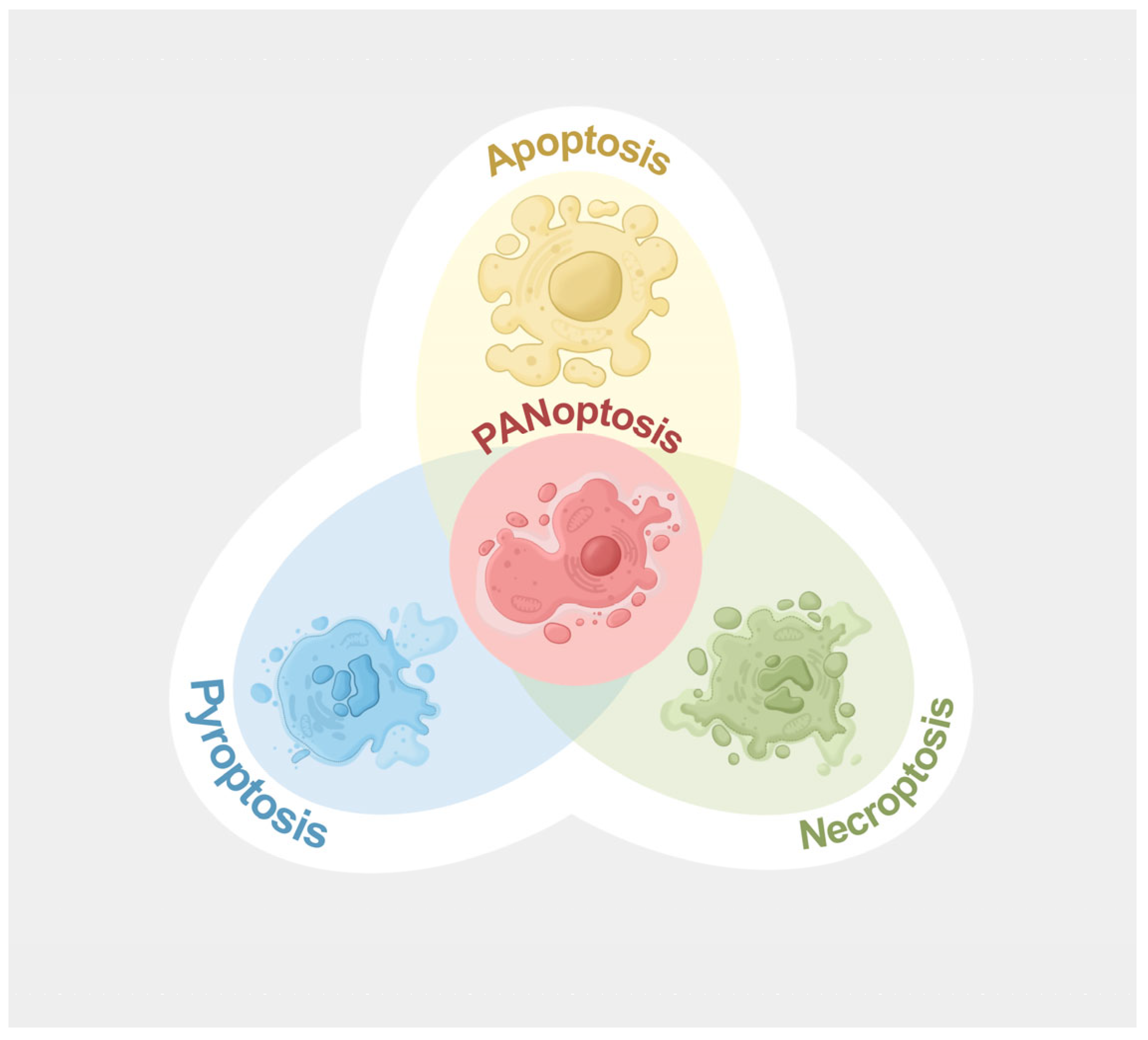
Figure 1. A simplified illustration of the interplay among PANoptosis, apoptosis, pyroptosis, and necroptosis. Figure created using Procreate v5.4.7.
Microorganisms 13 02560 g001
Figure 2. Molecular processes of PANoptosis. Diverse stimuli, such as viruses, bacteria, and heme plus PAMPs, lead to upregulation and/or activation of key upstream regulators of the PANoptosome (e.g., IRF1, caspase-8, and -6). These regulators then mediate the assembly of the ZBP-1 PANoptosome (ZBP-1, RIPK1, caspase-8, NLRP3, RIPK3, caspase-1, and ASC), AIM2 PANoptosome (AIM2, ZBP-1, Pyrin, RIPK1, caspase-8, RIPK3, caspase-1, ASC, and FADD), RIPK1 PANoptosome (RIPK1, caspase-8, NLRP3, caspase-1, RIPK3, ASC, and FADD), or NLRP12 PANoptosome (NLRP12, RIPK3, caspase-8, and ASC). The downstream effectors are subsequently activated, thereby contributing to plasma membrane pore formation and PANoptosis. Apoptosis-, pyroptosis-, and necroptosis-associated molecules are shown in red, green, and blue, respectively.
Microorganisms 13 02560 g002
Figure 3. Intrinsic and extrinsic mechanisms of apoptosis. In intrinsic apoptosis, activation of BH3-only proteins facilitates the formation and oligomerization of BAK and BAX, leading to the release of cytochrome c, apoptosome assembly, and apoptosis. In extrinsic apoptosis, extracellular stimuli activate the death receptors, contributing to death-inducing signaling complex formation, procaspase-8 activation, and effector caspase activation.
Microorganisms 13 02560 g003
Figure 4. Canonical and non-canonical pyroptosis pathways. In canonical pyroptosis, PAMPs and DAMPs promote inflammasome formation and caspase-1 activation. Activated caspase-1 cleaves GSDMD into N-GSDMD, perforating the plasma membrane and causing pyroptosis. In the non-canonical pathway, caspase-4, -5, and -11 undergo activation through binding with LPS, and these caspases subsequently cleave GSDMD for pore formation and pyroptosis. Meanwhile, active caspase-1, as well as active caspase-4, -5, and -11, contribute to the maturation of IL-1β and IL-18 directly or indirectly.
Microorganisms 13 02560 g004
Figure 5. Necroptosis signaling pathway. Activation of multiple receptors or intrinsic stimuli causes RIPK1 and RIPK3 phosphorylation and necrosome assembly (phosphorylated RIPK1 and RIPK3, FADD, and inactive caspase-8). Thereafter, MLKL undergoes phosphorylation and migrates to the plasma membrane, where it oligomerizes to drive pore formation and necroptosis. In the meantime, cell contents and DAMPs are released through pores to provoke inflammation.
Microorganisms 13 02560 g005
Figure 6. Molecular processes of ferroptosis. Iron accumulation (e.g., RSL3, increased transferrin expression, and declined ferroportin expression) and lipid peroxidation are two major contributors to ferroptosis. Lipid peroxidation occurs via the non-enzymatic (Fenton reaction) or lipoxygenase-mediated enzymatic pathway, resulting in membrane rupture and ferroptosis. Inhibition of GPX4 or CoQ10 also elicits ferroptosis.
Microorganisms 13 02560 g006
Table 1. Potential pharmacological compounds and small molecules targeting P. aeruginosa-induced PANoptosis.
Compound/
Small Molecule
Target
and Mechanism
Disease/ConditionOutcomeRefs
I-2IRF1
Forms hydrogen bonds and hydrophobic contacts with IRF; reduces IRF1 transcriptional activity
Radiation-induced inflammatory cell death and tissue injuryInhibits cleavage of PANoptotic molecules and mitigates cell death and tissue injury[138]
I-19IRF1
Targets DNA-binding domain of IRF1; reduces IRF1 transcriptional activity
Radiation-induced inflammatory cell death and tissue injuryInhibits cleavage of PANoptotic molecules and mitigates cell death and tissue injury[138]
Necrostatin-1caspase-8
Decreases caspase-8 cleavage
Cell death induced by P. aeruginosa-derived 3-oxo-C12-HSLSuppresses cell death in 3-oxo-C12-HSL-treated cells[53]
miR-29a-3pcaspase-8 and ZBP-1
Inhibits caspase-8 activation and ZBP-1 expression
Acute lung injuryReduces alveolar epithelial cell PANoptosis and alleviates disease severity[139]
Z-IETD-FMKcaspase-8
Inhibits caspase-8 cleavage
Bacterial peritonitis, pneumonia, and endotoxin shockEnhances neutrophil antimicrobial activity in the absence of increased cell death[140]
Z-VEID-FMKcaspase-6
Selectively and irreversibly inhibits caspase-6
Optic nerve injuryReduces apoptotic cell death and promotes cell survival[141]
ADAR1ZBP-1
Zα domain of ADAR1 limits ZBP1 activation
Embryonic lethality, intestinal cell death, and skin inflammationPrevents ZBP1-induced cell death and autoinflammation[142]
Cucurbitacin ECDK1/ZBP-1
Inhibits CDK1, a protein mediating PANoptosis via interaction with ZBP-1 PANoptosome
Adrenocortical carcinomaRegulates PANoptosis in cancer cells[143]
IFI16-βAIM2
Impairs AIM2-ASC interaction and AIM2-dsDNA sensing
Bacterial and viral infectionSuppresses AIM2 inflammasome activation and pro-inflammatory cytokine release[144]
J114AIM2
Disturbs AIM2-ASC interaction
AIM2-dependent inflammationReduces PANoptotic molecule expression and pro-inflammatory cytokine release[145]
4-Sulfonic calix[6]arene/suraminAIM2
Blocks DNA-binding HIN domain of AIM2
Post-stroke immunosuppressionPrevents AIM2 activation and AIM2-dependent cell death[146]
6E11RIPK1
Competes with RIPK1-ligand complexes
TNF-α-induced cell death and cold hypoxia/reoxygenation injuryProtects against RIPK1-dependent cell death[147]
ZB-R-55RIPK1
Occupies allosteric and ATP binding pockets of RIPK1
Sepsis and systemic inflammatory response syndromeAttenuates systemic inflammation and disease severity[148]
RIPA-56RIPK1
Inhibits RIPK1 phosphorylation and its kinase activity
Systemic inflammatory response syndromePrevents multiorgan damage and reduces mortality[149]
Spirodalesol analog 8AASC
Suppresses ASC speck formation and oligomerization
Endotoxemia, peritonitis, and gouty arthritisReduces mortality and multiorgan damage[150]
Tetracyclinecaspase-1
Reduces cleavage and activation of caspase-1
Acute lung injuryAttenuates lung injury and pulmonary inflammation; improves survival[151]
Unsaturated ester derivative-compound 9caspase-1
Blocks caspase-1 activity
Caspase-1-dependent cell deathProtects against caspase-1-dependent cell death[152]
Compound 42 (an analog of TAK-632)RIPK3
Inhibits RIPK3 phosphorylation and blocks necrosome formation
Systemic inflammatory response syndromeAlleviates disease symptoms and improves survival[153]
AZD5423RIPK3
Binds to the kinase domain of RIPK3 and decreases its activation
RIPK3-mediated cell injury and acute kidney injuryRestores cell viability and attenuates kidney injury and inflammation[154]
Necrosulfonamide (NSA)GSDMD
Binds to the Cys191 of GSDMD
MLKL
Binds to the Cys86 of human MLKL; inhibits MLKL phosphorylation
LPS-induced endotoxicity and sepsisPrevents cell death and reduces mortality[155]
Acute colitisAlleviates intestinal inflammation[38,157]
DisulfiramGSDMD
Modifies the Cys191 of human GSDMD
AIM2
Mechanism unknown
SepsisImproves survival and attenuates systemic inflammation[156]
SaracatinibMLKL
Disrupts the phosphorylation, translocation, and oligomerization of MLKL
Psoriasiform dermatitisAttenuates skin inflammation[159]
Disclaimer/Publisher’s Note: The statements, opinions and data contained in all publications are solely those of the individual author(s) and contributor(s) and not of MDPI and/or the editor(s). MDPI and/or the editor(s) disclaim responsibility for any injury to people or property resulting from any ideas, methods, instructions or products referred to in the content.