Article

Exploring Metal Ions as Potential Antimicrobial Agents to Combat Future Drug Resistance in Mycoplasma bovis

by
Mauida F. Hasoon Alkhallawi
*,
Majed H. Mohammed
,
Farhid Hemmatzadeh
and
Kiro Petrovski
*
Australian Center for Antimicrobial Resistance Ecology, School of Animal & Veterinary Sciences, The University of Adelaide, Rose Worthy Campus, Mudla Wirra Rd., Roseworthy, SA 5371, Australia
*
Authors to whom correspondence should be addressed.
Microorganisms 2025, 13(1), 169; https://doi.org/10.3390/microorganisms13010169
Submission received: 26 November 2024 / Revised: 30 December 2024 / Accepted: 9 January 2025 / Published: 15 January 2025
(This article belongs to the Special Issue Antimicrobial Testing (AMT), Third Edition)

Abstract

:
The rise in antimicrobial resistance (AMR) in Mycoplasma bovis underscores the urgent need for alternative treatments. This study evaluated the minimal inhibitory concentrations (MICs) of four metal ions (cobalt, copper, silver, and zinc) and colloidal silver against 15 clinical M. bovis isolates, alongside conventional antimicrobials (florfenicol, tetracycline, tulathromycin, and tylosin). Colloidal silver demonstrated the most effective antimicrobial activity, inhibiting 81.25% of isolates at 1.5 mg/L, while silver inhibited 93.7% of isolates at concentrations above 1.5 mg/L. Copper exhibited notable efficacy, inhibiting 37.5% of isolates at 1.5 mg/L, with a small proportion responding at 0.1 mg/L. Cobalt and zinc displayed variable activity, with MIC values ranging from 0.7 to 12.5 mg/L. In contrast, conventional antimicrobials showed limited effectiveness: tetracycline inhibited 31.25% of isolates at ≥16 mg/L, tylosin inhibited 25% at 16 mg/L, and tulathromycin MICs ranged from 0.5 to 8 mg/L. Time–kill assays revealed a reduction in M. bovis viability after eight hours of exposure to silver and colloidal silver, though higher concentrations (4×–8× MIC) were required for complete eradication. These findings highlight the significant potential of colloidal silver and copper as alternatives for treating M. bovis infections and combating AMR. Further research is essential to explore their standalone and synergistic applications for therapeutic use.

1. Introduction

Mycoplasma bovis is a significant veterinary medicine pathogen, posing considerable challenges to livestock health worldwide. Belonging to the class Mollicutes, these bacteria lack a cell wall, rendering them resistant to conventional antimicrobials targeting cell wall synthesis (e.g., beta-lactams). As a result, M. bovis infections often present a treatment challenge, leading to substantial economic losses in affected livestock populations [1]. In this introduction, we briefly delve into the background of M. bovis, its pathogenicity, diagnostic challenges, and the implications it holds for veterinary medicine [2]. Not only is the interaction of M. bovis with host cells a factor of its pathogenicity, but also it influences its response to antimicrobials. In the absence of an effective vaccine for the major bovine respiratory disease (BRD) bacterial pathogens, including M. bovis, antimicrobial therapy remains the main treatment [3]. Conventional antimicrobials such as phenicols, tetracyclines, and macrolides have been the cornerstone in treating M. bovis infections. These antimicrobials typically target protein synthesis and other bacterial biosynthetic pathways [4]. Antimicrobial resistance (AMR) occurs when pathogens no longer respond to medicines, making infections difficult to treat and increasing the risk of disease spread, severe illness, and death [5]. Traditional antimicrobials have been the mainstay of managing bacterial infections in both human and veterinary medicine. However, the overuse and misuse of these drugs in humans, animals, and agriculture are assumed to have accelerated the development of resistant strains. For M. bovis, this means that some of the standard treatments are becoming less effective, necessitating higher doses or the use of alternative, often more expensive or less-understood antimicrobials [1]. This not only increases the cost of treatment but also poses a risk of undesirable side effects in animals and potentially humans through the consumption of their products.
The resistance mechanisms are often linked to genetic mutations that alter drug targets or enhance efflux pumps, further diminishing the effectiveness of conventional treatments [6]. The development of resistance in BRD pathogens, such as M. bovis, is a major reason for poor response to treatment and is a growing issue worldwide [1,6]. With the development and spread of AMR, there are higher chances of treatment failure, leading to prolonged outbreaks and greater economic losses. The ability of M. bovis to develop resistance complicates the management of herd health and necessitates more stringent biosecurity and management measures [7]. Therefore, the emerging AMR in M. bovis isolates requires the exploration of alternative antimicrobial actives, including but not limited to metals. Some metals have been reported as promising agents in combating bacterial infections, especially silver (Ag), copper (Cu), iron (Fe), and zinc (Zn). They exhibit potent antibacterial properties by disrupting various cellular processes essential for bacterial survival and replication. The mechanisms underlying the antibacterial action of these metal ions are diverse and multifaceted, often involving the generation of reactive oxygen species (ROS), disruption of cell membranes, interference with protein and DNA functions, and modulation of metal ion homeostasis [8,9].
The mechanisms of copper antimicrobial activity are related to ion (Cu++) toxicity, leading to compromised structural integrity of cellular components, including disruption of cellular membrane integrity, disruption of microbial metabolic pathways, and activation of microbial stress responses. Compromised structural integrity of cellular components, related to copper exposure, may occur due to the generation of ROS. This leads to oxidative damage of cellular lipids, nucleic acids, and proteins, ultimately resulting in cell death [10,11,12]. The disruption of microbial cell membranes leads to increased permeability and leakage of cellular contents [13]. Disruption of microbial metabolic pathways may occur through copper binding to active sites or cofactors in essential enzymatic processes. Finally, microbial stress responses resulting from exposure to copper lead to the upregulation of defense mechanisms that can damage bacterial cells with prolonged stress, ultimately causing cell death [14].
Silver is the metal with the greatest association with antimicrobial activity, both historically and currently [15]. Various medical products include silver, such as bandages, ointments, and catheters [16]. Silver nanoparticles and colloidal silver (an alloy with the antimicrobial sulfadiazine) are commonly used [17]. The action of silver ions (Ag+) at the microbial cellular level and their potential as an alternative to conventional antimicrobials are critical areas of interest, especially in the face of rising AMR [16]. Despite the different forms, ions are generally believed to be the active component, and the antibacterial activity varies with size, shape, and surface characteristics (including surface coatings) of the silver. However, these differences are likely due to changes in the release kinetics of Ag+ and not because of the particles themselves [18]. The mechanisms of Ag⁺ toxicity involve compromising the structural integrity of cellular components, disrupting the cell membrane, and interfering with microbial metabolic pathways [19,20]. Compromised structural integrity of cellular components, related to silver exposure, may occur due to the high affinity to sulphur-containing compounds in the bacterial cells, particularly thiol groups in sulphur-containing amino acids. This results in damage to nucleic acids and proteins, ultimately resulting in cell death [21] or in the prevention of bacterial replication. The disruption of the microbial cell membranes leads to increased permeability and leakage of cellular content and/or penetration of environmental materials, ultimately leading to bacterial death [21,22].
Cobalt and zinc ions have also re-emerged as promising candidate agents for use as antimicrobials. The antimicrobial properties are related to the toxicity of ions (Co++ or Zn++), leading to disruption of cellular membrane integrity and disruption of bacterial metabolic pathways, and triggering a microbial stress response [21]. Additionally, cobalt and/or zinc ions, when combined with conventional antimicrobials, may boost effectiveness through increased uptake, potentiation of activity, or another synergism [8,23]. Cobalt is also a crucial component of vitamin B12, playing a vital role in bacterial metabolism. Synthetic analogs of vitamin B12, containing cobalt, may disrupt bacterial metabolic pathways [24].
The exploration of these metal ions as potential alternatives to antimicrobials is not without its challenges. Concerns over toxicity, environmental impact, and the development of resistance necessitate careful considerations [25]. Copper-based compounds, for example, have demonstrated low toxicity in in vivo models like Galleria mellonella larvae when injected directly into the haemocoel [25]. Similarly, silver compounds, while exhibiting low toxicity in controlled doses, present risks of acute and chronic adverse effects, such as gastrointestinal irritation and argyria, when overexposed. Therefore, due to toxicity concerns, their primary use is in topical formulations rather than systemic applications, such as silver sulfadiazine [25,26]. Additionally, the environmental impact of heavy metals should not be ignored.
Recent studies [27,28] have suggested that efflux pump mechanisms play a crucial role in bacterial responses to metal ions by regulating intracellular concentrations and mitigating their antimicrobial effects. Overexpression or mutations in efflux pump genes can enhance the expulsion of metal ions, potentially reducing their efficacy as antimicrobial agents. However, these resistance mechanisms vary significantly among bacterial species and isolates [29]. However, the potential benefits, including the reduced likelihood of resistance development and their broad-spectrum activity, make the pursuit of understanding and harnessing these metal ions as antimicrobials a compelling area of study [8].
The present study aimed to explore the efficacy of metal ions in inhibiting the in vitro growth of clinical isolates of M. bovis that have failed the conventional treatment, in comparison to conventional antimicrobials, by determining the minimum inhibitory concentrations (MICs) of various metal ions and time–kill kinetics assays. This comparative analysis not only highlights the antimicrobial potential of metal ions but also provides a foundation for future investigations into their mechanisms of action and application in livestock management practices.

2. Materials and Methods

2.1. Mycoplasma Isolates (Identification and Culture)

A total of 15 M. bovis isolates were obtained from government and private veterinary service laboratories in Australia and archived at the School of Animal and Veterinary Science, The University of Adelaide. The isolates were collected from cattle showing signs of the BRD complex and were either treated (n = 5), euthanized (n = 4), or had prolonged exposure to tetracycline-based metaphylactic treatment (n = 6) from 2021 to 2023. The identity of the isolates was confirmed by polymerase chain reaction (PCR) using specific primers for the Deoxy-ribodipyrimidine photolyase (uvrC) gene at F: 5′ AAG TTG AAG TTG ACC GGT TTG 3′ and R: 5′ TCC ATA TTT GGA CCT AGT CCT TT 3′ [30]. To confirm the identification, the PCR products were sent for sequencing (AGRF—https://www.agrf.org.au accessed on 9 December 2024).
Mycoplasma bovis Type Strain PG45 (ATCC 25523) was used as a reference strain for quality control assessment. The isolates (including the reference type strain) were cultured using Eaton broth/agar, prepared in-house with PPLO broth (Thermo Fisher Scientific, Melbourne, Australia) supplemented with 1% (w/v) yeast extract, glucose, sodium pyruvate, and 20% (v/v) horse serum (Sigma-Aldrich, Melbourne, Australia). Broths were incubated at 37 °C in a 5% CO2 atmosphere for 3–5 days and then plated to inspect for the typical fried-egg colony morphology of Mycoplasma. Before testing, each cultured broth was diluted to a known concentration (105 to 106 CFU/mL) following standard laboratory procedures for veterinary mollicutes [31].

2.2. Antimicrobial Susceptibility Testing

The methodology for determining the minimum inhibitory concentrations (MICs) of conventional antimicrobials and metal ions involved a modified broth microdilution method adapted from the standards recommended by the Clinical and Laboratory Standards Institute (CLSI, [32]) and adjustments proposed by Hasoon et al. [6]. Initially, Mycoplasma suspensions were prepared in Eaton broth, with the optical density adjusted to 0.08–0.12 at OD 600 nm using Spectrophotometer [Eppendorf BioPhotometer®, Hamburg, Germany]. These suspensions were then further diluted to achieve a final concentration of approximately 106 CFU/mL in each well of sterile 96-well polystyrene microtiter plates (Nunclon™; Thermo Fisher Scientific, Australia), with 50 µL of Eaton medium dispensed into each well. Metal powders were dissolved in 2% nitric acid and further diluted in Eaton broth to achieve the desired concentrations for stock solution preparation. The stock solutions of conventional antimicrobials (florfenicol, tetracycline, tulathromycin, and tylosin) and metals or the alloy (cobalt, copper, silver, zinc, and colloidal silver; metal hereafter) were prepared at a 10X concentration and stored at −80 °C until use. The antimicrobials were tested at concentrations ranging from 0.25 to 128 mg/L using two-fold serial dilutions, while metal ions were tested at concentration ranges between 0.1 mg/L to 100 mg/L. Each microtiter plate was set up by adding serial dilutions of each tested agent (antimicrobial or metal), followed by 50 µL of the Mycoplasma inoculum (106 CFU/mL) to each well. To validate the assay’s accuracy, the plates included both a positive control medium (without antimicrobials or metals) and a negative control medium (without Mycoplasma suspension). The assay plates were sealed and incubated until complete growth was seen in positive control wells (~3 days) at 37 °C in a 5% CO2 environment. To ensure the reliability and reproducibility of results, all tests were conducted in triplicates.

Interpretation of the MIC Testing

MIC values were determined as the lowest concentration of the antimicrobial agent that completely inhibited visible growth. The MIC50 and MIC90 values (concentrations inhibiting 50% and 90% of isolates, respectively) were calculated. The reference type strain (PG45) was used to standardize the work by comparing our MICs with values reported earlier [6].
Due to the lack of breakpoints for veterinary Mycoplasma spp., interpretation as susceptible, intermediate, or resistant was not possible. Therefore, the MIC results for the three antimicrobial groups (macrolides, tetracyclines, and phenicol), and five metal agents (cobalt, copper, silver, colloidal silver, and zinc) tested in this study were interpreted based on MICs for the M. bovis reference type strain PG45 (ATCC® 25523).

2.3. Metal Solvent Toxicity Testing

To ensure the 2% nitric acid used in metal stock solution preparation did not affect Mycoplasma growth, a separate toxicity test was conducted. Broths with and without nitric acid were incubated and examined for growth over 5–7 days at 37 °C in 5% CO2.

2.4. Time–Kill Kinetics Assay of Metal Ions

Time–kill kinetics assays for Co, Cu, Ag, Zn, and colloidal silver were carried out as previously described [33]. Briefly, selected Mycoplasma strains (those that exhibited high MIC to florfenicol, tetracycline, tulathromycin, and tylosin), together with PG45 ATCC strain, were prepared at an inoculum size of 1 × 10⁶ CFU/mL in a sterile Eaton broth medium, and seeded in 96-multiwell plates, followed by the addition of successive concentrations of each metal ion MIC (0.5× MIC, 1× MIC, 2× MIC, and 4× MIC), and then incubated (37 °C, 5% CO2). A total of 0.1 mL of cultures was taken at time intervals of 0, 8, 24, 48, and 72 h and spread onto Eaton agar plates and incubated at 37 °C for 48–72 h. The viable M. bovis cells were counted as CFU/mL, and data were reported for curve plotting. Each time–kill curve showed the percentage of Mycoplasma surviving over the time of development.

2.5. Statistical Analysis

To compare the inhibition percentages of different agents (antimicrobials and metals) against M. bovis, and to assess the time to kill Mycoplasma exposed to different concentrations (from 0.5 MIC to 8 MIC) of metals, ANOVA followed by Tukey’s HSD test was carried out in SAS version 9.4.

3. Results and Discussion

The current study aimed to assess the susceptibility of 15 Australian M. bovis isolates obtained from feedlot BRD cases to four antimicrobials (belonging to three different groups: macrolides, phenicol, and tetracyclines) and enable comparison with five metals (Ag, Cu, Co, Zn, and colloidal silver).
There is no certain specification of treatments considered yet for bovine mycoplasmas. However, The Australian Strategic and Technical Advisory Group on Antimicrobial Resistance (ASTAG) has developed a specific rating system for antimicrobials used in both human and veterinary settings according to their clinical importance, with the majority of antimicrobials registered for the treatment of BRD (amphenicols, macrolides, and tetracyclines) given a low importance rating [6]. Following the development of an antimicrobial stewardship program for Australian feedlots, a recent survey of antimicrobial use has shown an extensive reliance on these low-importance antimicrobials for treating BRD [3].

3.1. Mycoplasma Isolates Selection

Fifteen bovine Mycoplasma isolates were identified as M. bovis through PCR targeting a 106 bp fragment of the deoxy-ribodipyrimidine photolyase gene. Their characteristic fried-egg colony morphology was observed on PPLO agar using stereo microscope (Leica, Beijing, China).
The MIC results for the antimicrobials and metals tested in this study were categorised into “low MICs” and “higher MICs” (Antimicrobials such as florphenicol, tetracyclines, and tylosin showed predominantly higher MIC values, with most isolates exhibiting MICs at or above 4 mg/L. Conversely, colloidal silver demonstrated consistently low MICs, with 81.25% of isolates showing MICs at 1.5 mg/L. This categorization highlights the variability in antimicrobial and metal ion efficacy, offering valuable insights into the susceptibility patterns of Mycoplasma bovis isolates) based on the distribution of MIC values (Table 2 and Table 3).
The MIC data for the conventional antimicrobials and the metal agents evaluated in this study using (PG45) reference strain were consistent throughout the study and within the range of previous studies, except for tulathromycin, which yielded undetectable MIC values (Table 1).
Table 1. Minimum inhibitory concentrations (MICs) and the comparable MIC range tested by others worldwide, as well as the quality control Mycoplasma bovis strain PG45, for the tested conventional antimicrobials and metal ions (in mg/L).
Tested AgentsQuality Control Strain
M. bovis (PG45)
Acceptable Range of MICs Tested by Others *
Phenicol (Florphenicol)2.001.00–32.00
Tetracyclines (chlor- and oxy-tetracycline)0.25≤0.12–32.00
Macrolide–tylosin
Macrolide–tulathromycin
1.00
0.50
0.06–128.00
ND
Cobalt3.120.78–12.50
Copper1.560.19–12.50
Silver6.250.19–12.50
Zinc1.561.56–12.50
Colloidal silver *1.560.78–1.56
* References [6,34,35,36].
Table 2. The minimum inhibitory concentration (MIC) distribution percentages obtained for the 15 Mycoplasma bovis isolates against the commonly used conventional antimicrobials.
Tested AntimicrobialsPercentage of Mycoplasma bovis Isolates Showing MIC Values at Tested Antimicrobial Concentrations (mg/L)
0.250.51248163264128
Florphenicol06.256.2518.752543.750000
Tetracyclines6.256.256.2518.75031.25256.2500
Tulathromycin06.2512.5252531.250000
Tylosin006.25037.531.2525000

3.2. In Vitro Susceptibility Testing

The M. bovis susceptibility profile to conventional antimicrobial agents used in this study can be divided as follows:
Florfenicol (a phenicol) and tulathromycin (a macrolide) demonstrated consistently low MICs against M. bovis, ranging from 0.5 to 8 mg/L. Both antimicrobials exhibited uniform efficacy. These results are consistent with prior studies, where similar MIC ranges were reported for the PG45 reference strain (Table 1) [6,34,35]. The findings suggest that florfenicol and tulathromycin remain largely effective in inhibiting M. bovis at relatively low concentrations, highlighting their continued therapeutic potential.
Higher MICs for tylosin and tetracycline were observed. A tylosin MIC of 16 mg/L was applicable to 25% of the isolates. A small proportion of isolates (6.25%) displayed a high MIC of 32 mg/L to tetracyclines (Table 2). Tetracyclines, particularly oxytetracycline, have been among the most widely used antibiotics, especially for long-term, sequential treatments [37]. This extensive use exerts considerable selective pressure, probably contributing to the development of clinical resistant strains over time. Additional research is needed to confirm these hypotheses while also accounting for environmental influences. A consistent growth was observed in all positive control wells. Nitric acid (solvent) toxicity testing showed no effect on the growth rate of Mycoplasma after 24, 48, and 72 hrs of incubation at 37 °C and 5% CO2.
The antimicrobial susceptibility of M. bovis to conventional antimicrobials showed higher MIC values compared to those reported in a previous Australian study [6]. While the overuse or inappropriate use of antimicrobials in the livestock industry is often suggested as a main factor leading to changes in AMR patterns [38,39], this hypothesis has yet to be proven, particularly when the tested antimicrobials are used in the feedlots where the samples originated.
The metals’ MICs were variable (Table 3). Copper was able to inhibit 6.25% of the isolates at a concentration of 0.1 mg/L, while silver and zinc showed no inhibitory effect at this concentration. Increasing the concentration of copper to 1.5 mg/L caused a 37.5% inhibition of M. bovis isolates, and cobalt started showing an effect, inhibiting 12.5% of the isolates. A high percentage (81.25%) of M. bovis isolates were inhibited by colloidal silver at a concentration of 1.56 mg/L, while the zinc MIC ranged from 6.2 to 12.5 mg/L. No Mycoplasma growth was detected in the microwells at ≥25 mg/L for any of the five tested metals. The MIC50 and MIC90 were 1.5 mg/L for colloidal silver, or 3 and 6.2 mg/L for copper, and 6.2 and 12.5 mg/L for cobalt, silver, and zinc, respectively.
Table 3. The minimum inhibitory concentration (MIC) distribution percentages obtained for the 15 examined Mycoplasma bovis isolates against the tested metals.
Tested Metal AgentPercentage of Mycoplasma bovis Isolates Showing MIC Values at the Tested Metal Ion Concentrations (mg/L)
0.10.30.71.53.16.212.52550100
Cobalt0012.506.2556.2525000
Copper6.250037.518.7531.256.25000
Silver6.250006.2543.7543.75000
Colloidal silver0018.7581.25000000
Zinc00012.52531.2531.25000
In this study, isolates with high MICs (>8 mg/L) to tetracycline and macrolides, but a sensitive response to metals, can be interpreted through several microbiological and genetic mechanisms [39,40]. The observed patterns of MIC values indicated that the regulatory networks conferring AMR do not confer metal resistance. This highlighted the specificity and independence of the AMR mechanisms towards conventional antimicrobials and metals [40,41].
The absence of co-resistance between antimicrobials and metals in this study suggested that selective pressures from conventional antimicrobials had not coincided with or promoted resistance to metals. This may have resulted from different environmental exposures (like the isolates collected from treated cattle) or selective pressures in the bacteria’s natural or clinical settings (for the isolates collected after long exposure to metaphylactic treatment). Unfortunately, these hypotheses could not be confirmed due to a lack of information on the origin and management practices. This differential sensitivity profile is crucial for therapeutic strategies, suggesting that metal-based treatments might be an effective future alternative against M. bovis strains resistant to conventional antimicrobials. However, the use of metals in clinical settings must be carefully considered due to their potential toxicity and environmental impacts [39,40].
Low MICs to metals in the current study could be due to an absence or low efficiency of metal efflux systems, which suggests that M. bovis might lack effective resistance mechanisms against the cytotoxic effects of these ions [41,42]. It is also possible that the metabolic pathways or structural components of Mycoplasma are particularly vulnerable to disruption by metals, leading to cell damage or death even at lower concentrations [43]. The antimicrobial activity of metals against bacteria can be influenced by various factors, including the specific bacterial strain, growth conditions, and the experimental setup. Additionally, the mechanisms of metal interactions are complex and may involve multiple factors such as interference with efflux pumps, modulation of membrane permeability, or direct binding to bacterial components. For instance, copper and zinc have been shown to potentiate the activity of antibiotics by inhibiting efflux pumps and disrupting bacterial membranes [44]. Finally, some of these antibacterial properties may also be harmful to eukaryotic cells, and this should be an area of further research. To the best of our knowledge, this is the first time MICs and time–kill kinetics have been assessed for these metals against M. bovis [45]. Overall, the isolates tested in this study showed higher MIC90 values for conventional antimicrobials compared to the recent Australian study [6].

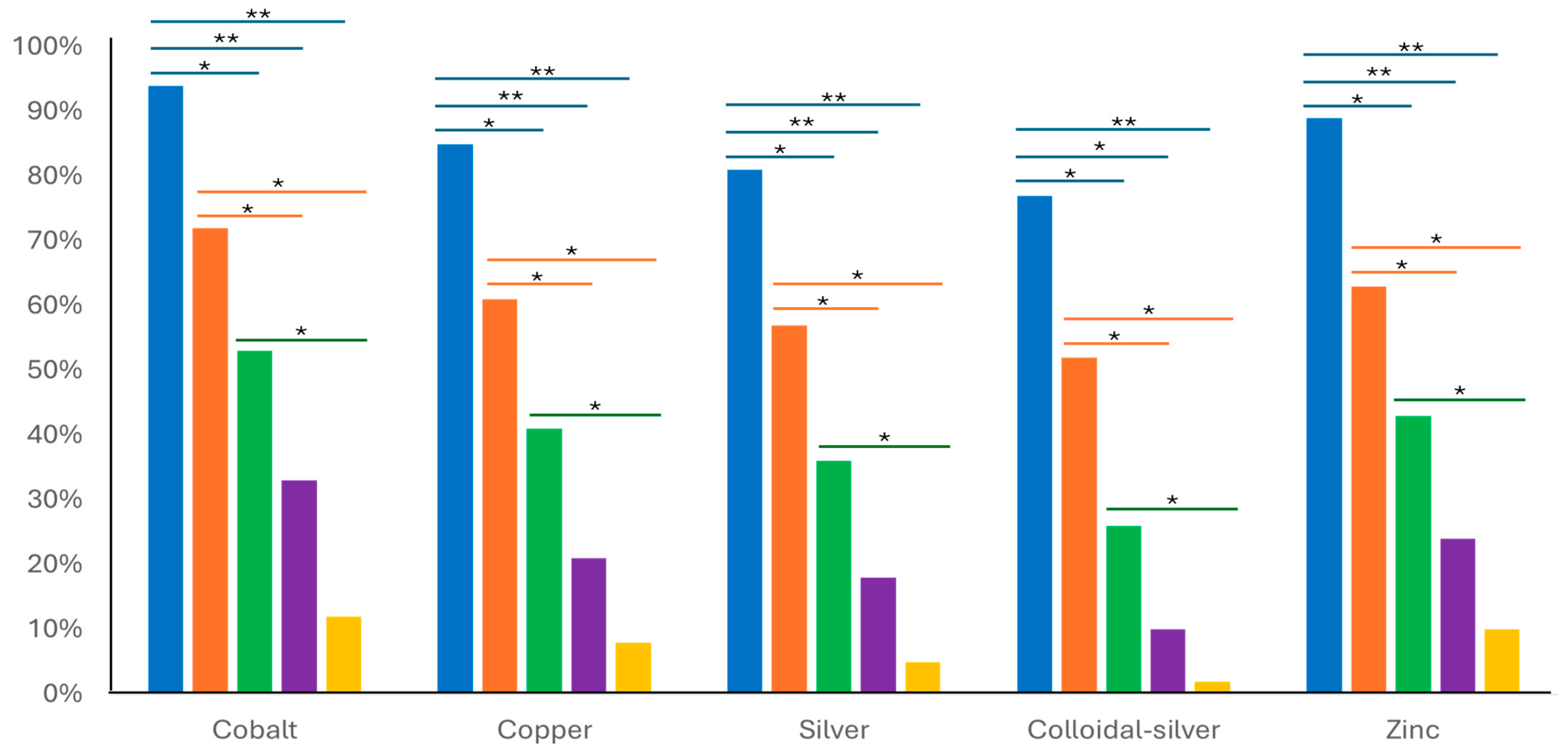
3.3. Time–Kill Kinetic Assay of Metal Ions

The time–kill measured across different points (0 h, 8 h, 24 h, 48 h, and 72 h) showed a reduction in M. bovis when treated with different ascending concentrations (0.5× MIC, 1× MIC, 2× MIC, 4× MIC, and 8× MIC) of metals (Figure 1). For example, cobalt and zinc demonstrated a pronounced bactericidal effect on M. bovis at 2× MIC, with the viable count being reduced by 50%, increasing to 73% at 4× MIC, and achieving an 88% reduction at 8× MIC within 24 h or longer. Silver and its colloidal alloy showed good effectiveness at 2× MIC, achieving a reduction of 63%, which increased to 73% and almost 90% at 4× MIC after 48 hrs. The bactericidal endpoint, where no viable M. bovis cells could be detected, was reached after 72 h of incubation at 8× MIC (12 mg/L) for colloidal silver. In contrast, cobalt and zinc did not consistently reach these bactericidal endpoints within the same time frame (Figure 1).
Our results showed that metals of colloidal silver and copper showed the most promising antimicrobial properties (Table 2). Similarly, the time–kill kinetics of colloidal silver and copper were indicative of the highest efficacy against M. bovis. The time–kill kinetics revealed a combination of concentration-dependent and time-dependant activity for all metals. Overall, the current study suggests that a metal may have some potential in the management of M. bovis, but further work is required before a definitive conclusion can be drawn. Interestingly, strains resistant to conventional antimicrobials (e.g., tetracyclines) had no resistance to metals, particularly colloidal silver.
The time–kill curves demonstrated that the bactericidal effects of metals are concentration- and time-dependent. Higher concentrations of metal ions generally resulted in faster and more complete inhibition of M. bovis growth. Colloidal silver exhibited the highest bactericidal effect, achieving complete inhibition at lower concentrations (4× MIC) and at the earliest time points (24 h) compared to other metals. Cobalt and copper also showed strong inhibition, particularly at higher concentrations. These observations suggest that while silver and copper are highly effective in reducing the viability of M. bovis over a shorter duration, the effectiveness of cobalt and zinc, particularly at higher concentrations, may be influenced by factors not captured in this dataset [46].
The p-value from the ANOVA was 0.05, indicating no difference in the inhibition effects of antimicrobials or metals. The results of the current study supported the hypothesis that the differential resistance profiles of conventional antimicrobials and metals highlight distinct microbiological adaptations within Mycoplasma. Tetracycline and macrolides, which inhibit protein synthesis by targeting ribosomal subunits, often encounter resistance through mechanisms like efflux pumps, ribosomal protection proteins, and less commonly, enzymatic inactivation [47].
This study has some limitations. One important limitation is the relatively small sample size. However, despite the study’s small sample size, the reliability of the findings was supported by the consistency in susceptibility of the reference strain, and the close alignment with the existing literature [48]. Another limitation is the lack of interpretive criteria for metals and some antimicrobials for M. bovis. Overall, metals showed similar or higher MIC90 compared to the tested conventional antimicrobials. Therefore, these concentrations are likely achievable in the biological medium of the host. However, as we have not carried out tests for the transition from viable to viable-but-not-culturable status, like most other studies, we cannot confirm that the M. bovis isolates did not undergo such a change.
The exploration of in vitro metal antimicrobial activity against veterinary Mycoplasma is an essential step in understanding the potential usefulness of these metals and/or their alloys in the treatment of infections caused by these pathogens. Despite the advantages of metals, metal-based compounds face challenges, including limited research on their pharmacological behavior and toxicity thresholds. High dosages of metal ions may exacerbate in vivo toxicity, as mammalian cells share similar targets with bacteria. Moreover, metals not naturally occurring as trace elements in the human body may pose toxicity risks. Finally, the potential environmental impact of heavy metal ions was not considered in this study. Addressing these concerns requires further in vivo and clinical studies to determine safe and effective routes of administration while ensuring their therapeutic potential [26,49,50].
Further in vitro and in vivo clinical studies are necessary to evaluate metal ions’ safety, optimal dosage, and therapeutic potential in animals and humans. To ensure effective and safe use, this additional research should determine their pharmacokinetics, toxicity thresholds, and practical applications.

4. Conclusions

Our results demonstrated that colloidal silver and copper exhibited inhibitory effects on tested M. bovis strains, with colloidal silver showing notable potential against tetracycline-resistant strains. While some metal ions displayed higher inhibition rates, the variability in responses limited their significance, except in time–kill kinetics assays. Despite this, the observed biological significance highlights the potential of these agents as effective alternatives in combating antimicrobial resistance. Integrating metal-based compounds into existing treatment strategies could provide promising options for veterinary medicine. However, further comprehensive research is needed to fully explore their therapeutic potential and address the broader challenges of antimicrobial resistance.

Author Contributions

Conceptualization, K.P. and M.F.H.A.; methodology, M.F.H.A.; validation, M.F.H.A. and M.H.M.; formal analysis, K.P.; investigation, M.F.H.A.; resources, F.H. and K.P.; writing—original draft preparation, M.F.H.A. and M.H.M.; writing—review and editing, K.P.; visualization; F.H.; supervision, K.P. and F.H.; funding acquisition, K.P. All authors have read and agreed to the published version of the manuscript.

Funding

This research received no external funding.

Institutional Review Board Statement

Not applicable.

Informed Consent Statement

Not applicable.

Data Availability Statement

A total of 15 isolates collected either from lung samples (swab, fresh tissue, or both) were obtained during 2021–2023 from post-mortem BRD-affected cattle in different states in Australia: Queensland (QLD), New South Wales (NSW), and South Australia (SA). Further details are provided in our previous publication 5 (Hasoon et al., 2023 [6]). These isolates were obtained from animals that had been previously treated with antimicrobials but failed to respond.

Acknowledgments

The authors acknowledge the support of Australian cattle feedlots and their veterinarians for supporting this research with the provision of samples. We also thank all lab technicians involved in the isolation, identification, and storage of Mycoplasma. Roy Kirkwood is thanked for the English editing.

Conflicts of Interest

The authors declare no conflicts of interest.

References

  1. Lysnyansky, I.; Ayling, R.D. Mycoplasma bovis: Mechanisms of Resistance and Trends in Antimicrobial Susceptibility. Front. Microbiol. 2016, 7, 595. [Google Scholar] [CrossRef] [PubMed]
  2. Rottem, S. Interaction of mycoplasmas with host cells. Physiol. Rev. 2003, 83, 417–432. [Google Scholar] [CrossRef] [PubMed]
  3. Badger, S.M.; Sullivan, K.F.; Jordan, D.; Caraguel, C.G.B.; Page, S.W.; Cusack, P.M.V.; Frith, D.; Trott, D.J. Antimicrobial use and stewardship practices on Australian beef feedlots. Aust. Vet. J. 2020, 98, 37–47. [Google Scholar] [CrossRef] [PubMed]
  4. Sulyok, K.M.; Kreizinger, Z.; Fekete, L.; Hrivnák, V.; Magyar, T.; Jánosi, S.; Schweitzer, N.; Turcsányi, I.; Makrai, L.; Erdélyi, K.; et al. Antibiotic susceptibility profiles of Mycoplasma bovis strains isolated from cattle in Hungary, Central Europe. BMC Vet. Res. 2014, 10, 256. [Google Scholar] [CrossRef]
  5. Prestinaci, F.; Pezzotti, P.; Pantosti, A. Antimicrobial Resistance: A Global Multifaceted Phenomenon. Pathog. Glob. Health 2015, 109, 309–318. [Google Scholar] [CrossRef]
  6. Hasoon, M.F.; Jarocki, V.M.; Mohammed, M.H.; Djordjevic, S.P.; Yip, H.Y.E.; Carr, M.; Khabiri, A.; Azari, A.A.; Amanollahi, R.; Jozani, R.J.; et al. Antimicrobial Susceptibility and Molecular Characteristics of Mycoplasma bovis Isolated from Cases of Bovine Respiratory Disease in Australian Feedlot Cattle. Vet. Microbiol. 2023, 283, 109779. [Google Scholar] [CrossRef]
  7. Helmy, Y.A.; Taha-Abdelaziz, K.; Hawwas, H.A.E.-H.; Ghosh, S.; AlKafaas, S.S.; Moawad, M.M.M.; Saied, E.M.; Kassem, I.I.; Mawad, A.M.M. Antimicrobial Resistance and Recent Alternatives to Antibiotics for the Control of Bacterial Pathogens with an Emphasis on Foodborne Pathogens. Antibiotics 2023, 12, 274. [Google Scholar] [CrossRef]
  8. Frei, A.; Verderosa, A.D.; Elliott, A.G.; Zuegg, J.; Blaskovich, M.A.T. Metals to Combat Antimicrobial Resistance. Nat. Rev. Chem. 2023, 7, 202–224. [Google Scholar] [CrossRef]
  9. Raja, F.N.S.; Worthington, T.; Martin, R.A. The Antimicrobial Efficacy of Copper, Cobalt, Zinc, and Silver Nanoparticles: Alone and in Combination. Biomed. Mater. 2023, 18, 045003. [Google Scholar] [CrossRef]
  10. Cervantes-Cervantes, M.P.; Calderón-Salinas, J.V.; Albores, A.; Muñoz-Sánchez, J.L. Copper Increases the Damage to DNA and Proteins Caused by Reactive Oxygen Species. Biol. Trace Elem. Res. 2005, 103, 229–248. [Google Scholar] [CrossRef]
  11. Hood, M.I.; Skaar, E.P. Nutritional Immunity: Transition Metals at the Pathogen–Host Interface. Nat. Rev. Microbiol. 2012, 10, 525–537. [Google Scholar] [CrossRef] [PubMed]
  12. Grass, G.; Rensing, C.; Solioz, M. Metallic Copper as an Antimicrobial Surface. Appl. Environ. Microbiol. 2011, 77, 1541–1547. [Google Scholar] [CrossRef] [PubMed]
  13. Ma, M.; Zhao, J.; Zeng, Z.; Wan, D.; Yu, P.; Cheng, D.; Gong, D.; Deng, S. Antibacterial Activity and Membrane-Disrupting Mechanism of Monocaprin against Escherichia coli and Its Application in Apple and Carrot Juices. LWT 2020, 131, 109794. [Google Scholar] [CrossRef]
  14. Gao, L.; Zhang, A. Copper-instigated modulatory cell mortality mechanisms and progress in oncological treatment investigations. Front. Immunol. 2023, 14, 1236063. [Google Scholar] [CrossRef]
  15. Eckhardt, S.; Brunetto, P.S.; Gagnon, J.; Priebe, M.; Giese, B.; Fromm, K.M. Nanobio Silver: Its Interactions with Peptides and Bacteria, and Its Uses in Medicine. Chem. Rev. 2013, 113, 4708–4754. [Google Scholar] [CrossRef]
  16. Sim, W.; Barnard, R.T.; Blaskovich, M.A.T.; Ziora, Z.M. Antimicrobial Silver in Medicinal and Consumer Applications: A Patent Review of the Past Decade (2007–2017). Antibiotics 2018, 7, 93. [Google Scholar] [CrossRef]
  17. Stanford, W.; Rappole, B.W.; Fox, C.L., Jr. Clinical Experience with Silver Sulfadiazine, a New Topical Agent for Control of Pseudomonas Infections in Burns. J. Trauma 1969, 9, 377–388. [Google Scholar] [CrossRef]
  18. Xiu, Z.M.; Zhang, Q.B.; Puppala, H.L.; Colvin, V.L.; Alvarez, P.J. Negligible Particle-Specific Antibacterial Activity of Silver Nanoparticles. Nano Lett. 2012, 12, 4271–4275. [Google Scholar] [CrossRef]
  19. Chen, M.; Yang, Z.; Wu, H.; Pan, X.; Xie, X.; Wu, C. Antimicrobial Activity and the Mechanism of Silver Nanoparticle Thermosensitive Gel. Int. J. Nanomed. 2011, 6, 2873–2877. [Google Scholar] [CrossRef]
  20. Jung, W.K.; Koo, H.C.; Kim, K.W.; Shin, S.; Kim, S.H.; Park, Y.H. Antibacterial Activity and Mechanism of Action of the Silver Ion in Staphylococcus aureus and Escherichia coli. Appl. Environ. Microbiol. 2008, 74, 2171–2178. [Google Scholar] [CrossRef]
  21. More, P.R.; Pandit, S.; De Filippis, A.; Franci, G.; Mijakovic, I.; Galdiero, M. Silver Nanoparticles: Bactericidal and Mechanistic Approach Against Drug-Resistant Pathogens. Microorganisms 2023, 11, 369. [Google Scholar] [CrossRef] [PubMed]
  22. Yin, I.X.; Zhang, J.; Zhao, I.S.; Mei, M.L.; Li, Q.; Chu, C.H. The Antibacterial Mechanism of Silver Nanoparticles and Its Application in Dentistry. Int. J. Nanomed. 2020, 15, 2555–2562. [Google Scholar] [CrossRef] [PubMed]
  23. Cuajungco, M.P.; Ramirez, M.S.; Tolmasky, M.E. Zinc: Multidimensional effects on living organisms. Biomedicines 2021, 9, 208. [Google Scholar] [CrossRef] [PubMed]
  24. González-Montaña, J.R.; Escalera-Valente, F.; Alonso, A.J.; Lomillos, J.M.; Robles, R.; Alonso, M.E. Relationship between vitamin B12 and cobalt metabolism in domestic ruminants: An update. Animals 2020, 10, 1855. [Google Scholar] [CrossRef] [PubMed]
  25. Evans, A.; Kavanagh, K.A. Evaluation of metal-based antimicrobial compounds for the treatment of bacterial pathogens. J. Med. Microbiol. 2021, 70, 001363. [Google Scholar] [CrossRef]
  26. Lansdown, A.B. Silver in health care: Antimicrobial effects and safety in use. Curr. Probl. Dermatol. 2006, 33, 17–34. [Google Scholar] [CrossRef]
  27. Gaurav, A.; Bakht, P.; Saini, M.; Pandey, S.; Pathania, R. Role of bacterial efflux pumps in antibiotic resistance, virulence, and strategies to discover novel efflux pump inhibitors. Microbiology 2023, 169, 001333. [Google Scholar] [CrossRef]
  28. Huang, L.; Wu, C.; Gao, H.; Xu, C.; Dai, M.; Huang, L.; Hao, H.; Wang, X.; Cheng, G. Bacterial Multidrug Efflux Pumps at the Frontline of Antimicrobial Resistance: An Overview. Antibiotics 2022, 11, 520. [Google Scholar] [CrossRef]
  29. Chetri, S. The culmination of multidrug-resistant efflux pumps vs. meager antibiotic arsenal era: Urgent need for an improved new generation of EPIs. Front. Microbiol. 2023, 14, 1149418. [Google Scholar] [CrossRef]
  30. Subramaniam, S.; Bergonier, D.; Poumarat, F.; Capaul, Y.; Schlatter, J.; Nicolet, J.; Frey, J. Species identification of Mycoplasma bovis and Mycoplasma agalactiae based on the uvrC genes by PCR. Mol. Cell. Probes 1998, 12, 161–169. [Google Scholar] [CrossRef]
  31. Hannan, P.C. Guidelines and recommendations for antimicrobial minimum inhibitory concentration (MIC) testing against veterinary Mycoplasma species. Int. Res. Programme Comp. Mycoplasmol. Vet. Res. 2000, 31, 373–395. [Google Scholar] [CrossRef] [PubMed]
  32. Performance Standards for Antimicrobial Disk and Dilution Susceptibility Tests for Bacterial Isolated from Animals; The Clinical and Laboratory Standards Institute: Wayne, PA, USA, 2018.
  33. Eleftheriadou, I.; Giannousi, K.; Protonotariou, E.; Skoura, L.; Arsenakis, M.; Dendrinou-Samara, C.; Sivropoulou, A. Cocktail of CuO, ZnO, or CuZn nanoparticles and antibiotics for combating multidrug-resistant Pseudomonas aeruginosa via efflux pump inhibition. ACS Appl. Nano Mater. 2021, 4, 9799–9810. [Google Scholar] [CrossRef]
  34. Hata, E.; Harada, T.; Itoh, M. Relationship between antimicrobial susceptibility and multilocus sequence type of Mycoplasma bovis isolates and development of a method for rapid detection of point mutations involved in decreased susceptibility to macrolides, lincosamides, tetracyclines, and spectinomycin. Appl. Environ. Microbiol. 2019, 85, 13. [Google Scholar] [CrossRef]
  35. Kong, L.-C.; Gao, D.; Jia, B.-Y.; Wang, Z.; Gao, Y.-H.; Pei, Z.-H.; Liu, S.-M.; Xin, J.-Q. Antimicrobial susceptibility and molecular characterization of macrolide resistance of Mycoplasma bovis isolates from multiple provinces in China. J. Vet. Med. Sci. 2016, 78, 293–296. [Google Scholar] [CrossRef]
  36. Totten, A.H.; Crawford, C.L.; Dalecki, A.G.; Xiao, L.; Wolschendorf, F.; Atkinson, T.P. Differential susceptibility of Mycoplasma and Ureaplasma species to compound-enhanced copper toxicity. Front. Microbiol. 2019, 10, 1720. [Google Scholar] [CrossRef]
  37. Ross, J.I.; Eady, E.A.; Cove, J.H.; Cunliffe, W.J. 16S rRNA mutation associated with tetracycline resistance in a gram-positive bacterium. Antimicrob. Agents Chemother. 1998, 42, 1702–1705. [Google Scholar] [CrossRef]
  38. Serwecińska, L. Antimicrobials and antibiotic-resistant bacteria: A risk to the environment and public health. Water 2020, 12, 3313. [Google Scholar] [CrossRef]
  39. van Duijkeren, E.; Schink, A.K.; Roberts, M.C.; Wang, Y.; Schwarz, S. Mechanisms of bacterial resistance to antimicrobial agents. Microbiol. Spectr. 2017, 6, ARBA-0019-2017. [Google Scholar] [CrossRef]
  40. Munita, J.M.; Arias, C.A. Mechanisms of antibiotic resistance. Microbiol. Spectr. 2016, 4, VMBF-0016-2015. [Google Scholar] [CrossRef]
  41. Godoy-Gallardo, M.; Eckhard, U.; Delgado, L.M.; de Roo Puente YJ, D.; Hoyos-Nogués, M.; Gil, F.J.; Perez, R.A. Antibacterial approaches in tissue engineering using metal ions and nanoparticles: From mechanisms to applications. Bioact. Mater. 2021, 6, 4470–4490. [Google Scholar] [CrossRef]
  42. Perálvarez-Marín, A.; Baranowski, E.; Bierge, P.; Pich, O.Q.; Lebrette, H. Metal utilization in genome-reduced bacteria: Do human mycoplasmas rely on iron? Comput. Struct. Biotechnol. J. 2021, 19, 5752–5761. [Google Scholar] [CrossRef] [PubMed]
  43. Margaryan, A.; Panosyan, H.; Birkeland, N.K. Heavy metal resistance in prokaryotes: Mechanism and application. In Microbial Communities and Their Interactions in the Extreme Environment; Springer: Singapore, 2021; pp. 273–313. [Google Scholar]
  44. Christena, L.R.; Mangalagowri, V.; Pradheeba, P.; Ahmed, K.B.A.; Shalini, B.I.S.; Vidyalakshmi, M.; Anbazhagan, V. Copper nanoparticles as an efflux pump inhibitor to tackle drug resistant bacteria. RSC Adv. 2015, 5, 12899–12909. [Google Scholar] [CrossRef]
  45. Shehabeldine, A.M.; Amin, B.H.; Hagras, F.A.; Ramadan, A.A.; Kamel, M.R.; Ahmed, M.A.; Atia, K.H.; Salem, S.S. Potential antimicrobial and antibiofilm properties of copper oxide nanoparticles: Time-kill kinetic essay and ultrastructure of pathogenic bacterial cells. Appl. Biochem. Biotechnol. 2023, 195, 467–485. [Google Scholar] [CrossRef] [PubMed]
  46. Ma, Y.; Lin, W.; Ruan, Y.; Lu, H.; Fan, S.; Chen, D.; Huang, Y.; Zhang, T.; Pi, J.; Xu, J.-F. Advances of Cobalt Nanomaterials as Anti-Infection Agents, Drug Carriers, and Immunomodulators for Potential Infectious Disease Treatment. Pharmaceutics 2022, 14, 2351. [Google Scholar] [CrossRef]
  47. Roberts, M.C. Tetracyclines: Mode of action and their bacterial mechanisms of resistance. In Bacterial Resistance to Antibiotics—From Molecules to Man; John Wiley & Sons Ltd.: Hoboken, NJ, USA, 2019; pp. 101–124. [Google Scholar]
  48. Gautier-Bouchardon, A.V.; Ferre, S.; Le Grand, D.; Paoli, A.; Gay, E.; Poumarat, F. Overall decrease in the susceptibility of Mycoplasma bovis to antimicrobials over the past 30 years in France. PLoS ONE 2014, 9, e87672. [Google Scholar] [CrossRef]
  49. Jomova, K.; Makova, M.; Alomar, S.Y.; Alwasel, S.H.; Nepovimova, E.; Kuca, K.; Rhodes, C.J.; Valko, M. Essential metals in health and disease. Chem.-Biol. Interact. 2022, 367, 110173. [Google Scholar] [CrossRef]
  50. Durán, N.; Marcato, P.D.; Conti, R.D.; Alves, O.L.; Costa, F.; Brocchi, M. Potential Use of Silver Nanoparticles on Pathogenic Bacteria, Their Toxicity and Possible Mechanisms of Action. J. Braz. Chem. Soc. 2010, 21, 949–959. [Google Scholar] [CrossRef]
Figure 1. Relative viable count percentages of Mycoplasma bovis exposed to different MIC multiples (0.5×, 1×, 2×, 4×, and 8× MIC) for each target metal ion (cobalt, copper, silver, colloidal silver, and zinc) assessed at different points of time–killing: 0 h in blue, 8 h in red, 24 h in green, 48 h in purple, and 72 h in orange-colored bars. A significance of <0.05 down to 0.001 is marked by a single asterisk (*), and significance <0.001 is marked by a double asterisk (**). Blue Bars (0 h): The blue bars indicate the baseline viable count of M. bovis at (0 h), before exposure to the tested metal ions. These bars provide a reference point for comparing bacterial survival over time and across different concentrations of metals. Yellow Bars (72 h): The yellow bars represent the viable count after 72 h of exposure to the tested metal ions at the specified MIC multiples. These bars illustrate the cumulative bactericidal effect over the entire incubation period.
Microorganisms 13 00169 g001
Disclaimer/Publisher’s Note: The statements, opinions and data contained in all publications are solely those of the individual author(s) and contributor(s) and not of MDPI and/or the editor(s). MDPI and/or the editor(s) disclaim responsibility for any injury to people or property resulting from any ideas, methods, instructions or products referred to in the content.

