Article

Comparative Genomics and Biosynthetic Cluster Analysis of Antifungal Secondary Metabolites of Three Strains of Streptomyces albidoflavus Isolated from Rhizospheric Soils

by
Adilene Gonzalez-Silva
1,
Magali San Juan-Mendo
1,
Gustavo Delgado-Prudencio
2,
Juan Alfredo Hernández-García
1,
Violeta Larios-Serrato
3,
César Aguilar
4,†,
Lourdes Villa-Tanaca
1 and
César Hernández-Rodríguez
1,*
1
Departamento de Microbiología, Escuela Nacional de Ciencias Biológicas, Instituto Politécnico Nacional, Prol. Carpio y Plan de Ayala S/N, Mexico City CP 11430, Mexico
2
Departamento de Medicina Molecular y Bioprocesos, Instituto de Biotecnología, Universidad Nacional Autónoma de México, Cuernavaca CP 62210, Mexico
3
Departamento de Bioquímica, Escuela Nacional de Ciencias Biológicas, Instituto Politécnico Nacional, Prol. Carpio y Plan de Ayala S/N, Mexico City CP 11430, Mexico
4
Department of Chemistry, Purdue University, 575 Stadium Mall Dr. West Lafayette, Indiana, IN 47907, USA
*
Author to whom correspondence should be addressed.
Current address: Industrial Genomics Laboratory, Centro de Biotecnología FEMSA, Escuela de Ingeniería y Ciencias, Tecnológico de Monterrey, Monterrey CP 64700, Mexico.
Microorganisms 2024, 12(12), 2637; https://doi.org/10.3390/microorganisms12122637
Submission received: 21 October 2024 / Revised: 1 December 2024 / Accepted: 17 December 2024 / Published: 19 December 2024
(This article belongs to the Special Issue Genomics Approaches in Microbial Ecology)

Abstract

:
Streptomyces is a genus of Gram-positive bacteria with high GC content. It remains attractive for studying and discovering new antibiotics, antifungals, and chemotherapeutics. Streptomyces genomes can contain more than 30 cryptic and expressed biosynthetic gene clusters (BGC) encoding secondary metabolites. In this study, three Streptomyces strains isolated from jungle rhizospheric soil exhibited supernatants that can inhibit sensitive and fluconazole-resistant Candida spp. The genomes of the strains Streptomyces sp. A1, J25, J29 ori2 were sequenced, assembled de novo, and analyzed. The genome assemblies revealed that the size of the genomes was 6.9 Mb, with linear topology and 73.5% GC. A phylogenomic approach identified the strains with high similitudes between 98.5 and 98.7% with Streptomyces albidoflavus SM254 and R-53649 strains, respectively. Pangenomic analysis of eight genomes of S. albidoflavus strains deposited in the Genomes database recognized 4707 core protein orthogroups and 745 abundant accessory and exclusive protein orthogroups, suggesting an open pangenome in this species. The antiSMASH software detected candicidin and surugamide BGC-encoding polyene and octapeptide antifungal secondary metabolites in other S. albidoflavus. CORASON software was used to compare the synteny, and the abundance of genes harbored in the clusters was used. In conclusion, although the three strains belong to the same species, each possesses a distinct genome, as evidenced by the different phenotypes, including antifungal and extracellular enzymatic activities.

1. Introduction

Soil and rhizosphere ecosystems are environments with a vast cultivable and non-cultivable microbial diversity, which participate in plant productivity, nutrient cycling, carbon storage, and biogeochemical cycles of carbon, nitrogen, sulfur, and phosphorus [1,2]. Plant growth-promoting rhizobacteria (PGPR) have gained importance as an alternative to sustainably promote crop growth and maintain soil fertility [3]. Several rhizospheric Streptomyces isolates establish mutualistic symbiosis with plants, promoting growth through the secretion of hydrolytic enzymes and excretion of phytohormones, siderophores, and secondary metabolites with antifungal activities [4].
The genus Streptomyces belongs to the phylum Actinomycetota, and it is a Gram-positive mycelial and spore-forming bacterium with high GC content, soil saprophytes with large and linear genomes (~8–10 Mb) [5], and often linear or circular plasmids [6]. The genomes of Streptomyces species harbor around 30 BGCs of secondary metabolites [7]. Under laboratory culture conditions, most of these clusters are strictly regulated, and many remain as “dormant” or “cryptic”. However, under specific environmental conditions, these clusters can be expressed [8,9]. Genome mining is a valuable tool for detecting BGCs encoding secondary metabolites of diverse chemical families, siderophores, and hydrolytic extracellular enzymes [10].
From an anthropocentric point of view, the secondary metabolites of Streptomyces have applications in health and agriculture as antibiotics, antifungals, chemotherapeutics, immunosuppressants, and anthelmintics, among others [7,11]. However, when it comes to the Streptomyces species, these substances also have a significant role in survival in highly competitive environments through the activation of biosynthesis of siderophores to iron absorption, UV-absorbing pigments to protect against ultraviolet radiation, and compounds with antioxidant activity [12]. Also, compatible solute, such as ectoin, prevents osmotic stress [13]. Meanwhile, γ-butyrolactone participates in chemical communication, conferring some adaptative advantages [14]. Other important secondary metabolites secreted with antifungal activity produced by Streptomyces spp. include broad chemical families of nucleoside analogs [15], nikkomycines [16] and polioxins [17], allylamines, and desotamides [18], which inhibit DNA replication, chitin synthetase, squalene epoxidase, and proteases, respectively. Also, compounds such as polyenes affect membrane activity by binding the ergosterol [19]. In the soil, Streptomyces secondary metabolites serve a chemical communication function and do not necessarily function as inhibitory agents against competing bacteria and fungi [20].
The organic nitrogen from microbial sources represents more than 80% of total soil nitrogen [21]. In particular, fungal and arthropod chitin, bacterial peptidoglycan, and microbial glycoproteins account for >60% of soil organic nitrogen [22,23]. Streptomyces spp. secrete many extracellular enzymes that recycle these macromolecules, producing simpler nitrogen-containing monomers such as N-acetylglucosamine (GlcNAc), N-acetylmuramic acid (MurNAc), and free amino acids [24]. Moreover, the amino sugars activate global sensory regulators of Streptomyces that induce the synthesis of secondary metabolites, such as GnrT and DasR protein activators. GlcNAc activates DasR and induces the production of antifungal metabolites; meanwhile, MurNAc induces antibacterial metabolites [14]. Other enzymes, such as cellulases, degrade cellulose, producing easily assimilable carbohydrates and stimulating soil microorganisms [25]. Also, multiple extracellular Streptomyces proteases are secreted. For example, S. coelicolor 56 annotated genome coding to 27 serine protease, 21 aminopeptidase, and eight metalloprotease genes [26]. In addition, Streptomyces can secrete other extracellular hydrolases, such as xylanase, chitinase, cellulase, amylase, and phosphatase [27].
In this study, the genomes of three Streptomyces strains with antifungal, enzymatic, and metal chelation abilities were sequenced, and BGC encoding to putative secondary metabolites with antifungal activity and extracellular enzymes genes was detected and related to phenotypic features.

2. Materials and Methods

2.1. Streptomyces Isolation, Culture, and Molecular Identification

Streptomyces strains were isolated and axenized from the jungle rhizospheric soil of Palenque, Chiapas, Mexico, conserved in the collection of the Laboratorio de Biología Molecular de Bacterias y Levaduras (LBMByL), Escuela Nacional de Ciencias Biológicas (ENCB), Instituto Politécnico Nacional (IPN). Also, the strains were deposited in the Colección de Microorganismos of Centro Nacional de Recursos Genéticos, Instituto Nacional de Investigaciones Forestales, Agrícolas y Pecuarias, Mexico. The Streptomyces sp. 102, 116, and J22 with no antifungal activity were included as negative controls in phenotypic tests. Streptomyces sp. A1, J25, and J29 ori2 with antifungal activity were recognized in this work as different strains of Streptomyces albidoflavus. From the rhizospheric soil samples, the bacteria were isolated by conducting the decimal serial dilution method using solid GAE medium: 20 g/L Glucose, 1 g/L asparagine, 0.5 g/L yeast extract, 0.5 g/L K2HPO4·7H2O, 0.5 g/L MgSO4·7H2O, 0.1 g/L FeSO4·7H2O, 15 g/L agar, pH 7 [28]. The Petri dishes were incubated at 28 °C for 10–20 days. Once the characteristic colonial morphology of actinobacteria was recognized, the strains were reisolated to obtain axenic cultures. Conventional Gram staining was used for the description of microscopic morphology. The mycelia and spores of pure strains harvested from cultures in solid media were conserved at −70 °C using 30% glycerol as the cryoprotective agent.
Primary identification of the strains was by recognition of the typical actinobacterial colony morphology, and molecular identification was performed by sequencing of the 16S rRNA gene. The primers used were 27F (5′-AGTTTGATCCTGGCTCAG-3′) and 1492R (5′-ACGGCTACCTACCTTGTACGACTT-3′) [29]. Amplification was detected by 1% (w/v) agarose gel electrophoresis using ultraviolet (UV) fluorescence with ethidium bromide, followed by purification of the reaction product using the Zymo Research DNA Clean & ConcentratorTM-5 Kit (Irvine, CA, USA). After purification, the amplified samples were sequenced by the Sanger method in the laboratories of Macrogen®, Seoul, Republic of Korea (http://www.macrogen.com/).

2.2. Candida spp. Cultures

Several sensitive (S) and fluconazole-resistant (R) Candida species were used in this work, including Candida albicans ATCC 10231 (S), Candida krusei ATCC 14423 (R), Candida glabrata CBS 138 (S), and Candida glabrata 43 (R). Yeasts were cultured in YPD medium (10 g/L yeast extract, 20 g/L peptone of casein, 20 g/L glucose), and yeast inhibition tests were assayed in Sabouraud Dextrose Agar (SDA) medium supplemented with 40 g/L glucose.

2.3. Fungal Susceptibility of Candida spp. Strains

Fungal sensitivity and resistance to fluconazole were performed according to the Clinical & Laboratory Standards Institute (CLSI) Reference Method for Broth Dilution Antifungal Susceptibility Testing of Yeasts M27-A3 [30].

2.4. Bioassay for Candida spp. Growth Inhibitory Activity of Streptomyces Strains

Pure Candida strains preserved in 30% glycerol were used as pre-inocula, which were prepared according to the CLSI Reference Method for Broth Dilution Antifungal Susceptibility Testing of Yeasts [30]. Subsequently, 100 µL of each Candida strain’s suspension were adjusted to an optical density of 0.5 at 600 nm and spread on SDA petri dishes.
Streptomyces spp. strains were seeded in massive culture on GAE solid medium and incubated at 28 °C for 20 days. After the incubation time, agar bites were cut with Oxford towers for an antibiotics assay from each of the Streptomyces cultures. Each agar bite was taken and placed on plates of SDA medium massively inoculated previously with the Candida strains mentioned above. Streptomyces growth was placed on top, so that no direct contact between the Streptomyces and the yeasts was produced, and only diffusible metabolites allowed Candida growth. The plates with the bites were refrigerated for 2 h for diffusion into the SDA plates of the metabolites produced by Streptomyces. Subsequently, the plates were incubated at 37 °C for 24 h. and the inhibition halos were measured with a vernier. Bites of an uninoculated GAE medium subjected to the same incubation treatments were used as negative inhibition controls.

2.5. Obtention and Lyophilisation of Streptomyces Supernatants

Each 125 mL flask with sterile GAE medium was inoculated with spores of each Streptomyces strain and incubated at 28 °C with constant orbital shaking at 150 rpm for 15 days. After incubation, the cultures were centrifuged at 13,000 rpm for 25 min, and the supernatants were collected in 50 mL conical tubes. The supernatants were filtered through a 0.45 and 0.22 µm membrane and then lyophilized. The lyophilized supernatants were dissolved in sterile water at a final concentration of 200 mg/mL. The resuspended supernatants were centrifuged at 13,000 rpm for 10 min and filtered again through 0.22 µm pore size membranes. The supernatants were stored at −70 °C until use.

2.6. Confirmation of Antifungal Activity of Supernatants

The CLSI standardized microdilution method M27-A3 was used to evaluate the inhibition profile of the previously obtained lyophilized supernatants. The antifungal activity against Candida strains was evaluated in the range of 19 to 10,000 µg/mL. The absorbance at 620 nm was obtained in a MultiSkan FC 3.1 (Thermo ScientificTM, Waltham, MA, USA), and the minimum inhibitory concentrations of each supernatant and strain were determined. Dose–response assays were performed in triplicate. Statistical analysis was performed by two-way ANOVA.

2.7. Statistical Analysis

For the agar inhibition bioassay, the Student’s t-test was applied with a confidence interval of 95%. The data shown represent the arithmetic average; error bars indicate standard deviations. The results of antifungal sensitivity and MICs determination of Streptomyces supernatants were analyzed through a two-way analysis of variance (ANOVA), followed by a Bonferroni post-test with a confidence interval >99%. Analyses were performed with GraphPad Prism version 10.0.0 for Windows, GraphPad Software, Boston, MA, USA, available on www.graphpad.com.

2.8. Library Preparation and Genome Sequencing of Streptomyces A1, J25, and J29 ori2

Streptomyces strains seeded on GAE solid medium were incubated at 28 °C for 4 days. After incubation, according to the manufacturer’s instructions, DNA was extracted with the ZR Soil Microbe DNA MiniPrepTM kit from Zymo Research. A 1% agarose gel electrophoresis was performed to verify DNA integrity. The genomic DNAs in the gels were visualized using ethidium bromide. The genomic DNA of each strain was fragmented to 150 bp, and libraries were constructed and sequenced using the Illumina HiSeq short read platform by Novogene Corporation Inc., Sacramento, CA, USA.

2.9. De Novo Assembly and Genome Annotation

Before assembly, the raw data were quality-controlled using the FastQC v. 0.11.9 program [31]. Adequate-quality reads were used to assemble genomes de novo following the workflow of the SPAdes v. 3.13.0 software [32]. Quality control of the assembled genomes was performed with QUAST software v. 5.3 [33]. Annotation was performed using Rapid Annotation using Subsystems Technology v. 2.0 (RAST) [34]. The genomes were visualized using the Proksee online server (https://proksee.ca/) [35].
Phylogenomic analyses were performed with OrthoFinder software (v2.5.4), and the ortho groups were defined as a set of genes descended from a single gene in the last common ancestor of all the species under consideration [36]. Other related Streptomyces genomes available at NCBI (National Center for Biotechnology Information) were collected. The phylogenomic tree was constructed with IQ TREE 2 software [37]. The Ortho Average Nucleotide Identity was calculated with ANI Calculator by EZBioCloud (https://www.ezbiocloud.net/) [38].

2.10. Comparative Genomics

Reference genomes were also annotated using Rapid Annotation using the Subsystem Technology (RAST) server v. 2.0. Comparative genomics for the search of orthologues was performed with the OrthoVenn3 online server (https://orthovenn3.bioinfotoolkits.net/) [39].

2.11. Bioassay for the Production of Extracellular Hydrolytic Enzymes and Siderophore of Streptomyces

The qualitative assays for extracellular enzymes were carried out in solid media and incubated at 37 °C for five days [40]. The diameters of hydrolysis halos were measured, and solubilization indices (SI) were calculated according to the formula:
SI = Colony diameter + Halo zone diameter/Colony diameter
In particular, qualitative assays for extracellular cellulase detection were carried out on Congo red plates (10 g/L carboxymethylcellulose; 1 g/L KH2PO4; 0.5 g/L MgSO4·7H2O; 0.5 g/L NaCl; 0.01 g/L FeSO4·7H2O; 0.01 g/L MnSO4·7H2O; 0.3 g/L NH4NO3; 15 g/L agar; 0.2 g/L Congo red). The diameter of transparent halos was measured. Extracellular amylases were detected on starch agar (8 g/L dehydrated nutrient broth; 25 g/L soluble starch; 15 g/L agar). After incubation time, the plates were developed with Lugol solution. Extracellular chitinases were inoculated on chitin-yeast extract-salt medium (5 g/L colloidal chitin; 0.5 g/L yeast extract; 2 g/L K2HPO4; 1 g/L MgSO4·7H2O; 0.1 g/L FeSO4·7H2O). After the incubation time, the presence of chitinases was evidenced by adding 0.1% of Congo red solution to reveal hydrolysis halos [41]. Extracellular proteases were detected on skim milk agar (8 g/L dehydrated nutrient broth; 25 g/L skim milk powder; 15 g/L agar). A translucent halo around the colonies evidenced the presence of proteases.
The putative genes encoding hydrolytic enzymes were mined in the RAST-annotated genomes [34]. The online server Gpos-mPLoc [42] was used to confirm the extracellular location of the extracellular hydrolytic enzymes of Gram-positive bacteria.
Siderophore production was revealed with the universal assay, Chromium Azurol S (CAS). GAE medium was used as a base. CAS was prepared by dissolving 60.5 mg of chromium azurol S in 50 mL of distilled H2O. Ten mL of Fe3+ solution (162 mg FeCl3 in 83.3 µL/HCl in 100 mL distilled H2O) was also added. On the other hand, 72.9 mg hexadecyltrimethyl ammonium bromide (CTAB) was dissolved in 40 mL distilled H2O. A color change around the colonies with an orange halo pointed to siderophore production [43].

2.12. Prediction and Genomic Context of BGCs of Antifungal Metabolites

Prediction of antifungal putative BGCs was performed with bacterial antiSMASH version v. 6.0 [44]. To determine the organization and genomic context of the putative BGCs, CORe Analysis of Synthenic Orthologues (CORASON) software (https://github.com/nselem/corason, accessed on 20 October 2024) was employed [10]. During the analysis, the reference genomes (previously used in the phylogenomic identification) were included. The reference clusters used during comparative genomics analysis were downloaded from the Minimum Information about a Biosynthetic Gene cluster repository 3.0 (MIBiG) [45].

3. Results

3.1. Colonial Morphology of Streptomyces Strains

The strains studied in this work were previously isolated from rhizosphere plants growing in jungle soils of south-eastern Mexico. The three strains that showed biological activity were incorporated after screening the LBMByL (ENCB-IPN) collection before this work. The purity of the strains was verified, and the characteristic colonial morphology was recognized, including circular colonies 4–8 mm in diameter with flat or convex elevation, filamentous edges, and a granular or powdery surface after 7 days of incubation. Particularly, isolate 116-B showed grey aerial and brown vegetative mycelia. Meanwhile, isolates 102, A1, J22, J25, and J29 ori2 showed white aerial and pale brown mycelia. Also, isolates J25 and J29 ori2 exhibited a brown diffusible pigment in the culture media (Figure 1). Also, typical microscopic Gram-positive mycelial morphology and spore chains were observed.
Phylogenetic identification based on the 16S rRNA gene revealed that isolates A1, J25 and J29 ori2 belong to the genus Streptomyces (Figure S1). However, it was not possible to assign them to species level with the similarity percentages obtained (96–98%) (Table S1).

3.2. Fungal Susceptibility of Candida spp. Strains to Determine Fluconazole Phenotype

The fluconazole sensitivity phenotype of Candida strains was determined according to the cut-off points established by CLSI, finding that C. albicans ATCC 10231 and C. glabrata CBS 138 have a sensitive phenotype, while C. krusei ATCC 14423 and C. glabrata 43 have a resistant phenotype (Figure S2).

3.3. Bioassay for Selection of Streptomyces spp. with Inhibitory Capacity for the Growth of Candida spp. Strains

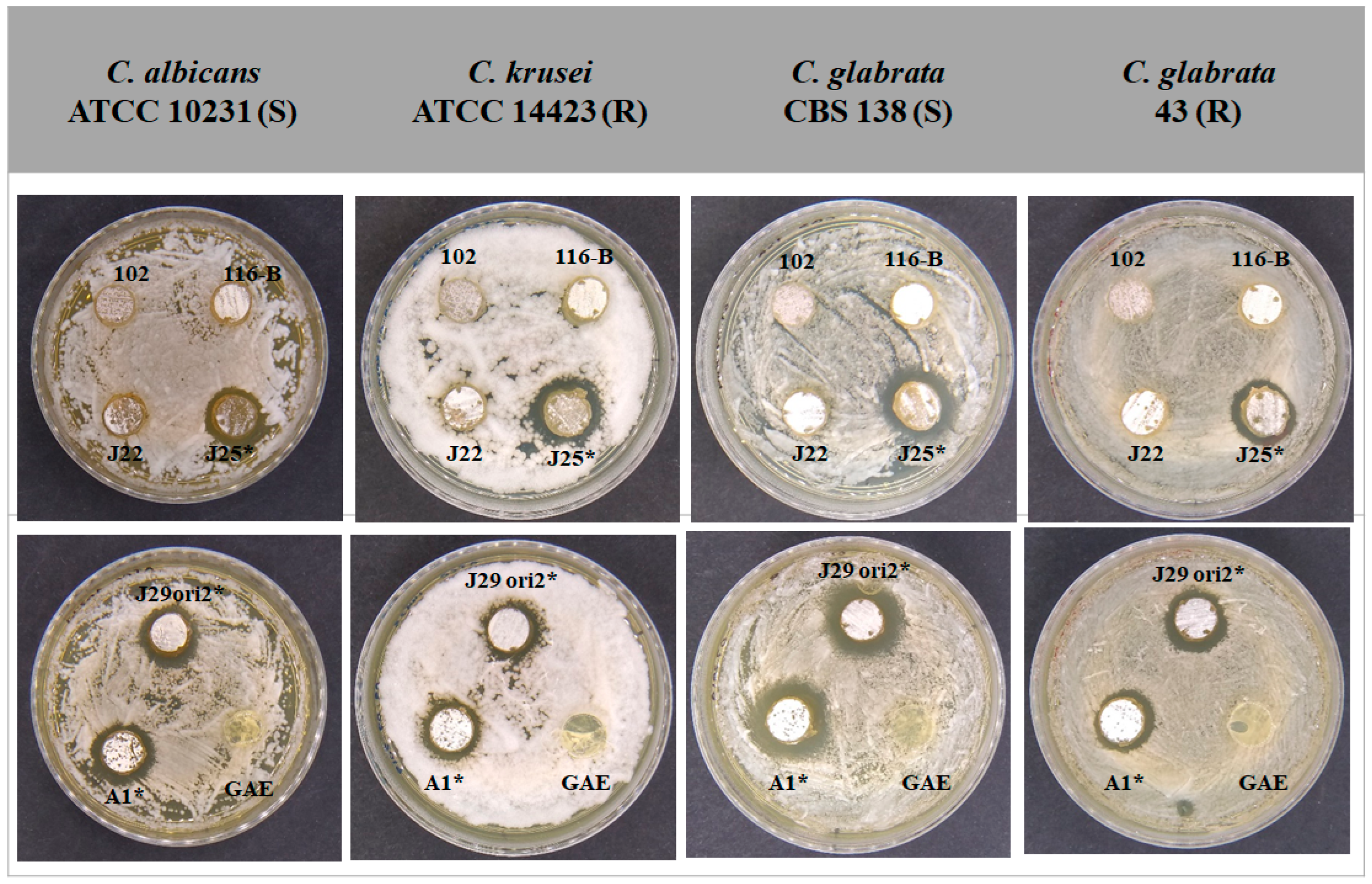
SDA plates were inoculated and incubated with the yeasts, and the diameter of the inhibition halos of Candida spp. by Streptomyces strains was measured (Figure 2).

3.4. Determination of the Minimum Inhibitory Concentration of Supernatants

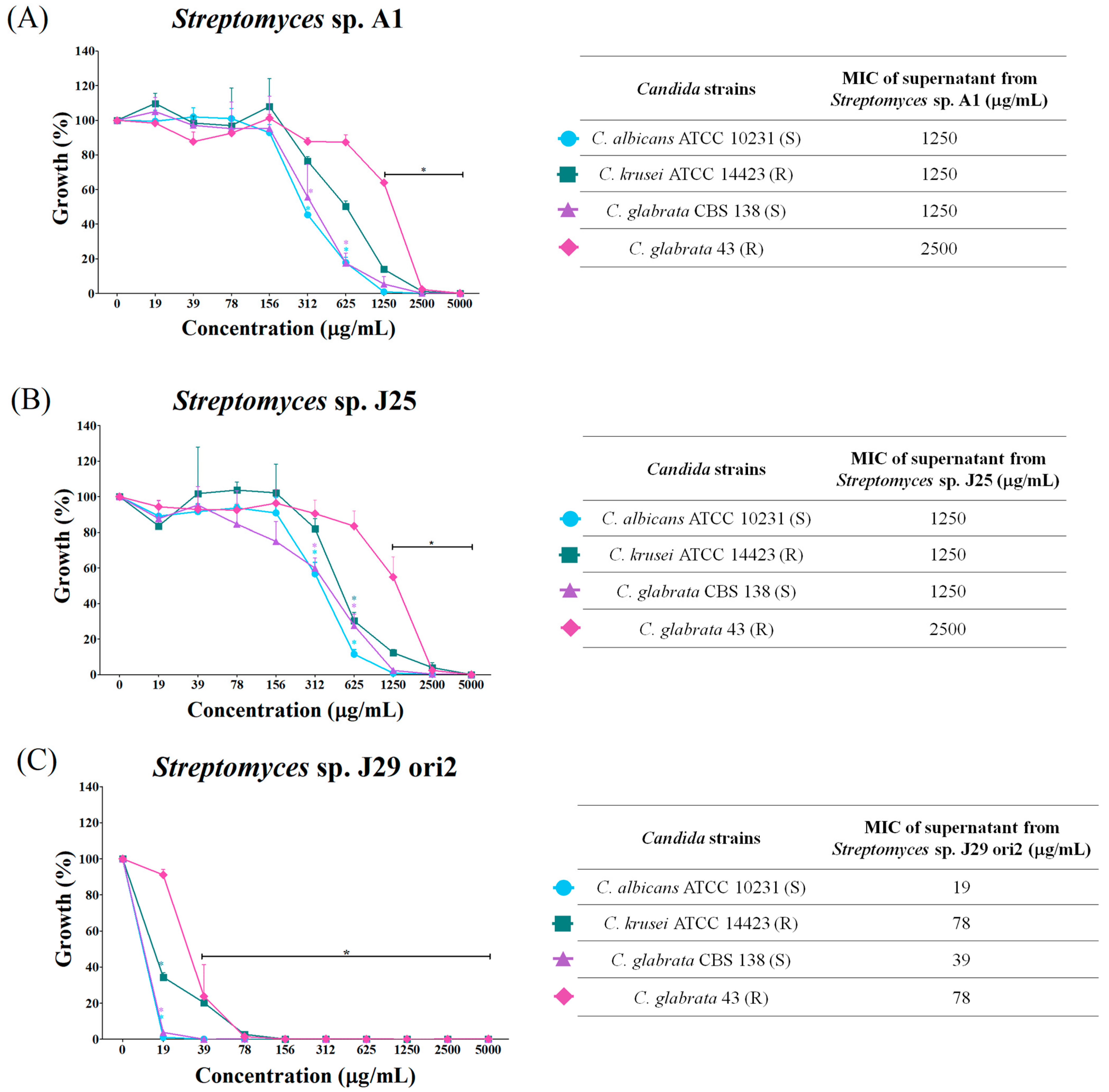
The lyophilized and rehydrated supernatants of Streptomyces spp. (200 mg/mL) were tested against strains of Candida spp. A dose–response effect between 19 and 5000 µg/mL was observed. The highest concentrations exhibited the smallest growth (Figure 3). The lyophilized supernatants of Streptomyces sp. A1 and J25 inhibited Candida species at MICs between 1250 and 2500 µg/mL (Figure 3A,B). Also, from the concentration of 1250 μg/mL onwards, a statistically significant difference was observed with respect to the growth control. The Streptomyces sp. J29 ori2 (Figure 3C) supernatant presented the most significant growth inhibitory capacity with MICs of 19, 39, 78, and 78 µg/mL for C. albicans ATCC 10231 (S), C. glabrata CBS 138 (S), C. krusei (R), and C. glabrata 43 (R), respectively. In this case, a statistically significant difference with respect to the growth control was observed at a concentration of 19 μg/mL.

3.5. Genome Sequencing, Assembly, and Annotation of S. albidoflavus Strains

All the reads analyzed by FastQC and filtering had a Phred scale >36. After assembly with SPAdes, and the coverage obtained was the 1×. Genome sizes were between 6.92 and 6.94 Mb, exhibiting typical linear topology, GC content (73.5%), numbers of coding genes between 6220 and 6257, and a total of contigs between 43 and 124 (Table S2).
The graphical representation of the strains’ genome (Figure S3) located the positions of non-encoding RNA, tRNA, rRNA, contigs, G-C content, and genes present in the genome. In addition, CRISPR regions and the origin of replication initiation (oriC) could be visualized. These figures also allowed comparison of the structural organization of the genes.

3.6. Genome-Based Taxonomic Identification of the Streptomyces Species

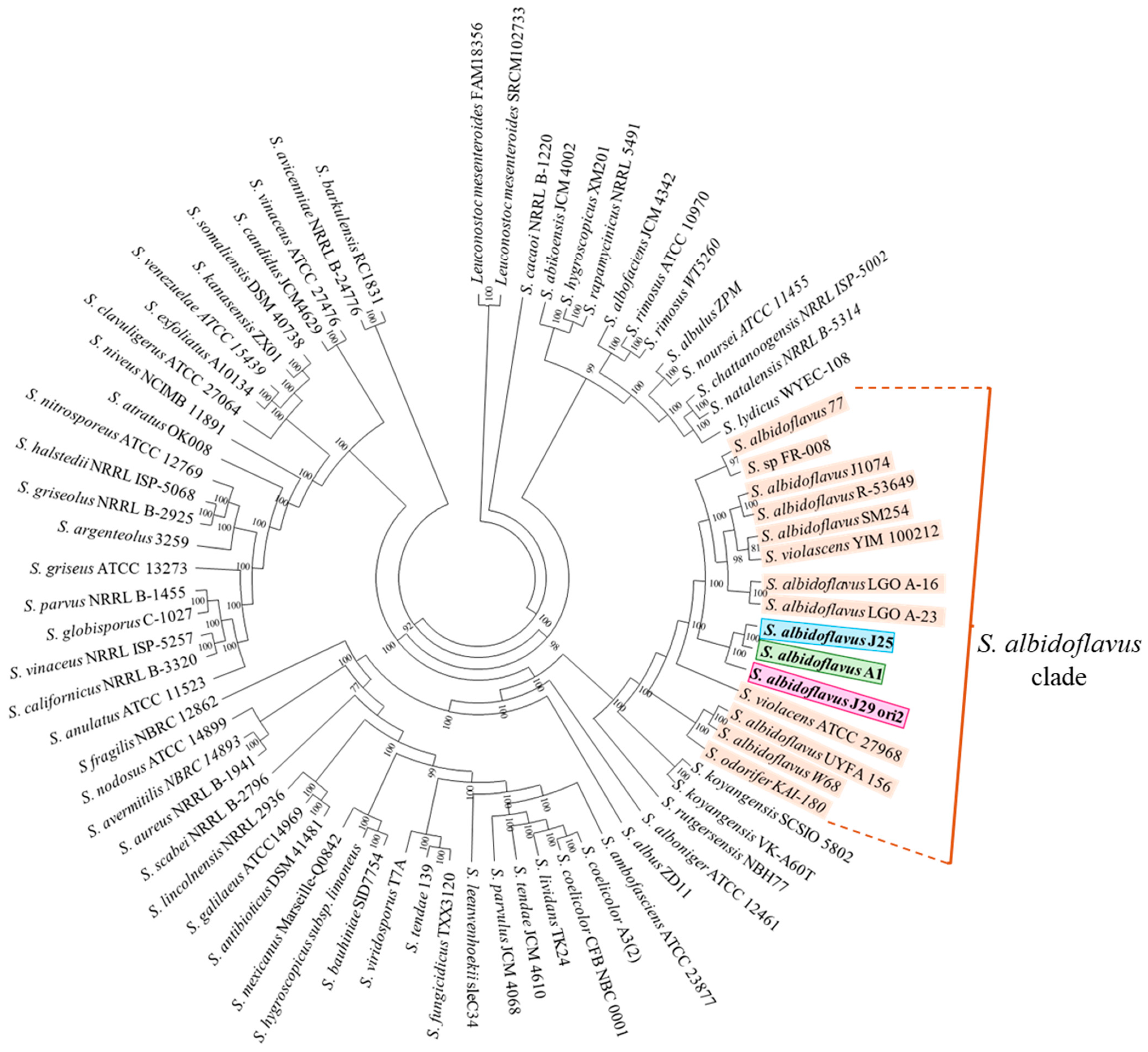
The phylogenetic approach based on the analysis of the 16S rRNA only allowed the identification of Streptomyces strains to genus level (Figure S1). The phylogenomic analyses of the Streptomyces strains performed in Orthofinder associated all the strains with the Streptomyces albidoflavus species, which formed a consistent clade (Figure 4). An Ortho Average Nucleotide Identity (ANI) of entire genomes revealed high similitudes in the range of 98.58 and 98.71 among S. albidoflavus strains, but minor similitudes with other related Streptomyces species (Table 1). Both criteria support the assignation of the strains to the S. albidoflavus species. OrthoANI compares orthologous genes in the genomes of S. albidoflavus strains, allowing a more robust analysis of nucleotide identity.

3.7. Comparative Genomics Among S. albidoflavus Strains

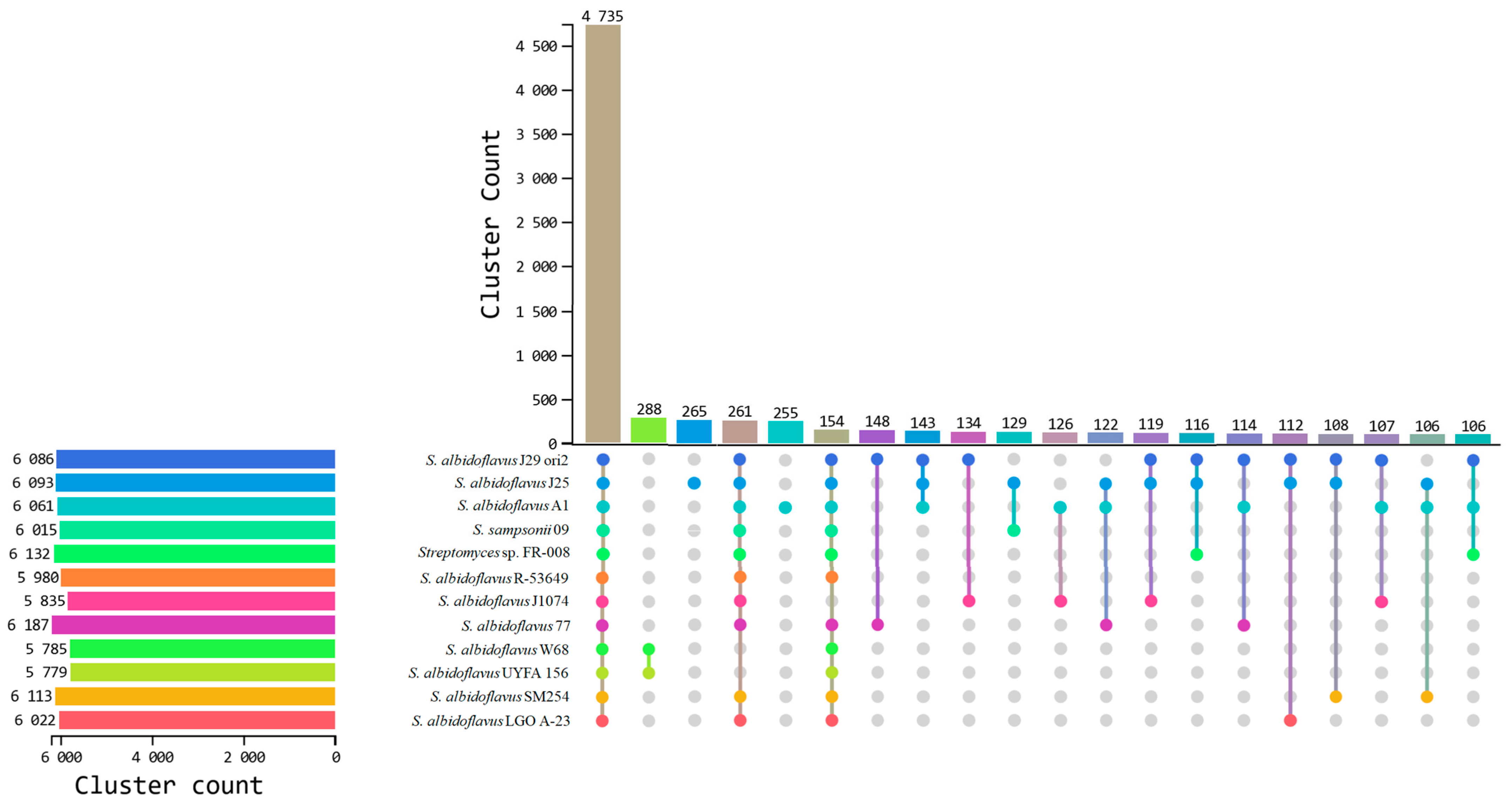
Orthovenn3 analysis recognized 4735 common orthogroups of three S. albidoflavus strains and nine strains from the database. Also, the strains contained between 106 and 288 accessory orthogroups. S. albidoflavus J25 and A1 strains harbored 265 and 255 exclusive orthogroups, respectively (Figure 5). In particular, only the strains S. albidoflavus J29 ori2, A1, and J25 included in this work shared 6014 common orthogroups and harbored 111, 7, and 24 exclusive orthogroups, respectively (Figure S4). Among these exclusive orthogroups a highlight was hypothetical proteins, glycosyltransferases involved in cell wall biogenesis and the transport of heavy metal-deduced proteins (Table S3).

3.8. Analysis of Extracellular Hydrolytic Enzymes and Siderophore Production of S. albidoflavus Strains

Phenotypic extracellular activities and their putative encoding genes for amylases, cellulases, chitinases, and proteases were identified in all three strains (Table 2). Frequently, more than one copy of each enzyme per genome was detected. More details of extracellular encoding enzymes, including enzyme class, function, contig ID, genome location, phenotypic activity, amino acid sequences, and solubilization indices (SI), were collected (Tables S4–S6, Figure S5).
In addition, the plate assay for siderophore production was positive, showing an orange halo around the colonies (Figure S6).

3.9. Analysis of BGCs of Metabolites with Antifungal Activity from S. albidoflavus Strains

The BGCs of secondary metabolite genes in S. albidoflavus J29 ori2, A1, and J25 genomes were detected by the antiSMASH bacterial version server. All three strains have BGC-encoding diverse chemical families of secondary metabolites with antibiotic, antifungal, antitumor, siderophore, and anticarcinogenic biological activities, among others (Tables S7–S9). According to phenotypical features, clusters encoding to antifungal compounds such as surugamides, polyenes such as candicidin, valinomycin, compound WS9326, and fluostatins were found in the three S. albidoflavus strains (Table 3).

3.10. Synteny Analysis of the BGC of Candicidin and Surugamide A/D of Streptomyces spp.

Based on the inhibitory activity of S. albidoflavus strains on Candida, synteny focused on analyzing candicidin and surugamide BGCs.
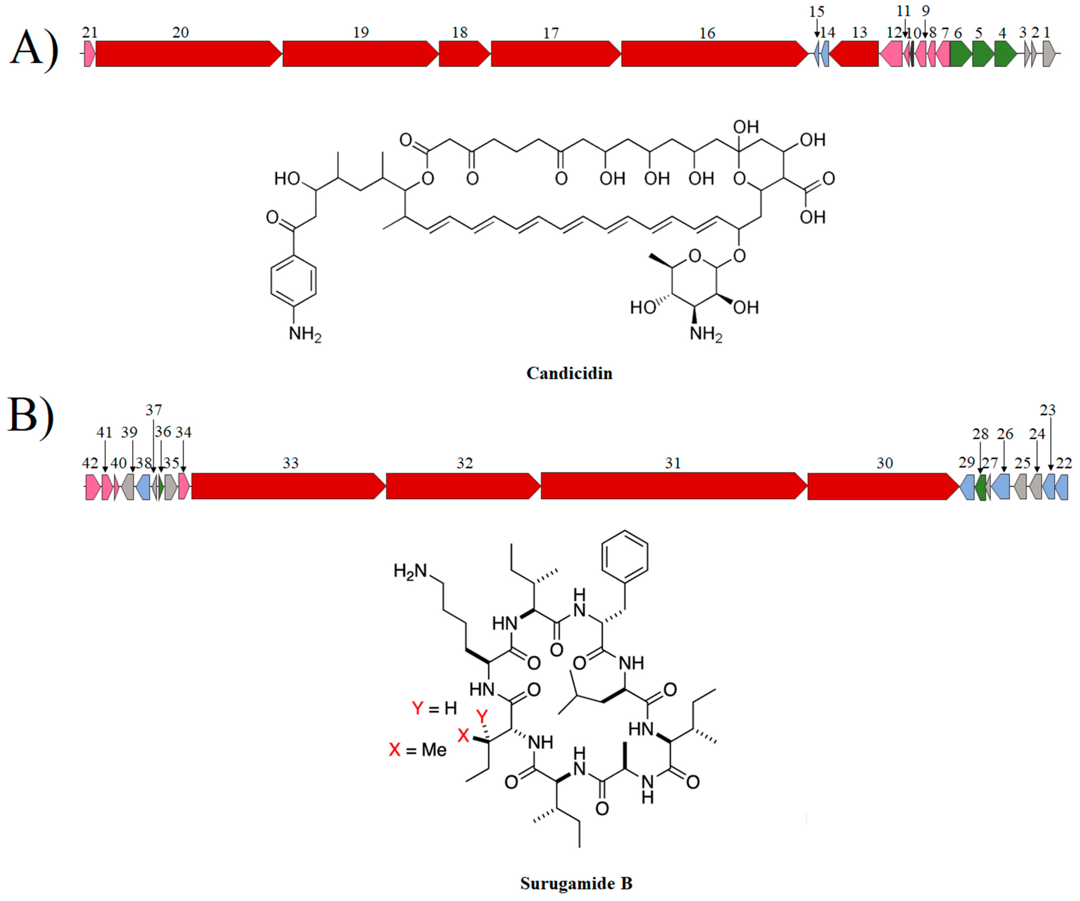
A synteny analysis of the candicidin and surugamide BGC with the highest percentage of similarity was performed to determine the genomic context, similitudes, and differences among these putative antifungal encoding clusters (Figure 6). The candicidin biosynthetic cluster (138.2 kb) contained six PKS type I genes (fscA-fscF) encoding to modular polyketide synthase enzymes which synthesize the macrolactone skeleton. Each deduced PKS enzyme contains ketosynthase repeat domains (KS), acyl transferase (AT), an acyl carrier protein (ACP), and, optionally, ketoreductase (KR), dehydratase (DH), and enoyl reductase (ER) domains. Also, the cluster harbors another 15 biosynthetic, transport, and regulatory genes that contribute to the final structure of the polyene. pabAB and pabC genes encode for 4-amino-4-deoxycorismate (ADC) synthase and ADC lyase, which participate in the synthesis of PABA, a precursor molecule that bonds to ACP to initiate candicidin biosynthesis. The fscTE gene encodes for a type II thioesterase, possibly responsible for removing aberrant intermediates and maintaining normal levels of candicidin synthesis. Also, the cluster includes fscTI-II, fscRI-IV, and fscO genes encoding to ABC type transporters, LuxR regulatory proteins, and a putative FAD-dependent monooxygenase, respectively.
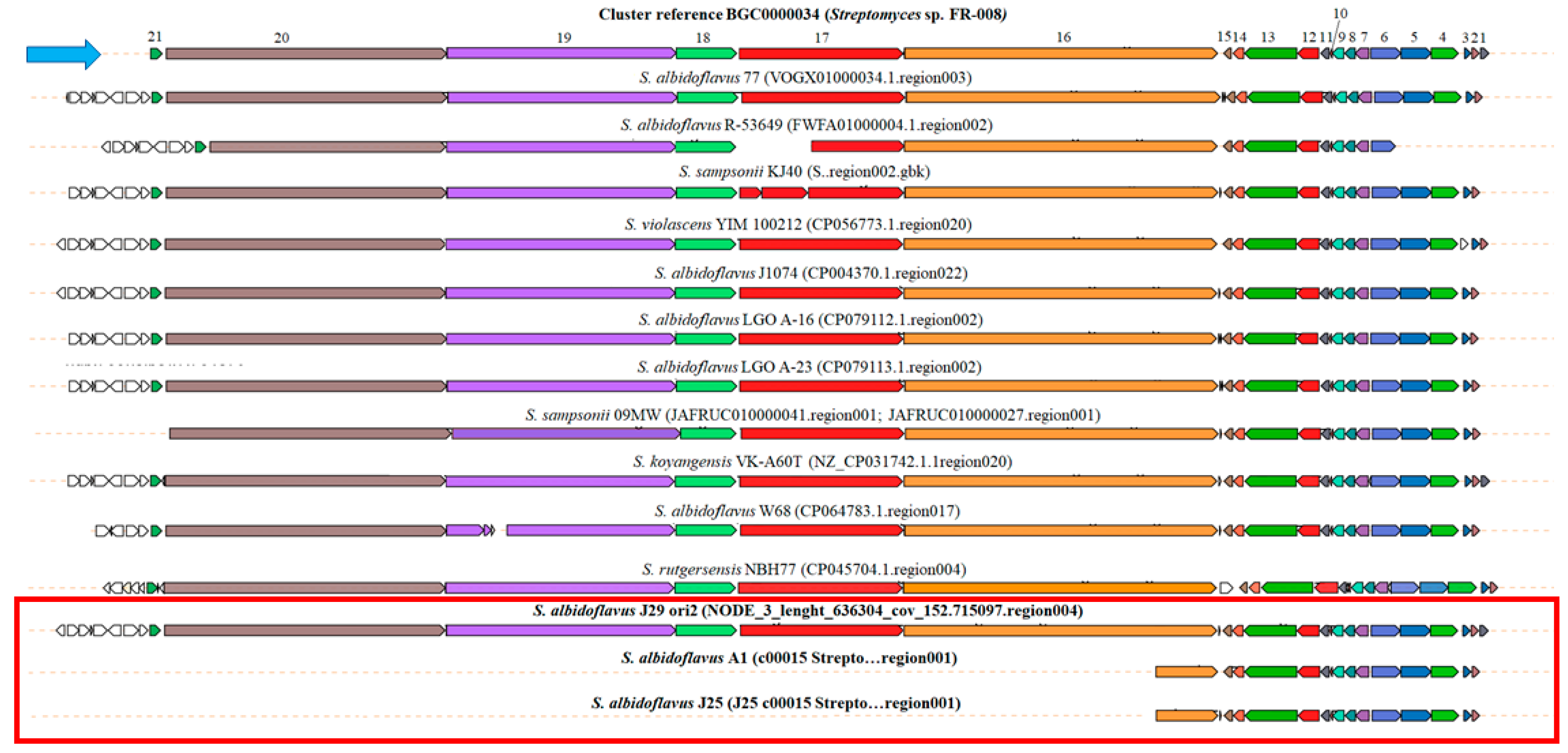
The CORASON analysis was performed with fscB (16,626 bp) and pabAB genes (2172 bp) encoding to type I PKS and isochorismate synthase, respectively, as query or center (Figure 7). The upload and download genes of the fscB and pabAB genes were ubicated according to the reference cluster of Streptomyces sp. FR-008. Clear differences between the clusters harbored in the three S. albidoflavus strains can be observed in the BGC synteny of candicidin. S. albidoflavus J29 ori2 has six PKSs genes (fscA-fscF) of candicidin cluster observed in the reference strain S. albidoflavus FR-008. Upstream of fscD, other sets of accessory genes of candicidin biosynthesis encoding amidases, oxidoreductases, and short-chain dehydrogenases complete the cluster. This candicidin cluster is broadly distributed among the S. albidoflavus clade and possibly encodes very similar candicidin structures.
On the other hand, S. albidoflavus A1 and S. albidoflavus J25 strains have only two PKS genes, a short fscC, and a conserved fscA. However, both strains maintain all regulatory genes, fscRI-IV, of the cluster. To our knowledge, the metabolite structure encoded by this cluster is unknown.
The clusters containing the biosynthetic enzymes for synthesizing surugamide were found in the three strains of this study and many other S. albidoflavus of the Genome database. The percentages of similarity estimated by antiSMASH of BSG of S. albidoflavus A1, J25, and J29 ori2 with the reference cluster were high (90–100%), and the syntenies were very similar (Figure 8). The cluster has a total size of 82,464 bp and harbors 21 genes, including essential surA-surD and surE genes, which encode to NRPS for the synthesis of this cyclic peptide. A transcriptional regulator AcrR, a putative helicase, and an uncharacterized MFS-like transporter were also identified in all genomes.

4. Discussion

Secondary metabolites are non-essential small molecules produced by microorganisms. However, they have relevant functions in chemical communication, competition for nutrients, survival, antagonism, and mutualism [46]. From the anthropocentric point of view, secondary metabolites have applications in various fields and in the treatment of human, animal, and plant diseases.
In this work, Streptomyces sp. A1, J25, and J29 ori2 showed in vitro inhibitory activity against fluconazole-sensitive and resistant Candida spp. when their genomes were sequenced, assembled and annotated. In addition to the Streptomyces strains used in this work, other S. albidoflavus strains with inhibitory capacity also present MICs in the order of µg/mL [47], the same as fluconazole, suggesting that these species harbor a high diversity of secondary metabolites with inhibitory capacity against filamentous and yeast-like fungi [48,49,50]. The 16S rRNA-based identification could not identify the strains at the species level. However, the identification by the phylogenomic approach and ANI percentages suggest that the strains under study are associated with the Streptomyces albidoflavus clade. Despite the apparent taxonomic closeness of the strains, they exhibited different colonial morphologies, diffusible pigment production, phenotypical antifungal capabilities, and genetic secondary metabolite profiles. Similar findings were observed among closely related Streptomyces griseus strains with differences in pigmentation and secondary metabolite profiles, which appear to be strain-specific [51].
Typically, Streptomyces linear genomes are among the largest in prokaryotes. For example, Streptomyces prasinosporus harbor a large genome of 14.99 Mb, S. coelicolor a moderate genome of 8.6 Mb, and Streptomyces gobiensis a small genome of 5.7 Mb (GCA_039535455.1; [26,52]. In particular, the S. albidoflavus clade, including the three strains studied in this work, harbored relatively small genomes of around 6.8–7.3 Mb and maintained high paired similitudes above 98.5% (GCA_007896905.1, GCA_019286295.1 GCA_900171555.1). However, the comparison of structure genomes exhibited many chromosomal rearrangements. Beyond that, the pangenomic approach reveals an extensive core genome but also many exclusive genes harbored in the S. albidoflavus strains, which suggest frequent horizontal gene transference (HGT) events drawing an intraspecific genetic polymorphism and open pangenome similar to other Streptomyces [53]. Although extensive homologous recombination has been documented in S. albidoflavus strains [54], to our knowledge, no specific events of HGT have been studied in S. albidoflavus. These events in Streptomyces are relatively frequent in an evolutive time scale and may shape the vast diversity of secondary metabolite and antimicrobial peptide clusters, strain fitness, and adaptation to environmental changes of this genus [55,56,57,58,59].
S. albidoflavus strains have been isolated from different natural and artificial environments. S. albidoflavus UYFA 156 is a stem endophyte of Festuca arundinacea grass, and S. albidoflavus SM254 was isolated from mining sediments with high copper concentrations [60,61]. S. albidoflavus A1. J25 and J29 ori2 were isolated from rhizosphere and soil as were other similar strains [3,62,63,64].
Cellulose, starch, peptidoglycan, chitin, and proteins are abundant macromolecules of soil containing carbon and nitrogen [23]. Extracellular enzymes must hydrolyze all these relatively stable compounds to mobilize, recycle, and make them available as nitrogen and carbon sources for plants and other organisms [23,65]. By their origin, the three strains of S. albidoflavus expressed phenotypically extracellular enzymatic activities and harbored genes encoding extracellular amylases, cellulases, chitinases, and proteases. These enzymes are broadly distributed among Streptomyces, particularly in S. albidoflavus [66,67,68].
Like S. coelicolor and Streptomyces lividans genomes, S. albidoflavus strains harbor multigene chitinase families and synergic chitin-binding proteins promoting chitin degradation. Streptomyces chitininases have been used as biological control of fungal phytopathogens [24,69,70]. The peptidoglycan, chitin, and protein turnover introduce carbon and nitrogen sources to soil [71,72].
The three genomes of S. albidoflavus strains harbored putative genes and extracellular enzymes to degrade polysaccharides and proteins, producing substrates used as carbon and nitrogen sources. Multiple copies of extracellular cellulases, amylases, chitinases, and proteases encoding genes per strain were detected. This genetic redundancy has also been observed in other Streptomyces species’ genomes [26]. Genetic redundancy in bacteria is commonly subjected to purifying selection [73]. However, some genes evolve to sub-functionalization, neo-functionalization, or differential expression depending on selective pressures under stressful or stable environmental conditions [73]. In S. coelicolor, some redundant gene families that encode the same enzymes and specialized metabolite gene clusters (actinorhodin, isorenieratane, coelichelin, and a cryptic NRPS) are differentially expressed at the transcriptional level when grown in tween, glucose, or other carbon sources [74].
Streptomyces genomes commonly harbor many BGCs of secondary metabolite [7,75]. In particular, the genomes of S. albidoflavus A1, J25, and J29 ori2 strains harbor 13, 12, and 13 putative complete BGC of secondary metabolites, among which are antibiotics, antifungal, siderophores, antitumoral, pigments, and signally compounds. Although antifungal activity and siderophores of the strains were detected in vitro, other expressed or cryptic biological activities have yet to be detected. Genes encoding siderophores desferrioxamine B were demonstrated in vitro, and their possible encoding genes were detected in genomes. These iron-chelating NRPs exhibit antifungal abilities by competence for iron-limiting fungal growth [76]. S. albidoflavus strains produce several polyenes, non-polyenic, and phenolic compounds with antifungal properties against both filamentous and yeast fungi, and their application as a fungal biocontrol agent has been suggested [46,47,48,49,62,77,78,79].
The genomes of the strains in this work harbor BGC encode to polyenes with recognized antifungal activities such as candicidin, surugamides, and fluostatin. Among Streptomyces species, BGCs encoding polyene antifungals, such as pimaricin, nystatin, amphotericin B, and candicidin, are broadly distributed [19]. The candicidin biosynthesis gene cluster is widely distributed among Streptomyces, although their importance in ecology is poorly understood [80].
Candicidin is an amphipathic polyene containing a macrolactone ring of 59 carbons with a D-mycosamine residue covalently attached to ergosterol, which forms pores in the membrane, causing membrane disruption and, eventually, cell death [81].
According to the antiSMASH software, genomes of S. albidoflavus A1, J25, and J29 ori2 harbor BGC of candicidin similar to the BGC of the Streptomyces FR008 strain [82]; however a more detailed analysis with CORASON software demonstrated that only S. albidoflavus A1 and J29 ori2 completed the biosynthetic cluster. Particularly, S. albidoflavus A1 has a BGC encoding a short type 1 PKS encoded by fscC, suggesting that the synthesized metabolite is a different polyene to candicidin.
The first candicidin BGC was initially described in Streptomyces griseus IMRU 3570, but the clusters are widely distributed among different Streptomyces species [80,83]. This antifungal compound shares the structure of a macrocyclic ring and a mycosamine residue with other polyenes, such as nystatin, amphotericin B, and pimaricin, but also has a 4-aminophenyl residue, which gives an aromatic character to the molecule [75]. This study compared the organization of BGC of candicidin S. albidoflavus-related species with Streptomyces sp. FR-008 was used as a reference cluster [76]. Only S. albidoflavus J29 ori2 harbored the complete PKS cluster, which suggests that it encodes a similar candicidin molecule to reference and other S. albidoflavus, S. rutgersensis, and S. violascens BGCs [84]. Also, the upstream genes encoding amidases, oxidoreductases, and short-chain dehydrogenases implicated in the bond saturations and high resonance of carbon polyene skeleton are present in all the strains [85,86,87].
In the genomes of S. albidoflavus A1 and S. albidoflavus J25 strains, only a short fscC gene and a fscA encoding to PKS and regulatory genes fscRI-IV participate in the biosynthesis of an unknown polyene. Evolutionary studies of PKSs show that when the rest of the biosynthetic enzymes are not contiguous, they probably were acquired by HGT. Furthermore, the length of PKSs can vary depending on the environmental stresses to which the bacteria are subjected. Thus, PKSs can selectively incorporate modules, changing their length, and the resulting products have different structures and bioactivities [88]. An evolutive approach will be necessary to define if the short BGC of this polyene is a product of a phenomenon of evolution towards complexity or reductiveness.
Surugamides are natural cyclic NRPs from 6 to 10 amino acids belonging to the desotamides family, which exhibit antibiotic and antifungal activities and anticancer properties of cathepsin B through the inhibition of proteases [18,89,90]. NRPs are biosynthesized by a modular enzymatic assembly line catalyzed by NRPs synthases and cyclized by a cyclase belonging to the β-lactamase superfamily [91,92]. S. albidoflavus strains of this work maintain BGCs of surugamides with high synteny similarity with the reference BGC of S. albidoflavus J1074 and other strains from marine sediments [84,93,94]. Downstream surD gene of surugamide BGC, and other genes encoding hypothetical proteins, transporters, and transcriptional regulators were identified. Surugamide BGC has been described as cryptic [18,93]. However, many “silent” BGCs of Streptomyces are not expressed because the signal molecules or environmental elicitors are not commonly provided in the synthetic culture media. Many in vitro environmental conditions or co-cultures are required to express BGCs of secondary metabolites of some Streptomyces species [95,96,97].
Considering the results of the MICs of the supernatants with the arrangements presented by the BGCs of secondary metabolites with antifungal activity, it could be suspected that the difference in inhibitory capacity between each of the strains is given by the arrangements in the organization of their clusters, since this will, in one way or another, have a direct consequence on the final structure of the metabolite.
The identification in the genome of the secondary metabolites, such as siderophores and antifungals, such as polyenes and surugamides, among others, suggests that S. albidoflavus strains A1, J25, and J29 ori2 harbor significant potential for use as PGPR bacteria.

5. Conclusions

In conclusion, in this work, the genome of Streptomyces albidoflavus A1, J25, and J29 ori2 strains isolated from jungle soils were fully identified to species level by a phylogenomic approach because the 16S rRNA gene sequence is not sufficient for an adequate taxonomic assignment. The comparative genomics of the strains allowed the recognition of a core and accessory genome in each strain. Also, phenotypic assays and genome mining revealed BGCs and genes encoding to secondary metabolites with potential antifungal activities, extracellular hydrolytic enzymes, and siderophores. The synteny of BGCs of the metabolites candicidin and surugamide suggest a common origin. However, several genomic rearrangements probably explain the difference in inhibitory activity between the strains. It will be possible to relate the intraspecific diversity detected in the genomes to the specific phenotypic characteristics of each strain through comparative genomics and analysis of the arrangement of BGCs in phylogenomically related strains.

Supplementary Materials

The following supporting information can be downloaded at: https://www.mdpi.com/article/10.3390/microorganisms12122637/s1, Table S1: Percentage similitude of the 16S rRNA gene of Streptomyces strains. Table S2: Features of linear S. albidoflavus genomes. Table S3: Exclusive orthogroups in each of the S. albidoflavus genomes. Table S4–S6: Features of extracellular enzymes phenotypically expressed of S. albidoflavus strains. Table S7–S9. Putative BGC of secondary metabolite from S. albidoflavus A1, J25, and J29 ori2. Figure S1: Phylogenetic tree based on the 16S rRNA gene of Streptomyces strains. Figure S2: Sensitivity profiles and determination phenotype to fluconazole of Candida spp. Figure S3: Graphical representation of S. albidoflavus strains linear chromosomes. Figure S4: Venn diagram of the number of orthogroups shared between S. albidoflavus A1, J25, and J29 ori2. Figure S5: Bioassay to produce extracellular hydrolytic enzymes of Streptomyces strains. Figure S6: Bioassay to siderophore production of Streptomyces strains.

Author Contributions

A.G.-S. and G.D.-P. performed isolation of Streptomyces strains, yeast inhibition bioassays and taxonomic identification of Streptomyces by 16SrRNA; A.G.-S., J.A.H.-G. and V.L.-S. analyzed, assembled and annotated bacterial genomes. A.G.-S. and M.S.J.-M. analyzed the genomes and detected putative BGCs of metabolites with antifungal activity in S. albidoflavus genomes. A.G.-S. determined the MICs of S. albidoflavus and performed phenotypic screens and a genomic search for extracellular hydrolytic enzymes. A.G.-S. and J.A.H.-G. performed phylogenomic identification. A.G.-S. and C.A. performed synteny analysis of putative BGCs for metabolites with antifungal activity. L.V.-T. and C.H.-R. designed and coordinated the study. A.G.-S. and C.H.-R. wrote the first draft of the manuscript. All authors have read and agreed to the published version of the manuscript.

Funding

The Secretaría de Investigación y Posgrado-IPN funded this research (SIP 20220742, 20220795, 20231480, 20231481, 20240945, 20240946) and CONACYT (CB 283225).

Data Availability Statement

The S. albidoflavus A1, J25 and J29 ori2 genome sequences herein generated were deposited in the GenBank. The identification number of the bioproject is PRJNA1171758. The accession numbers of the sequences are S. albidoflavus A1, S. albidoflavus J25 (SAMN44309340), and S. albidoflavus J29 ori2 (SAMN44309348).

Acknowledgments

Special thanks to Francisco Barona Gómez for the facilities provided and assistance A.G-S. for the use of the CORASON software. A.G.-S. is grateful to the Consejo Nacional de Humanidades, Ciencia y Tecnología (CONAHCyT), and BEIFI for the assistance. J.A.H.-G., L.V.-T. and C.H.-R. are fellows of EDI-IPN, COFFA-IPN, and SNI-CONAHCyT.

Conflicts of Interest

The authors declare no conflicts of interest.

References

  1. Youseif, S.H.; Abd El-Megeed, F.H.; Humm, E.A.; Maymon, M.; Mohamed, A.H.; Saleh, S.A.; Hirsch, A.M. Comparative Analysis of the Cultured and Total Bacterial Community in the Wheat Rhizosphere Microbiome Using Culture-Dependent and Culture-Independent Approaches. Microbiol. Spectr. 2021, 9, e0067821. [Google Scholar] [CrossRef]
  2. Osburn, E.D.; Yang, G.; Rillig, M.C.; Strickland, M.S. Evaluating the role of bacterial diversity in supporting soil ecosystem functions under anthropogenic stress. ISME Commun. 2023, 3, 66. [Google Scholar] [CrossRef]
  3. Nonthakaew, N.; Panbangred, W.; Songnuan, W.; Intra, B. Plant growth-promoting properties of Streptomyces spp. isolates and their impact on mung bean plantlets’ rhizosphere microbiome. Front. Microbiol. 2022, 13, 967415. [Google Scholar] [CrossRef]
  4. Park, H.S.; Nah, H.J.; Kang, S.H.; Choi, S.S.; Kim, E.S. Screening and Isolation of a Novel Polyene-Producing Streptomyces Strain Inhibiting Phytopathogenic Fungi in the Soil Environment. Front. Bioeng. Biotechnol. 2021, 9, 692340. [Google Scholar] [CrossRef]
  5. Bekker, V.; Dodd, A.; Brady, D.; Rumbold, K. Tools for metabolic engineering in Streptomyces. Bioengineered 2014, 5, 293–299. [Google Scholar] [CrossRef] [PubMed]
  6. Hwang, K.S.; Kim, H.U.; Charusanti, P.; Palsson, B.Ø.; Lee, S.Y. Systems biology and biotechnology of Streptomyces species for the production of secondary metabolites. Biotechnol. Adv. 2014, 32, 255–268. [Google Scholar] [CrossRef]
  7. Li, L.; Liu, X.; Jiang, W.; Lu, Y. Recent advances in synthetic biology approaches to optimize production of bioactive natural products in Actinobacteria. Front. Microbiol. 2019, 10, 2467. [Google Scholar] [CrossRef]
  8. Alam, K.; Hao, J.; Zhong, L.; Fan, G.; Ouyang, Q.; Islam, M.M.; Saiful, I.; Sun, H.; Zhang, Y.; Li, R.; et al. Complete genome sequencing and in silico genome mining reveal the promising metabolic potential in Streptomyces strain CS-7. Front. Microbiol. 2022, 13, 939919. [Google Scholar] [CrossRef] [PubMed]
  9. García-Gutiérrez, C.; Pérez-Victoria, I.; Montero, I.; Fernández-De la Hoz, J.; Malmierca, M.G.; Martín, J.; Salas, J.A.; Olano, C.; Reyes, F.; Méndez, C. Unearthing a cryptic biosynthetic gene cluster for the piperazic acid-bearing depsipeptide diperamycin in the ant-dweller Streptomyces sp. CS113. Int. J. Mol. Sci. 2024, 25, 2347. [Google Scholar] [CrossRef] [PubMed]
  10. Navarro-Muñoz, J.C.; Selem-Mojica, N.; Mullowney, M.W.; Kautsar, S.A.; Tryon, J.H.; Parkinson, E.I.; De Los Santos, E.L.C.; Yeong, M.; Cruz-Morales, P.; Abubucker, S.; et al. A computational framework to explore large-scale biosynthetic diversity. Nat. Chem. Biol. 2020, 16, 60–68. [Google Scholar] [CrossRef] [PubMed]
  11. Manteca, Á.; Yagüe, P. Streptomyces differentiation in liquid cultures as a trigger of secondary metabolism. Antibiotics 2018, 7, 41. [Google Scholar] [CrossRef]
  12. Sarmiento-Tovar, A.A.; Silva, L.; Sánchez-Suárez, J.; Diaz, L. Streptomyces-Derived Bioactive Pigments: Ecofriendly Source of Bioactive Compounds. Coatings 2022, 12, 1858. [Google Scholar] [CrossRef]
  13. Sadeghi, A.; Soltani, B.M.; Nekouei, M.K.; Jouzani, G.S.; Mirzaei, H.H.; Sadeghizadeh, M. Diversity of the ectoines biosynthesis genes in the salt tolerant Streptomyces and evidence for inductive effect of ectoines on their accumulation. Microbiol. Res. 2014, 169, 699–708. [Google Scholar] [CrossRef] [PubMed]
  14. van Bergeijk, D.A.; Terlouw, B.R.; Medema, M.H.; van Wezel, G.P. Ecology and genomics of Actinobacteria: New concepts for natural product discovery. Nat. Rev. Microbiol. 2020, 18, 546–558. [Google Scholar] [CrossRef]
  15. Zhang, M.; Kong, L.; Gong, R.; Iorio, M.; Donadio, S.; Deng, Z.; Sosio, M.; Chen, W. Biosynthesis of C-nucleoside antibiotics in actinobacteria: Recent advances and future developments. Microb. Cell Factories 2022, 21, 2. [Google Scholar] [CrossRef]
  16. Wu, Y.; Zhang, M.; Yang, Y.; Ding, X.; Yang, P.; Huang, K.; Hu, X.; Zhang, M.; Liu, X.; Yu, H. Structures and mechanism of chitin synthase and its inhibition by antifungal drug Nikkomycin Z. Cell Discov. 2022, 8, 129. [Google Scholar] [CrossRef] [PubMed]
  17. Osada, H. Discovery and applications of nucleoside antibiotics beyond polyoxin. J. Antibiot. 2019, 72, 855–864. [Google Scholar] [CrossRef]
  18. Xu, F.; Nazari, B.; Moon, K.; Bushin, L.B.; Seyedsayamdost, M.R. Discovery of a cryptic antifungal compound from Streptomycess albus J1074 using high-throughput elicitor screens. J. Am. Chem. Soc. 2017, 139, 9203–9212. [Google Scholar] [CrossRef]
  19. Caffrey, P.; De Poire, E.; Sheehan, J.; Sweeney, P. Polyene macrolide biosynthesis in streptomycetes and related bacteria: Recent advances from genome sequencing and experimental studies. Appl. Microbiol. Biotechnol. 2016, 100, 3893–3908. [Google Scholar] [CrossRef]
  20. Krespach, M.K.C.; Stroe, M.C.; Netzker, T.; Rosin, M.; Zehner, L.M.; Komor, A.J.; Beilmann, J.M.; Krüger, T.; Scherlach, K.; Kniemeyer, O.; et al. Streptomyces polyketides mediate bacteria-fungi interactions across soil environments. Nat. Microbiol. 2023, 8, 1348–1361. [Google Scholar] [CrossRef] [PubMed]
  21. Simpson, A.J.; Simpson, M.J.; Smith, E.; Kelleher, B.P. Microbially derived inputs to soil organic matter: Are cur-rent estimates too low? Environ. Sci. Technol. 2007, 41, 8070–8076. [Google Scholar] [CrossRef] [PubMed]
  22. Schulten, H.R.; Schnitzer, M. The chemistry of soil organic nitrogen: A review. Biol. Fertil Soils. 1997, 26, 1–15. [Google Scholar] [CrossRef]
  23. Hu, Y.; Zheng, Q.; Noll, L.; Zhang, S.; Wanek, W. Direct measurement of the in-situ decomposition of microbial-derived soil organic matter. Soil Biol. Biochem. 2020, 141, 107660. [Google Scholar] [CrossRef]
  24. Chater, K.F.; Biró, S.; Lee, K.J.; Palmer, T.; Schrempf, H. The complex extracellular biology of Streptomyces. FEMS Microbiol. Rev. 2010, 34, 171–198. [Google Scholar] [CrossRef] [PubMed]
  25. Book, A.J.; Lewin, G.R.; McDonald, B.R.; Takasuka, T.E.; Wendt-Pienkowski, E.; Doering, D.T.; Suh, S.; Raffa, K.F.; Fox, B.G.; Currie, C.R. Evolution of high cellulolytic activity in symbiotic Streptomyces through selection of expanded gene content and coordinated gene expression. PLoS Biol. 2016, 14, e1002475. [Google Scholar] [CrossRef]
  26. Bentley, S.D.; Chater, K.F.; Cerdeño-Tárraga, A.M.; Challis, G.L.; Thomson, N.R.; James, K.D.; Harris, D.E.; Quail, M.A.; Kieser, H.; Harper, D.; et al. Complete genome sequence of the model actinomycete Streptomyces coelicolor A3(2). Nature 2002, 417, 141–147. [Google Scholar] [CrossRef]
  27. Kumar, M.; Kumar, P.; Das, P.; Solanki, R.; Kapur, M.K. Potential applications of extracellular enzymes from Streptomyces spp. in various industries. Arch. Microbiol. 2020, 202, 1597–1615. [Google Scholar] [CrossRef] [PubMed]
  28. Méndez, C.; Braña, A.F.; Manzanal, M.B.; Hardisson, C. Role of substrate mycelium in colony development in Streptomyces. Can. J. Microbiol. 1985, 31, 446–450. [Google Scholar] [CrossRef] [PubMed]
  29. Stubner, S. Enumeration of 16S rDNA of Desulfotomaculum lineage 1 in rice field soil by real-time PCR with SybrGreen™ detection. J. Microbiol. Methods 2002, 50, 155–164. [Google Scholar] [CrossRef] [PubMed]
  30. CLSI Standard M27; Reference Method for Broth Dilution Antifungal Susceptibility Testing of Yeasts. 4th ed, Clinical and Laboratory Standards Institute: Malvern, PA, USA, 2017.
  31. Andrews, S. FastQC: A Quality Control Tool for High Throughput Sequence Data. 2010. Available online: https://www.bioinformatics.babraham.ac.uk/projects/fastqc/ (accessed on 1 August 2024).
  32. Bankevich, A.; Nurk, S.; Antipov, D.; Gurevich, A.A.; Dvorkin, M.; Kulikov, A.S.; Lesin, V.M.; Nikolenko, S.I.; Pham, S.; Prjibelski, A.D.; et al. SPAdes: A new genome assembly algorithm and its applications to single-cell sequencing. J. Comput. Biol. 2012, 19, 455–477. [Google Scholar] [CrossRef]
  33. Gurevich, A.; Saveliev, V.; Vyahhi, N.; Tesler, G. QUAST: Quality assessment tool for genome assemblies. Bioinformatics 2013, 29, 1072–1075. [Google Scholar] [CrossRef]
  34. Aziz, R.K.; Bartels, D.; Best, A.A.; DeJongh, M.; Disz, T.; Edwards, R.A.; Formsma, K.; Gerdes, S.; Glass, E.M.; Kubal, M.; et al. The RAST server: Rapid annotations using subsystems technology. BMC Genom. 2008, 9, 75. [Google Scholar] [CrossRef] [PubMed]
  35. Grant, J.R.; Enns, E.; Marinier, E.; Mandal, A.; Herman, E.K.; Chen, C.Y.; Graham, M.; Van Domselaar, G.; Stothard, P. Proksee: In-depth characterization and visualization of bacterial genomes. Nucleic Acids Res. 2023, 51, W484–W492. [Google Scholar] [CrossRef]
  36. Emms, D.M.; Kelly, S. OrthoFinder: Phylogenetic orthology inference for comparative genomics. Genome Biol. 2019, 20, 238. [Google Scholar] [CrossRef]
  37. Minh, B.Q.; Schmidt, H.A.; Chernomor, O.; Schrempf, D.; Woodhams, M.D.; Von Haeseler, A.; Lanfear, R. IQ-TREE 2: New models and efficient methods for phylogenetic inference in the genomic era. Mol. Biol. Evol. 2020, 37, 1530–1534. [Google Scholar] [CrossRef] [PubMed]
  38. Yoon, S.H.; Ha, S.M.; Lim, J.; Kwon, S.; Chun, J. A large-scale evaluation of algorithms to calculate average nucleotide identity. Antonie Van Leeuwenhoek 2017, 110, 1281–1286. [Google Scholar] [CrossRef]
  39. Sun, J.; Lu, F.; Luo, Y.; Bie, L.; Xu, L.; Wang, Y. OrthoVenn3: An integrated platform for exploring and visualizing orthologous data across genomes. Nucleic Acids Res. 2023, 51, W397–W403. [Google Scholar] [CrossRef] [PubMed]
  40. Suriyachadkun, C.; Chunhachart, O.; Srithaworn, M.; Tangchitcharoenkhul, R.; Tangjitjareonkun, J. Zinc-solubilizing Streptomyces spp. as bioinoculants for promoting the growth of soybean (Glycine max (L.) Merrill). J. Microbiol. Biotechnol. 2022, 32, 1435–1446. [Google Scholar] [CrossRef] [PubMed]
  41. Narayana, K.J.; Vijayalakshmi, M. Chitinase production by Streptomyces sp. ANU 6277. Braz. J. Microbiol. 2009, 40, 725–733. [Google Scholar] [CrossRef] [PubMed]
  42. Shen, H.B.; Chou, K.C. Gpos-mPLoc: A top-down approach to improve the quality of predicting subcellular localization of Gram-positive bacterial proteins. Protein Pept. Lett. 2009, 16, 1478–1484. [Google Scholar] [CrossRef]
  43. Rehan, M.; Barakat, H.; Almami, I.S.; Qureshi, K.A.; Alsohim, A.S. Production and Potential Genetic Pathways of Three Different Siderophore Types in Streptomyces tricolor Strain HM10. Fermentation 2022, 8, 346. [Google Scholar] [CrossRef]
  44. Blin, K.; Shaw, S.; Steinke, K.; Villebro, R.; Ziemert, N.; Lee, S.Y.; Medema, M.H.; Weber, T. antiSMASH 5.0: Updates to the secondary metabolite genome mining pipeline. Nucleic Acids Res. 2019, 47, W81–W87. [Google Scholar] [CrossRef] [PubMed]
  45. Terlouw, B.R.; Blin, K.; Navarro-Munoz, J.C.; Avalon, N.E.; Chevrette, M.G.; Egbert, S.; Lee, S.; Meijer, D.; Recchia, M.J.J.; Reitz, Z.L.; et al. MIBiG 3.0: A community-driven effort to annotate experimentally validated biosynthetic gene clusters. Nucleic Acids Res. 2023, 51, D603–D610. [Google Scholar] [CrossRef]
  46. Nofiani, R.; Rudiyansyah; Ardiningsih, P.; Rizky; Zahra, S.T.A.; Sukito, A.; Weisberg, A.J.; Chang, J.H.; Mahmud, T. Genome features and secondary metabolite potential of the marine symbiont Streptomyces sp. RS2. Arch. Microbiol. 2023, 205, 244. [Google Scholar] [CrossRef]
  47. Bautista-Crescencio, C.; Casimiro-Ramos, A.; Fragoso-Vázquez, M.J.; Correa-Basurto, J.; Olano, C.; Hernández-Rodríguez, C.; Villa-Tanaca, L. Streptomyces albidoflavus Q antifungal metabolites inhibit the ergosterol biosynthesis pathway and yeast growth in fluconazole-resistant Candida glabrata: Phylogenomic and metabolomic analyses. Microbiol. Spectr. 2023, 11, e0127123. [Google Scholar] [CrossRef] [PubMed]
  48. Yao, X.; Zhang, Z.; Huang, J.; Wei, S.; Sun, X.; Chen, Y.; Liu, H.; Li, S. Candicidin isomer production is assential for biocontrol of cucumber Rhizoctonia rot by Streptomyces albidoflavus W68. Appl. Environ. Microbiol. 2021, 87, e03078-20. [Google Scholar] [CrossRef]
  49. Martins, N.D.R.C.; Rodrigues da Silva, A.; Ratcliffe, N.; Evangelho, V.G.O.; Castro, H.C.; Quinn, G.A. Streptomyces: A natural source of anti-Candida agents. J. Med. Microbiol. 2023, 72, 001777. [Google Scholar] [CrossRef]
  50. Lai, J.; Liu, B.; Xiong, G.; Luo, Q.; Song, S.; Jiang, J.; Wei, H.; Wang, J. Inhibitory mechanism of 4-ethyl-1,2-dimethoxybenzene produced by Streptomyces albidoflavus strain ML27 against Colletotrichum gloeosporioides. Pestic. Biochem. Physiol. 2024, 204, 106086. [Google Scholar] [CrossRef] [PubMed]
  51. Sottorff, I.; Wiese, J.; Lipfert, M.; Preußke, N.; Sönnichsen, F.D.; Imhoff, J.F. Different secondary metabolite profiles of phylogenetically almost identical Streptomyces griseus strains originating from geographically remote locations. Microorganisms 2019, 7, 166. [Google Scholar] [CrossRef]
  52. Wen, Y.; Zhang, G.; Bahadur, A.; Liu, Y.; Zhang, Z.; Chen, T.; Liu, G.; Zhang, W. Streptomyces gobiensis sp. nov., an antimicrobial producing actinobacterium isolated from soil under black Gobi rocks. Int. J. Syst. Evol. Microbiol. 2022, 72, 005318. [Google Scholar] [CrossRef] [PubMed]
  53. Feng, N.X.; Li, D.W.; Zhang, F.; Bin, H.; Huang, Y.T.; Xiang, L.; Liu, B.L.; Cai, Q.Y.; Li, Y.W.; Xu, D.L.; et al. Biodegradation of phthalate acid esters and whole-genome analysis of a novel Streptomyces sp. FZ201 isolated from natural habitats. J. Hazard. Mater. 2024, 469, 133972. [Google Scholar] [CrossRef] [PubMed]
  54. Cheng, K.; Rong, X.; Pinto-Tomás, A.A.; Fernández-Villalobos, M.; Murillo-Cruz, C.; Huang, Y. Population genetic analysis of Streptomyces albidoflavus reveals habitat barriers to homologous recombination in the diversification of streptomycetes. Appl. Environ. Microbiol. 2015, 81, 966–975. [Google Scholar] [CrossRef] [PubMed]
  55. Deng, M.R.; Guo, J.; Li, X.; Zhu, C.H.; Zhu, H.H. Granaticins and their biosynthetic gene cluster from Streptomyces vietnamensis: Evidence of horizontal gene transfer. Antonie Van Leeuwenhoek 2011, 100, 607–617. [Google Scholar] [CrossRef]
  56. Li, J.; Zhao, Z.; Zhong, W.; Zhong, C.; Zong, G.; Fu, J.; Cao, G. Impacts of horizontal gene transfer on the compact genome of the clavulanic acid-producing Streptomyces strain F613-1. 3 Biotech 2018, 8, 472. [Google Scholar] [CrossRef] [PubMed]
  57. Ayala-Ruano, S.; Santander-Gordón, D.; Tejera, E.; Perez-Castillo, Y.; Armijos-Jaramillo, V. A putative antimicrobial peptide from Hymenoptera in the megaplasmid pSCL4 of Streptomyces clavuligerus ATCC 27064 reveals a singular case of horizontal gene transfer with potential applications. Ecol. Evol. 2019, 9, 2602–2614. [Google Scholar] [CrossRef]
  58. Tidjani, A.R.; Lorenzi, J.N.; Toussaint, M.; van Dijk, E.; Naquin, D.; Lespinet, O.; Bontemps, C.; Leblond, P. Massive gene flux drives genome diversity between sympatric Streptomyces conspecifics. MBio 2019, 10, e01533-19. [Google Scholar] [CrossRef]
  59. Choufa, C.; Tidjani, A.R.; Gauthier, A.; Harb, M.; Lao, J.; Leblond-Bourget, N.; Vos, M.; Leblond, P.; Bontemps, C. Prevalence and mobility of integrative and conjugative elements within a Streptomyces natural population. Front. Microbiol. 2022, 13, 970179. [Google Scholar] [CrossRef]
  60. Badalamenti, J.P.; Erickson, J.D.; Salomon, C.E. Complete genome sequence of Streptomyces albus SM254, a potent antagonist of bat white-nose syndrome pathogen Pseudogymnoascus destructans. Genome Announc. 2016, 4, e00290-16. [Google Scholar] [CrossRef]
  61. de los Santos, M.C.; Taulé, C.; Mareque, C.; Beracochea, M.; Battistoni, F. Identification and characterization of the part of the bacterial community associated with field-grown tall fescue (Festuca arundinacea) cv. SFRO Don Tomás in Uruguay. Ann. Microbiol. 2016, 66, 329–342. [Google Scholar] [CrossRef]
  62. Peng, F.; Zhang, M.Y.; Hou, S.Y.; Chen, J.; Wu, Y.Y.; Zhang, Y.X. Insights into Streptomyces spp. isolated from the rhizospheric soil of Panax notoginseng: Isolation, antimicrobial activity and biosynthetic potential for polyketides and non-ribosomal peptides. BMC Microbiol. 2020, 20, 143. [Google Scholar] [CrossRef]
  63. Du, Y.; Wang, T.; Jiang, J.; Wang, Y.; Lv, C.; Sun, K.; Sun, J.; Yan, B.; Kang, C.; Guo, L.; et al. Biological control and plant growth promotion properties of Streptomyces albidoflavus St-220 isolated from Salvia miltiorrhiza rhizosphere. Front. Plant Sci. 2022, 13, 976813. [Google Scholar] [CrossRef]
  64. Li, J.; Zhang, L.; Yao, G.; Zhu, L.; Lin, J.; Wang, C.; Du, B.; Ding, Y.; Mei, X. Synergistic effect of co-culture rhizosphere Streptomyces: A promising strategy to enhance antimicrobial activity and plant growth-promoting function. Front. Microbiol. 2022, 13, 976484. [Google Scholar] [CrossRef]
  65. Amaresan, N.; Kumar, K.; Naik, J.H.; Bapatla, K.G.; Mishra, R.K. Streptomyces in plant growth promotion: Mechanisms and role. In New and Future Developments in Microbial Biotechnology and Bioengineering; Elsevier: Amsterdam, The Netherlands, 2018; pp. 125–135. [Google Scholar] [CrossRef]
  66. Vijayalakshmi, M.; Narayana, K.J.P. Production of extracellular a-amylase by Streptomyces albidoflavus. Asian J. Biochem. 2008, 3, 194–197. [Google Scholar] [CrossRef]
  67. Vijayalakshmi, M.; Narayana, K.J.P. Production of extracellular protease by Streptomyces albidoflavus. Asian J. Biochem. 2008, 3, 198–202. [Google Scholar] [CrossRef]
  68. Sugimori, D.; Kano, K.; Matsumoto, Y. Purification, characterization, molecular cloning and extracellular production of a phospholipase A1 from Streptomyces albidoflavus NA297. FEBS Open Bio 2012, 2, 318–327. [Google Scholar] [CrossRef] [PubMed]
  69. Umar, A.A.; Hussaini, A.B.; Yahayya, J.; Sani, I.; Aminu, H. Chitinolytic and antagonistic activity of Streptomyces isolated from fadama soil against phytopathogenic fungi. Trop. Life Sci. Res. 2021, 32, 25–38. [Google Scholar] [CrossRef] [PubMed]
  70. Kotb, E.; Alabdalall, A.H.; Alghamdi, A.I.; Ababutain, I.M.; Aldakeel, S.A.; Al-Zuwaid, S.K.; Algarudi, B.M.; Algarudi, S.M.; Ahmed, A.A.; Albarrag, A.M. Screening for chitin degrading bacteria in the environment of Saudi Arabia and characterization of the most potent chitinase from Streptomyces variabilis Am1. Sci. Rep. 2023, 13, 11723. [Google Scholar] [CrossRef] [PubMed]
  71. Rillig, M.C.; Caldwell, B.A.; Wösten, H.A.B.; Sollins, P. Role of proteins in soil carbon and nitrogen storage: Controls on persistence. Biogeochemistry 2007, 85, 25–44. [Google Scholar] [CrossRef]
  72. Greenfield, L.M.; Hill, P.W.; Seaton, F.M.; Paterson, E.; Baggs, E.M.; Jones, D.L. Is soluble protein mineralisation and protease activity in soil regulated by supply or demand? Soil Biol. Biochem. 2020, 150, 108007. [Google Scholar] [CrossRef]
  73. Kim, J.; Copley, S.D. Why metabolic enzymes are essential or nonessential for growth of Escherichia coli K12 on glucose. Biochemistry 2007, 46, 12501–12511. [Google Scholar] [CrossRef]
  74. Schniete, J.K.; Reumerman, R.; Kerr, L.; Tucker, N.P.; Hunter, I.S.; Herron, P.R.; Hoskisson, P.A. Differential transcription of expanded gene families in central carbon metabolism of Streptomyces coelicolor A3(2). Access Microbiol. 2020, 2, acmi000122. [Google Scholar] [CrossRef]
  75. Belknap, K.C.; Park, C.J.; Barth, B.M.; Andam, C.P. Genome mining of biosynthetic and chemotherapeutic gene clusters in Streptomyces bacteria. Sci. Rep. 2020, 10, 2003. [Google Scholar] [CrossRef] [PubMed]
  76. Diabankana, R.G.C.; Frolov, M.; Keremli, S.; Validov, S.Z.; Afordoanyi, D.M. Genomic insights into the microbial agent Streptomyces albidoflavus MGMM6 for various biotechnology applications. Microorganisms 2023, 11, 2872. [Google Scholar] [CrossRef] [PubMed]
  77. Thekkangil, A.; George, B.; Prakash, S.M.U.; Suchithra, T.V. Mechanism of Streptomyces albidoflavus STV1572a derived 1-heneicosanol as an inhibitor against squalene epoxidase of Trichophyton mentagrophytes. Microb. Pathog. 2021, 154, 104853. [Google Scholar] [CrossRef]
  78. Ye, S.; Magadán-Corpas, P.; Pérez-Valero, Á.; Villar, C.J.; Lombó, F. Metabolic engineering strategies for naringenin production enhancement in Streptomyces albidoflavus J1074. Microb. Cell Factories 2023, 22, 167. [Google Scholar] [CrossRef]
  79. Abdelshafy Mohamad, O.A.; Liu, Y.H.; Huang, Y.; Kuchkarova, N.; Dong, L.; Jiao, J.Y.; Fang, B.Z.; Ma, J.B.; Hatab, S.; Li, W.J. Metabonomic analysis to identify exometabolome changes underlying antifungal and growth promotion mechanisms of endophytic actinobacterium Streptomyces albidoflavus for sustainable agriculture practice. Front. Microbiol. 2024, 15, 1439798. [Google Scholar] [CrossRef]
  80. Jørgensen, H.; Fjaervik, E.; Hakvåg, S.; Bruheim, P.; Bredholt, H.; Klinkenberg, G.; Ellingsen, T.E.; Zotchev, S.B. Candicidin biosynthesis gene cluster is widely distributed among Streptomyces spp. isolated from the sediments and the neuston layer of the Trondheim fjord, Norway. Appl. Environ. Microbiol. 2009, 75, 3296–3303. [Google Scholar] [CrossRef] [PubMed]
  81. Aparicio, J.F.; Caffrey, P.; Gil, J.A.; Zotchev, S.B. Polyene antibiotic biosynthesis gene clusters. Appl. Microbiol. Biotechnol. 2003, 61, 179–188. [Google Scholar] [CrossRef]
  82. Chen, S.; Huang, X.; Zhou, X.; Bai, L.; He, J.; Jeong, K.J.; Lee, S.Y.; Deng, Z. organizational and mutational analysis of a complete FR-008/candicidin gene cluster encoding a structural related polyene complex. Chem. Biol. 2003, 10, 1065–1076. [Google Scholar] [CrossRef] [PubMed]
  83. Gil, J.; Campelo-Diez, A. Candicidin biosynthesis in Streptomyces griseus. Appl. Microbiol. Biotechnol. 2003, 60, 633–642. [Google Scholar] [CrossRef] [PubMed]
  84. Olano, C.; García, I.; González, A.; Rodriguez, M.; Rozas, D.; Rubio, J.; Sánchez-Hidalgo, M.; Braña, A.F.; Méndez, C.; Salas, J.A. Activation and identification of five clusters for secondary metabolites in Streptomyces albus J1074. Microb. Biotechnol. 2014, 7, 242–256. [Google Scholar] [CrossRef]
  85. Liu, X.; Sun, X.; Wang, T.; Zhang, X.; Tian, X.; Zhuang, Y.; Chu, J. Enhancing candicidin biosynthesis by medium optimization and pH stepwise control strategy with process metabolomics analysis of Streptomyces ZYJ-6. Bioprocess Biosyst. Eng. 2018, 41, 1743–1755. [Google Scholar] [CrossRef]
  86. Szwarc, K.; Szczeblewski, P.; Sowiński, P.; Borowski, E.; Pawlak, J. The stereostructure of candicidin D. J. Antibiot. 2015, 68, 504–510. [Google Scholar] [CrossRef]
  87. Haeder, S.; Wirth, R.; Herz, H.; Spiteller, D. Candicidin-producing Streptomyces support leaf-cutting ants to protect their fungus garden against the pathogenic fungus Escovopsis. Proc. Natl. Acad. Sci. USA 2009, 106, 4742–4746. [Google Scholar] [CrossRef] [PubMed]
  88. Ridley, C.P.; Lee, H.Y.; Khosla, C. Evolution of polyketide synthases in bacteria. Proc. Natl. Acad. Sci. USA 2008, 105, 4595–4600. [Google Scholar] [CrossRef] [PubMed]
  89. Matsuda, K.; Kuranaga, T.; Sano, A.; Ninomiya, A.; Takada, K.; Wakimoto, T. The revised structure of the cyclic octapeptide surugamide A. Chem. Pharm. Bull. 2019, 67, 476–480. [Google Scholar] [CrossRef]
  90. Takeuchi, A.; Hirata, A.; Teshima, A.; Ueki, M.; Satoh, T.; Matsuda, K.; Wakimoto, T.; Arakawa, K.; Ishikawa, M.; Suzuki, T. Characterization of the surugamide biosynthetic gene cluster of TUA-NKU25, a Streptomyces diastaticus strain isolated from Kusaya, and its effects on salt-dependent growth. Biosci. Biotechnol. Biochem. 2023, 87, 320–329. [Google Scholar] [CrossRef]
  91. Attia, M.A.; Thibodeaux, C.J. Cycling back to biocatalysis. Nat. Catal. 2020, 3, 476–477. [Google Scholar] [CrossRef]
  92. Fazal, A.; Webb, M.E.; Seipke, R.F. The desotamide family of antibiotics. Antibiotics 2020, 9, 452. [Google Scholar] [CrossRef]
  93. Almeida, E.L.; Kaur, N.; Jennings, L.K.; Carrillo Rincón, A.F.; Jackson, S.A.; Thomas, O.P.; Dobson, A.D.W. Genome mining coupled with OSMAC-based cultivation reveal differential production of surugamide A by the marine sponge isolate Streptomyces sp. SM17 when compared to its terrestrial relative S. albidoflavus J1074. Microorganisms 2019, 7, 394. [Google Scholar] [CrossRef]
  94. Maw, Z.A.; Haltli, B.; Guo, J.J.; Baldisseri, D.M.; Cartmell, C.; Kerr, R.G. Discovery of acyl-surugamide A2 from marine Streptomyces albidoflavus RKJM-0023-a new cyclic nonribosomal peptide containing an N-E-acetyl-L-lysine residue. Molecules 2024, 29, 1482. [Google Scholar] [CrossRef] [PubMed]
  95. Schroeckh, V.; Scherlach, K.; Nützmann, H.W.; Shelest, E.; Schmidt-Heck, W.; Schuemann, J.; Martin, K.; Hertweck, C.; Brakhage, A.A. Intimate bacterial-fungal interaction triggers biosynthesis of archetypal polyketides in Aspergillus nidulans. Proc. Natl. Acad. Sci. USA 2009, 106, 14558–14563. [Google Scholar] [CrossRef]
  96. Onaka, H.; Mori, Y.; Igarashi, Y.; Furumai, T. Mycolic acid-containing bacteria induce natural-product biosynthesis in Streptomyces species. Appl. Environ. Microbiol. 2011, 77, 400–406. [Google Scholar] [CrossRef]
  97. Wu, C.; Zacchetti, B.; Ram, A.F.; van Wezel, G.P.; Claessen, D.; Hae Choi, Y. Expanding the chemical space for natural products by Aspergillus-Streptomyces co-cultivation and biotransformation. Sci. Rep. 2015, 5, 10868. [Google Scholar] [CrossRef] [PubMed]
Figure 1. Colonial and microscopic morphologies of axenic cultures of Streptomyces sp. strains. (AC) and (DF), colonial and microscopic morphologies of Streptomyces sp. 102, 116-B, and J22, respectively; (GI) and (JL), colonial and microscopic morphologies of Streptomyces sp. A1, J25 and J29 ori2, respectively. Conventional Gram staining was used for description of microscopic morphology and observed at 1000×. Bars in panels (AC) and (GI) represent colony size in mm. Bars in panels (DF,JL) represent the mycelial diameter in μm.
Microorganisms 12 02637 g001
Figure 2. Bioassay of growth inhibition after 24 h at 37 °C of Candida spp. by metabolites produced by Streptomyces sp. strains. Streptomyces sp. 102, 116-B, and J22 did not exhibit antifungal activities on Candida spp. Streptomyces sp. A1 displayed 10–13 mm inhibition halos, Streptomyces sp. J25 displayed 10–12 mm inhibition halos and Streptomyces sp. J29 ori2 displayed 10–13 mm inhibition halos. GAE fresh medium was used as a negative control. Inhibition halos were measured after 24 h of incubation at 37 °C. The symbol * represents the statistically significant difference. Fluconazole sensitive (S) and resistant yeast (R).
Microorganisms 12 02637 g002
Figure 3. Dose–response effect of lyophilized supernatants of Streptomyces spp. and determination of minimum inhibitory concentrations on Candida spp. by the microdilution plate method. Panel (A), graph, and table of MICs obtained for the supernatant of Streptomyces sp. A1; (B), graph, and table of MICs obtained for the supernatant of Streptomyces sp. J25, and (C), graph, and table of MICs obtained for the supernatant of Streptomyces sp. J29 ori2. Figures and line in blue represent growth of C. albicans ATCC 10231; in green, C. krusei ATCC 14423; in purple, C. glabrata CBS 138; and in pink, C. glabrata 43. The symbol on the bar (*) represents statistically significant difference by two-way ANOVA analysis (p < 0.001). Fluconazole sensitive (S) and resistant yeast (R).
Microorganisms 12 02637 g003
Figure 4. Phylogenomics identification tree by maximum likelihood of Streptomyces albidoflavus strains and related species. The core of 134 protein orthogroups concatenated were defined by Orthofinder. The amino acid substitution model was Q. insect + F + I + I + I + R10. The numbers in the nodes indicate the Ultra-Fast-bootstrap values with 1000 replicates. Leuconostoc mesenteroides strains were used as an outgroup.
Microorganisms 12 02637 g004
Figure 5. Upset plot of the comparative genomics to determine the number of orthogroups shared between S. albidoflavus strains. The colored bars on the left side represent the genomes of each S. albidoflavus strain and parents. The graph represents the number of orthogroups shared between them. The first bar is the number of orthogroups shared among all genomes or core genome of S. albidoflavus. The other columns show the orthogroups shared between groups of strains. For example, in the second column, S. albidoflavus W68 and S. albidoflavus UYFA 156 shared exclusively 288 orthogroups, and in the third column the blue dot represents the orthogroups unique to S. albidoflavus J25. The remaining columns can be interpreted in the same way. The gray dots mean the orthogroups are absent in the indicated genome. Comparative genomics and figure were performed on the OrthoVenn3 online server.
Microorganisms 12 02637 g005
Figure 6. Organization of the candicidin (A) and surugamide (B) BGCs from Streptomyces sp. FR-008 and S. albidoflavus J1074, respectively. The arrows in red indicate the core genes of biosynthesis; in pink, the accessory genes; in green, the regulatory genes; in blue, the genes associated with transport; and in gray, the genes with other functions. Candicidin genes: 1. fscO, putative FAD-dependent monooxygenase (1377 bp); 2. pabC, putative 4-amino-4-deoxychorismate lyase (744 bp); 3. fscRI, putative transcriptional activator (669 bp); 4. fscRII LuxR family transcriptional regulator (2829 bp), 5. fscRIII LuxR family transcriptional regulator (3045 bp); 6. fscRIV LuxR family transcriptional regulator (3018 bp); 7. fscMI glycosyltransferase, MGT family (1377 bp); 8. fscMII, DegT/ DnrJ/ Ery C1/ StrS aminotransferase (1059 bp); 9. fscP cytochrome P450 (1182 bp); 10. fscFE ferredoxin (194 bp); 11. fscTE thioesterase type II (858 bp); 12. pabAB isochorismate synthase (2172 bp); 13. fscA, beta-ketoacyl synthase (5232 bp); 14. fscTI ATP binding protein (transport) (1008); 15. fscTII ABC-2 type transporter (720 bp); 16. fscC acetyl-CoA acetyltransferase (31,878 bp); 17. fscB malonyl CoA-acyl carrier protein transacylase (16,626 bp); 18. fscF beta-ketoacyl synthase (6150 bp); 19. fscE beta-ketoacyl synthase (23,316 bp); 20. fscD beta-ketoacyl synthase (28,653 bp) and 21. fscMIII NAD-dependent epimerase/dehydratase (1209 bp). Surugamide genes: 22. XNR_3438, ABC transporter, permease protein (711 bp); 23. XNR_3439, ABC transporter, permease protein (762 bp); 24. XNR_3440, amino acid ABC transporter amino acid-binding protein (990 bp); 25. XNR_344 1, secreted protein (1221 bp); 26. XNR_3442, major facilitator superfamily permease (1575 bp); 27. XNR_3443, hypothetical protein (309 bp); 28. XNR_3444, TetR family transcriptional regulator (612 bp); 29. XNR_3445, drug resistance transporter EmrB/QacA subfamily protein (1347 bp); 30. surD, non-ribosomal peptide synthetase (12345 bp); 31. surC, non-ribosomal peptide synthetase (23,076 bp); 32. surB, ATP-dependent valine adenylase (12,798 bp); 33. surA, non-ribosomal peptide synthetase (17,202 bp); 34. surE, alpha/beta hydrolase MppK (1356 bp); 35. XNR_3451, putative membrane protein (1098 bp); 36. surR, transcriptional regulator, GntR family (417 bp); 37. XNR_3453, hypothetical protein (372 bp); 38. XNR_3454, ABC transporter ATP-binding protein (942 bp); 39. XNR_3455, ABC transport system membrane protein (798 bp); 40. XNR_3456, MbtH domain-containing protein (258 bp); 41. lipE, abhydrolase_6 biosynthetic-additional (810 bp), and 42. XNR_3458, succinate-semialdehyde dehydrogenase (1428 bp).
Microorganisms 12 02637 g006
Figure 7. Synteny of BGC of candicidin of S. albidoflavus and related species. The figure was composed with the genetic contexts detected using fscB PKS and pabAB genes as a query in the CORASON software. The blue arrow points to the reference cluster, and the red box points to the S. albidoflavus A1, J25, and J29 ori2 genomes described in this work. Candicidin genes: 1. fscO, putative FAD-dependent monooxygenase (1377 bp); 2. pabC, putative 4-amino-4-deoxychorismate lyase (744 bp); 3. fscRI, putative transcriptional activator (669 bp); 4. fscRII LuxR family transcriptional regulator (2829 bp), 5. fscRIII LuxR family transcriptional regulator (3045 bp); 6. fscRIV LuxR family transcriptional regulator (3018 bp); 7. fscMI glycosyltransferase, MGT family (1377 bp); 8. fscMII, DegT/DnrJ/Ery C1/StrS aminotransferase (1059 bp); 9. fscP cytochrome P450 (1182 bp); 10. fscFE ferredoxin (194 bp); 11. fscTE thioesterase type II (858 bp); 12. pabAB isochorismate synthase (2172 bp); 13. fscA, beta-ketoacyl synthase (5232 bp); 14. fscTI ATP binding protein (transport) (1008); 15. fscTII ABC-2 type transporter (720 bp); 16. fscC acetyl-CoA acetyltransferase (31,878 bp); 17. fscB malonyl CoA-acyl carrier protein transacylase (16,626 bp); 18. fscF beta-ketoacyl synthase (6150 bp); 19. fscE beta-ketoacyl synthase (23,316 bp); 20. fscD beta-ketoacyl synthase (28,653 bp) and 21. fscMIII NAD-dependent epimerase/dehydratase (1209 bp).
Microorganisms 12 02637 g007
Figure 8. Synteny of biosynthetic gene clusters of surugamide A of S. albidoflavus and related species. The figure was composed with the genetic contexts detected using the surB gene as a query in the CORASON software. The data were taken from the antiSMASH comparison of the BGC query with the cluster reported in the MIBiG. The blue arrow points to the reference cluster, and the red box points to the S. albidoflavus A1, J25, and J29 ori2 genomes described in this work. Surugamide genes: 22. XNR_3438, ABC transporter, permease protein (711 bp); 23. XNR_3439, ABC transporter, permease protein (762 bp); 24. XNR_3440, amino acid ABC transporter amino acid-binding protein (990 bp); 25. XNR_3441, secreted protein (1221 bp); 26. XNR_3442, major facilitator superfamily permease (1575 bp); 27. XNR_3443, hypothetical protein (309 bp); 28. XNR_3444, TetR family transcriptional regulator (612 bp); 29. XNR_3445, drug resistance transporter EmrB/QacA subfamily protein (1347 bp); 30. surD, non-ribosomal peptide synthetase (12,345 bp); 31. surC, non-ribosomal peptide synthetase (23,076 bp); 32. surB, ATP-dependent valine adenylase (12,798 bp); 33. surA, non-ribosomal peptide synthetase (17,202 bp); 34. surE, alpha/beta hydrolase MppK (1356 bp); 35. XNR_3451, putative membrane protein (1098 bp); 36. surR, transcriptional regulator, GntR family (417 bp); 37. XNR_3453, hypothetical protein (372 bp); 38. XNR_3454, ABC transporter ATP-binding protein (942 bp); 39. XNR_3455, ABC transport system membrane protein (798 bp); 40. XNR_3456, MbtH domain-containing protein (258 bp); 41. lipE, abhydrolase_6 biosynthetic-additional (810 bp), and 42. XNR_3458, succinate-semialdehyde dehydrogenase (1428 bp).
Microorganisms 12 02637 g008
Table 1. Average Nucleotide Identity by Orthology (OrthoANI) of whole genomes of Streptomyces albidoflavus strains and related species.
Reference Genome
(GeneBank Access Number)
Streptomyces sp. A1Streptomyces sp. J25Streptomyces sp. J29 ori2
Streptomyces albidoflavus 77 (GCA_007896905.1)98.6298.6298.60
Streptomyces albidoflavus J1074 (GCA_000359525.1)98.6298.6198.65
Streptomyces albidoflavus LGO A-23 (GCA_019286295.1)98.6798.6498.63
Streptomyces albidoflavus SM254 (GCA_001577385.1)98.7198.6798.65
Streptomyces albidoflavus R-53649 (GCA_900171555.1)98.6898.6498.66
Streptomyces sp. FR-008 (GCA_001431765.1)98.6198.5998.60
Streptomyces koyangensis VK-A60T (GCA_003428925)95.7995.7895.75
Streptomyces odorifer KAI-180 (GCA_013363465.1)95.9895.9596.05
Streptomyces vinaceus ATCC 27476 (GCA_008704935.1)78.2578.2678.13
Streptomyces violascens ATCC 27968 (GCA_009429105.1)96.0095.9995.98
Table 2. Extracellular enzymes phenotypically expressed and their encoding genes of S. albidoflavus strains.
S. albidoflavus StrainPhenotypic Extracellular EnzymeNumber of Encoding GenesSolubilization Index (SI)
A1Amylase14.93
Cellulase32.28
Chitinase112.16
Protease223.26
J25Amylase15.23
Cellulase32.13
Chitinase112.17
Protease253.16
J29 ori2Amylase36.17
Cellulase22.22
Chitinase102.10
Protease284.24
Table 3. Putative BGCs of metabolites with antifungal activity from S. albidoflavus A1, J25, and J29 ori2.
TypeActivityMost Similar Known ClusterSimilitude
Percentage (%)
S. albidoflavus A1
T1PKS, NRPSAF, ABSGR PTMs100
T1PKS, NRPS-like AFCandicidin90
T2PKSAF, ATM, ABFredericamycin A60
NRPS, RRE-containingAFSurugamide A/Surugamide D90
SiderophoreQ, AFDesferrioxamin B100
SiderophoreAF, ABFicellomycin5
LAP, ThiopeptideAFFluostatins M-Q4
S. albidoflavus J25
T1PKS, NRPSAF, ABSGR PTMs100
T1PKS, NRPS-like, NRPS, lanthipeptide Class IIAFCandicidin95
T1PKSAFMediomycin A28
T2PKSAF, ATM, ABFredericamycin A60
NRPS, RRE-containingAFSurugamide A/Surugamide D100
SiderophoreAF, ABFicellomycin5
SiderophoreQ, AFDesferrioxamin B100
LAP, ThiopeptideAFFluostatins M-Q4
S. albidoflavus J29 ori2
T1PKS, NRPS-like, NRPS, lanthipeptide Class IIAFCandicidin100
T1PKS, NRPSAF, ABSGR PTMs/SGR PTM Compound b-d100
T2PKS, NRPSAF, ATM, ABFredericamycin A96
NRPSAF, ABCyclofaulknamycin75
NRPS, LAPAFSurugamide A/Surugamide D100
NI-SiderophoreQ, AFDesferrioxamin B100
LAP, thiopeptide, RRE-containingAFFluostatins M-Q4
AF, antifungal; AB, antibacterial; ATM, antitumoral; Q, chelator; T1PKS, polyketide synthase type I; T2PKS, polyketide synthase type II; T3PKS, polyketide synthase type III; NRPS, non-ribosomal peptide synthetase; NRPS-like, NRPS-like fragment; LAP, Linear azol(in)e-containing peptides; RRE-containing, RRE-element containing cluster; RiPP-like, other unspecified ribosomally synthesized and post-translationally modified peptide product (RiPP); PKS-like, other types of PKS.
Disclaimer/Publisher’s Note: The statements, opinions and data contained in all publications are solely those of the individual author(s) and contributor(s) and not of MDPI and/or the editor(s). MDPI and/or the editor(s) disclaim responsibility for any injury to people or property resulting from any ideas, methods, instructions or products referred to in the content.