Communication

Application of Lactobacillus plantarum and Pediococcus lactis on Lipid Metabolism, Anti-Inflammatory, and Fecal Microbiota in Cats

1
Key Laboratory of Feed Biotechnology of Ministry of Agriculture and Rural Affairs, Institute of Feed Research, Chinese Academy of Agricultural Sciences, No. 12 Zhong Guan Cun South Street, Haidian District, Beijing 100081, China
2
School of Veterinary Medicine, China Agricultural University, Beijing 100193, China
3
College of Animal Science and Technology, Jiangxi Agricultural University, No. 1225, Zhimin Avenue, Xinjian District, Nanchang 330045, China
*
Authors to whom correspondence should be addressed.
These authors contributed equally to this work.
Microorganisms 2024, 12(12), 2446; https://doi.org/10.3390/microorganisms12122446
Submission received: 23 October 2024 / Revised: 18 November 2024 / Accepted: 26 November 2024 / Published: 28 November 2024
(This article belongs to the Section Veterinary Microbiology)

Abstract

:
Probiotics have been used in functional foods and dietary supplements, and in recent years, they have become more widely used in pets. In our previous experiment, Lactobacillus plantarum L-27-2 and Pediococcus lactis L-14-1 were isolated from cat feces and proved to have positive effects on lipid metabolism in mice. To further discuss their possible effects in cats, a total of 12 healthy cats (British Shorthair) were randomly divided into two groups. One group was fed Pediococcus lactis L-14-1 (1 × 10⁹ CFU/kg/d, n = 6), and the other group was fed Lactobacillus plantarum L-27-2 (1 × 10⁹ CFU/kg/d, n = 6), and the experiment was conducted for 28 days. Blood and feces were collected on days 0 and 28 separately. ELISA was used to detect blood biochemical indexes in cats. The results showed that L-27-2 and L-14-1 could reduce the content of TG (triglyceride, p < 0.05) and LDL-C (low-density lipoprotein cholesterol, p < 0.01) in the blood, increase the content of HDL-C (high-density lipoprotein, p < 0.01), and L-27-2 could significantly reduce the content of IL-6 (p < 0.01). The diversity of feces microbiota was also tested. On the phylum level, there was no significance in the phylum level of Firmicutes and Bacteroidetes (p > 0.05), but on the genus level, in the L-14-1 group, the abundance of Lantiplantibacillus and Cetobacterium was increased (p < 0.05), and the abundance of Ruminococcus, Olsenella, and Labanicoccus was decreased (p < 0.05), while in the L-27-2 group, the abundance of Libanicoccus was also decreased in L-14-1 (p < 0.05). Above all, L-27-2 and L-14-1 can be considered potential probiotics to improve cat gut health and lipid metabolism.

1. Introduction

In recent years, gut health has become more important for pets. Recent efforts have focused on certain functional food probiotics, which are live microorganisms, showing beneficial effects on the gut health of the host [1]. Lactic acid bacteria are widely used on humans and livestock, many species of which could improve nutrient absorption and gut function, modulate lipid metabolism and obesity, inhibit the growth of pathogens, and even improve immune function [2,3,4,5].
Obesity, causing many diseases in pets, is a serious problem, and it is increasing [6], especially in the elderly. The obesity problem was closely related to the lipid metabolism of pets. Adipose tissue, recognized as an endocrine organ, actively participates in carbohydrate and lipid metabolism, energy regulation, and the inflammatory and coagulation cascades [7]. Some studies have shown that probiotics regulate the mechanism of lipid metabolism [8], reduce animal weight and liver health, improve blood glucose levels, and reduce adipose tissue weight [9,10]. With the improvement of the living levels of humans and pets, obesity poses an increasing risk to the health of cats; the studies of obesity have expanded from humans to cats [11,12,13].
An imbalance in the gut microbiota is associated with obesity, with one study demonstrating that the gut microbiota of overweight and obese cats is significantly different compared to lean cats [14]. The addition of Lactobacillus acidophilus D2/CSL (CECT 4529) could also improve fecal quality in healthy cats, increase the number of Lactobacillus, and reduce the number of Escherichia coli, thereby enhancing gut health [15]. However, it is worth noting that the research on the effects of probiotics on overweight and obese cats is not adequate. In a recent study, probiotics had no significant effect on food intake, body weight, and metabolic parameters in overweight and obese cats [16]. Therefore, further studies are needed to evaluate different types and strains of probiotics and verify the effects of probiotics on lipid metabolism and gut microbiota in cats.
Probiotics began to be applied in cats in recent years. The previous studies showed that after feeding constipated cats with the multi-strain probiotic SLAB51TM, containing Lactobacilli, Bifidobacteria, and Streptococcus species, the abundance of Lactobacillus spp. (p = 0.03) and Bacteroidetes (p < 0.05) was increased, and the symptoms of constipation and potential anti-inflammatory effects were markedly improved [17]. Also, the use of multi-strain probiotics could promote gut health by modulating gut microbes, improving microbiota-derived short-chain fatty acid production, reducing inflammation, and improving antioxidant status in healthy, short-haired domestic cats [18], which showed that probiotics could probably be beneficial for cats. In previous studies, microbiota were focused on, and lipid metabolism was less studied at the same time. In our previous research, it has been proved that Lactobacillus plantarum L-27-2 and Pediococcus lactis L-14-1 isolated from cats’ feces have positive effects on decreasing cholesterol and inhibiting pathogenic bacteria in mice [19].
In this study, in order to observe the effect of the two strains on healthy cats, we first fed the two strains to healthy cats, aiming to evaluate the effects of the two isolates on the blood biochemistry and fecal microbiota of adult cats. Also, provide more evidence for the safer use of these strains in cat clinics.

2. Materials and Methods

2.1. Preparation of Lyophilized Lactic Acid Bacteria

In our previous research, two newly isolated strains, Lactobacillus plantarum L-27-2 (hereinafter referred to as L-27-2) and Pediococcus lactis L-14-1 (hereinafter referred to as L-14-1), were isolated from healthy cat feces [19], which were deposited in the General Microbiology Center of the China Microbial Culture Collection Administration Committee with the deposit numbers CGMCC No. 27193 and CGMCC No. 27676, respectively.
For functional foods containing probiotics to be eligible to make health claims, microorganisms must be able to resist the processing operations, handling, storage, and finally, the passage through the gastrointestinal tract [20]. In order to protect probiotics, a series of processes such as processing, storage, and digestion will cause probiotics to lose their vitality, among which freeze-drying is an indispensable and valuable preservation method to ensure the long-term stability of bioactive products and has become a reference process for the preservation of lactic acid bacteria [21]. So, in this experiment, L-27-2 and L-14-1 were freeze-dried. L-27-2 and L-14-1 were inoculated at 1% inoculum in MRS (Solarbio, Beijing, China) and cultured at 37 °C for 24 h. L-27-2 and L-14-1 were fermented (MRS liquid medium, 37 °C, 48 h) and concentrated (6000× g/min, 10 min), and then lyophilized adult powder (1012 CFU/g) with a lyophilizer (Sihuan Bioengineering Co., Ltd., Beijing, China).

2.2. Animals and Experimental Design

The animal test was implemented according to the Animal Care and Use Committee of the Institute of Feed Research of the Chinese Academy of Agricultural Sciences (CAAS) and was approved by the Laboratory Animal Ethical Committee and its inspection of the Institute of Feed Research of CAAS (IFR-CAAS-20231027).
In this study, 12 healthy adult cats (British shorthair, aged 2–5 years) with normal weight (2–4 kg) were selected and obtained from Tianjin Cat Experimental Base. All cats were not treated with antibiotics prior to the trial. During the experiment, they were kept separately in cages (length: 162 cm; width: 62 cm; and height: 62 cm); only staple food and lactic acid bacteria were fed, without any other food, and all cats were fed by the ad libitum feeding method, with no restriction on drinking water, and no antibiotics were used during the test.
Twelve cats were randomly divided into two groups, with six cats in each group (male/female = 2:4). A within-subject study design was used to remove an important source of between-subject variation, with each cat representing its own control [22], and L-27-2 and L-14-1 lyophilized bacterial powders were administered to the L-27-2 group and L-14-1 group for 28 days continuously, and the dose was 1 × 10⁹ CFU/kg/d, and the lyophilized bacterial powders were thoroughly stirred and mixed with the cat’s basal diet (100 g) every day and then fed. All cats were free to eat and drink. The fecal and blood samples from the cat were collected.

2.3. Detection of Blood Lipid-Related Indexes and Inflammation-Related Indicators

Blood serum was taken for analysis on days 0 and 28. Approximately 1–1.5 mL of blood is collected from the cat’s saphenous vein by venipuncture. After the whole blood was naturally coagulated and precipitated into a light yellow and bright liquid, the serum was prepared by centrifugation at about 2000× g for 10 min at 4 °C, and ELISA (Jiangsu Meimian Industrial Co., Ltd., Yancheng, China) was used to detect inflammatory cytokines, including IL-6 (interleukin-6), TNF-α (tumor necrosis factor-alpha), and lipid metabolism-related indicators, including TC (total cholesterol), TG (triglyceride), HDL-C (high-density lipoprotein cholesterol), and LDL-C (low-density lipoprotein cholesterol).

2.4. Extraction of Fecal DNA

Feces collection is performed on days 0 and 28. All feces were collected immediately after defecation using clean, sterile 50 mL centrifuge tubes. Part of the feces was divided into sterile and clean 2 mL centrifuge tubes and stored in a −80 °C refrigerator for DNA extraction. Microbial genomic DNA was extracted from cats’ fecal samples using the E.Z.N.A. Mag-Bind Soil DNA Kit (Omega, M5635-02, San Antonio, TX, USA), and the concentration of the DNA samples was detected with the Quibit dsDNA HS kit (Thermo, Waltham, MA, USA).

2.5. PCR Amplification

PCR products were detected by electrophoresis. The V3-V4 region of the 16S rDNA was amplified and sequenced by second-generation sequencing technology at Sangon Biotech Co., Ltd. (Shanghai, China).
The sequence of the forward primer was CCTACGGGNGGCWGCAG, while the reverse primer was GACTACHVGGGTATCTAATCC. PCR testing was amplified twice. The first PCR reaction conditions referred to the method [23]. The amplification procedure was as follows: pre-denaturation at 95 °C for 3 min, 27 cycles (denaturation at 95 °C for 30 s, annealing at 55 °C for 30 s, extension at 72 °C for 30 s), followed by stable extension at 72 °C for 10 min, and finally storage at 4 °C.

2.6. Library Construction and Hands-On Sequencing

A total of 1 μg of genomic DNA was taken, and the fragments with a length of about 350 bp were randomly interrupted by the Covaris ultrasonic breaker, and the library was constructed, and the whole library was prepared by end repair, A-tailing, sequencing adapter, purification, PCR amplification, etc. Once the library was constructed, it was initially quantified using Qubit 2.0, diluted to 2 ng/μL, and then the insert size of the library was detected using the Agilent 2100, and the effective concentration of the library was accurately quantified using Q-PCR (>3 nM) to ensure library quality. After passing the library inspection, Illumina PE150 sequencing was performed after pooling different libraries according to the requirements of effective concentration and target data volume.

2.7. Data Analysis

Principal coordinate analysis (PCoA) was used to determine beta diversity, which adopted unweighted UniFrac. The alpha diversity index was tested by T-test and Wilcox rank sum test to determine whether there were significant differences in species diversity between groups. Differences were compared using STAMP version 2.1.3 and LefSe (version 1.1.0) software to identify features with significantly different abundances between groups. In addition to the 16S rRNA data described, additional data obtained in this study were performed using IBM SPSS statistical software (version 19.0, IBM, Armonk, NY, USA), and a one-way analysis of variance (ANOVA) was used for differences between more than two groups, followed by a Newman–Keuls post hoc test.
It should be noted that p < 0.05 is considered statistically significant. Use the following p-values: * p < 0.05, ** p < 0.01.

3. Results

3.1. Blood Biochemistry

Both L-14-1 and L-27-2 could significantly increase the contents of TC and HDL-C (p < 0.01) (Figure 1a,e), decrease the content of LDL-C (p < 0.01) (Figure 1f), and decrease the content of TG (p < 0.05) (Figure 1b). Detection of inflammatory cytokines in the blood showed that L-27-2 significantly reduced IL-6 levels (p < 0.01) (Figure 1c). There was no significant effect on TNF-α content in either bacteria (p > 0.05) (Figure 1d).
These results showed that both strains could cause changes in blood lipids in healthy cats.

3.2. Fecal Microbiota

There was no significant difference in the diversity of fecal microbiota on the 28th day compared with day 0 in both groups for both isolates (p > 0.05) (Figure 2), but the overall trend program showed a downward trend.
There were significant differences in five genera on day 28 compared to day 0 after using L-14-1, namely Lactiplantibacillus (p = 0.039), Ruminococcus-gauvreauii-group (p = 0.046), Libanicoccus (p = 0.015), Olsenella (p = 0.044), Cetobacterium (p = 0.013), and Fournierella (p = 0.049) (Figure 3a–c). In the L-27-2 group, the abundance of Libanicoccus (p = 0.014) in the fecal microbiota of the 28th trolls was significantly reduced compared to day 0 (Figure 3d,e). On the genus level, in both groups, Libanicoccus was significantly lower in the intestinal microbiota on the 28th day than on day 0 (Figure 3f).

4. Discussion

4.1. Lactobacillus plantarum and Pediococcus lactis Reduced Blood Lipid Levels

Probiotics have been increasingly used in companion animals, and current evidence suggests that probiotics have a beneficial role in promoting health and disease prevention in dogs and cats. However, it remains underexplored, particularly with regard to lipid metabolism, when compared to research on humans and livestock [24,25,26]. To date, there have been few studies on the use of probiotics on lipid metabolism in cats, and the efficacy of probiotics in cats cannot be inferred from studies in dogs due to differences in host physiology and diet [22]. Therefore, we used the strains screened from cat feces to do the experiment.
In our former studies, it has been proved that dyslipidemia, gut microbiome, and liver metabolism in rats with a high-fat diet (HFD) induced hyperlipidemia can be modulated by feeding Lactobacillus plantarum [19]. Increases in TC, TG, and LDL-C were strongly associated with hyperlipidemia, obesity, and other diseases [27]. Obesity is often associated with dyslipidemia, such as elevated TC, TG, and LDL-C levels and decreased HDL-C levels [28]. Therefore, the serum levels of TC, TG, LDL-C, and HDL-C in cats were measured after 28 days of feeding to assess their lipid metabolism. In this study, two probiotics, Pediococcus lactis L-14-1 and Lactobacillus plantarum L-27-2, had significant effects on TG, HDL-C, and LDL-C of cats, and the mechanisms of these two probiotics on lipid modulation need further validation.

4.2. Lactobacillus plantarum and Pediococcus lactis Reduced the Levels of Blood Inflammatory Factors

Previous studies showed that Lactobacillus plantarum and Pediococcus lactis had a certain beneficial effect in cats, which could improve the intestinal health and immune function of cats to a certain extent [19,27,29,30]. Studies have shown that Lactococcus can promote the development of the immune system, enhance antioxidant capacity, increase the diversity of intestinal microorganisms, and increase the abundance of lactic acid bacteria [31,32]. Compared with day 0, the pro-inflammatory factor (IL-6) of Lactobacillus plantarum L-27-2 was significantly reduced after 28 days, and its effect on the treatment of inflammation had a certain effect, but the specific treatment effect still needs to be further studied.

4.3. Lactobacillus plantarum and Pediococcus lactis Altered the Microbial Composition of Cat Feces

In this study, the microbiota in the feces of cats were mainly Actinomycetes and Firmicutes at the phylum level. The fecal microbiota of healthy cats is mainly Firmicutes, Proteobacteria, Bacteroidetes, Fusobacteria, and Actinobacteria [26,33]. In the L-14-1 group, the abundance of Ruminococcus-gauvreauii-group, Libanicoccus, and Fournierella was significantly decreased on day 28 compared to day 0, and the abundance of Libanicoccus on day 28 was significantly decreased in the two groups on day 28. Studies have found that the Ruminococcus_gauvreauii_group and Fournierella are associated with metabolic disorders and obesity [34,35,36]. Cetobacterium has been studied in the gut microbiota of fish and is generally used to improve glucose homeostasis and increase insulin expression [37]. As an anaerobic bacterium, Cetobacterium can participate in various metabolic activities, synthesize vitamins [38], reduce intestinal inflammation, and increase the body’s antiviral ability [39]. This experiment proved that Lactobacillus plantarum L-14-1 had a better colonization effect in cats, and at the same time, it increased the abundance of Cetobacterium and Lactiplantibacillus, the core beneficial bacteria in the gut, and exerted more probiotic effects.
According to the results of intestinal microbiota analysis, the changes in fecal microbiota on day 0 and day 28 of the two groups had a certain correlation with the blood biochemical results, and the probiotic effect of Pediococcus lactis L-14-1 on the fecal microbiota and lipid metabolism of cats was more obvious.
In summary, we found that both L-27-2 and L-14-1 can influence blood lipid levels, inflammatory markers, and the microbial composition of cat feces. In this experiment, 12 cats were used with a pre-and post-test design, which helped reduce the impact of individual variability and improved the reliability of the results. However, with only 6 samples per group, the limited sample size restricted our ability to conduct more in-depth statistical analysis and further exploration of the data. Future studies will aim to increase the sample size to enable more comprehensive analyses, such as metabolomics, to better understand the underlying mechanisms.

5. Conclusions

This experiment demonstrated that L-27-2 and L-14-1 have potential probiotic effects on inflammation, lipid metabolism, and fecal microbiota in cats. These findings suggest that these strains may offer a promising approach to improving cat health, particularly in terms of gut health and obesity management. Future research should focus on exploring the underlying mechanisms through techniques such as metabolomics and genomic analysis, as well as evaluating the long-term effects of these probiotics.

Author Contributions

Conceptualization, S.L. and B.H.; methodology, J.S. and X.W.; software, J.S. and X.W.; validation, S.L. and B.H.; formal analysis, S.L. and B.H.; investigation, J.S. and X.W.; resources, H.T., J.S. and X.W.; data curation, H.T. and Y.Z.; writing—original draft preparation, B.H. and Y.Z.; writing—review and editing, B.H. and X.G.; visualization, Z.W.; project administration, B.H., Y.Z. and J.W. All authors have read and agreed to the published version of the manuscript.

Funding

This work was supported by the Science and Technology Innovation Engineering Fund of the Feed Research Institute of the Chinese Academy of Agricultural Sciences (CAAS-ASTIP-2023-IFR-14).

Institutional Review Board Statement

The animal experiment was performed according to the Animal Care and Use Committee of the Institute of Feed Research of Chinese Academy of Agricultural Sciences (CAAS) and approved by the Laboratory Animal Ethical Committee and its Inspection of the Institute of Feed Research of CAAS (IFR-CAAS-20231027).

Data Availability Statement

The data presented in this study are available upon request from the corresponding author.

Conflicts of Interest

The authors declare no conflicts of interest.

References

  1. Khare, A.; Gaur, S. Cholesterol-Lowering Effects of Lactobacillus Species. Curr. Microbiol. 2020, 77, 638–644. [Google Scholar] [CrossRef] [PubMed]
  2. Seo, M.; Park, E.; Ko, S.; Choi, E.; Kim, S. Therapeutic effects of kefir grain Lactobacillus-derived extracellular vesicles in mice with 2,4,6-trinitrobenzene sulfonic acid-induced inflammatory bowel disease. J. Dairy Sci. 2018, 101, 8662–8671. [Google Scholar] [CrossRef] [PubMed]
  3. Thiennimitr, P.; Yasom, S.; Tunapong, W.; Chunchai, T.; Wanchai, K.; Pongchaidecha, A.; Lungkaphin, A.; Sirilun, S.; Chaiyasut, C.; Chattipakorn, N.; et al. Lactobacillus paracasei HII01, xylooligosaccharides, and synbiotics reduce gut disturbance in obese rats. Nutrition 2018, 54, 40–47. [Google Scholar] [CrossRef] [PubMed]
  4. Wu, X.; Wen, Z.; Hua, J. Effects of dietary inclusion of Lactobacillus and inulin on growth performance, gut microbiota, nutrient utilization, and immune parameters in broilers. Poult. Sci. 2019, 98, 4656–4663. [Google Scholar] [CrossRef] [PubMed]
  5. Shoukat, S. Potential anti-carcinogenic effect of probiotic and lactic acid bacteria in detoxification of benzo[a]pyrene: A review. Trends Food Sci. Technol. 2020, 99, 450–459. [Google Scholar] [CrossRef]
  6. Clark, M.; Hoenig, M. Metabolic Effects of Obesity and Its Interaction with Endocrine Diseases. Vet. Clin. N. Am. Small Anim. Pract. 2016, 46, 797–815. [Google Scholar] [CrossRef] [PubMed]
  7. Kershaw, E.E.; Flier, J.S. Adipose Tissue as an Endocrine Organ. J. Clin. Endocrinol. Metab. 2004, 89, 2548–2556. [Google Scholar] [CrossRef]
  8. Falcinelli, S.; Rodiles, A.; Hatef, A.; Picchietti, S.; Cossignani, L.; Merrifield, D.L.; Unniappan, S.; Carnevali, O. Influence of Probiotics Administration on Gut Microbiota Core: A Review on the Effects on Appetite Control, Glucose, and Lipid Metabolism. J. Clin. Gastroenterol. 2018, 52, S50–S56. [Google Scholar] [CrossRef]
  9. Beau, A.; Benoit, B.; Le Barz, M.; Meugnier, E.; Penhoat, A.; Calzada, C.; Pinteur, C.; Loizon, E.; Chanon, S.; Vieille-Marchiset, A.; et al. Inhibition of intestinal FXR activity as a possible mechanism for the beneficial effects of a probiotic mix supplementation on lipid metabolism alterations and weight gain in mice fed a high fat diet. Gut Microbes 2023, 15, 2281015. [Google Scholar] [CrossRef] [PubMed]
  10. Wu, Q.; Zhuang, M.; Guo, T.; Bao, S.; Wu, S.; Ke, S.; Wang, X.; Wang, A.; Zhou, Z. Gut microbiota, host lipid metabolism and regulation mechanism of high-fat diet induced mice following different probiotics-fermented wheat bran intervention. Food Res. Int. 2023, 174, 113497. [Google Scholar] [CrossRef]
  11. Hamper, B. Current Topics in Canine and Feline Obesity. Vet. Clin. N. Am. Small Anim. Pract. 2016, 46, 785–795. [Google Scholar] [CrossRef] [PubMed]
  12. Teng, K.T.; McGreevy, P.D.; Toribio, J.-A.L.M.L.; Dhand, N.K. Positive attitudes towards feline obesity are strongly associated with ownership of obese cats. PLoS ONE 2020, 15, e0234190. [Google Scholar] [CrossRef] [PubMed]
  13. Shepherd, M. Canine and Feline Obesity Management. Vet. Clin. N. Am. Small Anim. Pract. 2021, 51, 653–667. [Google Scholar] [CrossRef]
  14. Yeon, K.-D.; Kim, S.-M.; Kim, J.-H. Association between Gut Microbiota and Metabolic Health and Obesity Status in Cats. Animals 2024, 14, 2524. [Google Scholar] [CrossRef] [PubMed]
  15. Fusi, E.; Rizzi, R.; Polli, M.; Cannas, S.; Giardini, A.; Bruni, N.; Marelli, S.P. Effects of Lactobacillus acidophilus D2/CSL (CECT 4529) supplementation on healthy cat performance. Vet. Rec. Open 2019, 6, e000368. [Google Scholar] [CrossRef]
  16. Kathrani, A.; Larsen, J.A.; Kass, P.H.; Fascetti, A.J. Effect of short-term probiotic Enterococcus faecium SF68 dietary supplementation in overweight and obese cats without comorbidities. Vet. Rec. Open 2016, 3, e000164. [Google Scholar] [CrossRef]
  17. Rossi, G.; Jergens, A.; Cerquetella, M.; Berardi, S.; Di Cicco, E.; Bassotti, G.; Pengo, G.; Suchodolski, J. Effects of a probiotic (SLAB51™) on clinical and histologic variables and microbiota of cats with chronic constipation/megacolon: A pilot study. Benef. Microbes 2018, 9, 101–110. [Google Scholar] [CrossRef] [PubMed]
  18. Li, Y.; Ali, I.; Lei, Z.; Li, Y.; Yang, M.; Yang, C.; Li, L. Effect of a Multistrain Probiotic on Feline Gut Health through the Fecal Microbiota and Its Metabolite SCFAs. Metabolites 2023, 13, 228. [Google Scholar] [CrossRef]
  19. Liang, S.; Kang, Y.; Zhao, Y.; Sun, J.; Wang, X.; Tao, H.; Wang, Z.; Wang, J.; Zhong, Y.; Han, B. Characterization and potential lipid-lowering effects of lactic acid bacteria isolated from cats. Front. Microbiol. 2024, 15, 1392864. [Google Scholar] [CrossRef] [PubMed]
  20. Souza, B.V.C.; Rocha, P.O.; Brito, C.A.R.S.; Galvão, L.M.V.; Nunes, L.C.C. Scientific and technological prospection on microencapsulation of probiotics by spray drying. Rev. Geintec-Gestao Inovacao E Tecnol. 2019, 9, 4919–4928. [Google Scholar] [CrossRef]
  21. Fonseca, F.; Girardeau, A.; Passot, S. Freeze-Drying of Lactic Acid Bacteria: A Stepwise Approach for Developing a Freeze-Drying Protocol Based on Physical Properties. Methods Mol. Biol. 2021, 2180, 703–719. [Google Scholar] [CrossRef] [PubMed]
  22. Marshall-Jones, Z.V.; Baillon, M.-L.A.; Croft, J.M.; Butterwick, R.F. Effects of Lactobacillus acidophilus DSM13241 as a probiotic in healthy adult cats. Am. J. Vet. Res. 2006, 67, 1005–1012. [Google Scholar] [CrossRef] [PubMed]
  23. Biagi, G.; Cipollini, I.; Pompei, A.; Zaghini, G.; Matteuzzi, D. Effect of a Lactobacillus animalis strain on composition and metabolism of the intestinal microflora in adult dogs. Vet. Microbiol. 2007, 124, 160–165. [Google Scholar] [CrossRef]
  24. Grześkowiak, L.; Endo, A.; Beasley, S.; Salminen, S. Microbiota and probiotics in canine and feline welfare. Anaerobe 2015, 34, 14–23. [Google Scholar] [CrossRef]
  25. Schmitz, S.; Werling, D.; Allenspach, K. Effects of Ex-Vivo and In-Vivo Treatment with Probiotics on the Inflammasome in Dogs with Chronic Enteropathy. PLoS ONE 2015, 10, e0120779. [Google Scholar] [CrossRef] [PubMed]
  26. Pilla, R.; Suchodolski, J.S. The Gut Microbiome of Dogs and Cats, and the Influence of Diet. Vet. Clin. N. Am. Small Anim. Pract. 2021, 51, 605–621. [Google Scholar] [CrossRef] [PubMed]
  27. Shekh, S.L.; Boricha, A.A.; Chavda, J.G.; Vyas, B.R.M. Probiotic potential of lyophilized Lactobacillus plantarum GP. Ann. Microbiol. 2020, 70, 16. [Google Scholar] [CrossRef]
  28. Hunter, P.M.; Hegele, R.A. Functional foods and dietary supplements for the management of dyslipidaemia. Nat. Rev. Endocrinol. 2017, 13, 278–288. [Google Scholar] [CrossRef] [PubMed]
  29. Yoon, J.-W.; Kang, S.-S. In Vitro Antibiofilm and Anti-Inflammatory Properties of Bacteriocins Produced by Pediococcus acidilactici Against Enterococcus faecalis. Foodborne Pathog. Dis. 2020, 17, 764–771. [Google Scholar] [CrossRef] [PubMed]
  30. Jaiswal, S.; Pradhan, S.N.; Jain, D.; Peter, M.P.D.; Antony, U. Probiotic and Functional Characterization of Pediococcus acidilactici Isolated from Bhaati jaanr, Traditional Fermented Rice Porridge. Appl. Biochem. Biotechnol. 2022, 194, 5734–5747. [Google Scholar] [CrossRef]
  31. Zhao, M.; Liu, K.; Zhang, Y.; Li, Y.; Zhou, N.; Li, G. Probiotic characteristics and whole-genome sequence analysis of Pediococcus acidilactici isolated from the feces of adult beagles. Front. Microbiol. 2023, 14, 1179953. [Google Scholar] [CrossRef]
  32. Zhao, M.; Zhang, Y.; Li, Y.; Liu, K.; Bao, K.; Li, G. Impact of Pediococcus acidilactici GLP06 supplementation on gut microbes and metabolites in adult beagles: A comparative analysis. Front. Microbiol. 2024, 15, 1369402. [Google Scholar] [CrossRef]
  33. Xiao, G.; Hu, Z.; Jia, C.; Yang, M.; Li, D.; Xu, A.; Jiang, J.; Chen, Z.; Li, Y.; Li, S.; et al. Deciphering the mechanisms of Yinlan Tiaozhi capsule in treating hyperlipidemia by combining network pharmacology, molecular docking and experimental verification. Sci. Rep. 2023, 13, 6424. [Google Scholar] [CrossRef] [PubMed]
  34. Tang, W.; Yao, X.; Xia, F.; Yang, M.; Chen, Z.; Zhou, B.; Liu, Q.; Tang, W.; Yao, X.; Xia, F.; et al. Modulation of the Gut Microbiota in Rats by Hugan Qingzhi Tablets during the Treatment of High-Fat-Diet-Induced Nonalcoholic Fatty Liver Disease. Oxid. Med. Cell. Longev. 2018, 2018, 7261619. [Google Scholar] [CrossRef] [PubMed]
  35. Wan, X.-Z.; Ai, C.; Chen, Y.-H.; Gao, X.-X.; Zhong, R.-T.; Liu, B.; Chen, X.-H.; Zhao, C. Physicochemical Characterization of a Polysaccharide from Green Microalga Chlorella pyrenoidosa and Its Hypolipidemic Activity via Gut Microbiota Regulation in Rats. J. Agric. Food Chem. 2019, 68, 1186–1197. [Google Scholar] [CrossRef] [PubMed]
  36. Wang, K.; Li, B.; Fu, R.; Jiang, Z.; Wen, X.; Ni, Y. Bentong ginger oleoresin mitigates liver injury and modulates gut microbiota in mouse with nonalcoholic fatty liver disease induced by high-fat diet. J. Food Sci. 2022, 87, 1268–1281. [Google Scholar] [CrossRef]
  37. Wang, A.; Zhang, Z.; Ding, Q.; Yang, Y.; Bindelle, J.; Ran, C.; Zhou, Z. Intestinal Cetobacterium and acetate modify glucose homeostasis via parasympathetic activation in zebrafish. Gut Microbes 2021, 13, 1–15. [Google Scholar] [CrossRef]
  38. Zhang, Z.; Fan, Z.; Yi, M.; Liu, Z.; Ke, X.; Gao, F.; Cao, J.; Wang, M.; Chen, G.; Lu, M. Characterization of the core gut microbiota of Nile tilapia (Oreochromis niloticus): Indication of a putative novel Cetobacterium species and analysis of its potential function on nutrition. Arch. Microbiol. 2022, 204, 690. [Google Scholar] [CrossRef]
  39. Xie, M.; Xie, Y.; Li, Y.; Zhou, W.; Zhang, Z.; Yang, Y.; Olsen, R.E.; Ringø, E.; Ran, C.; Zhou, Z. Stabilized fermentation product of Cetobacterium somerae improves gut and liver health and antiviral immunity of zebrafish. Fish Shellfish. Immunol. 2022, 120, 56–66. [Google Scholar] [CrossRef] [PubMed]
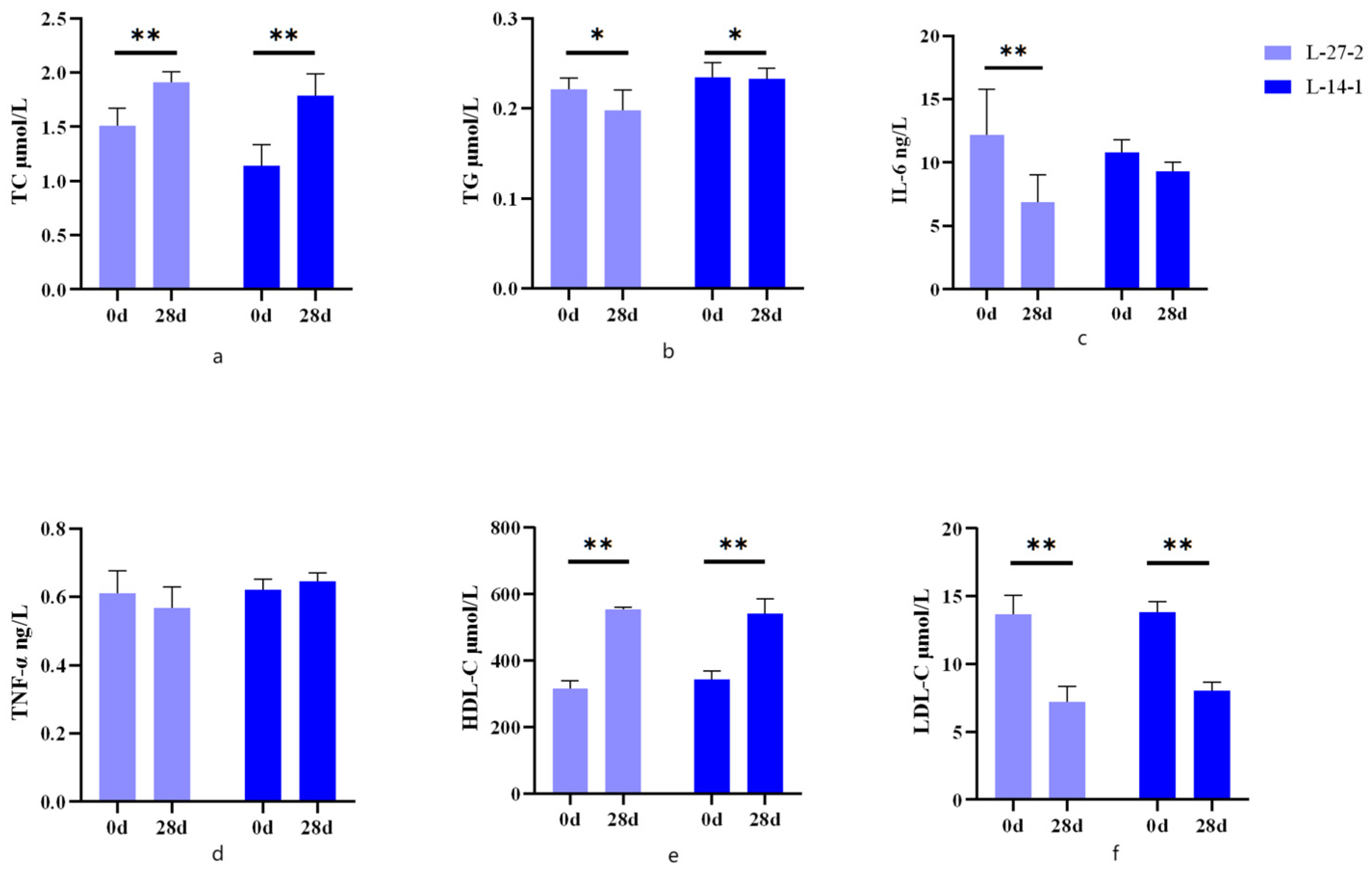
Figure 1. Changes in blood chemistry on days 0 and 28 in cats. (a) serum TC content; (b) serum TG content; (c) serum IL-6 content; (d) serum TNF-α content; (e) serum HDL-C content; and (f) serum LDL-C content. * p < 0.05. ** p < 0.01.
Microorganisms 12 02446 g001
Figure 2. Fecal Microbiota Analysis. (a) α-diversity (Shannon index) in the L-14-1 group; (b) PCoA analysis of the L-14-1 group; (c) α-diversity (Shannon index) in the L-27-2 group; and (d) PCoA analysis of the L-27-2 group.
Microorganisms 12 02446 g002
Figure 3. (a) Abundance of phylum level of fecal microbiota on day 0 and 28 of the L-14-1 group; (b) abundance of genus level of fecal microbiota on day 0 and 28 of the L-14-1 group; (c) significance of the genus differences in the L-14-1 group; (d) abundance of phylum level of fecal microbiota on day 0 and 28 of the L-27-2 group; (e) abundance of genus level of fecal microbiota on day 0 and 28 of the L-27-2 group; and (f) significance of the genus differences in the L-27-2 group.* p < 0.05.
Microorganisms 12 02446 g003
Disclaimer/Publisher’s Note: The statements, opinions and data contained in all publications are solely those of the individual author(s) and contributor(s) and not of MDPI and/or the editor(s). MDPI and/or the editor(s) disclaim responsibility for any injury to people or property resulting from any ideas, methods, instructions or products referred to in the content.