Article

Feline Vector-Borne Diseases and Their Possible Association with Hematological Abnormalities in Cats from Midwestern Brazil

by
Stephani Félix Carvalho
1,
Gracielle Teles Pádua
1,
Warley Vieira de Freitas Paula
1,
Mariana Avelar Tavares
1,
Lucianne Cardoso Neves
1,
Brenda Gomes Pereira
1,
Rayane Almeida Santos
1,
Gabriel Cândido dos Santos
1,
Ennya Rafaella Neves Cardoso
1,
Andriele Ferreira Qualhato
2,
Raphaela Bueno Mendes Bittencourt
1,
Nicolas Jalowitzki de Lima
1,
Danieli Brolo Martins
2,
Filipe Dantas-Torres
3 and
Felipe da Silva Krawczak
1,*
1
Laboratório de Doenças Parasitárias (LADOPAR), Setor de Medicina Veterinária Preventiva, Escola de Veterinária e Zootecnia, Universidade Federal de Goiás—UFG, Goiânia 74690-900, GO, Brazil
2
Laboratório de Patologia Clínica Veterinária, Escola de Veterinária e Zootecnia, Universidade Federal de Goiás—UFG, Goiânia 74690-900, GO, Brazil
3
Departamento de Imunologia, Instituto Ageu Magalhães—IAM, Fundação Oswaldo Cruz (Fiocruz), Recife 50740-465, PE, Brazil
*
Author to whom correspondence should be addressed.
Microorganisms 2024, 12(11), 2171; https://doi.org/10.3390/microorganisms12112171
Submission received: 6 October 2024 / Revised: 21 October 2024 / Accepted: 25 October 2024 / Published: 29 October 2024
(This article belongs to the Special Issue Detection, Diagnosis, and Host Interactions of Animal Mycoplasmas)

Abstract

:
Among the parasitic and infectious diseases affecting cats, those caused by vector-borne pathogens deserve attention due to their ability to cause nonspecific clinical signs and clinicopathological abnormalities. We studied the presence of Cytauxzoon spp., Ehrlichia spp., and Mycoplasma spp. in blood samples from 135 cats referred to the veterinary teaching hospital of the Federal University of Goiás in midwestern Brazil. We also investigated co-infections with Feline Immunodeficiency Virus (FIV) and Feline Leukemia Virus (FeLV) as well as the correlation between Mycoplasma spp. infection and cat variables, including age, sex, breed, and complete blood count abnormalities. Upon PCR testing, 20.7% (28/135) of samples were positive for Mycoplasma spp., 1.5% (2/135) for Cytauxzoon spp., and none for Ehrlichia spp. Co-infections with Mycoplasma spp. and Cytauxzoon spp. were detected in the two cats with the latter infection. Mycoplasma spp. infection was statistically associated with the simultaneous presence of thrombocytopenia and leukocytosis. This study confirms a high frequence of Mycoplasma spp. infection, with both M. haemofelis and ‘Candidatus Mycoplasma haemominutum’ circulating in this cat population. The clinical significance of Mycoplasma spp. infection in cats should be further explored and this infection should eventually be included in the differential diagnosis of thrombocytopenia and leukocytosis in otherwise apparently healthy cats.

1. Introduction

Feline vector-borne diseases (FVBDs) affect cats worldwide but are often underestimated as most of the infected cats present a subclinical infection. When present, clinical signs and clinicopathological abnormalities of FVBDs may be nonspecific, thus making it difficult for veterinary practitioners to diagnose this group of diseases [1,2,3]. Cats are more frequently affected by fleas than ticks, sometimes participating in the transmission cycle of flea-borne bacteria of zoonotic concern [4,5].
One of most frequent FVBDs is mycoplasmosis, a disease caused by pleomorphic bacteria that locate on the surface of erythrocytes [6], sometimes inducing hemolytic anemia and fever [7,8]. The prevalence of Mycoplasma spp. infection varies widely and co-infections with other vector-borne agents (e.g., Bartonella henselae, Cytauxzoon felis, Leishmania infantum) have been reported [2,9,10]. Recent studies indicate that Mycoplasma spp. are transmitted by multiple pathways, including through vectors, blood transfusion, and aggressive interactions [11].
Domestic cats are mainly infected by three species of hemotropic mycoplasmas (Mycoplasma haemofelis, ‘Candidatus Mycoplasma haemominutum’, and ‘Candidatus Mycoplasma turicensis’), which may cause different clinical signs in infected cats, but are often associated with subclinical infection [7,12]. These three species have been detected in cats in Brazil [13]. Among other vector-borne agents detected in cats worldwide, Ehrlichia spp. and Cytauxzoon spp. may also be associated with nonspecific clinical signs, making the use of molecular techniques essential for accurate diagnosis and subsequent appropriate treatment [1,14].
FVBDs are endemic in all Brazilian regions, but a relatively low number of studies on Cytauxzoon, Ehrlichia, and Mycoplasma infections in cats have been conducted in midwestern Brazil [15,16,17,18]. Moreover, no study has assessed the association of infection by Mycoplasma spp. and complete blood count (CBC) abnormalities in a population of cats. In this perspective, this molecular survey aimed to detect Cytauxzoon spp., Ehrlichia spp., and Mycoplasma spp. in cats referred to a teaching veterinary hospital in midwestern Brazil. We also investigated existing co-infections with Feline Immunodeficiency Virus (FIV) and Feline Leukemia Virus (FeLV) and assessed the correlation between Mycoplasma spp. positivity and cat variables, including age, sex, breed, and CBC abnormalities.

2. Materials and Methods
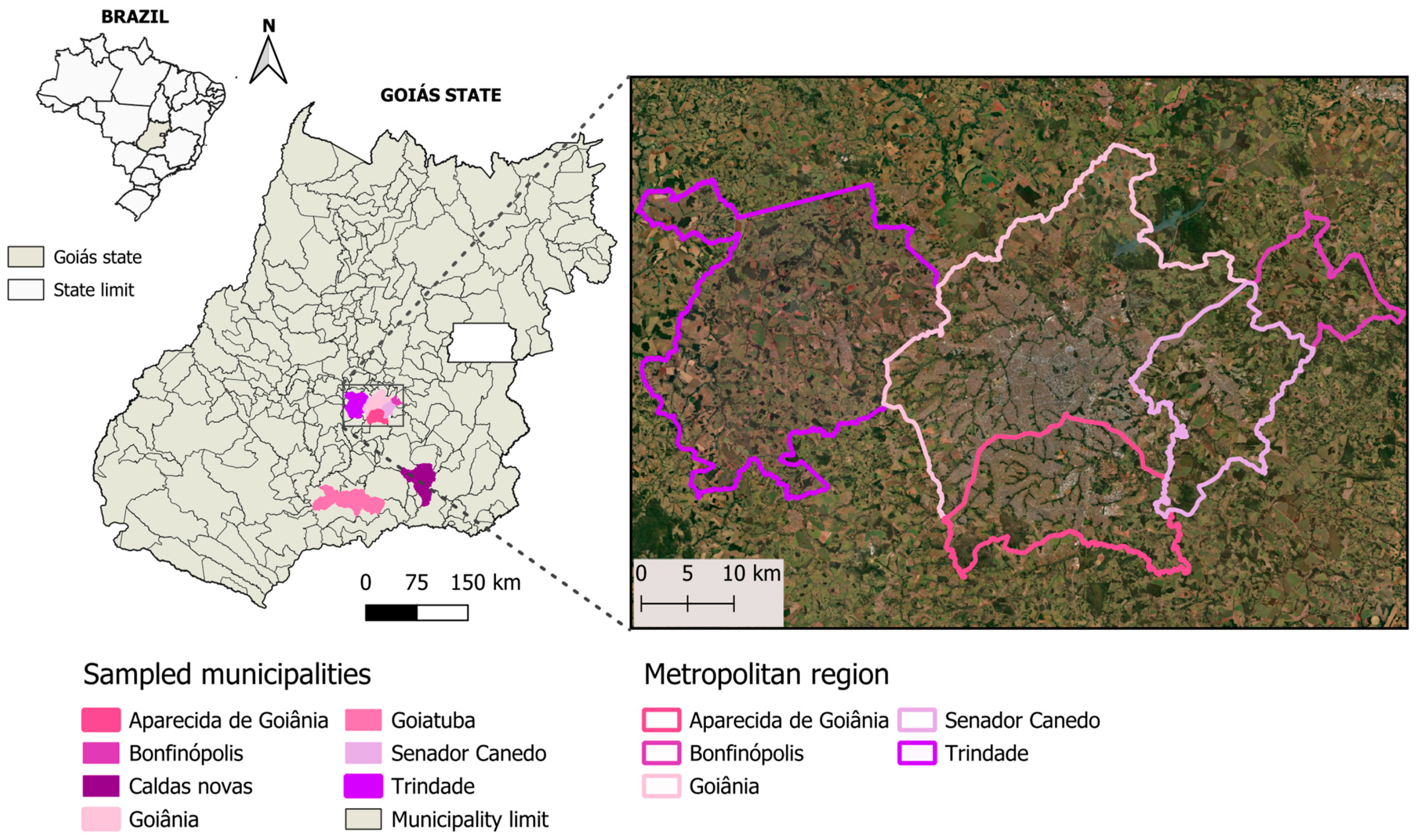
2.1. Study Area

The study was conducted in the state of Goiás in midwestern Brazil. This region has a tropical climate with two well-defined seasons characterized by a dry period between May and September and a rainy period between October and April. The blood samples from cats included in this study were provided by the veterinary teaching hospital of the Federal University of Goiás. The hospital is located in Goiânia city (16°40′ S, 49°15′ W), Goiás state, but cats came from several municipalities: Goiânia (n = 120), Aparecida de Goiânia (n = 6), Trindade (n = 3), Senador Canedo (n = 3), Caldas Novas (n = 1), Bonfinópolis (n = 1) and Goiatuba (n = 1) (Figure 1). Cats were referred to the hospital for routine procedures or other reasons, not necessarily due to suspicion of FVBDs.

2.2. Sample Collection

Blood samples were collected (convenience sampling) between January 2022 and December 2023, as part of the routine veterinary care provided by the veterinary teaching hospital. The samples were processed for CBC and then stored at −20 °C until DNA extraction, as described below.
A total of 135 EDTA blood samples (minimum volume, 150 μL) were randomly selected, regardless of the cat’s history and clinical suspicion. Data including municipality of residence, sex, breed, age, and serological results for feline retroviruses (FIV and FeLV) were obtained for each cat included in this study.
The Ethics Committee on Animal Use of the Federal University of Goiás (CEUA/UFG) approved this study (protocol number MB105/23), which was conducted in accordance with the ethical principles of animal experimentation.

2.3. CBC Analysis

The CBC analysis included red blood cells (×106/μL), hemoglobin (g/dL), mean corpuscular volume (MCV) (fl), mean corpuscular hemoglobin concentration (MCHC) (%), platelets (×103/μL), and white blood cells (μL), which were measured using an automated cell counter (Celltac Alpha/MEK-6550®, Nihon Kohden, Shinjuku City, Japan). A blood smear was also performed on all samples to check the results of the automatic count, to carry out the differential count of white blood cells, to determine the morphology of red blood cells, leukocytes, and platelets, and to analyze the presence of blood pathogens. The packed cell volume (PCV) (%) was determined by the microhematocrit method.
The CBC reference values considered in this study were based on the literature [19] as follows: 5.50–10.00 × 10⁶/μL for red blood cells, 8.0–15.0 g/dL for hemoglobin, 24–45% for hematocrit, 300–800 × 103/μL for platelets, and 5500–19,000/μL for white blood cells.

2.4. Immunochromatography for FIV and FeLV

During clinical care, 40% (54/135) of cats were tested for the detection of antibodies to FIV and antigens of FeLV; the assay was performed using the SNAP® ELISA FIV/FELV Combo (IDEXX Laboratories, INC, Westbrook, ME, USA), according to the manufacturer’s guidelines. This ELISA detects FeLV antigens and anti-FIV antibodies. The test was conducted on cats without a history of being tested. The test results were extracted from the electronic medical record of the veterinary teaching hospital of the Federal University of Goiás (HV/EVZ/UFG).

2.5. DNA Extraction

DNA was extracted from whole blood samples using the DNeasy Blood and Tissue Kit (QIAGEN, Chatsworth, CA, USA), following the manufacturer’s instructions. After extraction, the DNA samples were stored at −20 °C. The concentration and purity of the obtained DNA samples were assessed using a spectrophotometer (Nanodrop®, Thermo Fisher Scientific, Waltham, MA, USA).

2.6. PCR Assays

DNA samples were tested by conventional PCR (cPCR) assays for the detection of Cytauxzoon spp. and Ehrlichia spp. and a real-time PCR (qPCR) for Mycoplasma spp. If positive in the qPCR for Mycoplasma, the sample was further tested by a cPCR targeting a fragment (≈600 bp) of the 16S rRNA gene of Mycoplasma spp. Details on the target genes, primers, and the expected fragment are reported in Table 1.
cPCR assays were performed using 5 μL of DNA template in a mix containing 12.5 μL of DreamTaq® (Green PCR Master Mix 2X, Thermo Fisher Scientific), 1.5 μL of each primer (forward and reverse) at a concentration of 10 pmol, and 4.5 μL of nuclease-free ultrapure water, totaling a 25 μL reaction volume. The qPCR assays were conducted using the StepOnePlusTM Real-Time PCR System (Applied Biosystems, Foster City, CA, USA), as described by Willi et al. [20]. The 20 μL reaction mixture contained 1.5 μL of DNA template, 10 μL of SYBR Green real-time PCR Master Mix (Applied Biosystems, Thermo Fisher Scientific), 0.5 μL of each primer (forward and reverse) at a concentration of 10 pmol, and 7.5 μL of nuclease-free ultrapure water.
Table 1. Primers used for the detection of Cytauxzoon, Ehrlichia, and Mycoplasma DNA in cats from Goiás state, midwestern Brazil.
Agents (Target Gene)PrimersPrimer SequencesProduct Size (bp)References
Cytauxzoon spp. (18S rRNA) bCytaux FGCGAATCGCATTGCTTTATGCT284[21]
Cytaux RCCAATTGATACTCCGGAAAGAG
Ehrlichia spp. (dsb) bDsb-330 (F)GATGATGTCTGAAGATATGAAACAAAT409[22]
Dsb-728 (R)CTGCTCGTCTATTTTACTTCTTAAAGT
Mycoplasma spp. (16S rRNA) aSYBR_ForAGCAATRCCATGTGAACGATGAA 134[20] a
SYBR_Rev1TGGCACATAGTTWGCTGTCACTT
Mycoplasma spp. (16S rRNA) bHBT-FATACGGCCCATATTCCTACG595–620[23] b
HBT-RTGCTCCACCACTTGTTCA
a qPCR; b cPCR.
No-template controls (PCR-grade water, Sigma-Aldrich, St. Louis, MO, USA) and an appropriate positive control sample (DNA of Cytauxzoon, Ehrlichia or Mycoplasma) were included in each PCR run. Negative samples were further tested using a cPCR assay targeting the cytochrome b gene (cytB) of mammals [24,25] to validate the DNA extraction protocol. If a sample did not produce the expected product in this cPCR assay, the sample was excluded from analysis. cPCR products were stained with SYBR Safe (Invitrogen, Thermo Fisher Scientific, Waltham, MA, USA), following the manufacturer’s recommendations, and visualized by electrophoresis in 1.5% agarose gel with an ultraviolet transilluminator.

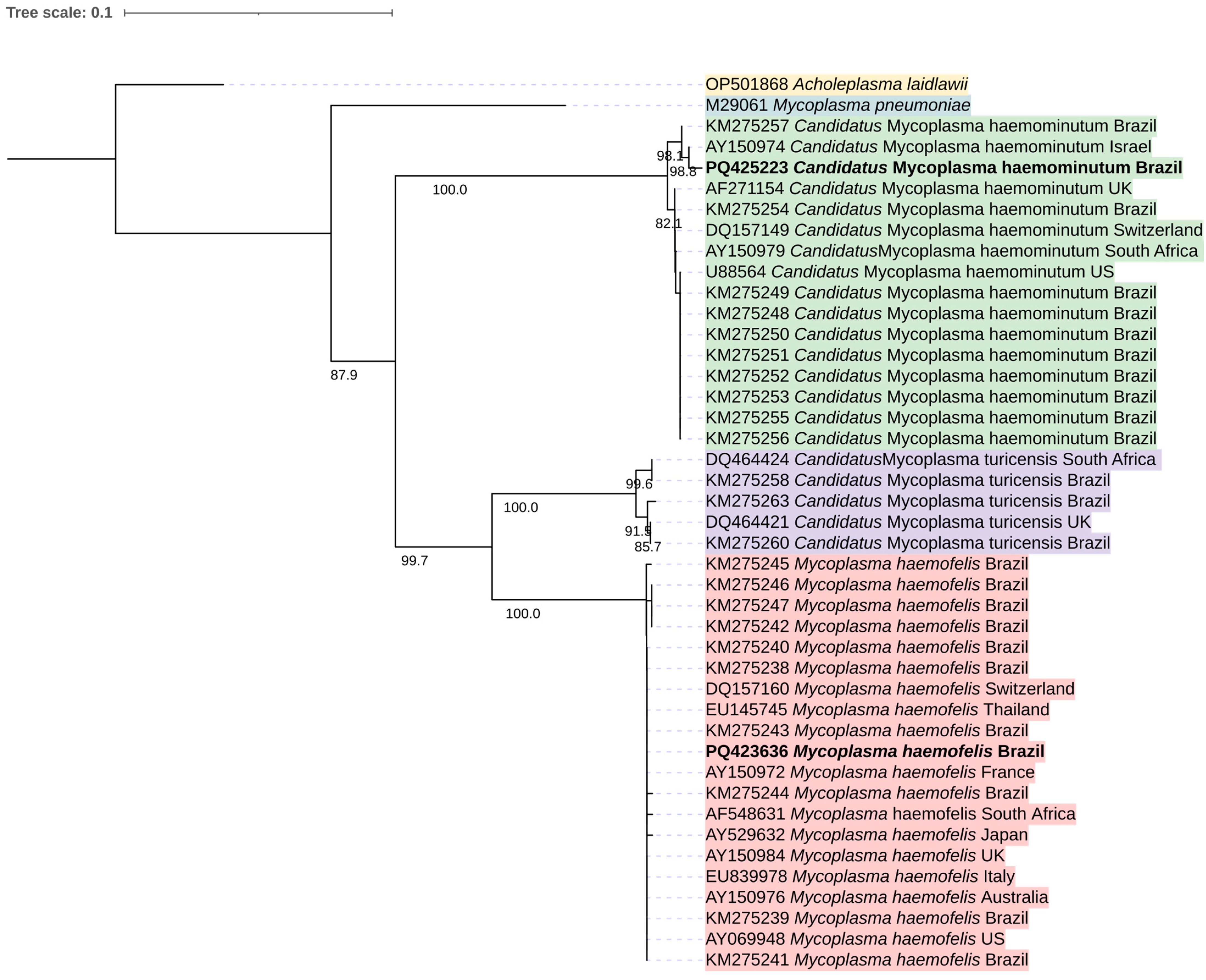
2.7. Partial Mycoplasma spp. 16S rRNA Sequencing and Analysis

Ten PCR amplicons were purified using PureLink™ PCR Micro Kit (Invitrogen, Carlsbad, CA, USA) and prepared for sequencing using BigDye terminator v3.1 (Applied Biosystems, Foster City, CA, USA). Sequencing was conducted in both directions and using the same primers as for PCR, in a 3500xL genetic analyzer (Applied Biosystems, Foster City, CA, USA).
Sequence editing and alignment were conducted using Geneious Prime® 2023.2.1. In brief, low-quality ends from both forward and reverse sequences were trimmed (error probability limit value of 0.05). Then, sequences were verified by eye, edited manually, and aligned using ClustalW (version 2.1). Consensus sequences were compared to sequences available in GenBank using the Basic Local Alignment Search Tool (BLASTn; http://blast.ncbi.nlm.nih.gov/Blast.cgi accessed on 29 September 2024).
16S rRNA sequences from hemotropic Mycoplasma spp. detected in cats in different countries [12] were exported from GenBank and aligned using ClustalW. The evolutionary history was inferred using the neighbor-joining method [26]. The percentage of replicate trees in which the associated taxa clustered together in the bootstrap test (10,000 replicates) are shown above the branches [27]. The evolutionary distances were computed using the p-distance method [28] and are in the units of the number of base differences per site. This analysis involved 43 sequences. All ambiguous positions were removed for each sequence pair (pairwise deletion option). There were 524 positions in the final dataset. Evolutionary analyses were conducted in MEGA11 [29,30]. Acholeplasma laidlawii (accession number: OP501868) and Mycoplasma pneumoniae (accession number: M29061) were used as outgroups. The final phylogenetic three was edited using iTOL v.6 (https://itol.embl.de/ accessed on 30 September 2024).

2.8. Statistical Analysis

The cat breed and age group distributions were assessed using a One-way Chi-squared test in MedCalc Software Ltd. (https://www.medcalc.org/calc/chisquared-1way.php accessed on 30 September 2024). The association between Mycoplasma spp. positivity and other variables (sex, age group, CBC abnormalities, or seropositivity for feline retroviruses) was investigated using a Chi-squared test implemented in QuickCalcs of GraphPad (https://www.graphpad.com/quickcalcs/ accessed on 30 September 2024). A p-value < 0.05 was considered statistically significant.

3. Results

The cat population investigated herein consisted of 70 (51.9%) males and 65 (48.1%) females. Most cats (n = 115) were crossbreeds, and the remaining cats (n = 20) belonged to four breeds: Siamese (n = 14), Persian (n = 4), Angora (n = 1), and Bengal (n = 1) (One-way Chi-squared test = 362.7407; df = 4; p < 0.0001). Most of the cats were 1–6 years old (One-way Chi-squared test = 59.9892; df = 3; p < 0.0001; cats with unknown age excluded from this analysis) (Table 2).
All samples that were negative for Cytauxzoon spp., Ehrlichia spp., and Mycoplasma spp. tested positive in the cPCR assay for the detection of the mammalian endogenous control (cytB gene) and, therefore, were included in the analyses. Out of 135 cats tested, 28 (20.7%) were positive for Mycoplasma spp., 2 (1.5%) for Cytauxzoon spp., and none for Ehrlichia spp. The positivity for Mycoplasma spp. was significantly higher in males (34.3%) than in females (6.2%) (Two-way Chi-squared test = 16.226, df = 1, p = 0.0001), but no differences were found in relation to age group (Two-way Chi-squared test = 3.5224, df = 3, p = 0.3179; cats with unknown age excluded) or breed (Two-way Chi-squared test = 3.2767, df = 4, p = 0.5126).
Out of 28 cats positive for Mycoplasma spp. by qPCR, 27 were positive by cPCR. Ten positive samples with more DNA (larger and brighter positive bands in the agarose gel) were selected for sequencing, but only two partial 16S rRNA gene sequences were successfully generated, one (491 bp) corresponding to Mycoplasma haemofelis (100% query coverage and identity with several sequences in GenBank: MN240855.1, MK632350.1, PP494713.1) and the other (450 bp) to ‘Candidatus Mycoplasma haemominutum’ (100% query coverage and identity with several sequences in GenBank: MK632390.1, OQ397117.1, MW598400.1). The low number (only two) of sequences obtained can be attributed to the low number of copies of the target DNA in the tested samples. Our sequences were deposited in GenBank under the accession numbers PQ423636 (Mycoplasma haemofelis) and PQ425223 (‘Candidatus Mycoplasma haemominutum’). Our sequences clustered with sequences of M. haemofelis and ‘Candidatus Mycoplasma haemominutum’ previously detected in cats from Brazil and from other countries (Figure 2).
Out of fifty-four cats tested for feline retroviruses, nine (16.7%) were positive for FeLV, one (1.8%) for FIV, and two (3.7%) for both species. Among the Mycoplasma spp.-positive cats, four (14.3%) were co-infected with FIV, FeLV, or both (Table 3).
Most of the cats (80.7%) presented at least one CBC abnormality. The results of CBC alterations related to Mycoplasma spp. qPCR positivity and FIV/FeLV seropositivity are shown in Table 4. Mycoplasma qPCR positivity was statistically associated with the simultaneous presence of thrombocytopenia and leukocytosis (Table 5).
Through the cytological examination of stained blood smears, only two cats (1.5%) were positive for FVBDs showing structures suggestive of Mycoplasma spp.; these animals were positive in the qPCR assay for Mycoplasma spp.

4. Discussion

The prevalence (20.7%) of Mycoplasma spp. infection in cats examined herein is consistent with that reported in some studies conducted in Brazil, but it is acknowledged that prevalence data vary widely among studies [13]. Indeed, such comparisons should be made with caution due to the differences in terms of the study populations (e.g., healthy versus sick cats) and diagnostic techniques [13]. For example, our study assessed a cat population that sought veterinary care, and this may have influenced our results. Because we did not evaluate a healthy cat population, our results may not represent the actual prevalence of Mycoplasma spp. in the overall cat population in the region. Similarly, the low positivity detected by cytological examination of blood smears highlights its lower sensitivity as compared to PCR and emphasizes the importance of using molecular tools for a reliable diagnosis of Mycoplasma spp. infection in cats [15]. In fact, due to cyclic bacteremia and the chronic nature of Mycoplasma spp. in cats, cytology is not reliable for diagnosing this infection [13,31].
Although species-specific PCR assays have been developed for Mycoplasma spp., their use as screening tools is limited due to the diversity of species [20]. For this reason, we used an SYBR Green-based qPCR assay targeting a fragment of the 16S rRNA gene of Mycoplasma spp., which presents 98.2% sensitivity and 92.1% specificity for feline hemoplasmas [20]. This assay was chosen for initial screening due to its ability to detect several Mycoplasma spp., including potentially new species [20].
The higher prevalence of hemotropic mycoplasmas in male cats has been reported in other studies and can partly be explained by their territorial behavior, which results in frequent intraspecific aggressions, thus increasing the possibility of bite-related transmission [11]. Furthermore, Mycoplasma spp. infections are more common in stray cats or those that roam freely between the house and the street [13].
Thrombocytopenia and leukocytosis were associated with Mycoplasma spp. infection in the present study, although actual species involved in most of these cases could not be determined. Thrombocytopenia is not a feature of M. haemofelis infection in cats, but a previous study also found an association between thrombocytopenia and M. haemofelis infection in southeastern Brazil [8]. The changes in the leukogram of cats with mycoplasmosis usually do not follow a defined pattern [32]. However, the leukocytosis observed in the present study may be related to the inflammatory process. Leukocytosis was also observed in felines affected by M. haemofelis, which showed increased inflammatory biomarkers in response to endothelial glycocalyx damage, a regulator of leukocyte adhesion and migration within the blood vessel [33]. M. haemofelis is the most pathogenic Mycoplasma sp. in cats and may cause hemolytic anemia, even in immunocompetent cats [8]. The lack of association between anemia and M. haemofelis in some studies, including herein, has been attributed to the inclusion of chronically infected asymptomatic cats, considering that anemia is most frequent in the acute phase of the infection [8].
One of the factors that can reduce the frequence of vector-borne pathogens in cats is their grooming behavior that reduces the prevalence and intensity of ectoparasite infestation, especially by ticks [34]. The early removal of ticks by cats may also help prevent pathogen transmission, as ticks need to attach for some time before they can transmit certain pathogens [35]. For instance, the transmission time may vary from 3 to 50 h for Ehrlichia spp. [35,36] and from 36 to 48 h for Cytauxzoon spp. [37].
Another possible reason for the low positivity of Cytauxzoon spp. in our study may be related to the low pathogenicity of the species infecting felids in Brazil [38], as is the case of the recently described Cytauxzoon brasiliensis [39]. Indeed, clinical cases of cytauxzoonosis are considered uncommon in this country and it is plausible to infer that cats included in this study were not referred to the hospital due to clinical suspicion of cytauxzoonosis.
Despite the low positivity, this is the first report of Cytauxzoon spp. infecting domestic cats in Goiás, midwestern Brazil, where another study detected Cytauxzoon felis in free-ranging Puma concolor [40]. It is known that cytauxzoonosis has ixodid ticks as definitive hosts and felids as intermediate hosts [16]. For this reason, this is a disease more commonly detected in wild felids. Concerning ixodid ticks, Amblyomma parvum was found to parasitize Leopardus pardalis infected with Cytauxzoon sp. (phylogenetically related to C. felis). However, the collected ticks were negative for Cytauxzoon spp. [41].
In a recent study, Fagundes-Moreira et al. [42] highlighted that Amblyomma sculptum could be a possible vector of Cytauxzoon spp., as several ticks of this species were found on infected wild felids in central-western Brazil. The ecological characteristics of Amblyomma spp. make their parasitism on domestic cats relatively rare, as they are typically found in forests and pastures. The positive cats found in this study were free of ticks, and their domiciled or semi-domiciled lifestyle makes their infestation by Amblyomma spp. ticks unlikely.
The absence of Ehrlichia spp. in the studied cats is consistent with other studies conducted in Rio Grande do Sul [43] and São Paulo [17], but some studies have reported the presence of E. canis in cats in Brazil [1,35]. In a study carried out in different Brazilian states, infected cats were detected in Minas Gerais and São Paulo, but not in Rondônia [1]. Overall, 2% of the 306 cats tested positive for the Ehrlichia spp. dsb gene and all sequences were assigned to E. canis [1]. Studies conducted in other countries such as Spain, Portugal, and Germany have reported the molecular detection of Ehrlichia spp. in 0 to 5.4% of the examined cats [2,44,45,46]. While we did not detect positive cats, Paula et al. [47] conducted a study in the same area investigated herein and reported a seroprevalence of 59.1% for E. canis in dogs. The high prevalence of E. canis in dogs in areas where R. linnaei predominate is a common occurrence in Brazil [48]. In this regard, adults of R. linnaei are more efficient than nymphs for transmitting E. canis [49], but are rarely found parasitizing cats [4]. The lack of exposure of cats to R. linnaei adults and nymphs may be the reason for the absence of E. canis-positive cats in our study.
This work presents a main limitation, which is the fact that cats included in the analyses were referred for veterinary care, which could result in an overestimation of the CBC abnormalities. Indeed, CBC abnormalities in naturally infected cats should be interpreted with caution due to the possibility of comorbidities that may not have been deeply investigated herein. This is particularly true for leukocytosis, which is not a common feature of Mycoplasma spp. infection in cats.

5. Conclusions

This study reports the presence of Cytauxzoon spp. and Mycoplasma spp. in cats in Goiás, expanding the known geographical distribution of these FVBD agents in South America. We also found an association between Mycoplasma qPCR positivity and the simultaneous presence of thrombocytopenia and leukocytosis, indicating that the clinical significance of Mycoplasma spp. infection in Brazilian cats should be further explored. Eventually, Mycoplasma spp. infection should be considered as a differential diagnosis of thrombocytopenia and leukocytosis in otherwise apparently healthy cats.

Author Contributions

Conceptualization, S.F.C. and F.d.S.K.; methodology, S.F.C. and F.d.S.K.; investigation, S.F.C., G.T.P., W.V.d.F.P., M.A.T., L.C.N., B.G.P., R.A.S., G.C.d.S., E.R.N.C., A.F.Q., R.B.M.B., N.J.d.L., D.B.M. and F.D.-T.; resources, D.B.M., F.D.-T. and F.d.S.K.; data curation, S.F.C., F.D.-T. and F.d.S.K.; writing—original draft preparation, S.F.C., F.D.-T. and F.d.S.K.; writing—review and editing, S.F.C., F.D.-T., N.J.d.L. and F.d.S.K.; visualization, F.D.-T. and F.d.S.K.; supervision, F.d.S.K.; project administration, S.F.C.; funding acquisition, F.d.S.K. All authors have read and agreed to the published version of the manuscript.

Funding

This research was funded by the Fundação de Amparo à Pesquisa do Estado de Goiás. F.d.S.K. received a productivity scholarship from Conselho Nacional de Desenvolvimento Científico e Tecnológico (CNPq-process number: 317557/2021-1).

Data Availability Statement

Data supporting the conclusions of this study are included in the manuscript. The 16S rRNA sequences generated herein were deposited in GenBank (accession numbers: PQ423636; PQ425223).

Conflicts of Interest

The authors declare no conflicts of interest.

References

  1. André, M.R.; Calchi, A.C.; Furquim, M.E.C.; de Andrade, I.; Arantes, P.V.C.; de Melo Lopes, L.C.; Demarchi, I.K.L.D.N.; Figueiredo, M.A.P.; de Paula Lima, C.A.; Machado, R.Z. Molecular detection of tick-borne agents in cats from southeastern and northern Brazil. Pathogens 2022, 11, 106. [Google Scholar] [CrossRef] [PubMed]
  2. Latrofa, M.S.; Iatta, R.; Toniolo, F.; Furlanello, T.; Ravagnan, S.; Capelli, G.; Schunack, B.; Chomel, B.; Zatelli, A.; Mendoza-Roldan, J.; et al. A molecular survey of vector-borne pathogens and haemoplasmas in owned cats across Italy. Parasite Vectors 2020, 13, 116. [Google Scholar] [CrossRef] [PubMed]
  3. Lopes, M.G.; Krawczak, F.D.S.; de Lima, J.T.R.; Fournier, G.F.D.S.R.; Acosta, I.D.C.L.; Ramirez, D.G.; Marcili, A.; Labruna, M.B.; Gennari, S.M. Occurrence of Ehrlichia canis and Hepatozoon canis and probable exposure to Rickettsia amblyommatis in dogs and cats in Natal, RN. Rev. Bras. Parasitol. Vet. 2019, 28, 151–156. [Google Scholar] [CrossRef] [PubMed]
  4. Maggi, R.G.; Halls, V.; Krämer, F.; Lappin, M.; Pennisi, M.G.; Peregrine, A.S.; Roura, X.; Schunack, B.; Scorza, V.; Tasker, S.; et al. Vector-borne and other pathogens of potential relevance disseminated by relocated cats. Parasite Vectors 2022, 15, 415. [Google Scholar] [CrossRef] [PubMed]
  5. Morelli, S.; Diakou, A.; Di Cesare, A.; Colombo, M.; Traversa, D. Canine and feline parasitology: Analogies, differences, and relevance for human health. Clin. Microbiol. Rev. 2021, 34, e0026620. [Google Scholar] [CrossRef]
  6. de Santis, A.G.G.A.; Herrera, H.M.; Sousa, K.C.M.; Gonçalves, L.R.; Denardi, N.C.B.; Domingos, I.H.; Campos, J.B.V.; Machado, R.Z.; André, M.R. Molecular detection of hemotrophic mycoplasmas among domiciled and free-roraming cats in Campo Grande, state of Mato Grosso do Sul, Brazil. Rev. Bras. Parasitol. Vet. 2014, 23, 231–236. [Google Scholar] [CrossRef]
  7. Tasker, S. Hemotropic Mycoplasma. Vet. Clin. N. Am. Small. Anim. Pract. 2022, 52, 1319–1340. [Google Scholar] [CrossRef]
  8. Tasker, S.; Hofmann-Lehmann, R.; Belák, S.; Frymus, T.; Addie, D.D.; Pennisi, M.G.; Boucraut-Baralon, C.; Egberink, H.; Hartmann, K.; Hosie, M.J.; et al. Haemoplasmosis in cats: European guidelines from the ABCD on prevention and management. J. Feline Med. Surg. 2018, 20, 256–261. [Google Scholar] [CrossRef]
  9. Barker, E.N. Update on feline hemoplasmosis. Vet. Clin. N. Am. Small Anim. Pract. 2019, 49, 733–743. [Google Scholar] [CrossRef]
  10. Maia, L.M.P.; Cerqueira, A.d.M.F.; Macieira, D.; de Souza, A.M.; Moreira, N.S.; da Silva, A.V.; Messick, J.B.; Ferreira, R.F.; Almosny, N.R.P. Cytauxzoon felis and “Candidatus Mycoplasma haemominutum” coinfection in a brazilian domestic cat (Felis catus). Rev. Bras. Parasitol. Vet. 2013, 22, 289–291. [Google Scholar] [CrossRef]
  11. Munhoz, A.D.; Simões, I.G.P.C.; Calazans, A.P.F.; Macedo, L.S.; Cruz, R.D.S.; Lacerda, L.C.; Said, R.A.; André, M.R. Hemotropic mycoplasmas in naturally infected cats in northeastern Brazil. Rev. Bras. Parasitol. Vet. 2018, 27, 446–454. [Google Scholar] [CrossRef] [PubMed]
  12. Aquino, L.C.; Hicks, C.A.E.; Scalon, M.C.; Lima, M.G.d.M.; Lemos, M.d.S.; Paludo, G.R.; Helps, C.R.; Tasker, S. Prevalence and phylogenetic analysis of haemoplasmas from cats infected with multiple species. J. Microbiol. Methods 2014, 107, 189–196. [Google Scholar] [CrossRef] [PubMed]
  13. Biondo, A.W.; dos Santos, A.P.; Guimarães, A.M.S.; Vieira, R.F.d.C.; Vidotto, O.; Macieira, D.d.B.; Almosny, N.R.P.; Molento, M.B.; Timenetsky, J.; de Morais, H.A.; et al. A review of the occurrence of hemoplasmas (Hemotrophic mycoplasmas) in Brazil. Rev. Bras. Parasitol. Vet. 2009, 18, 1–7. [Google Scholar] [CrossRef] [PubMed]
  14. Lappin, M.R. Update on flea and tick associated diseases of cats. Vet. Parasitol. 2018, 254, 26–29. [Google Scholar] [CrossRef]
  15. André, M.R.; Filgueira, K.D.; Calchi, A.C.; de Sousa, K.C.M.; Gonçalves, L.R.; Medeiros, V.B.; Ximenes, P.A.; Lelis, V.C.N.G.; de Meireles, M.V.N.; Machado, R.Z. Co-infection with arthropod-borne pathogens in domestic cats. Rev. Bras. Parasitol. Vet. 2017, 26, 525–531. [Google Scholar] [CrossRef]
  16. André, M.R.; Herrera, H.M.; de Jesus Fernandes, S.; de Sousa, K.C.M.; Gonçalves, L.R.; Domingos, I.H.; de Macedo, G.C.; Machado, R.Z. Tick-borne agents in domesticated and stray cats from the city of Campo Grande, state of Mato Grosso do Sul, midwestern Brazil. Ticks Tick-Borne Dis. 2015, 6, 779–786. [Google Scholar] [CrossRef]
  17. André, M.R.; Baccarim Denardi, N.C.; Marques de Sousa, K.C.; Gonçalves, L.R.; Henrique, P.C.; Grosse Rossi Ontivero, C.R.; Lima Gonzalez, I.H.; Cabral Nery, C.V.; Fernandes Chagas, C.R.; Monticelli, C.; et al. Arthropod-borne pathogens circulating in free-roaming domestic cats in a zoo Environment in Brazil. Ticks Tick-Borne Dis. 2014, 5, 545–551. [Google Scholar] [CrossRef]
  18. Pedrassani, D.; Biolchi, J.; Gonçalves, L.R.; Mendes, N.S.; Zanatto, D.C.d.S.; Calchi, A.C.; Machado, R.Z.; André, M.R. Molecular detection of vector-borne agents in cats in southern Brazil. Rev. Bras. Parasitol. Vet. 2019, 28, 632–643. [Google Scholar] [CrossRef]
  19. Weiss, D.J.; Wardrop, K.J. (Eds.) Schalm’s Veterinary Hematology, 6th ed.; Blackwell Publishing: Ames, IA, USA, 2010; ISBN 9780470961834. [Google Scholar]
  20. Willi, B.; Meli, M.L.; Lüthy, R.; Honegger, H.; Wengi, N.; Hoelzle, L.E.; Reusch, C.E.; Lutz, H.; Hofmann-Lehmann, R. Development and application of a universal hemoplasma screening assay based on the SYBR Green PCR principle. J. Clin. Microbiol. 2009, 47, 4049. [Google Scholar] [CrossRef]
  21. Birkenheuer, A.J.; Marr, H.; Alleman, A.R.; Levy, M.G.; Breitschwerdt, E.B. Development and evaluation of a PCR assay for the detection of Cytauxzoon felis DNA in feline blood samples. Vet. Parasitol. 2006, 137, 144–149. [Google Scholar] [CrossRef]
  22. Doyle, C.K.; Labruna, M.B.; Breitschwerdt, E.B.; Tang, Y.W.; Corstvet, R.E.; Hegarty, B.C.; Bloch, K.C.; Li, P.; Walker, D.H.; McBride, J.W. Detection of medically important Ehrlichia by quantitative multicolor TaqMan real-time polymerase chain reaction of the Dsb gene. J. Mol. Diagn. 2005, 7, 504–510. [Google Scholar] [CrossRef] [PubMed]
  23. Criado-Fornelio, A.; Martinez-Marcos, A.; Buling-Saraña, A.; Barba-Carretero, J.C. Presence of Mycoplasma haemofelis, Mycoplasma haemominutum and Piroplasmids in cats from southern Europe: A molecular study. Vet. Microbiol. 2003, 93, 307–317. [Google Scholar] [CrossRef] [PubMed]
  24. Kocher, T.D.; Thomas, W.K.; Meyer, A.; Edwards, S.V.; Paabo, S.; Villablanca, F.X.; Wilson, A.C. Dynamics of Mitochondrial DNA Evolution in animals: Amplification and sequencing with conserved primers. Proc. Natl. Acad. Sci. USA 1989, 86, 6196. [Google Scholar] [CrossRef]
  25. Steuber, S.; Abdel-Rady, A.; Clausen, P.H. PCR-RFLP Analysis: A promising technique for host species identification of blood meals from Tsetse flies (Diptera: Glossinidae). Parasitol. Res. 2005, 97, 247–254. [Google Scholar] [CrossRef] [PubMed]
  26. Saitou, N.; Nei, M. The neighbor-joining method: A new method for reconstructing phylogenetic trees. Mol. Biol. Evol. 1987, 4, 406–425. [Google Scholar] [CrossRef]
  27. Felsenstein, J. Confidence limits on phylogenies: An approach using the bootstrap. Evolution 1985, 39, 783–791. [Google Scholar] [CrossRef]
  28. Nei, M.; Kumar, S. Molecular Evolution and Phylogenetics; Oxford University Press: Oxford, UK, 2000. [Google Scholar] [CrossRef]
  29. Stecher, G.; Tamura, K.; Kumar, S. Molecular evolutionary genetics analysis (MEGA) for MacOS. Mol. Biol. Evol. 2020, 37, 1237–1239. [Google Scholar] [CrossRef]
  30. Tamura, K.; Stecher, G.; Kumar, S. MEGA11: Molecular evolutionary genetics analysis version 11. Mol. Biol. Evol. 2021, 38, 3022–3027. [Google Scholar] [CrossRef]
  31. Raimundo, J.M.; Guimarães, A.; Rodrigues, R.B.; Botelho, C.F.M.; Peixoto, M.P.; Pires, M.S.; Machado, C.H.; Santos, H.A.; Massard, C.L.; André, M.R.; et al. Hematological changes associated with hemoplasma infection in cats in Rio de Janeiro, Brazil. Rev. Bras. Parasitol. Vet. 2016, 25, 441–449. [Google Scholar] [CrossRef]
  32. Maciel, A.d.R.; Biezus, G.; de Cristo, T.G.; Miletti, L.C.; da Costa Maciel, U.; Medeiros, A.L.V.; Xavier, M.G.N.; Casagrande, R.A. Mycoplasma haemofelis infection and its correlation with Feline Leukemia Virus (FeLV) and Feline Immunodeficiency Virus (FIV) in cats in southern Brazil. Comp. Immunol. Microbiol. Infect. Dis. 2023, 93, 101941. [Google Scholar] [CrossRef]
  33. Ider, M.; Ceylan, C.; Naseri, A.; Ceylan, O.; Durgut, M.K.; Ok, M.; Iyigun, S.S.; Erol, B.B.; Sahin, H.B.; Kilickaya, M.C. Evaluation of endothelial glycocalyx injury biomarkers in feline hemotropic mycoplasmosis. Sci. Rep. 2024, 14, 12931. [Google Scholar] [CrossRef] [PubMed]
  34. Eckstein, R.A.; Hart, B.L. Grooming and control of fleas in cats. Appl. Anim. Behav. Sci. 2000, 68, 141–150. [Google Scholar] [CrossRef]
  35. Richards, S.L.; Langley, R.; Apperson, C.S.; Watson, E. Do tick attachment times vary between different tick-pathogen systems? Environments 2017, 4, 37. [Google Scholar] [CrossRef]
  36. Tahir, D.; Meyer, L.; Fourie, J.; Jongejan, F.; Mather, T.; Choumet, V.; Blagburn, B.; Straubinger, R.K.; Varloud, M. Interrupted blood feeding in ticks: Causes and consequences. Microorganisms 2020, 8, 910. [Google Scholar] [CrossRef]
  37. Thomas, J.E.; Ohmes, C.M.; Payton, M.E.; Hostetler, J.A.; Reichard, M.V. Minimum transmission time of Cytauxzoon felis by Amblyomma americanum to domestic cats in relation to duration of infestation, and investigation of ingestion of infected ticks as a potential route of transmission. J. Feline Med. Surg. 2017, 20, 67–72. [Google Scholar] [CrossRef] [PubMed]
  38. de Oliveira, C.M.; Yang, S.; Duarte, M.A.; Figueiredo, D.M.; do Rosario Batista, L.M.; Marr, H.; McManus, C.M.; André, M.R.; Birkenheuer, A.J.; Paludo, G.R. Piroplasmid infection is not associated with clinicopathological and laboratory abnormalities in cats from midwestern Brazil. Parasitol. Res. 2022, 121, 2561–2570. [Google Scholar] [CrossRef]
  39. Duarte, M.A.; de Oliveira, C.M.; Honorato, S.M.; do Rosario Batista, L.M.; Mendonça, J.T.; de Sousa, D.E.R.; Hirano, L.Q.L.; André, M.R.; de Castro, M.B.; Paludo, G.R. Cytauxzoon brasiliensis Sp. Nov. (Apicomplexa: Theileriidae), a new species infecting a little-spotted-cat (Leopardus tigrinus) (Carnivora: Felidae) from Brazil. Syst. Parasitol. 2024, 101, 53. [Google Scholar] [CrossRef] [PubMed]
  40. de Paula, L.G.F.; Weck, B.C.; Neves, L.C.; Paula, W.V.d.F.; Araújo, L.B.d.M.; Martins, D.B.; Peres, P.C.d.O.; Labruna, M.B.; Krawczak, F.d.S. Natural infection and molecular detection of Cytauxzoon felis in a free-ranging Puma concolor in the State of Goiás, Brazil. Ciência Rural 2022, 52, e20210577. [Google Scholar] [CrossRef]
  41. de Sousa, K.C.M.; Fernandes, M.P.; Herrera, H.M.; Freschi, C.R.; Machado, R.Z.; André, M.R. Diversity of piroplasmids among wild and domestic mammals and ectoparasites in Pantanal wetland, Brazil. Ticks Tick-Borne Dis. 2018, 9, 245–253. [Google Scholar] [CrossRef]
  42. Fagundes-Moreira, R.; Souza, U.A.; May-Junior, J.A.; Baggio-Souza, V.; Berger, L.; Wagner, P.G.C.; Mazim, F.D.; Peters, F.B.; Favarini, M.O.; Tortato, M.A.; et al. Epidemiological compatibility of Amblyomma sculptum as possible vector and Panthera onca as reservoir of Cytauxzoon spp. in midwestern Brazil. Ticks Tick-Borne Dis. 2022, 13, 102021. [Google Scholar] [CrossRef]
  43. Malheiros, J.; Costa, M.M.; do Amaral, R.B.; de Sousa, K.C.M.; André, M.R.; Machado, R.Z.; Vieira, M.I.B. Identification of vector-borne pathogens in dogs and cats from southern Brazil. Ticks Tick-Borne Dis. 2016, 7, 893–900. [Google Scholar] [CrossRef] [PubMed]
  44. Villanueva-Saz, S.; Martínez, M.; Nijhof, A.M.; Gerst, B.; Gentil, M.; Müller, E.; Fernández, A.; González, A.; Yusuf, M.S.M.; Greco, G.; et al. Molecular survey on vector-borne pathogens in clinically healthy stray cats in Zaragoza (Spain). Parasite Vectors 2023, 16, 428. [Google Scholar] [CrossRef] [PubMed]
  45. Maia, C.; Ramos, C.; Coimbra, M.; Bastos, F.; Martins, Â.; Pinto, P.; Nunes, M.; Vieira, M.L.; Cardoso, L.; Campino, L. Bacterial and Protozoal Agents of feline vector-borne diseases in domestic and stray cats from southern Portugal. Parasite Vectors 2014, 7, 115. [Google Scholar] [CrossRef] [PubMed]
  46. Bergmann, M.; Englert, T.; Stuetzer, B.; Hawley, J.R.; Lappin, M.R.; Hartmann, K. Prevalence of selected rickettsial infections in cats in southern Germany. Comp. Immunol. Microbiol. Infect. Dis. 2015, 42, 33–36. [Google Scholar] [CrossRef]
  47. Paula, W.V.d.F.; Taques, Í.I.G.G.; Miranda, V.C.; Barreto, A.L.G.; de Paula, L.G.F.; Martins, D.B.; Damasceno, A.D.; Muñoz-Leal, S.; Sevá, A.d.P.; Dantas-Torres, F.; et al. Seroprevalence and hematological abnormalities associated with Ehrlichia canis in dogs referred to a veterinary teaching hospital in central-western Brazil. Ciência Rural 2021, 52, e20201131. [Google Scholar] [CrossRef]
  48. Dantas-Torres, F.; Figueredo, L.A.; Sales, K.G.D.S.; Miranda, D.E.D.O.; Alexandre, J.L.D.A.; Da Silva, Y.Y.; Da Silva, L.G.; Valle, G.R.; Ribeiro, V.M.; Otranto, D.; et al. Prevalence and incidence of vector-borne pathogens in unprotected dogs in two brazilian regions. Parasite Vectors 2020, 13, 195. [Google Scholar] [CrossRef]
  49. Moraes-Filho, J.; Krawczak, F.S.; Costa, F.B.; Soares, J.F.; Labruna, M.B. Comparative evaluation of the vector competence of four south american populations of the Rhipicephalus sanguineus group for the bacterium Ehrlichia canis, the agent of canine monocytic ehrlichiosis. PLoS ONE 2015, 10, e0139386. [Google Scholar] [CrossRef]
Figure 1. Urban locations in the state of Goiás, midwestern Brazil, where animals were sampled in the present study.
Microorganisms 12 02171 g001
Figure 2. Phylogenetic tree reconstruction of the Mycoplasma genus based on a fragment of the 16S rRNA gene using a dataset of 43 sequences (5 species). There were 524 positions in the final dataset. The evolutionary history was inferred using the neighbor-joining method and analyses were conducted in MEGA11. Bootstrap values < 70% are not shown. Acholeplasma laidlawii and Mycoplasma pneumoniae were used as outgroups. The final tree was edited in iTOL v.6.
Microorganisms 12 02171 g002
Table 2. Classification of the cats according to age groups.
Age Groupsn%
Kitten (<1 year)64.4
Young adult (1–6 years)5440
Adult (7–9 years)118.2
Senior (>9 years)2216.3
Unknown4231.1
n = number.
Table 3. Cytauxzoon and retrovirus positivity status in Mycoplasma spp.-positive cats.
Cat NumberMycoplasma qPCR PositivityCytauxzoon cPCR PositivityFIV SeropositivityFELV Seropositivity
1+
2+
3+++
4+ntnt
5+++
6+ntnt
7+
8+ntnt
9+ntnt
10+ntnt
11+
12++ntnt
13+ntnt
14++
15+ntnt
16+ntnt
17+
18+ntnt
19+
20+ntnt
21++
22+ntnt
23+ntnt
24+ntnt
25+
26+
27++
28+ntnt
nt, not tested; +, positive; −, negative.
Table 4. Complete blood count (CBC) abnormalities in cats positive for Mycoplasma qPCR, feline retrovirus (FIV/FeLV) serology, or both.
CBC Abnormalitiesn%Mycoplasma-PositiveFIV/FELV-
Positive
Mycoplasma- and FIV/FELV-Positive
n%n%n%
Anemia21/13515.56/2128.64/2119.01/214.8
Thrombocytopenia71/13552.619/7126.86/718.43/714.2
Thrombocytosis3/1352.20/300/300/30
Leukopenia11/1358.11/119.10/1100/110
Leukocytosis30/13522.210/3033.31/303.31/303.3
Anemia + thrombocytopenia10/1357.44/10403/10301/1010
Anemia + thrombocytosis2/1351.50/200/200/20
Anemia + leukopenia2/1351.50/200/200/20
Anemia + leukocytosis5/1353.73/5601/5201/520
Thrombocytopenia + leukopenia3/1352.20/300/300/30
Thrombocytopenia + leukocytosis15/13511.18/1553.31/156.71/156.7
Thrombocytosis + leukopenia1/1350.70/100/100/10
n = number.
Table 5. Association between the most frequent complete blood count (CBC) abnormalities (anemia, thrombocytopenia, leukocytosis, and the combination of the last two) and Mycoplasma qPCR positivity in cats.
CBC AbnormalitiesPresent?Mycoplasma qPCRChi-Squared Testp-Value
PositiveNegative
AnemiaYes6150.9280.3355
No2292
ThrombocytopeniaYes19523.3010.0692
No955
LeukocytosisYes10203.7210.0537
No1887
Thrombocytopenia + leukocytosisYes8710.9050.0010
No20100
Disclaimer/Publisher’s Note: The statements, opinions and data contained in all publications are solely those of the individual author(s) and contributor(s) and not of MDPI and/or the editor(s). MDPI and/or the editor(s) disclaim responsibility for any injury to people or property resulting from any ideas, methods, instructions or products referred to in the content.

