
Enterocytozoon hepatopenaei Infection in Shrimp: Diagnosis, Interventions, and Food Safety Guidelines

Abstract
:1. Introduction
2. EHP Diagnostic Methods
2.1. Histopathology
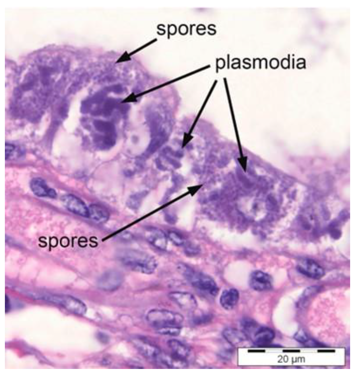
2.1.1. Microscopy
2.1.2. In Situ Hybridisation (ISH)
2.2. Molecular Detection Methods
2.2.1. Polymerase Chain Reaction (PCR)
2.2.2. Recombinase Polymerase Amplification (RPA)
2.2.3. Loop-Mediated Isothermal Amplification (LAMP)
3. Suggested Future EHP Detection Methods
4. Interventions in Managing EHP
5. Food Safety
Systematic Guidelines for Shrimp Food Safety
6. Conclusions
Author Contributions
Funding
Data Availability Statement
Acknowledgments
Conflicts of Interest
References
- Wurmann, C.G.; Madrid, R.M.; Brugger, A.M. Shrimp farming in Latin America: Current status, opportunities, challenges, and strategies for Sustainable Development. Aquac. Econ. Manag. 2004, 8, 117–141. [Google Scholar] [CrossRef]
- Food and Agriculture Organization of the United Nations-Globefish. Fishmeal Market Report. 2009. Available online: http://www.globefish.org (accessed on 2 March 2023).
- Thitamadee, S.; Prachumwat, A.; Srisala, J.; Jaroenlak, P.; Salachan, P.V.; Sritunyalucksana, K.; Flegel, T.W.; Itsathitphaisarn, O. Review of current disease threats for cultivated penaeid shrimp in Asia. Aquaculture 2016, 452, 69–87. [Google Scholar] [CrossRef]
- Kmmari, S.; Rathlavath, S.; Pillai, D.; Rajesh, G. Hepatopancreatic microsporidiasis (HPM) in Shrimp culture: A review. Int. J. Curr. Microbiol. Appl. Sci. 2018, 7, 3208–3215. [Google Scholar] [CrossRef]
- Jaroenlak, P.; Sanguanrut, P.; Williams, B.A.; Stentiford, G.D.; Flegel, T.W.; Sritunyalucksana, K.; Itsathitphaisarn, O. A nested PCR assay to avoid false positive detection of the Microsporidian Enterocytozoon hepatopenaei (EHP) in environmental samples in shrimp farms. PLoS ONE 2016, 11, e0166320. [Google Scholar] [CrossRef] [PubMed]
- Chaijarasphong, T.; Munkongwongsiri, N.; Stentiford, G.D.; Aldama-Cano, D.J.; Thansa, K.; Flegel, T.W.; Sritunyalucksana, K.; Itsathitphaisarn, O. The shrimp microsporidian Enterocytozoon hepatopenaei (EHP): Biology, pathology, diagnostics, and control. J. Invertebr. Pathol. 2021, 186, 107458. [Google Scholar] [CrossRef] [PubMed]
- Jaroenlak, P.; Boakye, D.W.; Vanichviriyakit, R.; Williams, B.A.; Sritunyalucksana, K.; Itsathitphaisarn, O. Identification, characterization and heparin binding capacity of a spore-wall, virulence protein from the shrimp microsporidian, Enterocytozoon hepatopenaei (EHP). Parasites Vectors 2018, 11, 177. [Google Scholar] [CrossRef] [PubMed]
- Kim, B.-S.; Jang, G.-I.; Kim, S.-M.; Kim, Y.-S.; Jeon, Y.-G.; Oh, Y.-K.; Hwang, J.-Y.; Kwon, M.-G. First Report of Enterocytozoon hepatopenaei infection in Pacific whiteleg shrimp (Litopenaeus vannamei) cultured in Korea. Animals 2021, 11, 3150. [Google Scholar] [CrossRef]
- Caro, L.F.A.; Mai, H.N.; Schofield, P.; Alenton, R.R.R. A laboratory challenge model for evaluating Enterocytozoon hepatopenaei susceptibility in selected lines of Pacific Whiteleg shrimp Penaeus Vannamei. J. Invertebr. Pathol. 2022, 196, 107853. [Google Scholar] [CrossRef]
- Dhar, A.K.; Mai, H.N. Acute hepatopancreatic necrosis disease (AHPND) and hepatopancreatic microsporidiosis (HPM): Two threats to sustainable shrimp aquaculture. In Proceedings of the International Workshop on the Promotion of Sustainable Aquaculture, Aquatic Animal Health, and Resource Enhancement in Southeast Asia, Iloilo City, Philippines, 25–27 June 2019; Aquaculture Department, Southeast Asian Fisheries Development Center: Iloilo, Philippines, 2021; pp. 200–216. [Google Scholar]
- Kumar, T.S.; Praveena, P.E.; Sivaramakrishnan, T.; Rajan, J.J.S.; Makesh, M.; Jithendran, K.P. Effect of Enterocytozoon hepatopenaei (EHP) infection on physiology, metabolism, immunity, and growth of Penaeus Vannamei. Aquaculture 2022, 553, 738105. [Google Scholar] [CrossRef]
- Tang, K.F.J.; Pantoja, C.R.; Redman, R.M.; Han, J.E.; Tran, L.H.; Lightner, D.V. Development of in situ hybridisation and PCR assays for the detection of Enterocytozoon Hepatopenaei (EHP), a microsporidian parasite infecting penaeid shrimp. J. Invertebr. Pathol. 2015, 130, 37–41. [Google Scholar] [CrossRef]
- Tang, K.F.J.; Aranguren, L.F.; Piamsomboon, P.; Han, J.E.; Maskaykina, I.Y.; Schmidt, M.M. Detection of the Microsporidian Enterocytozoon hepatopenaei (EHP) and Taura syndrome virus in Penaeus Vannamei cultured in Venezuela. Aquaculture 2017, 480, 17–21. [Google Scholar] [CrossRef]
- Hou, Z.-H.; Yu, J.-Y.; Wang, J.-J.; Li, T.; Chang, L.-R.; Fang, Y.; Yan, D.-C. Development of a PCR assay for the effective detection of Enterocytozoon Hepatopenaei (EHP) and investigation of EHP prevalence in Shandong Province, China. J. Invertebr. Pathol. 2021, 184, 107653. [Google Scholar] [CrossRef] [PubMed]
- Wang, L.; Lv, Q.; He, Y.; Gu, R.; Zhou, B.; Chen, J.; Fan, X.; Pan, G.; Long, M.; Zhou, Z. Integrated qPCR and staining methods for detection and quantification of Enterocytozoon hepatopenaei in shrimp Litopenaeus Vannamei. Microorganisms 2020, 8, 1366. [Google Scholar] [CrossRef] [PubMed]
- Wang, Y.; Zhou, J.; Yin, M.; Ying, N.; Xiang, Y.; Liu, W.; Ye, J.; Li, X.; Fang, W.; Tan, H. A modification of nested PCR method for detection of Enterocytozoon Hepatopenaei (EHP) in giant freshwater prawn Macrobrachium Rosenbergii. Front. Cell. Infect. Microbiol. 2022, 12, 1013016. [Google Scholar] [CrossRef] [PubMed]
- Kralik, P.; Ricchi, M. A basic guide to real time PCR in microbial diagnostics: Definitions, parameters, and everything. Front. Microbiol. 2017, 8, 108. [Google Scholar] [CrossRef]
- Ma, C.; Fan, S.; Wang, Y.; Yang, H.; Qiao, Y.; Jiang, G.; Lyu, M.; Dong, J.; Shen, H.; Gao, S. Rapid detection of Enterocytozoon hepatopenaei infection in shrimp with a real-time isothermal recombinase polymerase amplification assay. Front. Cell. Infect. Microbiol. 2021, 11, 631960. [Google Scholar] [CrossRef]
- Kumar, T.S.; Radhika, K.; Rajan, J.J.S.; Makesh, M.; Alavandi, S.V.; Vijayan, K.K. Closed-tube field-deployable loop-mediated isothermal amplification (LAMP) assay based on spore wall protein (SWP) for the visual detection of Enterocytozoon Hepatopenaei (EHP). J. Invertebr. Pathol. 2021, 183, 107624. [Google Scholar] [CrossRef]
- Kumar, T.S.; Krishnan, A.N.; Sahaya, J.J.; Makesh, M.; Jithendran, K.P.; Alavandi, S.V.; Vijayan, K.K. Visual loop-mediated isothermal amplification (LAMP) for the rapid diagnosis of Enterocytozoon hepatopenaei (EHP) infection. Parasitol. Res. 2018, 117, 1485–1493. [Google Scholar] [CrossRef]
- Tangprasittipap, A.; Srisala, J.; Chouwdee, S.; Somboon, M.; Chuchird, N.; Limsuwan, C.; Srisuvan, T.; Flegel, T.W.; Sritunyalucksana, K. The Microsporidian Enterocytozoon hepatopenaei is not the cause of white faeces syndrome in white leg shrimp Penaeus (Litopenaeus) vannamei. BMC Vet. Res. 2013, 9, 139. [Google Scholar] [CrossRef]
- Zhou, S.; Wang, M.; Liu, M.; Jiang, K.; Wang, B.; Wang, L. Rapid detection of Enterocytozoon hepatopenaei in shrimp through an isothermal recombinase polymerase amplification assay. Aquaculture 2020, 521, 734987. [Google Scholar] [CrossRef]
- Lei, S.; Chen, S.; Zhong, Q. Digital PCR for accurate quantification of pathogens: Principles, applications, challenges, and future prospects. Int. J. Biol. Macromol. 2021, 184, 750–759. [Google Scholar] [CrossRef]
- Zhang, H.; Gong, H.Y.; Cao, W.W.; Que, M.Y.; Ye, L.; Shi, L. Duplex droplet digital PCR method for the detection of Enterocytozoon hepatopenaei and Vibrio parahaemolyticus acute hepatopancreatic necrosis disease. J. Fish Dis. 2022, 45, 761–769. [Google Scholar] [CrossRef] [PubMed]
- Jiang, S.; Chen, Y.; Han, S.; Lv, L.; Li, L. Next-generation sequencing applications for the study of fungal pathogens. Microorganisms 2022, 10, 1882. [Google Scholar] [CrossRef] [PubMed]
- Thomsen, P.F.; Willerslev, E. Environmental DNA—An emerging tool in conservation for monitoring past and present biodiversity. Biol. Conserv. 2015, 183, 4–18. [Google Scholar] [CrossRef]
- Li, M.; Shan, X.; Wang, W.; Ding, X.; Dai, F.; Lv, D.; Wu, H. Qualitative and quantitative detection using eDNA Technology: A case study of Fenneropenaeus chinensis in the Bohai Sea. Aquac. Fish. 2020, 5, 148–155. [Google Scholar] [CrossRef]
- Buchatip, S.; Ananthanawat, C.; Sithigorngul, P.; Sangvanich, P.; Rengpipat, S.; Hoven, V.P. Detection of the shrimp pathogenic bacteria, Vibrio harveyi, by a quartz crystal microbalance-specific antibody-based sensor. Sens. Actuators B Chem. 2010, 145, 259–264. [Google Scholar] [CrossRef]
- Roy, S.; Baruah, K.; Bossier, P.; Vanrompay, D.; Norouzitallab, P. Induction of transgenerational innate immune memory against Vibrio infections in a brine shrimp (Artemia franciscana) model. Aquaculture 2022, 557, 738309. [Google Scholar] [CrossRef]
- Kumar, S.; Verma, A.K.; Singh, S.P.; Awasthi, A. Immunostimulants for shrimp aquaculture: Paving pathway towards shrimp sustainability. Environ. Sci. Pollut. Res. 2022. [Google Scholar] [CrossRef]
- Li, H.; Yang, Y.; Hong, W.; Huang, M.; Wu, M.; Zhao, X. Applications of genome editing technology in the targeted therapy of human diseases: Mechanisms, advances, and prospects. Signal Transduct. Target. Ther. 2020, 5. [Google Scholar] [CrossRef]
- Malavia, D.; Gow, N.A.; Usher, J. Advances in molecular tools and in vivo models for the study of human fungal pathogenesis. Microorganisms 2020, 8, 803. [Google Scholar] [CrossRef]
- Nargesi, S.; Kaboli, S.; Thekkiniath, J.; Heidari, S.; Keramati, F.; Seyedmousavi, S.; Hedayati, M.T. Recent advances in genome editing tools in Medical Mycology Research. J. Fungi 2021, 7, 257. [Google Scholar] [CrossRef] [PubMed]
- Kim, J.H.; Cheng, L.W.; Land, K.M. Advances in antifungal development: Discovery of new drugs and drug repurposing. Pharmaceuticals 2022, 15, 787. [Google Scholar] [CrossRef] [PubMed]
- Pagán-Mercado, G.; Rivera-Ruiz, M.E.; Segarra-Román, F.; Rodríguez-Medina, J.R. Antifungal research strategies aiming for new targets. Puerto Rico Health Sci. J. 2009, 28, 220. [Google Scholar]
- Soo, T.C.; Bhassu, S. Biochemical indexes and gut microbiota testing as diagnostic methods for Penaeus monodon health and physiological changes during AHPND infection with food safety concerns. Food Sci. Nutr. 2022, 10, 2694–2709. [Google Scholar] [CrossRef] [PubMed]
- Flegel, T.W. Review of disease transmission risks from prawn products exported for human consumption. Aquaculture 2009, 290, 179–189. [Google Scholar] [CrossRef]
- Karunasagar, I. Food safety and public health risks associated with products of aquaculture. In Understanding and Applying Risk Analysis in Aquaculture; Bondad-Reantaso, M.G., Arthur., J.R., Subasinghe, R.P., Eds.; FAO Fisheries and Aquaculture Technical Paper; FAO: Rome, Italy, 2008; pp. 9–25. [Google Scholar]
- Food and Agriculture Organization of the United Nations-Globefish. The State of World Fisheries and Aquaculture 2022. Towards Blue Transformation; FAO: Rome, Italy, 2022. [Google Scholar] [CrossRef]
- Irkin, L.C. The effects of shellfish consumption frequency for human health. In Update on Malacology; Intechopen: London, UK, 2022. [Google Scholar] [CrossRef]
- Han, B.; Pan, G.; Weiss, L.M. Microsporidiosis in humans. Clin. Microbiol. Rev. 2021, 34, e0001020. [Google Scholar] [CrossRef]


| Method | Primer Name | Primer Sequence (5′–3′) | Reference |
|---|---|---|---|
One-step PCR
| EHP-510F | GCCTGAGAGATG GCTCCCACGT | [12] |
| EHP-510R | GCGTACTATCCCCAGAGCCCGA | ||
qPCR
| EHP-PTP2-F | GCAGCACTCAAGGAATGGC | [15] |
| EHP-PTP2-R | TTTCGTTAGGCTTACCCTGTGA | ||
Nested PCR
| SWP_1F | TTGCAGAGTGTTGTTAAGGGTTT | [5] |
| SWP_1R | CACGATGTGTCTTTGCAATTTTC | ||
| SWP_2F | TTGGCGGCACAATTCTCAAACA | ||
| SWP_2R | GCTGTTTGTCTCCAACTGTATTTGA | ||
| SWP_2F’ | GCAGAGTGTTGTTAAGGGTTTAAG | [16] | |
| SWP_2R’ | GCTGTTTGTCWCCAACTGTATT | ||
Nested PCR
| ENF779 | CAGCAGGCGCGAAAATTGTCCA | [21] |
| ENR779 | AAGAGATATTGTATTGCGCTTGCTG | ||
| ENF176 | CAACGCGGGAAAACTTACCA | ||
| ENR176 | ACCTGTTATTGCCTTCTCCCTCC | ||
RPA
| F2 | CATTGAGTTTGTTGAGAGTAGCGGAACGGAT | [22] |
| R2 | CTAAGAGCATCGCTTTCGCCTCCGTTGGTC |
Disclaimer/Publisher’s Note: The statements, opinions and data contained in all publications are solely those of the individual author(s) and contributor(s) and not of MDPI and/or the editor(s). MDPI and/or the editor(s) disclaim responsibility for any injury to people or property resulting from any ideas, methods, instructions or products referred to in the content. |
© 2023 by the authors. Licensee MDPI, Basel, Switzerland. This article is an open access article distributed under the terms and conditions of the Creative Commons Attribution (CC BY) license (https://creativecommons.org/licenses/by/4.0/).
Share and Cite
Govindasamy, T.; Bhassu, S.; Raju, C.S. Enterocytozoon hepatopenaei Infection in Shrimp: Diagnosis, Interventions, and Food Safety Guidelines. Microorganisms 2024, 12, 21. https://doi.org/10.3390/microorganisms12010021
Govindasamy T, Bhassu S, Raju CS. Enterocytozoon hepatopenaei Infection in Shrimp: Diagnosis, Interventions, and Food Safety Guidelines. Microorganisms. 2024; 12(1):21. https://doi.org/10.3390/microorganisms12010021
Chicago/Turabian StyleGovindasamy, Thenmoli, Subha Bhassu, and Chandramathi Samudi Raju. 2024. "Enterocytozoon hepatopenaei Infection in Shrimp: Diagnosis, Interventions, and Food Safety Guidelines" Microorganisms 12, no. 1: 21. https://doi.org/10.3390/microorganisms12010021
APA StyleGovindasamy, T., Bhassu, S., & Raju, C. S. (2024). Enterocytozoon hepatopenaei Infection in Shrimp: Diagnosis, Interventions, and Food Safety Guidelines. Microorganisms, 12(1), 21. https://doi.org/10.3390/microorganisms12010021