Communication

Metagenome and Resistome Analysis of Beta-Lactam-Resistant Bacteria Isolated from River Waters in Surabaya, Indonesia

1
Department of Infectious Diseases, Kobe Institute of Health, Kobe 650-0046, Japan
2
Department of Medical Technology, Kobe Tokiwa University, Kobe 653-0838, Japan
3
Division of Advanced Medical Science, Kobe University Graduate School of Science, Technology and Innovation, Kobe 650-0017, Japan
4
Department of Microbiology, Faculty of Medicine, Airlangga University, Surabaya 60132, Indonesia
5
Institute of Tropical Disease, Airlangga University, Surabaya 60286, Indonesia
6
Division of Urology, Department of Organ Therapeutics, Faculty of Medicine, Kobe University Graduate School of Medicine, Kobe 650-0017, Japan
*
Author to whom correspondence should be addressed.
Microorganisms 2024, 12(1), 199; https://doi.org/10.3390/microorganisms12010199
Submission received: 5 December 2023 / Revised: 5 January 2024 / Accepted: 16 January 2024 / Published: 18 January 2024
(This article belongs to the Section Antimicrobial Agents and Resistance)

Abstract

:
Antimicrobial agents are administered to humans and livestock, and bacterial antimicrobial resistance (AMR) and antimicrobial agents are released into the environment. In this study, to investigate the trend of AMR in humans, livestock, and the environment, we performed a metagenomic analysis of multidrug-resistant bacteria with CHROMagar ESBL in environmental river water samples, which were collected using syringe filter units from waters near hospitals, downtown areas, residential areas, and water treatment plants in Surabaya, Indonesia. Our results showed that Acinetobacter, Pseudomonas, Aeromonas, Enterobacter, Escherichia, and Klebsiella grew in CHROMagar ESBL; they were most frequently detected in water samples from rivers surrounding hospitals contaminated with various AMR genes (ARGs) in high levels. These results identified bacteria as ARG reservoirs and revealed that hospitals could be sources for various ARGs disseminated into the environment. In conclusion, this study details a novel metagenomic analysis of collected bacteria in environmental water samples using a syringe filter unit for an AMR epidemiological study based on the One Health approach.

1. Introduction

According to an antimicrobial resistance (AMR) review by the UK government in 2014 [1], the number of annual deaths owing to bacterial AMR will increase to 10 million by 2050 if appropriate measures are not taken. Another predictive statistical method estimated 4.95 and 1.27 million deaths associated with and attributable to bacterial AMR worldwide in 2019, respectively [2]. As antimicrobial agents are administered to humans as well as livestock, and bacterial AMR and antimicrobial agents are released into the environment, adopting a One Health approach is increasingly important to investigate trends in AMR that cover humans, livestock, and the environment [3,4,5]. In Indonesia, the One Health approach is essential for investigating AMR trends because inexpensive antimicrobial agents are over used, sewage infrastructure is inadequate, and poultry consumption is high.
Recent advances in genetic analysis technologies, such as next-generation sequencing, have enabled whole-genome sequencing of bacteria, enabling the investigation of the molecular epidemiology of AMR genes (ARGs) and bacterial AMR in further detail [6,7]. In addition, metagenomic analysis using next-generation sequencing allows a comprehensive investigation of the microbiota in humans, livestock, and the environment (e.g., river water) [8]. Because the administration of antimicrobial agents and their leakage into the environment directly affects the ARG profile (resistome) in humans and livestock, comprehensively analyzing ARGs in the environment will help to clarify the mechanism of AMR spread [7].
In this study, we performed a metagenome and resistome analysis of multidrug-resistant bacteria stored in syringe filter units from environmental river water samples located near hospitals, downtown areas, residential areas, and water treatment plants in Surabaya, Indonesia. Employing metagenome and resistome analyses for analyzing the environmental contaminating bacteria, especially in river water, enabled the creation of a novel comprehensive molecular epidemiological method under the One Health approach. This method is expected to identify ARG reservoirs and transmission vehicles in the environment and provide new insights into the molecular mechanisms of AMR spread.

2. Materials and Methods

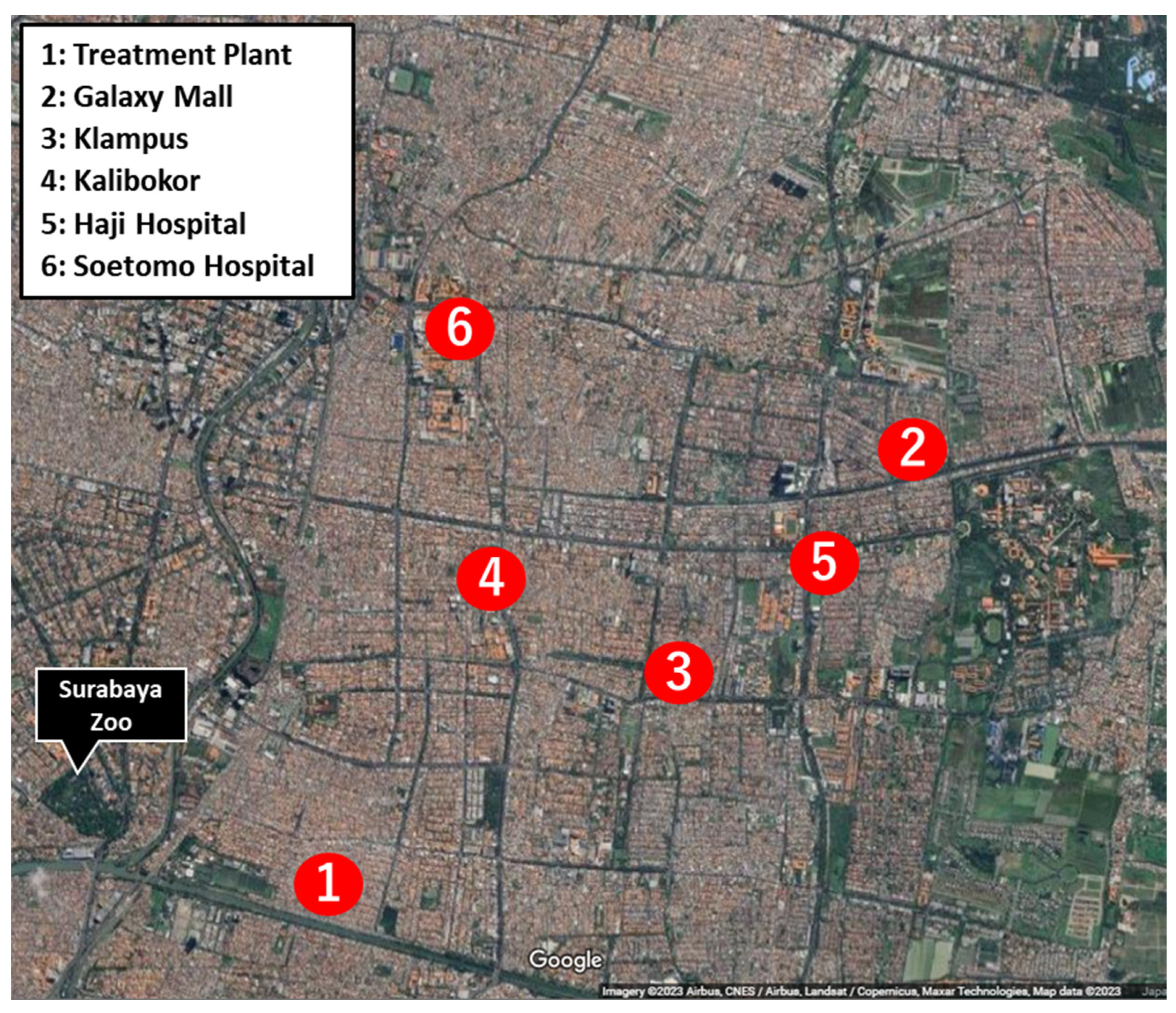
2.1. Sample Collection and Culture Condition

River water samples were collected from six locations in the urban areas of Surabaya, Indonesia, in December 2019 (nearby; (1) Treatment Plant: TP, (2) Galaxy-Mall (downtown): GM, (3) Klampus (residential area): KL, (4) Kalibokor (residential area): KA, (5) Haji-Hospital: HH, (6) Soetomo-Hospital: SH; Figure 1). To trap and collect bacteria, 30 mL water samples were filtered with a Millex-GV Syringe Filter Unit, 0.22 µm pore size, PVDF-33 mm membrane, and stored using the Syringe Filter Unit (Milliporesigma, Bedford, MA, USA). The syringe filters were attached to an empty 10 mL syringe, and 5 mL phosphate-buffered saline (PBS) was aspirated to suspend the bacteria adsorbed on the filter. The aspirate was collected in a 15 mL tube and centrifuged at 10,000× g for 10 min at 4 °C. The supernatant was discarded and the pellet was resuspended in PBS (0.5 mL). Then, 200 µL of the suspension was seeded into CHROMagar ESBL plates (CHROMagar, Paris, France) and cultured overnight at 37 °C. After incubation, 2 mL of PBS was added to the CHROMagar ESBL plates and all colonies were collected. Of the collected bacterial suspension, 200 µL was centrifuged and the pellet was resuspended in 1 mL PBS. DNA extraction was performed on 200 µL of the resuspension solution and metagenomic DNA was extracted using the Nucleospin Tissue Kit (MACHEREY-NAGEL, Düren, Germany), as per manufacturer’s instructions.

2.2. Metagenomic DNA-Seq Analysis of Extended Spectrum Beta-Lactamase (ESBL)-Producing Bacteria

A metagenomic DNA-seq library was prepared using the QIAseq FX DNA library kit (QIAGEN, Hilden, Germany), and sequencing was performed using the Illumina MiSeq system (Illumina, San Diego, CA, USA) with v3 chemistry (2 × 300-bp format). Adapter and low-quality sequences in short-read data were trimmed using fastp v0.23.2 [9] with default settings. Metagenomic analysis was performed using the MePIC software v2.0 [10] with default parameters, and the results were visualized using MEGAN [11] to determine the distribution of operational taxonomic units (OTUs).

2.3. Resistome Analysis

ARGs were predicted using the Resistance Gene Identifier (RGI v5.2.0; default parameters of RGI bwt mode) and the Comprehensive Antibiotic Resistance Database (card v3.1.4) [12]. OTUs of ARGs in the database were created via clustering at ≥90% sequence identity and ≥80% coverage. The detected genes were summarized for each OTU. For normalization, per kilobase per million mapped reads (RPKM) were calculated using the following formula:
RPKM = number of detected reads against OTUs/(average gene length of the detected OTUs (bp) × total number of trimmed reads) × 109.

3. Results

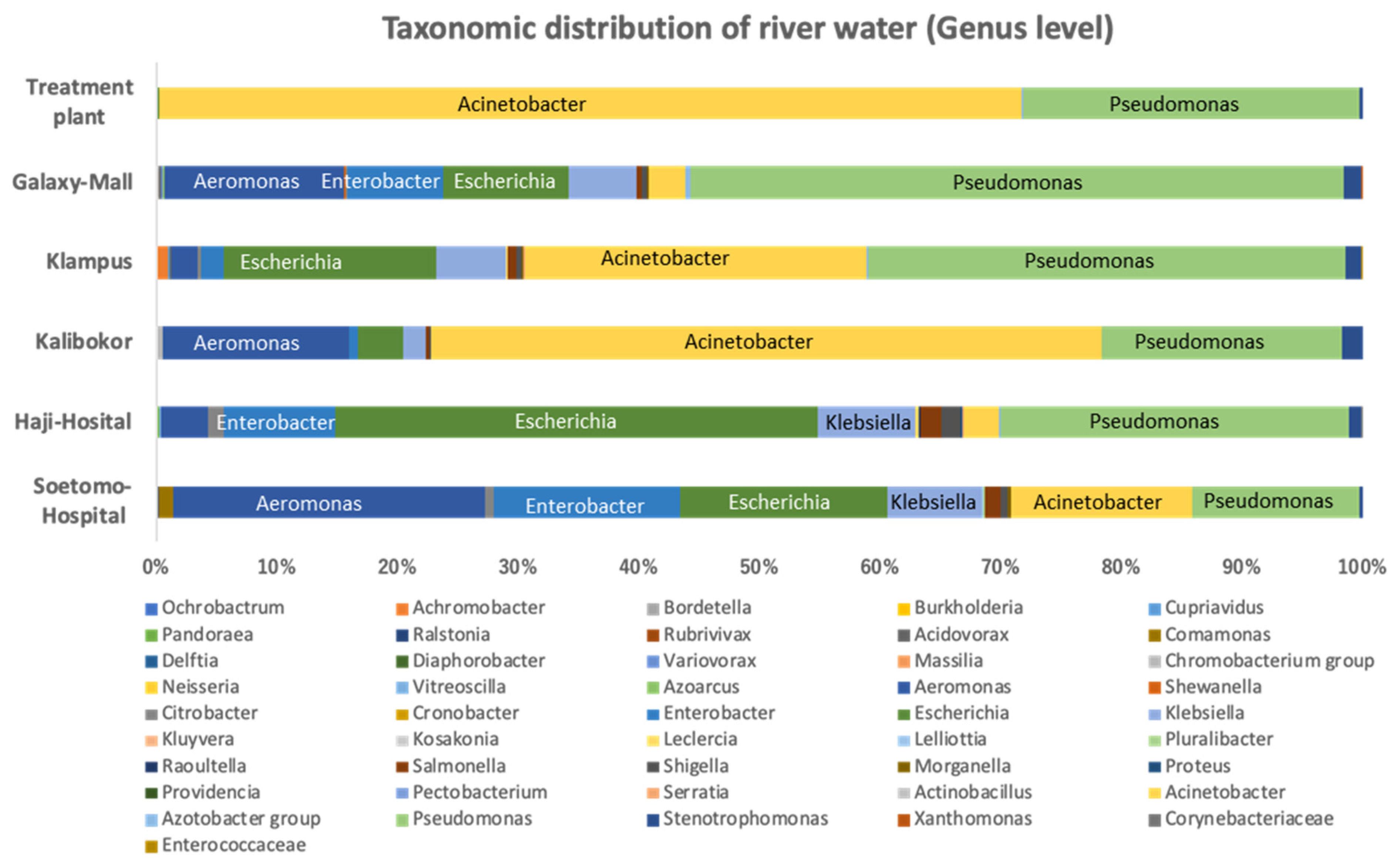
3.1. Bacterial Proportion of Strains Growing on CHROMagar ESBL Plate from Indonesian River Water

The taxonomic classification of cultured bacteria from Indonesian river water samples on CHROMagar ESBL plates at the genus level is shown in Figure 2. Acinetobacter and Pseudomonas were the most frequent bacterial genera that grew in CHROMagar ESBL from the water collected at all sampling sites. Bacterial genera differed among the samples. The bacterial proportion of the river water sample from TP was the poorest in diversity, with Acinetobacter (71.5%) and Pseudomonas (27.9%) being the dominant genera. Acinetobacter and Pseudomonas were detected in river water samples from GM, KL, and KA, with high abundance ratios of 75.6%, 68.0%, and 57.3%, respectively, whereas in samples from HH and SH surrounding the hospital, the abundance ratios of both species were comparatively lower at 28.9% and 31.8%, respectively. Bacterial genera, such as Acinetobacter, Pseudomonas, Aeromonas, Escherichia, Enterobacter, and Klebsiella were the dominant constituents in river water culture samples from HH and SH, and the representative ESBL-producing bacteria, Escherichia, Enterobacter, and Klebsiella, had a higher proportion ratio than at the other sampling sites (Figure 2 and Table S1).

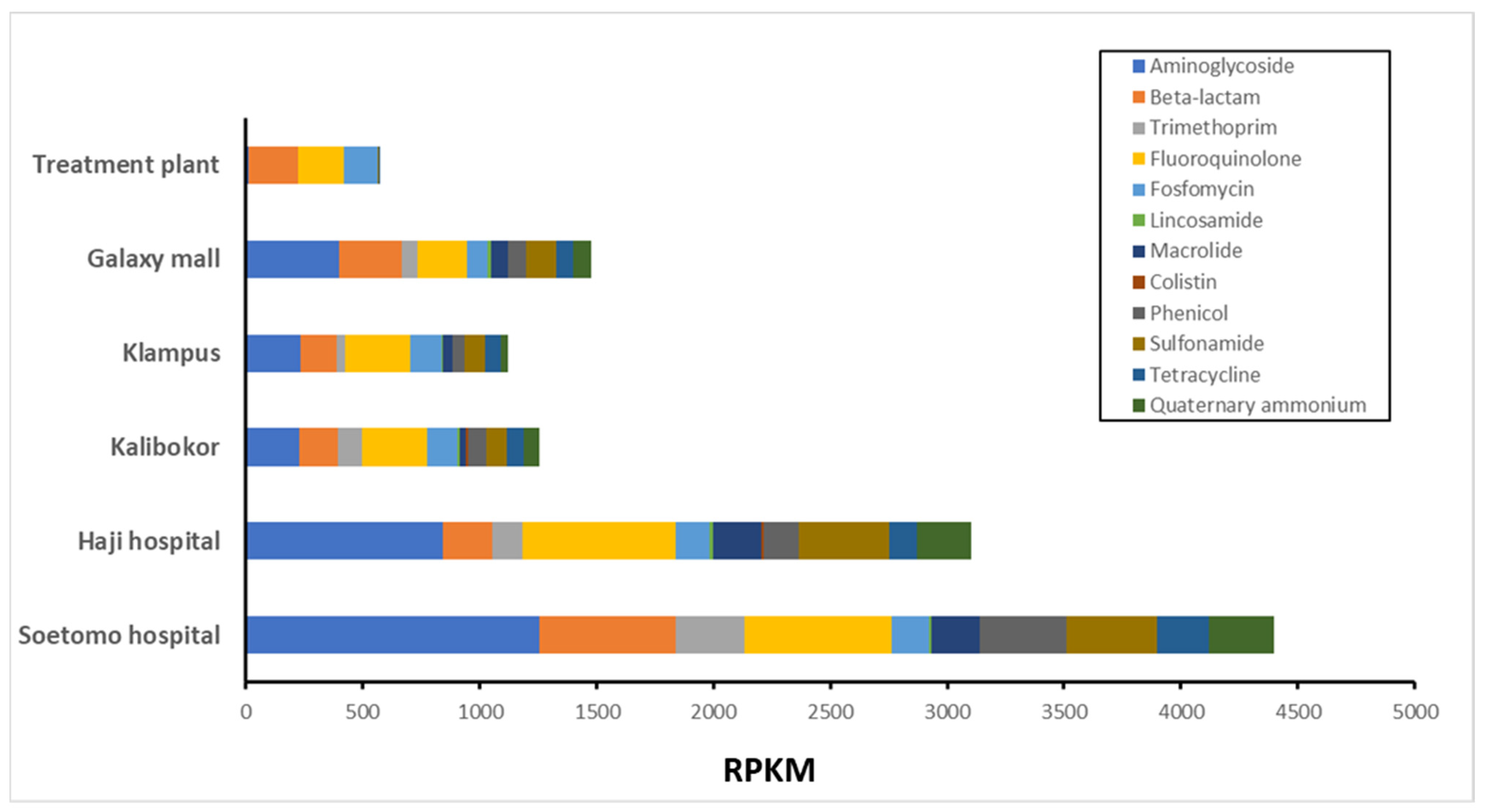
3.2. ARG Proportion of Growing on CHROMagar ESBL Plate from Indonesian River Water

The metagenomic DNA-seq short reads were analyzed using RGI software v5.1.0 in the CARD AMR gene database under normalization using reads per kilobase of exon per million mapped reads (RPKM). ARGs to quaternary aminoglycoside (AMG), β-lactam, quaternary ammonium compounds (QAC), sulfonamide (SA), and macrolide were detected substantially in culture samples from all water collection points (Figure 3). Resistome analysis showed that drug resistance genes for aminoglycoside and β-lactam antibiotics were predominantly detected in all samples. The most abundant ARGs in the total RPKMs were detected in the river water samples from SH, followed by samples from HH. In contrast, river water from TP had the lowest total RPKM values, and the ARGs detected were mostly derived from Acinetobacter and Pseudomonas, with limited diversity (Figure 3).
ARGs detected in each AMR category were further classified as orthologous genes (or gene families) (Figure 4 and Table S2). The SA resistance gene sul1 was most frequently detected at 660.9 RPKM (Table S2); sul2 detection was slightly lower at 398.7 RPKM, whereas that of sul3 was much lower at 29.1 RPKM. The second most detected ARG was the QAC resistance gene qacEΔ1 at 643.8 RPKM (Table S2), suggesting that basic gene sets (sul1 and qacEΔ1) in the class 1 integron [13] were predominant in the detected ARGs. Multiple AMG resistance genes were identified in the clinically important ARGs (Figure 4). The four most predominant AMG resistance genes belonged to the aac and aph gene families (Table S2), which contribute to streptomycin resistance, implying that streptomycin could be the most potent selective agent among aminoglycosides. Following this, high RPKM of kdpE and acr, efflux pumps located on the chromosomes of E. coli, were detected. Regarding the β-lactamase gene type, blaOXA, and blaTEM were the most considerably detected (Figure 4), followed by POM-1 and ADC, which were present in Pseudomonas and Acinetobacter, respectively. The blaCTX-M cluster, an ESBL gene commonly detected in clinical settings, was most predominant in river water culture samples around hospitals such as HH and SH. In particular, the blaNDM gene cluster was also detected in samples from SH (Figure 4, Table S2).
In addition, mcr genes contributing to colistin resistance were detected in KA and HH river water samples with extremely low RPKM values.

4. Discussion

In this study, a metagenomic analysis was conducted on bacterial strains collected from river water using syringe filter units and cultured in CHROMagar ESBL. In developing countries such as Indonesia, the widespread use of antimicrobials has become a public health problem because their components and antimicrobial-resistant bacteria are released into environmental rivers; however, only few studies report on these issues [14]. Filtering large quantities of water using specialized filtration equipment is necessary to investigate the presence of antimicrobial-resistant bacteria in rivers. In particular, investigating antimicrobial-resistant bacteria in areas lacking well-developed transportation is challenging. Therefore, we determined that environmental water could be easily collected using syringe filter units and that bacteria could be easily recovered from the units. Previously, the sensitivity of CHROMagar ESBL medium for the detection of ESBL-producing Enterobacteriaceae was evaluated using spiked stools, and 93.9% of the sensitivity of the CHROMagar ESBL medium was confirmed [15]. In addition, Vurayai and his colleagues investigated the environmental ESBL bioburden in neonatal intensive care units (NICU) using CHROMagar ESBL, and they concluded that the CHROMagar ESBL is useful to identify ESBL reservoirs and transmission vehicles [16]. Similarly, in our study, metagenomic analysis of bacteria collected via syringe filter units and screened using the CHROMagar ESBL medium revealed reservoirs and transmission vehicles of ESBL and various ARGs in rivers near urban and residential areas, water treatment plants, and hospitals.
Throughout this analysis, Acinetobacter and Pseudomonas were the most frequently detected bacterial genera; they were even isolated from water samples of treatment plants, which are treated water sources (Figure 2). Multidrug-resistant Acinetobacter baumannii and Pseudomonas aeruginosa often cause infections that are difficult to treat. The isolation of these organisms from drinking water sources should be alarming [17,18]. Enterobacteriaceae, including Escherichia, Enterobacter, and Klebsiella, were also detected at high frequencies in rivers and wastewater, particularly near hospitals (Figure 2). ESBL-producing E. coli is a multidrug-resistant organism targeted by the WHO’s Tricycle Project based on the One Health approach and should continue to be closely monitored [19]. In this study, Aeromonas growing on CHROMagar ESBL was also detected in most water samples (Figure 2). Recently, Aeromonas carrying the bla-NDM-1 gene has been reported to cause community-acquired pneumonia, suggesting that Aeromonas plays an important role in the spread of multidrug-resistant bacteria as a reservoir of ARGs in the community and environment [20].
In this study, resistome analysis revealed that drug-resistant genes to antibiotics commonly used in clinical practice were detected at a high frequency with a high diversity of bacterial proportions and high total RPKM values in river water around hospitals (Figure 2 and Figure 3). In addition, ARGs that pose a serious clinical threat, such as NDM and MCR, were detected in rivers near hospitals (Figure 4 and Table S2). ARG effluents from these facilities have a considerable impact on the surrounding environment. The use of antimicrobial agents is the primary cause of AMR epidemics and worldwide consumption of antimicrobial agents reportedly increased by 65% between 2000 and 2015 [21]. Currently, proper usage of antimicrobial agents is strictly required in healthcare settings, and antimicrobial use in developed countries is decreasing; however, in emerging countries, their use continues to increase rapidly [22]. Antimicrobial abuse is the major causative factor of the spread of AMR, as exposure to antimicrobial agents increases selection pressure for AMR and promotes the horizontal spread of ARGs [23].

5. Conclusions

In conclusion, a novel metagenomic and resistome analysis of environmental waters using CHROMagar ESBL revealed that Acinetobacter, Pseudomonas, Aeromonas, Enterobacter, Escherichia, and Klebsiella were important reservoirs and spreaders of ARGs, and water surrounding hospitals was contaminated with various ARGs at high levels. This novel metagenomic analysis of environmental water could be a powerful tool for AMR molecular epidemiological studies based on the One Health approach.

Supplementary Materials

The following supporting information can be downloaded at: https://www.mdpi.com/article/10.3390/microorganisms12010199/s1, Table S1: Summary of OTUs detected in CHROMagar ESBL culture samples from Indonesian river water. Table S2: Summary of RPKM values of operational taxonomy unit (OUT) of antimicrobial resistance genes.

Author Contributions

Conceptualization, R.N., K.O., K.K. (Kuntaman Kuntaman) and T.S.; methodology, R.N., K.O., K.K. (Koichi Kitagawa), N.N. and T.S.; software, R.N. and N.N.; formal analysis, R.N. and N.N.; investigation, R.N., S.K. and T.S.; resources, R.N., S.K., R.S., D.R., K.K. (Kuntaman Kuntaman) and T.S.; data curation, R.N. and N.N.; writing—original draft preparation, R.N., K.O., N.N., D.R. and T.S.; writing—review and editing, S.K., K.K. (Koichi Kitagawa), R.S., M.F. and K.K. (Kuntaman Kuntaman); supervision, M.F.; project administration, R.N., K.O., K.K. (Kuntaman Kuntaman) and T.S.; funding acquisition, R.N., K.O., K.K. (Kuntaman Kuntaman) and T.S. All authors have read and agreed to the published version of the manuscript.

Funding

This study was supported by the following grants: grant no. 1149/UN3/2018 from the Tahir Professorship of Airlangga University for K.K. (Kuntaman Kuntaman); the Japan Initiative for the Japan Agency for Medical Research and Development (AMED) (Grant No. JP23wm0125009 and JP23fk0108664 for T.S., JP22fk0108636 and JP23fk0108636 for R.N.); and Japan Society for the Promotion of Science (JSPS) KAKENHI Grant No. 21K10423 for K.O.

Institutional Review Board Statement

Institutional Review Board Statement: The study was approved by the Ethical Committee from the Dr. Soetomo Hospital (No. 0353/KEPK/VI/2018) and Airlangga University Hospital (No.193/KEH/2018), the ethical committee on health research of the Kobe University Graduate School of Health Sciences (No. 393-3), and Kobe Tokiwa University (No.19-11).

Data Availability Statement

All metagenome read data obtained as part of this study were deposited in DDBJ/EMBL/GenBank under the BioProject accession number PRJDB16871. The DRA accession numbers were DRR513858 (TP), DRR513859 (GM), DRR513860 (KL), DRR513861 (KA), DRR513862 (HH), and DRR513863.

Conflicts of Interest

The authors declare no conflicts of interest.

References

  1. O’Neill, J. Review on Antimicrobial Resistance. Antimicrobial Resistance: Tackling a Crisis for the Health and Wealth of Nations; Welcome Trust: London, UK, 2014; pp. 1–16. [Google Scholar]
  2. Murray, C.J.L.; Ikuta, K.S.; Sharara, F.; Swetschinski, L.; Robles Aguilar, G.; Gray, A.; Han, C.; Bisignano, C.; Rao, P.; Wool, E.; et al. Global burden of bacterial antimicrobial resistance in 2019: A systematic analysis. Lancet 2022, 399, 629–655. [Google Scholar] [CrossRef]
  3. McEwen, S.A.; Collignon, P.J. Antimicrobial resistance: A one health perspective. Microbiol. Spectr. 2018, 6, 521–547. [Google Scholar] [CrossRef]
  4. Aslam, B.; Khurshid, M.; Arshad, M.I.; Muzammil, S.; Rasool, M.; Yasmeen, N.; Shah, T.; Chaudhry, T.H.; Rasool, M.H.; Shahid, A.; et al. Antibiotic resistance: One health one world outlook. Front. Cell. Infect. Microbiol. 2021, 11, 771510. [Google Scholar] [CrossRef]
  5. Mackenzie, J.S.; Jeggo, M. The one health approach—Why is it so important? Trop. Med. Infect. Dis. 2019, 4, 88. [Google Scholar] [CrossRef]
  6. Su, M.; Satola, S.W.; Read, T.D. Genome-based prediction of bacterial antibiotic resistance. J. Clin. Microbiol. 2019, 57, e01405–e01418. [Google Scholar] [CrossRef] [PubMed]
  7. de Abreu, V.A.C.; Perdigão, J.; Almeida, S. Metagenomic approaches to analyze antimicrobial resistance: An overview. Front. Genet. 2021, 11, 575592. [Google Scholar] [CrossRef] [PubMed]
  8. Chen, K.; Pachter, L. Bioinformatics for whole-genome shotgun sequencing of microbial communities. PLoS Comput. Biol. 2005, 1, 106–112. [Google Scholar] [CrossRef]
  9. Chen, S.; Zhou, Y.; Chen, Y.; Gu, J. Fastp: An ultra-fast all-in-one FASTQ preprocessor. Bioinformatics 2018, 34, i884–i890. [Google Scholar] [CrossRef]
  10. Takeuchi, F.; Sekizuka, T.; Yamashita, A.; Ogasawara, Y.; Mizuta, K.; Kuroda, M. MePIC, metagenomic pathogen identification for clinical specimens. Jpn. J. Infect. Dis. 2014, 67, 62–65. [Google Scholar] [CrossRef]
  11. Huson, D.H.; Auch, A.F.; Qi, J.; Schuster, S.C. MEGAN analysis of metagenomic data. Genome Res. 2007, 17, 377–386. [Google Scholar] [CrossRef]
  12. Alcock, B.P.; Raphenya, A.R.; Lau, T.T.Y.; Tsang, K.K.; Bouchard, M.; Edalatmand, A.; Huynh, W.; Nguyen, A.V.; Cheng, A.A.; Liu, S.; et al. CARD 2020: Antibiotic resistome surveillance with the comprehensive antibiotic resistance database. Nucleic Acids Res. 2020, 48, D517–D525. [Google Scholar] [CrossRef] [PubMed]
  13. Gillings, M.R. Integrons: Past, present, and future. Microbiol. Mol. Biol. Rev. 2014, 78, 257–277. [Google Scholar] [CrossRef] [PubMed]
  14. Muurinen, J.; Muziasari, W.I.; Hultman, J.; Pärnänen, K.; Narita, V.; Lyra, C.; Fadlillah, L.N.; Rizki, L.P.; Nurmi, W.; Tiedje, J.M.; et al. Antibiotic resistomes and microbiomes in the surface water along the code river in Indonesia reflect drainage basin anthropogenic activities. Environ. Sci. Technol. 2022, 56, 14994–15006. [Google Scholar] [CrossRef]
  15. Girlich, D.; Grosperrin, V.; Naas, T.; Dortet, L. CHROMagar™ ESBL/mSuperCARBA bi-plate medium for detection of ESBL- and carbapenemase-producing Enterobacteriaceae from spiked stools. Diagn. Microbiol. Infect. Dis. 2019, 95, 107–112. [Google Scholar] [CrossRef] [PubMed]
  16. Vurayai, M.; Strysko, J.; Kgomanyane, K.; Bayani, O.; Mokomane, M.; Machiya, T.; Arscott-Mills, T.; Goldfarb, D.M.; Steenhoff, A.P.; McGann, C.; et al. Characterizing the bioburden of ESBL-producing organisms in a neonatal unit using chromogenic culture media: A feasible and efficient environmental sampling method. Antimicrob. Resist. Infect. Control 2022, 11, 14. [Google Scholar] [CrossRef] [PubMed]
  17. Carvalheira, A.; Silva, J.; Teixeira, P. Acinetobacter spp. In food and drinking water—A review. Food Microbiol. 2021, 95, 103675. [Google Scholar] [CrossRef]
  18. Mena, K.D.; Gerba, C.P. Risk assessment of Pseudomonas aeruginosa in water. Rev. Environ. Contam. Toxicol. 2009, 201, 71–115. [Google Scholar] [CrossRef]
  19. Puspandari, N.; Sunarno, S.; Febrianti, T.; Febriyana, D.; Saraswati, R.D.; Rooslamiati, I.; Amalia, N.; Nursofiah, S.; Hartoyo, Y.; Herna, H.; et al. Extended spectrum beta-lactamase-producing Escherichia coli surveillance in the human, food chain, and environment sectors: Tricycle project (pilot) in Indonesia. One Health 2021, 13, 100331. [Google Scholar] [CrossRef]
  20. Luo, X.; Mu, K.; Zhao, Y.; Zhang, J.; Qu, Y.; Hu, D.; Jia, Y.; Dai, P.; Weng, J.; Wang, D.; et al. Emergence of blaNDM-1-carrying Aeromonas caviae K433 isolated from patient with community-acquired pneumonia. Front. Microbiol. 2022, 13, 825389. [Google Scholar] [CrossRef]
  21. Klein, E.Y.; Van Boeckel, T.P.; Martinez, E.M.; Pant, S.; Gandra, S.; Levin, S.A.; Goossens, H.; Laxminarayan, R. Global increase and geographic convergence in antibiotic consumption between 2000 and 2015. Proc. Natl. Acad. Sci. USA 2018, 115, E3463–E3470. [Google Scholar] [CrossRef]
  22. Ayukekbong, J.A.; Ntemgwa, M.; Atabe, A.N. The threat of antimicrobial resistance in developing countries: Causes and control strategies. Antimicrob. Resist. Infect. Control 2017, 6, 47. [Google Scholar] [CrossRef] [PubMed]
  23. Aarestrup, F.M. The livestock reservoir for antimicrobial resistance: A personal view on changing patterns of risks, effects of interventions and the way forward. Philos. Trans. R. Soc. B 2015, 370, 20140085. [Google Scholar] [CrossRef] [PubMed]
Figure 1. Map of water sampling sites in the Surabaya area. Surabaya Zoo was marked as a landmark.
Microorganisms 12 00199 g001
Figure 2. Taxonomic classification at the genus level of river water culture samples on CHROMagar ESBL plates based on metagenomic DNA-Seq analysis results.
Microorganisms 12 00199 g002
Figure 3. Metagenomic analysis for AMR genes detection for each culture sample of river water. Metagenomic DNA-seq short reads were analyzed using RGI software v5.1.0 in CARD database, followed by normalization with RPKM. AMR, antimicrobial resistance; RPKM, per kilobase per million mapped reads.
Microorganisms 12 00199 g003
Figure 4. AMR gene profiling based on AMR categories. Trends in AMR gene variations; detected genes in each AMR category at every sampling site. AMR, antimicrobial resistance.
Microorganisms 12 00199 g004
Disclaimer/Publisher’s Note: The statements, opinions and data contained in all publications are solely those of the individual author(s) and contributor(s) and not of MDPI and/or the editor(s). MDPI and/or the editor(s) disclaim responsibility for any injury to people or property resulting from any ideas, methods, instructions or products referred to in the content.

