Article

Probiotics as an Alternative to Antibiotics: Genomic and Physiological Characterization of Aerobic Spore Formers from the Human Intestine

1
Department of Biology, Federico II University of Naples, 80125 Naples, Italy
2
National Biodiversity Future Center (NBFC), 90133 Palermo, Italy
3
Department of Molecular Medicine and Medical Biotechnology, Federico II University of Naples, 80131 Naples, Italy
*
Author to whom correspondence should be addressed.
These authors contributed equally to this work.
Microorganisms 2023, 11(8), 1978; https://doi.org/10.3390/microorganisms11081978
Submission received: 7 July 2023 / Revised: 28 July 2023 / Accepted: 30 July 2023 / Published: 31 July 2023
(This article belongs to the Special Issue Microbes at the Root of Solutions for Anthropocene Challenges)

Abstract

:
A total of thirty-two aerobic spore former strains were isolated from intestinal samples of healthy children and analyzed for their hemolytic and antibiotic-resistant activities. Four strains selected as non-hemolytic and sensitive to all antibiotics recommended as relevant by regulatory agencies were short-listed and evaluated for their in silico and in vitro probiotic potentials. The four selected strains were assigned to the Bacillus velezensis (MV4 and MV11), B. subtilis (MV24), and Priestia megaterium (formerly Bacillus megaterium) (MV30) species. A genomic analysis indicated that MV4, MV11, and MV24 contained a homolog of the gene coding for the fibrinolytic enzyme nattokinase while only MV30 encoded a glutamic acid decarboxylase essential to synthesize the neurotransmitter GABA. All four strains contained gene clusters potentially coding for new antimicrobials, showed strong antioxidant activity, formed biofilm, and produced/secreted quorum-sensing peptides able to induce a cytoprotective stress response in a model of human intestinal (HT-29) cells. Altogether, genomic and physiological data indicate that the analyzed strains do not pose safety concerns and have in vitro probiotic potentials allowing us to propose their use as an alternative to antibiotics.

1. Introduction

Antibiotic resistance is a global health threat, predicted to cause, by 2050, a million annual deaths due to an increased incidence of infectious diseases that will, in turn, affect many medical and surgical procedures [1]. So far, the identification and design of new antibiotics, and the development of novel vaccines and probiotics have been proposed as alternatives to current antibiotics [2]. Probiotics, live bacteria that exert a beneficial health effect, are recognized as potential candidates to substitute antibiotics for the capacity of some bacteria to protect the animal body against bacterial and viral infections. Inhibition of bacterial adhesion, enhanced mucosal barrier function, modulation of the immune systems, and secretion of antimicrobial metabolites are the main mechanisms proposed to explain the anti-infectious activity of probiotics [3].
Several species of the Bifidobacterium, Lactobacillus, and Bacillus genera have long been used as commercial probiotics [4], while other bacteria, such as Faecalibacterium prausnitzii and Akkermansia muciniphila have been recently proposed as next-generation probiotics [5,6]. In this context, the case of Bacillus-based probiotics is peculiar since all commercial preparations do not contain vegetative cells but metabolically quiescent spores [7]. For these organisms, spore formation is a survival strategy since spores are extremely stable cells, resistant to harsh conditions such as high temperatures, low pH, absence of water and nutrients, presence of lytic enzymes, toxic chemicals, and UV irradiations [7]. In response to the renewed presence of water, nutrients, and of favorable environmental conditions, spores germinate, originating vegetative cells able to grow and eventually sporulate again [8]. Also, due to their resistance properties, Bacillus spores are ubiquitous in nature and can be isolated from common and extreme environments [7,8]. Bacillus spores are also abundantly present in the gut of animals, where they enter as contaminants of food and water and safely transit the gastric barrier. In the intestine, some of the ingested spores germinate [9,10,11,12], and the germination-derived cells grow and temporarily colonize that niche, before re-sporulating in the terminal part of the intestine and leaving the body as spores [13,14].
Ingested spores of various Bacillus species have health-beneficial effects [9,11] and provide protection against bacterial [15] and viral infections [16]. Such probiotic effects have been ascribed to various mechanisms, including the promotion of the development of the Gut-Associated Lymphoid Tissue (GALT) [17], the induction of the production of cytokines in mesenteric lymph nodes (MLN) (IL-1, IL-5, IL-6, IFN-γ, and TNF-α) and spleen (IFN-γ and TNF-α) [18], the upregulation of the expression of the Toll-like receptors TLR-2 and TLR-4 [19], and the protection of human intestinal cells from oxidative stress [20] by inducing the nuclear translocation of the transcriptional factor Nrf-2 that, in turn, activates stress-response genes [21]. More recently, B. subtilis cells have been shown to have regulatory effects on epithelial differentiation by inhibiting the Notch pathway and, as a consequence, shifting differentiation toward a secretory cell fate [22]. Such a Toll-like receptor 2-dependent effect causes an increased number of Paneth and goblet cells in the intestine that, in turn, results in the production of antimicrobial peptides [22]. An additional mechanism by which spores exert their beneficial effects is the modulation of the microbial composition of the gut, exerted by favoring the prevalence of other potentially beneficial bacteria such as Faecalibacterium prausnitzii [23], Akkermansia muciniphila, and Bifidobacterium spp. [24].
In the intestine, germination-derived cells produce a variety of metabolites that mediate the interaction with the host’s intestinal and immune cells. Examples of such beneficial molecules produced by several Bacillus strains are enzymes, such as the NattoKinase (NK), a serine protease characterized by a potent thrombolytic activity and proposed for the treatment of multiple cardiovascular diseases [25], and the Glutamic Acid Decarboxylase (GAD), able to catalyze the synthesis of γ-aminobutyric acid (GABA), the most common inhibitory neurotransmitter in the central nervous system that also regulates cardiovascular functions such as blood pressure and heart rate [26]. In addition, peptides and lipopeptides with antimicrobial activity [27,28] and quorum-sensing peptides [29] are also produced by several Bacillus strains. An example is the Competence and Sporulation Factor (CSF), a quorum-sensing pentapeptide secreted by B. subtilis cells that is also recognized by human epithelial cells in which it induces the synthesis of the heat-shock (HS) proteins [29]. In a murine model, CSF prevents oxidant-induced intestinal epithelial injuries and loss of barrier function, exerting a cytoprotective activity [30]. CSF is only produced by members of the B. subtilis species but other species produce different peptides (CSF-like) similarly able to induce the synthesis of the heat-shock (HS) proteins in human cells in vitro [31]. More recently, CSF has been recognized as a key factor for the anti-neurodegenerative activity of B. subtilis in the model organism Caenorhabditis elegans [32]. B. subtilis strains able to produce biofilm, CSF, and nitric oxide (NO) were shown to extend the longevity of C. elegans by downregulating the insulin-like signaling system, thus opening the possibility that ingested spores of B. subtilis could contribute to delay the development of age-related diseases [33,34].
In this work, several Bacillus strains were isolated from intestinal samples of healthy children not under probiotic or antibiotic treatment. Four of these strains which were not hemolytic and not resistant to antibiotics, were assigned to the B. velezensis (MV4 and MV11), B. subtilis (MV24), and Priestia megaterium (formerly B. megaterium) (MV30) species and characterized at the genomic and physiological level.

2. Materials and Methods

2.1. Bacterial Isolation and Characterization

Fecal samples of healthy children (from ten months to ten years old) that had not been under antibiotic or probiotic treatment in the previous three months were collected and about 1 g of feces homogenized in sterile Phosphate-buffered saline (PBS) and heat-treated at 80 °C for 20 min. The samples were then serially diluted and plated on Difco Sporulation Medium (DSM, for 1 L: 8 g/L Nutrient Broth, 1 g/L KCl, 1 mM MgSO4, 1 mM Ca(NO3)2, 10 μM MnCl2, 1 μM FeSO4, Sigma-Aldrich, Darmstadt, Germany) and incubated overnight at 37 °C. The colonies were selected and isolated as previously described [35]. All isolates were tested for hemolytic activity by spotting 10 µL of growing cells on Columbia Agar plates supplemented with 5% defibrinated horse blood (ThermoScientific, Waltham, MA, USA) and, after 24 h of incubation at 37 °C, the clear halo around the colonies indicated a positive result. The non-hemolytic strains were then tested for their antibiotic susceptibility by plating 0.5 McFarland of bacteria on LB agar plates (for 1 L: 10 g/L Bacto-Tryptone, 5/L g Bacto yeast extract, 10 g/L NaCl, pH 7.0). Antibiotic-impregnated MIC test strips (Liofilchem®, Roseto, Italy) were then placed on the plates and incubated overnight at 37 °C. The minimum inhibitory concentration (MIC) was measured at the intersection of the inhibition halo and the antibiotic strip. The strains were indicated as resistant (R) if the antibiotic concentration exceeded the threshold level set by the EFSA for Bacillus strains [36].

2.2. Whole-Genome Sequencing and Bioinformatic Analysis

Bacterial genomic DNA was extracted from exponentially growing cells as previously reported [37]. Genome sequencing of MV4, MV11, MV24, and MV30 was performed with Illumina MiSeq Sequencing System by GenProbio (Parma, Italy). Genome assemblies were performed with SPAdes v3.14.0 by means of a MEGAnnotator pipeline [38]. The genomes of MV4, MV11, MV24, and MV30 have been deposited in GenBank as BioProject numbers PRJNA977871, PRJNA977867, PRJNA977868, PRJNA977866, and accession numbers CP127102, JASNWD000000000, JASPFH000000000, JASPFG000000000, respectively. For phylogenetic analyses, all genomic sequences were imported into the KBase system to create a genome set that was subsequently employed to build the tree, using Insert Genome Into SpeciesTree-v2.2.0. The phylogenomic trees were then visualized on the Interactive Tree of Life (iTOL, platform v6). Average Nucleotide Identity (ANI) values between the sequenced genomes and the closest bacteria were obtained using the Ezbiocloud tool (https://www.ezbiocloud.net/tools/ani (accessed on 3 October 2022)).
For the detection of toxin-associated genes, the genome of the strains was analyzed using the VF analyzer pipeline [39] as described above [35].
The presence of homologs of nattokinase and glutamate decarboxylase was investigated in aminoacid sequences of MV strains using protein BLAST alignment (https://blast.ncbi.nlm.nih.gov/Blast.cgi?PAGE=Proteins (accessed on 3 October 2022)).
Biosynthetic gene clusters for the secondary metabolites and predicted core structure peptides were obtained using the web-based genome mining tool antiSMASH (http://antismash.secondarymetabolites.org (accessed on 7 November 2022)).
Functional annotations of MV genomes were obtained using EggNOG-mapper v2 (http://eggnog-mapper.embl.de, (accessed on 7 November 2022)). Similarly, the genomes were also scanned using the dbCAN2 server (https://bcb.unl.edu/dbCAN2/blast.php (accessed on 7 November 2022)) that integrated three tools/databases for automated CAZyme annotation [40].

2.3. Antimicrobial and Biofilm Production Assays

Antimicrobial activity was assessed as previously described [40]. Briefly, 10 µL of supernatant was spotted onto LB agar plates and the plates were air dried. Then, 100 µL of an exponential culture of each of the target strains was mixed with 10 mL of 0.7% LB agar and plated. Fresh medium was used as a negative control. The plates were incubated overnight at 37 °C and the antimicrobial activity was determined as the diameter of the inhibition halo.
Biofilm formation was tested as previously reported [41]. Briefly, growing cells were inoculated into 24-well culture plates with three different media: LB, DSM [42], and S7 minimal medium (for 1 L: 50 mM morpholine-propane-sulfonic acid (MOPS) (adjusted to pH 7.0 with KOH), 10 mM (NH4)2SO4, 5 mM potassium phosphate (pH 7.0), 2 mM MgCl2, 0.9 mM CaCl2, 50 μM MnCl2, 5 μM FeCl3, 10 μM ZnCl2, 2 μM thiamine hydrochloride, 20 mM sodium glutamate, 1% glucose, 0.1 mg/mL phenylalanine, and 0.1 mg/mL tryptophan) [43]. The plates were incubated for 48 h at 37 °C, and then biofilm production was assessed by crystal violet assay as previously described in [41]. The experiment was performed in triplicate.

2.4. Antioxidant Activity Tests

2.4.1. Hydrogen Peroxide Scavenging Assay

The hydrogen peroxide stability was measured by following absorbance at 240 nm of 1 mL of fresh hydrogen peroxide solution (50 mM Potassium Phosphate Buffer, pH 7.0; 0.036% (w/w) H2O2). Quantitative determination of H2O2 scavenging activity of cells of MV strains was measured by the loss of absorbance at 240 nm as previously described [20]. Briefly, 107, 108, and 109 cells/mL of selected strains were incubated at room temperature in 1 mL of 0.036% of hydrogen peroxide solution. After 30 min, the hydrogen peroxide concentration in the supernatant was determined by measuring the absorbance at 240 nm. The percentage of peroxide removed was calculated as reported below:
H2O2 removed (%) = (1 − Asample/Acontrol) × 100

2.4.2. DPPH Assay

The α,α-diphenyl-β-picrylhydrazyl (DPPH) free radical scavenging method was used to evaluate the potential antioxidant activity of MV strains [20]. Different concentrations of the cells of each strain (107, 108, and 109 cells/mL) were incubated in a final volume of 1 mL of 50 mM of citrate phosphate buffer (pH 7) containing 0.1 mM of freshly prepared DPPH (giving absorbance ≤ 1.0). The reaction was allowed to proceed for 30 min in the dark at room temperature. The DPPH free radical scavenging activity was then monitored by determining the absorbance at 515 nm and calculated according to the following equation:
DPPH radical scavenging activity (%) = (1 − Asample/Acontrol) × 100
where Asample is the absorbance of the reacted mixture of DPPH with the extract sample, and Acontrol is the absorbance of the DPPH solution.

2.5. Growth and Treatment of HT-29 Cells with Bacterial Conditioned Medium

HT-29 cells (ATCC HTB-38), derived from colon adenocarcinoma, were grown in RPMI 1640 medium (EuroClone S.p.A., Pero, Italy) supplemented with 10% fetal bovine serum and 1% penicillin-streptomycin. HT-29 cells were cultured at 37 °C in a humidified atmosphere of 5% CO2 [40]. At 80% confluence, cells were harvested and diluted in 6-well plates to a total concentration of 5.5 × 10⁶ cells per well. Bacterial conditioned medium (CM), required for the treatment of the HT-29 cells, was obtained by growing bacterial cells in minimal S7 medium, aerobically, for 16 h at 37 °C [44]. The culture was then centrifuged (7000 rpm for 10 min) and the supernatant (CM) was filtered through a 0.22 µm low protein binding filter (Millex, Millipore, Bedford, MA, USA). The obtained CM was used for HT-29 cells treatment at a concentration of 20% v/v in a complete growth medium as previously reported [44]. After 24 h incubation of HT-29 cells with 20% CM, the cells were harvested, lysed, and cell extracts were prepared for Western blot analysis as described below. A heat-shock treatment (45 °C for 30 min) was performed prior to cell lysis for heat-shock proteins induction in HT-29 cells (not treated with CM), used as a positive control. The experiment was repeated in triplicate.

2.6. Protein Extraction, SDS-PAGE, and Western Blot Analysis

Cells were scraped and harvested in lysis buffer (50 mM Tris-HCl pH 7.5, 5 mM EDTA, 150 mM NaCl, 1% NP-40, 1 mM phenylmethylsulfonyl fluoride, 0.5% sodium deoxycholate and protease inhibitors) and the cell lysates were incubated on ice for 40 min followed by centrifugation at 13,000 rpm for 30 min to remove cellular debris. Protein concentration was determined using the Bio-Rad protein assay (Bio-Rad, Segrate, Milano, Italy). Subsequently, 2X Laemmli buffer (Sigma Aldrich, Milan, Italy) was added and proteins were boiled at 100 °C for 5 min and separated on a 12% SDS-PAGE gel. Proteins were transferred (2 h at 100 V, 4 °C) to polyvinylidene fluoride membranes (Millipore, Burlington, MA, USA). The membranes were blocked in a PBS-Tween20 (0.2%) buffer, with 5% (w/v) Bovine Serum Albumin (BSA, PanReac AppliChem, Darmstadt, Germany) and incubated with the primary antibodies anti-HSP27 and anti-β-Actin (Elabscience®, Wuhan China), diluted 1:500 and 1:1000, respectively, in a PBS-Tween20-2.5% BSA solution overnight at 4 °C. After several washes with PBS-Tween20 buffer, the membranes were incubated with 1:7000 anti-rabbit secondary antibody (SantaCruz, Santa Cruz, CA, USA) for 2 h and then visualized by enhanced chemiluminescence (ECL; GE-Healthcare, Little Chalfont, UK) using horseradish peroxidase-conjugated secondary antibody (Santa-Cruz Biotechnology) and analyzed using Quantity One® software from the ChemiDoc™ XRS system (version 4.62, Bio-Rad, Hercules, CA, USA). Band intensity was evaluated using ImageQuant analysis. Values were normalized to the β-Actin and band intensity was measured as follows: [Reference sample β-Actin/Treated sample β-Actin] × Band quantity (https://www.bio-rad.com/it-it/applications-technologies/image-analysis-quantitation-for-western-blotting?ID=PQEERM9V5F6X accessed on 6 February 2023)).

2.7. β-Galactosidase Activity

The β-galactosidase activity was evaluated as previously described [31]. Briefly, growing cells of strain ED5 (comQ::kan phrC::cat srfA::lacZ) were supplemented with 50% (v/v) of the CM of each intestinal isolated Bacillus strain (MV4, MV11, MV24, MV30). The growth was followed at 37 °C under aerobic conditions, until the stationary phase of growth. Then, 1 mL of these cultures were harvested and centrifuged at 4000 rpm for 5 min. The pellet was resuspended in 1 mL of Z-buffer (0.06 M Na2HPO4, 0.04 M NaH2PO4, 0.01 M KCl, 0.001 M MgSO4, and 0.05 M β-mercaptoethanol) and 10 µL of toluene. Tubes were incubated at 30 °C for 20 min and vortexed every 5 min. The enzymatic reaction was started by adding 200 µL of Ortho-nitrophenyl-β-galactoside (ONPG) solution (4 mg/mL ONPG in Z-Buffer without β-mercaptoethanol) and incubated at 30 °C. When the color was clearly yellow (A 420 nm~0.3), the reaction was stopped by adding 500 µL of 1 M Na2CO3, and the reaction time was recorded. Tubes were then centrifuged at 10,000 rpm for 5 min and the absorbance at 420 nm was recorded for each sample immediately. The β-galactosidase activity was expressed in Miller Units, as described in [45].

3. Results

3.1. Preliminary Characterization and Selection of Strains with Probiotic Potentials

Fecal samples of healthy children (Mat. Meth) were collected, heat-treated as previously reported [35], plated on Difco Sporulation (DS) medium, and incubated aerobically at 37 °C for 24–48 h. Putative siblings, colonies of apparently identical morphology originating from the same intestinal sample, were eliminated. All colonies that did not show a free spore or a forming spore still within a sporangium were also eliminated.
A total of thirty-two aerobic spore formers (MV2-MV33) were selected for further characterization. Cells of all thirty-two strains showed a rod-shaped morphology under the light microscope and were able to form phase-bright ellipsoidal endospores localized in central or sub-terminal positions. Only strain MV19 formed unusual spores that have been described elsewhere [41]. In addition to spores, strains MV19 [41], MV21, MV26, and MV30 formed multiple parasporal granules (possibly inclusion bodies) (Supplementary Materials Figure S1).
All thirty-two isolates were screened for hemolytic activity on blood agar plates. Isolates appearing greenish were considered able to oxidize the iron of the hemoglobin molecules within red blood cells and indicated as alpha-hemolytic; isolates forming a clear halo around their colonies were considered able to lyse red blood cells and indicated as beta-hemolytic; isolates that did not appear greenish and did not form any halo were considered not hemolytic and indicated as gamma-hemolytic. As shown in Supplementary Materials Table S1, ten out of thirty-two isolates were not hemolytic.
The ten not hemolytic strains were analyzed for their resistance to the panel of eight antibiotics indicated as relevant for members of the Bacillus genus by the European Food Safety Authority, (EFSA, 2012). As shown in Table 1, four of the ten considered strains (MV4, MV11, MV24, and MV30) were sensitive to all tested antibiotics, four (MV6, MV14, MV20, and MV26) were resistant to a single antibiotic and the remaining two strains were resistant to three (MV15) and four (MV17) antibiotics over the EFSA breakpoint values, respectively. The four strains sensitive to all antibiotics (MV4, MV11, MV24, and MV30) were considered as potential probiotics and used for further characterization.

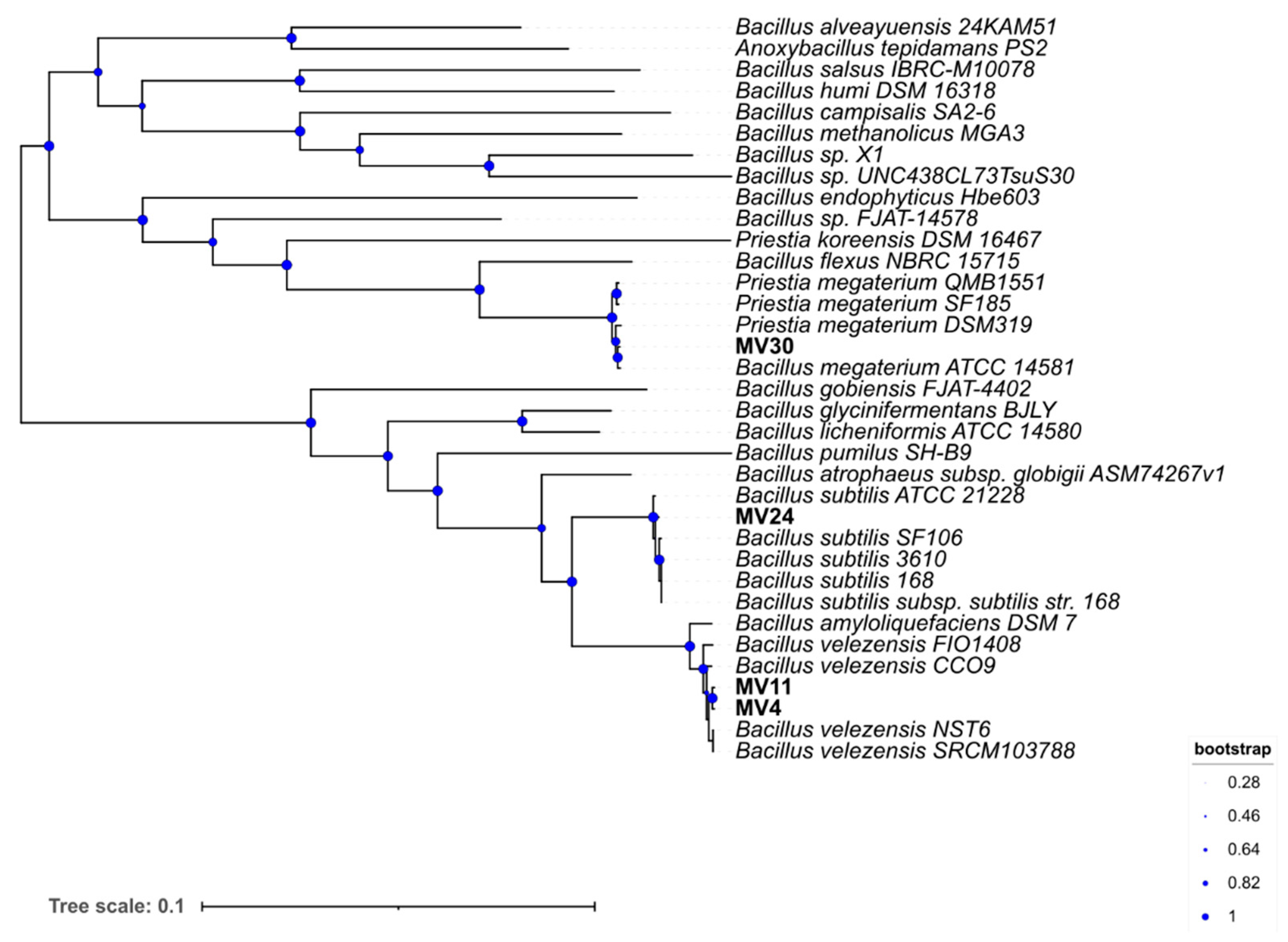
3.2. Species Assignment and Phylogenetic Analysi

The four isolates were used to extract total DNA and the genomic sequences were obtained with a coverage of about 30×. The genomes were approximately 4.0 Mbp long for MV4, MV11, and MV24, and about 5.8 Mbp for MV30 (Table 2). The total number of ORFs was 3860, 3956, and 4380 for MV4, MV11, and MV24, respectively, and higher for MV30 (6003) (Table 2). No plasmid sequences were detected in any of the strains (not shown). All genome sequences were deposited in the GenBank database (Methods).
Based on the analysis of the sequence of the 16S RNA genes, MV4 and MV11 were tentatively considered as belonging to the B. velezensis species while MV24 and MV30 were to the B. subtilis and Priestia megaterium (formerly Bacillus megaterium) species, respectively. The Average Nucleotide Identity (ANI) of the four genomes was determined against the genome of a type strain of each of the putative species and confirmed the species assignment suggested by the 16S RNA gene analysis (Table 3).
The phylogeny of the four strains was further characterized by comparing forty-nine universally conserved genes (Mat.&Meth) with those of several other strains of related species. A phylogenetic tree was generated by Insert Genome into Species Tree app in KBase (Figure 1).

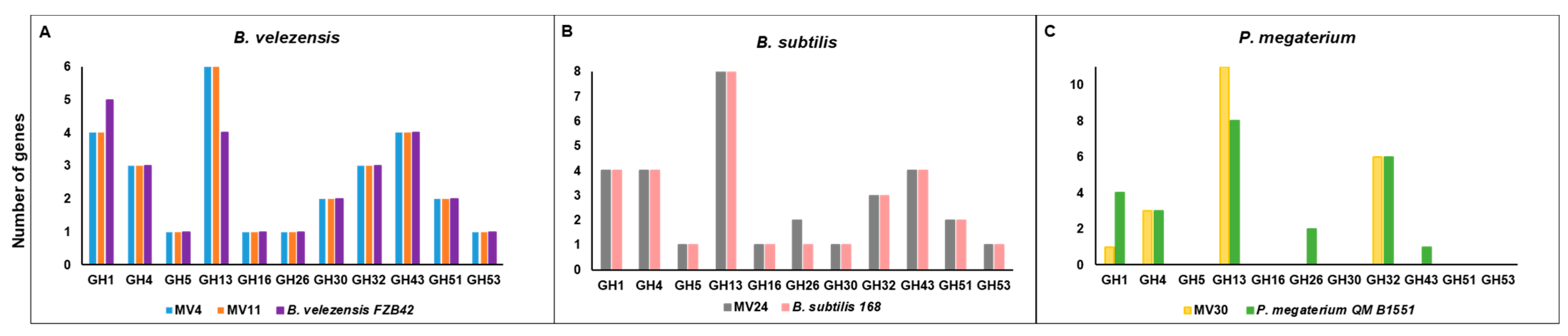
3.3. Genome Analyses

MV4, MV11, MV24, and MV30 genomes were screened for the presence of toxin genes previously found in various other Bacillus strains [31,41]. As reported in Supplementary Materials Table S2, homologs of most of the genes coding for virulence factors were not found in MV4, MV11, MV24, or MV30. Homologs of the hlyIII gene, coding for the channel-forming protein Hemolysin III, were found in all four genomes as well as in the non-pathogenic strains B. clausii KSM-K16 and B. subtilis 168 (Supplementary Materials Table S2). The two B. velezensis strains and MV30 also contained homologs of the cysC gene coding for an adenylyl-sulfate-kinase similar to the Pseudomonas adenylyl-sulfate-kinase phytotoxin (Supplementary Materials Table S2). Only the genome of MV30 contains two homologs of the cylR2 gene, coding for a DNA binding protein of Enterococcus acting as a repressor of the cytolysin operon (Supplementary Materials Table S2).
The functional annotation of the four genomes was performed by using the EggNOG (evolutionary genealogy of genes: Non-supervised Orthologous Groups) database, a public resource classification database able to provide Cluster Orthologous Groups (COGs) of proteins with functional annotations (Supplementary Materials Figure S2). For all four strains, the majority of the annotated genes were classified into the “Unknown Category” (S category in Supplementary Materials Figure S2). According to this annotation system, the next most represented classes for all four strains were “amino acid transport and metabolism”, “transcription”, and “carbohydrate transport and metabolism” (respectively, E, K, and G categories in Supplementary Materials Figure S2).
The genomes of the four isolates were also annotated using the Carbohydrate-Active enZymes (CAZymes) database in comparison with the genome of a reference strain of the respective species. MV4 and MV11 showed a number of genes putatively coding for glycosyl-hydrolase (GH) and a total number of CAZymes bigger than a reference strain of B. velezensis (Table 4). MV24 and MV30 showed, respectively, a slightly bigger and smaller number of CAZymes than their reference strains (Table 4). The GH category contains various hydrolases that act on the glycosidic bond and is divided into families. Some families such as GH1, GH4, GH5, GH16, and GH32 contain enzymes involved in the degradation of cellulose, starch, and sucrose while the GH26, GH30, GH43, GH51, and GH53 contain enzymes specialized in the degradation of hemicellulose, xylan, and cellobiose [46]. Figure 2 reports the abundance of the GH families mainly involved in the degradation of plant polysaccharides found in the four MV genomes. With respect to a reference strain of the same species, MV4, MV11, and MV30 contain more genes for the GH13 family and fewer genes for the GH1 family, respectively, containing enzymes specialized in the degradation of starch or monosaccharides (Figure 2).
MV30 also does not contain GH26 and GH43, present in the P. megaterium reference strain and coding for enzymes involved in the degradation of mannans or arabinose/xylose, respectively. The GH profile of the B. subtilis strain MV24 was very similar to that of the reference strain with the only difference in the number of GH26 (degradation of mannans) higher in MV24 than in B. subtilis 168 (Figure 2).
The genome sequences of the four strains were also analyzed to search for homologs of NK (EC 3.4.21.62), using as a query the protein sequence found in Bacillus subtilis subsp. natto BEST195 (accession No. BAI84580.1). Homologs were present in MV4 (85.86% identity, MV4_3335), MV11 (86.13%, Contig_6_158), and MV24 (99.21%, MV24_0328), while no homologs of the NK coding gene were present in the MV30.
The presence of homologs of the GAD, an enzyme that converts glutamate to GABA, was also investigated and a homolog with an identity of 77.9% with the GAD of other Bacillus species (WP_049107856.1) was found only in the MV30 genome (MV30_2943).

3.4. Antimicrobial and Antioxidant Activities

The four genomes were analyzed to identify genes coding for antimicrobials and antioxidants. The antiSMASH (Antibiotics and Secondary Metabolite Analysis Shell) database was used to assess the presence of clusters of genes associated with the synthesis of antimicrobials and secondary metabolites. As reported in Supplementary Materials Table S3 and schematically summarized in Figure 3, various gene clusters were identified in all four strains. In particular, seven, five, and four clusters with at least 80% similarity with known genes were found, respectively, in MV4, MV11, and MV24 while no homologous clusters were found in MV30 (Supplementary Materials Table S3). Based on this, both B. velezensis strains potentially produce macrolactin H, bacillaene, fengycin, bacillibactin, and bacilysin while only MV4 also codes for difficidin and surfactin while the genome of the B. subtilis strain MV24 codes for bacillibactin, bacilysin, subtilosin A, and fengycin (Supplementary Materials Table S3). In addition, several gene clusters of all four strains showed low or no similarity with previously identified genes, suggesting that they may code for variants of known molecules or totally new antimicrobials (Supplementary Materials Table S3). As an example, clusters coding for surfactin homologs of MV4 and MV11 were compared with the well-characterized surfactin locus of B. velezensis FZB42 [47]. The surfactin cluster of MV4 (82% similarity with that of FZB42) and the two surfactin clusters of MV11 (47 and 39% of similarity with that of FZB42) were predicted to produce very different molecules (Figure 4), confirming that the exploitation of these genomes may provide useful information for the identification of novel antimicrobials.
To validate the genomic predictions on the potential production of antimicrobials, the MV strains were tested against a panel of target microorganisms by agar diffusion assay with cell-free supernatants. MV30 did not show any antimicrobial activity against the target bacteria while the other three strains were active against L. monocytogenes and B. cereus. Only strains of the B. velezensis species (MV4 and MV11) were weakly active against some other bacteria and a strain of Candida albicans (Table 5).
All four genomes were screened for the presence of genes coding for antioxidants. Homologs of the enzymes catalase (CAT; EC:1.11.1.6) and superoxide dismutase (SOD; EC:1.15.1.1) were present in all four genomes. In particular, MV4, MV11, and MV24 contain three CAT (MV4_0856, MV4_0864, MV4_1319 in MV4; Contig_4_193; Contig_4_217 Contig_6_12 in MV11; MV24_3061, MV24_0468, MV24_1926 in MV24) and three SOD (MV4_0856, MV4_1319, MV4_0864 in MV4; 1_870, 2_130, 1_878 in MV11; MV24_2302, MV24_1406, MV24_2295) homologs. MV30 genome only contains one homolog of each enzyme (CAT:MV30_1299 and SOD:MV30_1877).
The four strains were then tested for their antioxidant activity. Vegetative cells (collected after 24 h of aerobic growth at 37 °C) (Methods) were tested for the scavenging activity against hydrogen peroxidase (H2O2) or free radicals (by α,α-diphenyl-β-picrylhydrazyl (DPPH) method), as previously reported [20]. As shown in Figure 5, the H2O2-scavenging activity was stronger than the anti-free radicals activity with between 80 and 90% of H2O2 elimination with 1 × 10⁸ cells and a total H2O2 elimination with 1 × 10⁹ cells of all four strains. Over 60% of free radical elimination was observed with 1 × 10⁹ cells of MV4, MV11, and MV24 (Figure 5A–C), while about 40% of free radicals reduction was observed with the same number of MV30 cells (Figure 5D).

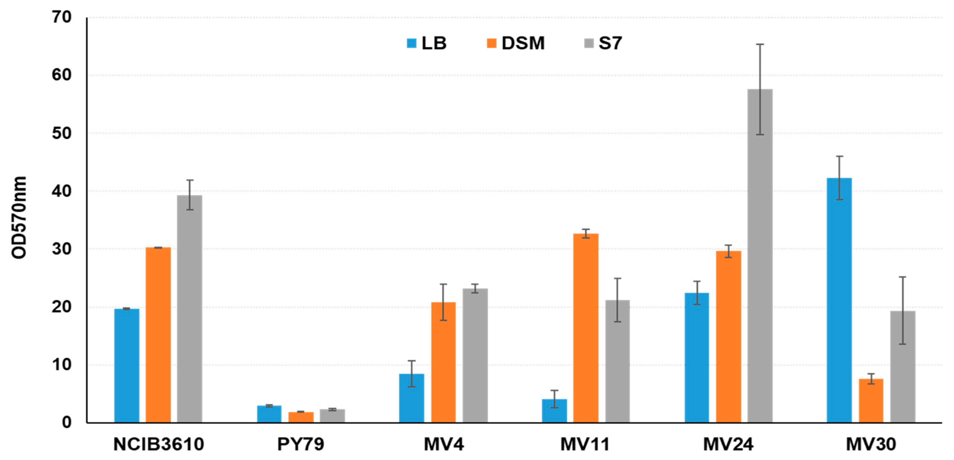
3.5. Matrix Formation

In B. subtilis, the model system for spore formers, the biofilm matrix components include secreted proteins, extracellular DNA (cDNA), and an exopolysaccharide (EPS) [48]. The genes coding for the secreted proteins and the EPS are clustered in the epsA-O and tapA-sipW-tasA operons that are controlled by the master regulators SinI-SinR and Spo0A [48]. Also, in B. cereus products of the pur operon, coding for enzymes needed for purine biosynthesis is required for biofilm formation [49]. The genomes of MV4, MV11, MV24, and MV30 were analyzed for the presence of all these genes involved in matrix formation. As reported in Supplementary Materials Table S4, all four genomes contain homologs of the epsA-O and tapA-sipW-tasA operons, of their regulators, and of the pur operon.
All four strains were analyzed for their ability to produce biofilm in different media in comparison with a B. subtilis laboratory collection strain producing minimal amounts of biofilm (PY79) and a natural strain of the same species known to produce large amounts of biofilm (NCIB3610) [50]. In our experimental conditions (aerobic growth at 37 °C) all four strains produced different amounts of matrix in different media (Figure 6). The two B. velezensis strains produced a low amount of biofilm in rich (LB) medium and higher amounts in sporulation-inducing (DSM) or minimal (LB) media (Figure 6). Similarly, to the reference strain of B. subtilis NCBI3610, B. subtilis MV24 produced the highest amount of matrix in a minimal (S7) medium. However, while the amount of biofilm produced by the two strains was almost identical in LB and DSM media, in minimal medium MV24 produced significantly more biofilm than NCIB3610 (Figure 6). MV30 showed a different pattern of biofilm formation with the highest amount produced in the LB medium and the lowest in the sporulation-inducing medium (Figure 6).

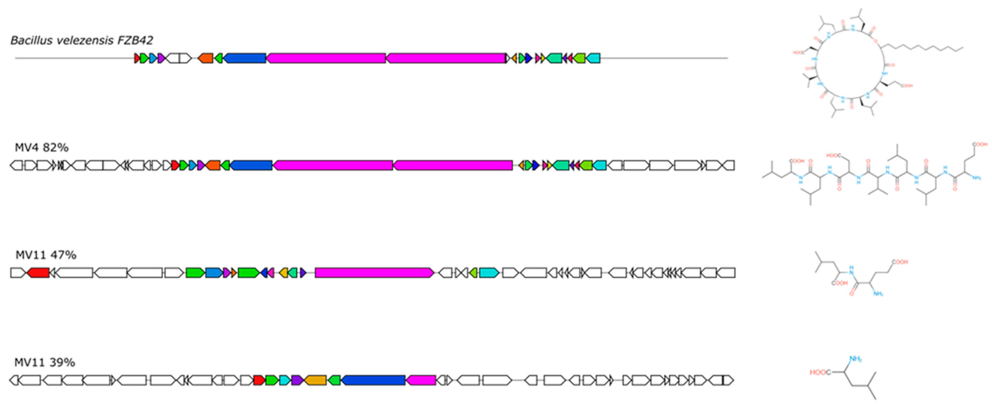
3.6. Production of CSF or CSF-like Peptides

Several Bacillus strains produce and secrete peptides sensed by human epithelial cells, such as the CSF pentapeptide of B. subtilis which is able to induce a cytoprotective heat-shock response [28,30]. The four MV strains were evaluated for their ability to induce a heat-shock response in human epithelial cells. HT-29 colon carcinoma cells were either heat-shocked (30 min at 45 °C) or treated with 20% v/v of cell-free conditioned medium (CM) from the four MV strains and Hps27 induction evaluated by Western blotting with anti-Hsp27 antibody (Figure 7).
After normalization of the intensity of the obtained signals with those of the β-actin extracted from the same cells, the levels of Hsp27 after the treatment with the CM of each of the four strains appeared higher than the threshold level obtained with uninduced HT-29 cells (Figure 7). Although the experiment was not quantitative, it suggested that the CM of all four MV strains was able to induce the expression of the heat-shock protein Hsp27 in the HT-29 cells and, therefore, that all four strains secreted CSF or CSF-like peptides.
A quantitative evaluation of the amount of secreted CSF was obtained by using a recombinant strain of B. subtilis carrying a null mutation in the gene coding for CSF (phrC) and a gene fusion between the promoter of the CSF-controlled gene rapA and the coding part of the reporter gene lacZ of Escherichia coli [30]. The recombinant strain ED5, not producing CSF, does not express the gene fusion but responds to exogenous CSF producing β-galactosidase (Figure 8A) [30]. Cells of ED5 growing in LB medium were supplemented (20% v/v) with the CM of the four MV and control strains, and tested for β-galactosidase production, as previously reported [30]. The B. subtilis isolate MV24 induced β-galactosidase at levels about two-fold higher than the laboratory collection strain of the same species (PY79) (Figure 8B). As expected, the three strains not belonging to the B. subtilis species did not induce β-galactosidase, suggesting that they produce CSF-like peptides able to induce a heat-shock response in epithelial cells [30] (Figure 5) but are unable to activate rapA transcription in B. subtilis (Figure 8B).

4. Discussion

Four aerobic spore former strains isolated from the intestinal samples of healthy children were selected as potential probiotics for not being hemolytic and sensitive to all antibiotics indicated as relevant for spore formers by the EFSA. The four strains were unambiguously assigned to species that are in the Qualified Presumption of Safety (QPS) list of the EFSA (https://www.efsa.europa.eu/en/topics/topic/qualified-presumption-safety-qps (accessed on 5 September 2022)) and are marketed as probiotics for human or animal use [9,12], therefore indicating that they could be considered as safe for human use. This assumption is supported by the in-silico observation that the four MV genomes do not carry putative genes coding for known Bacillus toxins. Indeed, the only gene present in all four genomes coding for a putative toxin is the hlyIII gene, common to pathogenic and non-pathogenic bacteria. The product of hlyIII was, however, not sufficient to cause an alpha- or a beta-hemolytic phenotype in the four MV strains. A gene coding for a putative phytotoxin, cysC, is carried by MV4, MV11, and MV30 while only the latter strain contains the cylR2 gene, a homolog of a transcriptional repressor of a cytolysin operon of Enterococcus.
All four strains formed biofilm and showed antioxidant activity in vitro. Both these properties are relevant for a probiotic strain, favoring the colonization of the intestinal environment and protecting the intestinal cells [20]. In addition, MV4, MV11, and MV30 have a gene coding for an NK homolog while MV30 has a gene coding for the enzyme GAD, able to convert glutamate into GABA. NK is a serine protease of the subtilisin family that when orally ingested exhibits fibrinolytic, antithrombotic, and antihypertensive effects [50]. GABA is an inhibitory neurotransmitter that improves various physiological illnesses, including diabetes, hypertension, and depression in humans [51]. Therefore, the potential to produce NK and/or GABA is considered a relevant property for a potential probiotic strain.
The four MV strains considered in this study also produced and secreted CSF (MV24) or CSF-like (MV4, MV11, and MV30) peptides. In addition to inducing anti-inflammatory and cytoprotective responses both in vitro and in vivo [28,29], these molecules have a role in the anti-neurodegenerative activity of B. subtilis in the model organism C. elegans [31,32,33]. Ingested spores of strains producing such molecules could have beneficial effects for the treatment of inflammatory and age-related neurodegenerative diseases.
Although the production of antimicrobials is not the main mechanism of action of probiotics against infectious diseases, the observed production of an array of secondary metabolites with potential antimicrobial activity by the four strains is certainly an additional relevant point for a probiotic to be used as an alternative to antibiotics. In this context, the identification of genes coding for products with low or no similarity with known antimicrobials is particularly promising. Such genes may encode either variants of previously identified molecules with different targets and mechanisms of action or totally new antimicrobials. The detailed characterization of these genes, which will be a future research challenge, is expected to provide useful information for the identification of novel antimicrobials.
MV4, MV11, MV24, and MV30 strains do not pose safety concerns and have beneficial properties, therefore they can be considered for in vivo experiments aimed at testing their role as an alternative to antibiotics for humans and animals.

Supplementary Materials

The following supporting information can be downloaded at: https://www.mdpi.com/article/10.3390/microorganisms11081978/s1.

Author Contributions

Conceptualization, E.R., L.B. and R.I.; in silico analysis, M.V. and A.S.; validation, M.V. and A.S.; data curation, M.V. and A.S.; writing—original draft preparation, E.R., M.V. and A.S.; writing—review and editing, L.B. and R.I. All authors have read and agreed to the published version of the manuscript.

Funding

This research received no external funding.

Data Availability Statement

Not applicable.

Conflicts of Interest

The authors declare no conflict of interest.

References

  1. Browne, K.; Chakraborty, S.; Chen, R.; Willcox, M.D.; Black, D.S.; Walsh, W.R.; Kumar, N. A New era of antibiotics: The clinical potential of antimicrobial peptides. Int. J. Mol. Sci. 2020, 21, 7047. [Google Scholar] [CrossRef] [PubMed]
  2. Zhao, P.; Xue, Y.; Gao, W.; Li, J.; Zu, X.; Fu, D.; Bai, X.; Zuo, Y.; Hu, Z.; Zhang, F. Bacillaceae-derived peptide antibiotics since 2000. Peptides 2018, 101, 10–16. [Google Scholar] [CrossRef] [PubMed]
  3. Liu, Y.; Tran, D.Q.; Rhoads, J.M. Probiotics in disease prevention and treatment. J. Clin. Pharmacol. 2018, 58 (Suppl. 10), S164–S179. [Google Scholar] [CrossRef] [PubMed] [Green Version]
  4. Sanders, M.E.; Merenstein, D.J.; Reid, G.; Gibson, G.R.; Rastall, R.A. Probiotics and prebiotics in intestinal health and disease: From biology to the clinic. Nat. Rev. Gastroenterol. Hepatol. 2019, 16, 605–616. [Google Scholar] [CrossRef]
  5. Martín, R.; Miquel, S.; Benevides, L.; Bridonneau, C.; Robert, V.; Hudault, S.; Chain, F.; Berteau, O.; Azevedo, V.; Chatel, J.M.; et al. Functional Characterization of novel Faecalibacterium prausnitzii strains isolated from healthy volunteers: A step forward in the use of F. prausnitzii as a Next-Generation Probiotic. Front. Microbiol. 2017, 8, 1664-302x. [Google Scholar] [CrossRef] [Green Version]
  6. Depommier, C.; Everard, A.; Druart, C.; Plovier, H.; Van Hul, M.; Vieira-Silva, S.; Falony, G.; Raes, J.; Maiter, D.; Delzenne, N.M.; et al. Supplementation with Akkermansia muciniphila in overweight and obese human volunteers: A proof-of-concept exploratory study. Nat. Med. 2019, 25, 1096–1103. [Google Scholar] [CrossRef]
  7. Nicholson, W.L.; Munakata, N.; Horneck, G.; Melosh, H.J.; Setlow, P. Resistance of Bacillus endospores to extreme terrestrial and extraterrestrial environments. Microbiol. Mol. Biol. Rev. 2000, 64, 548–572. [Google Scholar] [CrossRef] [Green Version]
  8. Christie, G.; Setlow, P. Bacillus spore germination: Knowns, unknowns and what we need to learn. Cell Signal 2020, 74, 109729. [Google Scholar] [CrossRef]
  9. Cutting, S.M. Bacillus probiotics. Food Microbiol. 2011, 28, 214–220. [Google Scholar] [CrossRef]
  10. Egan, M.; Dempsey, E.; Ryan, C.A.; Ross, R.P.; Stanton, C. The sporobiota of the human gut. Gut Microbes 2021, 13, e1863134. [Google Scholar] [CrossRef]
  11. Saggese, A.; Baccigalupi, L.; Ricca, E. Spore formers as beneficial microbes for humans and animals. Appl. Microbiol. 2021, 1, 498–509. [Google Scholar] [CrossRef]
  12. Casula, G.; Cutting, S.M. Bacillus probiotics: Spore germination in the gastrointestinal tract. Appl. Environ. Microbiol. 2002, 68, 2344–2352. [Google Scholar] [CrossRef] [PubMed] [Green Version]
  13. Bernardeau, M.; Lehtinen, M.J.; Forssten, S.D.; Nurminen, P. Importance of the gastrointestinal life cycle of Bacillus for probiotic functionality. J. Food Sci. Technol. 2017, 54, 2570–2584. [Google Scholar] [CrossRef]
  14. Tam, N.K.; Uyen, N.Q.; Hong, H.A.; Duc, L.H.; Hoa, T.T.; Serra, C.R.; Henriques, A.O.; Cutting, S.M. The intestinal life cycle of Bacillus subtilis and close relatives. J. Bacteriol. 2006, 188, 2692–2700. [Google Scholar] [CrossRef] [Green Version]
  15. Thirabunyanon, M.; Thongwittaya, N. Protection activity of a novel probiotic strain of Bacillus subtilis against Salmonella Enteritidis infection. Res. Vet. Sci. 2012, 93, 74–81. [Google Scholar] [CrossRef]
  16. Paparo, L.; Tripodi, L.; Bruno, C.; Pisapia, L.; Damiano, C.; Pastore, L.; Berni Canani, R. Protective action of Bacillus clausii probiotic strains in an in vitro model of Rotavirus infection. Sci. Rep. 2020, 10, 12636. [Google Scholar]
  17. Rhee, K.-J.; Sethupathi, P.; Driks, A.; Lanning, D.K.; Knight, K.L. Role of commensal bacteria in development of gut-associated lymphoid tissues and preimmune antibody repertoire. J. Immunol. 2004, 172, 1118. [Google Scholar] [CrossRef] [PubMed] [Green Version]
  18. Huang, J.M.; La Ragione, R.; Nunez, A.; Cutting, S.M. Immunostimulatory activity of Bacillus spores. FEMS Immunol. Med. Microbiol. 2008, 53, 195–203. [Google Scholar] [CrossRef] [Green Version]
  19. Kotowicz, N.; Bhardwaj, R.K.; Ferreira, W.T.; Hong, H.A.; Olender, A.; Ramirez, J.; Cutting, S.M. Safety and probiotic evaluation of two Bacillus strains producing antioxidant compounds. Benef. Microbes 2019, 10, 759–771. [Google Scholar] [CrossRef]
  20. Mazzoli, A.; Donadio, G.; Lanzilli, M.; Saggese, A.; Guarino, A.M.; Rivetti, M.; Crescenzo, R.; Ricca, E.; Ferrandino, I.; Iossa, S.; et al. Bacillus megaterium SF185 spores exert protective effects against oxidative stress in vivo and in vitro. Sci. Rep. 2014, 9, 12082. [Google Scholar] [CrossRef] [Green Version]
  21. Petruk, G.; Donadio, G.; Lanzilli, M.; Isticato, R.; Monti, D.M. Alternative use of Bacillus subtilis spores: Protection against environmental oxidative stress in human normal keratinocytes. Sci. Rep. 2018, 8, 1745. [Google Scholar] [CrossRef] [Green Version]
  22. Hou, Q.; Jia, J.; Lin, J.; Zhu, L.; Xie, S.; Yu, Q.; Li, Y. Bacillus subtilis programs the differentiation of intestinal secretory lineages to inhibit Salmonella infection. Cell Rep. 2022, 40, 111416. [Google Scholar] [CrossRef]
  23. Ji, L.; Zhang, L.; Shen, J.; Zhang, Y.; Lu, L.; Zhang, X.; Ma, X. Bacillus subtilis M6 improves intestinal barrier, antioxidant capacity and gut microbial composition in AA broiler. Front. Nutr. 2022, 9, 965310. [Google Scholar] [CrossRef]
  24. Marzorati, M.; Van den Abbeele, P.; Bubeck, S.; Bayne, T.; Krishnan, K.; Young, A. Treatment with a spore-based probiotic containing five strains of Bacillus induced changes in the metabolic activity and community composition of the gut microbiota in a SHIME® model of the human gastrointestinal system. Food Res. Intern. 2021, 149, 110676. [Google Scholar] [CrossRef] [PubMed]
  25. Altaf, F.; Wu, S.; Kasim, V. Role of fibrinolytic enzymes in anti-thrombosis therapy. Front. Mol. Biosci. 2021, 8, 680397. [Google Scholar] [CrossRef] [PubMed]
  26. Kim, H.; Irorita Fugaban, J.I.; Holzapfel, W.H.; Todorov, S.D. Selection of beneficial bacterial strains with potential as oral probiotic candidates. Probiotics Antimicrob. Proteins 2022, 14, 1077–1093. [Google Scholar] [CrossRef]
  27. Mnif, I.; Ghribi, D. Review lipopeptides biosurfactants: Mean classes and new insights for industrial, biomedical, and environmental applications. Biopolymers 2015, 104, 129–147. [Google Scholar] [CrossRef] [PubMed]
  28. Saggese, A.; De Luca, Y.; Baccigalupi, L.; Ricca, E. An antimicrobial peptide specifically active against Listeria monocytogenes is secreted by Bacillus pumilus SF214. BMC Microbiol. 2022, 22, 3. [Google Scholar] [CrossRef]
  29. Fujita, M.; Musch, M.W.; Nakagawa, Y.; Hu, S.; Alverdy, J.; Kohgo, Y.; Schneewind, O.; Jabri, B.; Chang, E.B. The Bacillus subtilis quorum-sensing molecule CSF contributes to intestinal homeostasis via OCTN2, a host cell membrane transporter. Cell Host Microbe 2007, 1, 299–308. [Google Scholar] [CrossRef] [Green Version]
  30. Okamoto, K.; Fujiya, M.; Nata, T.; Ueno, N.; Inaba, Y.; Ishikawa, C.; Ito, T.; Moriichi, K.; Tanabe, H.; Mizukami, Y.; et al. Competence and sporulation factor derived from Bacillus subtilis improves epithelial cell injury in intestinal inflammation via immunomodulation and cytoprotection. Int. J. Colorectal Dis. 2012, 27, 1039–1046. [Google Scholar] [CrossRef] [PubMed] [Green Version]
  31. Di Luccia, B.; D’Apuzzo, E.; Varriale, F.; Baccigalupi, L.; Ricca, E.; Pollice, A. Bacillus megaterium SF185 induces stress pathways and affects the cell cycle distribution of human intestinal epithelial cells. Benef. Microbes 2016, 7, 609–620. [Google Scholar] [CrossRef]
  32. Cogliati, S.; Clementi, V.; Francisco, M.; Crespo, C.; Argañaraz, F.; Grau, R. Bacillus subtilis delays neurodegeneration and behavioral impairment in the Alzheimer’s disease model Caenorhabditis elegans. J. Alzheimers Dis. 2020, 73, 1035–1052. [Google Scholar] [CrossRef]
  33. Donato, V.; Rodrıguez Ayala, F.; Cogliati, S.; Bauman, C.; Costa, J.C.; Lenini, C.; Grau, R. Bacillus subtilis biofilm extends Caenorhabditis elegans longevity through downregulation of the insulin-like signalling pathway. Nat. Commun. 2017, 8, 14332. [Google Scholar] [CrossRef] [PubMed]
  34. Rodrıguez Ayala, F.; Bauman, C.; Cogliati, S.; Lenini, C.; Bartolini, M.; Grau, R. Microbial flora, probiotics, Bacillus subtilis and the search for a long and healthy human longevity. Microbial. Cell 2017, 4, 133–136. [Google Scholar] [CrossRef] [PubMed]
  35. Fakhry, S.; Sorrentini, I.; Ricca, E.; De Felice, M. and Baccigalupi, L. Characterisation of spore forming Bacilli isolated from the human gastrointestinal tract. J. App. Microbiol. 2008, 105, 2178–2186. [Google Scholar] [CrossRef] [PubMed]
  36. EFSA Panel on Additives and Products or Substances used in Animal Feed (FEEDAP). Guidance on the assessment of bacterial susceptibility to antimicrobials of human and veterinary importance. EFSA J. 2012, 10, 2740. [Google Scholar]
  37. Saggese, A.; Isticato, R.; Cangiano, G.; Ricca, E.; Baccigalupi, L. CotG-like modular proteins are common among spore-forming Bacilli. J. Bacteriol. 2016, 198, 1513–1520. [Google Scholar] [CrossRef] [Green Version]
  38. Lugli, G.A.; Milani, C.; Mancabelli, L.; Van Sinderen, D.; Ventura, M. MEGAnnotator: A user-friendly pipeline for microbial genomes assembly and annotation. FEMS Microbiol. Lett. 2016, 363, fnw049. [Google Scholar] [CrossRef] [Green Version]
  39. Liu, B.; Zheng, D.; Jin, Q.; Chen, L.; Yang, J. VFDB 2019: A comparative pathogenomic platform with an interactive web interface. Nucleic Acids Res. 2019, 47, D687–D692. [Google Scholar] [CrossRef]
  40. Saggese, A.; Giglio, R.; D’Anzi, N.; Baccigalupi, L.; Ricca, E. Comparative genomics and physiological characterization of two aerobic spore formers isolated from human ileal samples. Int. J Mol. Sci. 2022, 23, 14946. [Google Scholar] [CrossRef]
  41. Vittoria, M.; Saggese, A.; Di Gregorio Barletta, G.; Castaldi, S.; Isticato, R.; Baccigalupi, L.; Ricca, E. Sporulation efficiency and spore quality in a human intestinal isolate of Bacillus cereus. Res. Microbiol. 2023, 2, 104030. [Google Scholar] [CrossRef] [PubMed]
  42. Isticato, R.; Sirec, T.; Vecchione, S.; Crispino, A.; Saggese, A.; Baccigalupi, L.; Notomista, E.; Driks, A.; Ricca, E. The direct interaction between two morphogenetic proteins is essential for spore coat formation in Bacillus subtilis. PLoS ONE 2015, 10, e0141040. [Google Scholar] [CrossRef] [PubMed] [Green Version]
  43. Zanfardino, A.; Migliardi, A.; D’Alonzo, D.; Lombardi, A.; Varcamonti, M.; Cordone, A. Inactivation of MSMEG_0412 gene drastically affects surface related properties of Mycobacterium smegmatis. BMC Microbiol 2016, 16, 267. [Google Scholar] [CrossRef] [PubMed] [Green Version]
  44. Di Luccia, B.; Acampora, V.; Saggese, A.; Calabrò, V.; Vivo, M.; Angrisano, T.; Baccigalupi, L.; Ricca, E.; Pollice, A. Modulation of intestinal epithelial cell proliferation and apoptosis by Lactobacillus gasseri SF1183. Sci. Rep. 2022, 12, 20248. [Google Scholar] [CrossRef] [PubMed]
  45. Guinebretiere, M.H.; Broussolle, V.; Nguyen-The, C. Enterotoxigenic profiles of food-poisoning and food-borne Bacillus cereus strains. J. Clin. Microbiol. 2002, 40, 3053–3056. [Google Scholar] [CrossRef] [Green Version]
  46. Phelps, R.J.; McKillip, J.L. Enterotoxin production in natural isolates of Bacillaceae outside the Bacillus cereus group. Appl. Environ. Microbiol. 2002, 68, 3147–3151. [Google Scholar] [CrossRef] [Green Version]
  47. Li, Y.; Lei, L.; Zheng, L.; Xiao, X.; Tang, L.; Luo, C. Genome sequencing of gut symbiotic Bacillus velezensis LC1 for bioethanol production from bamboo shoots. Biotechnol. Biofuels 2020, 13, 34. [Google Scholar] [CrossRef] [Green Version]
  48. Arnaouteli, S.; Bamford, N.C.; Stanley-Wall, N.R.; Kovacs, A.T. Bacillus subtilis biofilm formation and social interactions. Nat. Rev. Microbiol. 2021, 19, 600–614. [Google Scholar]
  49. Vilain, S.; Pretorius, J.M.; Theron, J.; Brozel, V.S. DNA as an adhesin: Bacillus cereus requires extracellular DNA to form biofilm. Appl. Environ. Microbiol. 2009, 75, 2861–2868. [Google Scholar] [CrossRef] [Green Version]
  50. Nijland, R.; Burgess, J.G.; Errington, J.; Veening, J.W. Transfromation of environmental Bacillus subtilis isolates by transiently inducing genetic competence. PLoS ONE 2010, 5, e9724. [Google Scholar] [CrossRef] [Green Version]
  51. Ero, M.P.; Ng, C.M.; Mihailovski, T.; Harvey, N.R.; Lewis, B.H. A pilot study on the serum pharmacokinetics of nattokinase in humans following a single, oral, daily dose. Altern. Ther. Health Med. 2013, 19, 16–19. [Google Scholar] [PubMed]
Figure 1. Phylogenetic tree. The genomes were imported into the Kbase system and the phylogenetic tree was constructed using Insert Genome Into SpeciesTree-v2.2.0. Bootstrap confidence values were generated using 1000 permutations.
Microorganisms 11 01978 g001
Figure 2. Number of predicted genes encoding cellulose/hemicellulose degradation enzymes found in the genome of MV4, MV11 (A), MV24 (B), and MV30 (C) compared to the indicated reference strains.
Microorganisms 11 01978 g002
Figure 3. Abundance of clusters of genes coding for antimicrobials and other secondary metabolites in MV4, MV11, MV24, and MV30. The identified clusters were divided into functional classes. NRPS = Non-ribosomal peptide synthetase; PK = polyketide synthase; RiPP = Ribosomally synthesised and post-translationally modified peptide product; Saccharide = possible saccharide; Terpene = possible terpene; Other = Cluster containing a secondary metabolite-related protein that does not fit into any other category; Unknown = unknown BCG clusters.
Microorganisms 11 01978 g003
Figure 4. Surfactin loci of MV4, MV11, and the reference strain FZB42. The same colors indicate genes coding for products with similar functions. On the right is reported the predicted structure of each gene cluster.
Microorganisms 11 01978 g004
Figure 5. Evaluation of the ability of MV4 (A), MV11 (B), MV24 (C), and MV30 (D) cells to reduce hydrogen peroxide (H2O2, grey line) and the radical 2,2-diphenyl-1-picrylhydrazyl (DPPH, blue line). The assay was performed using suspensions of 107, 108, and 109 cells and was repeated in triplicate.
Microorganisms 11 01978 g005
Figure 6. Biofilm production assay performed on the four bacterial strains in three different media: LB (blue bars), DSM (orange bars), and S7 (grey bars). The assay was performed in triplicate. B. subtilis strains NCIB3610 and PY79 were used as positive and negative controls, respectively.
Microorganisms 11 01978 g006
Figure 7. Effect of the conditioned medium (CM) of the four strains on Hsp27 production in HT-29 intestinal cells. Representative Western blot was performed with anti-Hsp27 antibodies (A) and a densitometric analysis was obtained by the average of three different experiments (B). Untreated (Co) and heat-shocked (HS) cells were used as a negative and positive control, respectively. The band intensity of each sample was evaluated by Image Quant analysis and normalized for the β-Actin intensity of the same sample. The black continuous line indicates the uninduced level of Hsp27 in HT-29 cells.
Microorganisms 11 01978 g007
Figure 8. Production of CSF pentapeptide. (A) Schematic representation of the mutant strain of B. subtilis ED5 unable to produce CSF (phrC null) carrying the rapA::lacZ gene fusion. The mutant alone does not produce endogenous CSF but responds to exogenous CSF producing β-galactosidase. (B) Beta-galactosidase activity was measured with ED5 cells treated with the supernatants of MV4, MV11, MV24, and MV30. Supernatants of PY79 (B. subtilis wild-type) and ED5 (isogenic phrC deletion mutant) were used as a positive and negative control, respectively. The β-galactosidase is expressed in Miller Units and data are the average of three independent experiments.
Microorganisms 11 01978 g008
Table 1. Minimum inhibitory concentration (MIC) values in mg/L.
StrainVanGenKanStrepEryClinTetChl
MV41.5000.5000.7508.0000.0940.7508.0001.000
MV60.5000.1252.000R 10.1250.1901.0000.250
MV110.1900.7500.5001.0000.0470.1252.0002.000
MV143.0000.1250.7508.0000.2504.000R 15.000
MV150.3800.250R 12.0000.380R 10.250R 1
MV170.0470.500R 1R 1R 10.7500.750R 1
MV200.1900.2500.5001.0000.125R 11.0000.750
MV241.5000.5001.5003.0000.3800.3800.0944.000
MV260.1250.0470.1251.0000.064R 10.2502.000
MV300.1250.0470.1908.0000.0644.0000.5001.500
EFSA
Breakpoints
4.0004.0008.0008.0004.0004.0008.0008.000
1 R = resistant (over the EFSA breakpoint value).
Table 2. General features of the MV4, MV11, MV24, and MV30 genomes.
MV4MV11MV24MV30
Size (bp)3,987,6954,035,8474,116,4005,853,282
Number of contigs365811235
Average Coverage582341133
Number of predicted ORFs3860395643806003
Average GC (percentage)46.3546.243.2637.46
Table 3. Average Nucleotide Identity (ANI) (%) based on whole genome alignments.
B. velezensis
FZB42
B. subtilis
168
P. megaterium
QM B1551
MV498.3677.368.77
MV1198.3677.0468.44
MV2477.398.5468.97
MV3068.3168.5897.42
Table 4. Number of genes putatively coding for the six CAZyme categories.
Species and StrainGHGTPLCECBMAATotal
B. velezensis MV44737311163117
B. velezensis MV114837311163118
B. velezensis FZB424035313145110
B. subtilis MV245639714223141
B. subtilis 1685634714234138
P. megaterium MV304942120235140
P. megaterium QM B15515342124215146
Table 5. Antimicrobial activity of MV4, MV11, MV24, and MV30 against a panel of target strains.
MV4MV11MV24MV30
Listeria monocytogenes ATCC 7644+++-
Bacillus cereus ATCC 10987+++++-
Enterococcus faecalis ATCC 29212----
Streptococcus faecalis ATCC 33186+/-+/---
Escherichia coli ATCC 25922+/-+/---
Salmonella enterica subsp. enterica serovar
Typhimurium ATCC 14028
+/-+/---
Citrobacter rodensis ATCC 14580+/-+/---
Shigella sonnei ATCC 25931----
Candida albicans ATCC 14028+/-+/---
Lacticaseibacillus rhamnosus GG 1----
+/- inhibition halo < 1 cm; + inhibition halo > 1 cm e < 2 cm; ++ inhibition halo > 2 cm. 1 This representative probiotic strain was used to validate the selectivity of the antimicrobial properties of the isolated bacteria.
Disclaimer/Publisher’s Note: The statements, opinions and data contained in all publications are solely those of the individual author(s) and contributor(s) and not of MDPI and/or the editor(s). MDPI and/or the editor(s) disclaim responsibility for any injury to people or property resulting from any ideas, methods, instructions or products referred to in the content.

